Université de Montréal
The role of the peptidyl prolyl isomerase Rrd1 in the transcriptional stress response
par
Jeremie Poschmann
Département de Microbiologie et Immunologie Faculté de Médecine
Thèse présentée à la Faculté des études supérieures en vue de l’obtention du grade de Philosophiae Doctor (Ph.D)
en Microbiologie et Immunologie
Février 2010
Université de Montréal
Faculté des études supérieures
Cette these intitulée:
The role of the peptidyl prolyl isomerase Rrd1 in the transcriptional stress response
présentée par : Jeremie Poschmann
a été évaluée par un jury composé des personnes suivantes :
Dr. George Szatmari (président-rapporteur) Dr. Dindial Ramotar (directeur de recherche) Dr. Marc Drolet (membre du jury) Dr. Malcolm Whiteway (examinateur externe) Dr. Gerardo Ferbeyre (représentant du doyen)
RÉSUMÉ
La régulation de la transcription est un processus complexe qui a évolué pendant des millions d’années permettant ainsi aux cellules de s’adapter aux changements environnementaux. Notre laboratoire étudie le rôle de la rapamycine, un agent immunosuppresseur et anticancéreux, qui mime la carence nutritionelle. Afin de comprendre les mécanismes impliqués dans la réponse a la rapamycine, nous recherchons des mutants de la levure Saccaromyces cerevisiae qui ont un phenotype altérée envers cette drogue. Nous avons identifié le gène RRD1, qui encode une peptidyl prolyl isomérase et dont la mutation rend les levures très résistantes à la rapamycine et il semble que se soit associé à une réponse transcriptionelle alterée. Mon projet de recherche de doctorat est d’identifier le rôle de Rrd1 dans la réponse à la rapamycine. Tout d’abord nous avons trouvé que Rrd1 interagit avec l’ARN polymérase II (RNAPII), plus spécifiquement avec son domaine C-terminal. En réponse à la rapamycine, Rrd1 induit un changement dans la conformation du domaine C-terminal in vivo permettant la régulation de l’association de RNAPII avec certains gènes. Des analyses in vitro ont également montré que cette action est directe et probablement liée à l’activité isomérase de Rrd1 suggérant un rôle pour Rrd1 dans la régulation de la transcription. Nous avons utilisé la technologie de ChIP sur micropuce pour localiser Rrd1 sur la majorité des gènes transcrits par RNAPII et montre que Rrd1 agit en tant que facteur d’élongation de RNAPII. Pour finir, des résultats suggèrent que Rrd1 n’est pas seulement impliqué dans la réponse à la rapamycine mais aussi à differents stress environnementaux, nous permettant ainsi d’établir que Rrd1 est un facteur d’élongation de la transcription requis pour la régulation de la transcription via RNAPII en réponse au stress.
Mots clés: ARN polymérase II, rapamycine, peptidyl-prolyl isomérase, Immuno-precipitiation de chromatine sur micropuce, régulation transcriptionelle, élongation
ABSTRACT
Transcriptional regulation is a complex process that has evolved over millions of years of evolution. Cells have to sense environmental conditions and adapt to them by altering their transcription. Herein, we study the role of rapamycin, an immunosuppressant and anticancer molecule that mimics cellular starvation. To understand how the action of rapamycin is mediated, we analyzed gene deletion mutants in the yeast Saccharomyces cerevisiae that have an altered response to this drug. Deletion of RRD1, a gene encoding a peptidyl prolyl isomerase, causes strong resistance to rapamycin and this was associated with a role of Rrd1 in the transcriptional response towards rapamycin. The main focus of my PhD was therefore to unravel the role of Rrd1 in response to rapamycin. First, we discovered that Rrd1 interacts with RNA polymerase II (RNAPII), more specifically with its C-terminal domain and we showed that in response to rapamycin, Rrd1 alters the structure of this C-terminal domain. This phenomenon was confirmed to be directly mediated by Rrd1 in vitro, presumably through its peptidyl prolyl isomerase activity. Further, we demonstrated that Rrd1 is capable of altering the occupancy of RNAPII on genes in vivo and in vitro. With the use of ChIP on chip technology, we show that Rrd1 is actually a transcription elongation factor that is associated with RNAPII on actively transcribed genes. In addition, we demonstrate that Rrd1 is indeed required to regulate the expression of a large subset of genes in response to rapamycin. This data let us propose a novel mechanism by which Rrd1 regulates RNAPII during transcription elongation. Finally, we provide evidence that Rrd1 is not only required for an efficient response towards rapamycin but to a larger variety of environmental stress conditions, thus establishing Rrd1 as a transcriptional elongation factor required to fine tune the transcriptional stress response of RNAPII.
Keywords: RNA polymerase II, transcriptional regulation, peptidyl prolyl isomerase, Chromatin immunoprecipitation and chip analyis, elongation, rapamycin
TABLE OF CONTENTS
RÉSUMÉ ... IV ABSTRACT ... V TABLE OF CONTENTS ... VI LIST OF FIGURES ... VIII ABREVIATIONS ... IX DEDICATION ... XII ACKNOWLEDGMENTS ... XII
1 INTRODUCTION ... 1
1.1 TRANSCRIPTION ... 1
1.1.1 Gene conservation during evolution ... 1
1.1.2 The transcriptional machinery ... 4
1.1.2.1 RNAPII structure ... 5 1.1.2.2 Transcription factors ... 6 1.1.3 Transcription initiation ... 8 1.2.4 Transcription elongation ... 9 1.1.3.1 SAGA ... 9 1.1.3.2 TFIIS ... 9
1.1.3.3 TFIIF, Elongin, Ell and Csb ... 10
1.1.3.4 PTEFb ... 10
1.1.3.5 The Paf1 complex ... 10
1.1.3.6 DSIF and NELF ... 10
1.1.3.7 Elongator and FACT ... 11
1.1.3.8 CTD-phosphatases ... 11
1.1.4 Transcription termination and polyadenylation ... 12
1.1.5 A central role of the RNAPII CTD during transcription ... 14
1.1.6 RNAPII arrest during transcription ... 15
1.1.6.1 Transcriptional blocks caused by DNA lesions ... 16
1.1.6.2 RNAPII ubiquitylation during transcriptional arrest ... 17
1.1.7 Transcriptional regulation of RNAPII ... 18
1.1.7.1 The target of rapamycin (TOR) signaling pathway ... 18
1.1.7.1.1 TORC1 regulates transcription of specific regulons ... 19
1.1.7.1.2 TORC1 regulates ribosome biogenesis ... 20
1.1.7.1.3 Differences between mammalian and yeast cells ... 21
1.1.7.2 The environmental stress response (ESR) ... 21
1.2 RRD1 ANDITSBIOLOGICALROLE ... 23
1.2.1 Introduction ... 23
1.2.3 Discovery of PTPA, the human homologue of Rrd1 ... 25
1.2.4 Structure and function of PP2A phosphatase complexes ... 25
1.2.5 Rrd1 is a peptidyl prolyl isomerase ... 26
1.2.6 RRD2 ... 30
1.2.7 Structure of Rrd1 ... 31
1.2.8 Rrd1 interacts with phosphatases in vivo ... 33
1.2.9 Rrd1 is involved in transcriptional regulation in response to rapamycin ... 34
1.2.10 Role of Rrd1 in rapamycin resistance ... 35
2 ARTICLES ... 36
2.1 RRD1 ISOMERIZES RNA POLYMERASE II INRESPONSETORAPAMYCIN ... 36
2.2 THEYEASTPEPTIDYLPROLYLISOMERASE RRD1 ISANELONGATIONFACTORREQUIREDFORTRANSCRIPTIONALSTRESSRESPONSES ... 67
ABSTRACT ... 69
3 DISCUSSION ... 112
3.1 RRD1 ACTSINDEPENDENTLYOFTHE SIT4-TAP42 COMPLEX ... 112
3.2 RRD1 INTERACTSWITH RNAPII ... 113
3.3 RRD1 ISOMERIZESTHE CTD OF RNAPII ... 114
3.4 RRD1 MODULATESTRANSCRIPTIONINRESPONSETORAPAMYCIN ... 115
3.5 RRD1 ISATRANSCRIPTIONELONGATIONFACTOR ... 116
3.6 RRD1 ISREQUIREDFORABROADRANGEOFSTRESSES ... 118
3.7 MODELOF RNAPII REGULATION ... 119
3.8 THECATALYTICACTIVITYOF RRD1 ... 122
3.9 ALTERNATIVETARGETSOF RRD1 ... 123
3.10 INVOLVEMENTOF RRD1 INREGULATORYPATHWAYS ... 124
3.11 S.CEREVISIAEASAMODEL ... 127
3.12 ISTHE TORC1 COMPLEXREGULATEDBYSUPEROXIDEANIONS? ... 128
4 CONCLUSION ... 131
4.1 RRD1 ISATRANSCRIPTIONELONGATIONFACTORREQUIREDFORTHE ESR INYEAST ... 131
4.2 THEREASONWHYRRD1Δ MUTANTSHAVEPLEIOTROPICPHENOTYPES ... 132
4.3 RRD1 ISTHESECOND PPIASETHATISOMERIZESTHE CTD OF RNAPII ... 133
4.4 THENEWROLEOF RRD1 MIGHTBECONSERVEDDURINGEVOLUTION ... 134
4.5 RRD1 ASAPOTENTIALDRUGTARGET ... 135
4.6 RRD1 HASMULTIPLECELLULARROLES ... 135
5 PERSPECTIVES ... 137
LIST OF FIGURES
FIGURE 1 PRE-INITIATION COMPLEX FORMATION AT THE PROMOTER ...7
FIGURE 2 TRANSCRIPTION CYCLE OF RNAPII...15
FIGURE 3 REGULATION OF TRANSCRIPTION BY TOR1 SIGNALING...20
FIGURE 4 MODEL OF PROLINE CIS- TRANS ISOMERISATION BY PPIASES ...27
FIGURE 5 PHYLOGRAM OF PEPTIDYL PROLYL ISOMERASE FAMILY ...30
FIGURE 6 CRYSTAL STRUCTURE PTPA...33
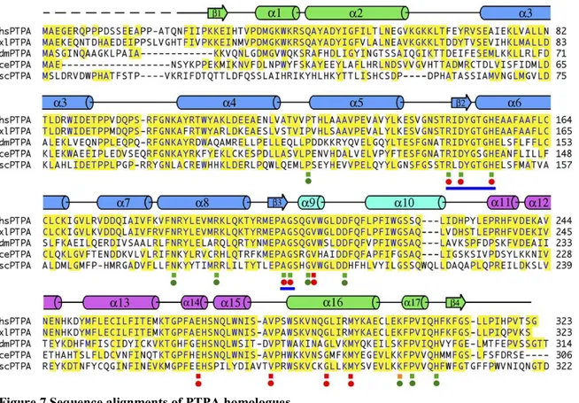
FIGURE 7 SEQUENCE ALIGNMENTS OF PTPA HOMOLOGUES...33
FIGURE 8 MODEL OF RNAPII REGULATION...122
FIGURE 9 THE RRD1 CATALYTIC MUTANT (D200G) ...123
ABREVIATIONS
4NQO 4-nitro quinolone oxide
5.8S 5.8 Svedberg
6-4PP 6-4 photoproduct
6AU 6-azauracil
ATP Adenosine triphosphate
Ca2+
Calcium
CD circular dichroism
ChIP chromatin immunoprecipitation CPD cyclobutane pyrimidine dimers Crf1 corepressor of Fhl1
CSA cocayne syndrome factor A (yeast gene is Rad28) CSB cocayne syndrome factor B (yeast gene is Rad26) CTD C-terminal domain of RNAPII
CUT cryptic unstable transcript Def1 RNAPII degradation factor DNA desoxyribonucleic acid
DSB double strand break
DSIF Elongation factor formed by Spt4 and Spt5
EF elongation factor
Ell elongation factor (eleven–nineteen-lysine-rich leukemia) ESR environmental stress response
Ess1 Peptidyl prolyl isomerase in yeast homologue of Pin1 FACT facilitates chromatin transcription
Fcp1 CTD phosphatase in yeast Fhl1 transcripion activator
FK506 Tacrolimus (an immunosupressant) FKBP peptidyl prolyl isomerase family
Gal4 Transcription factor that activates GAL genes
GGR global genomic repair
Gln3 Transcription factor that regulates NCR genes GST glutathione S-transferase
GTF general transcription factor
GTP guanine triphosphate
HAT Histone acetyl transferase
HDAC Histone deacetylase
Ifh1 interactor of Fhl1
Mep2 ammonium permease induced by NCR
MMS Methyl-Methane sulfonate
MPA Mycophenolic acid
mRNA messenger RNA
Msn2/Msn4 Transcription factor activating STRE
NaAs sodium arsenite
NaCl sodium chloride
NAD+ oxydized form of nicotinamide dinucleotide NCR nitrogen catabolite repression
NER nucleotide excision repair NMR nuclear magnetic resonance
Npr1 kinase that prevents nitrogen permease degradation PAF RNAPII associated factor
PI3K phosphoinositide-3 kinase PIC pre-initiation complex
Pin1 PPIase, human homologue of Ess1
PP2A phosphatase 2A
Ppg1 PP2A like phosphatase involved in glycogen metabolism Pph3 PP2A like phosphatase involved in DNA repair
PPIase peptidyl prolyl isomerase PTPA PP2A phosphotase activator
RNA ribonucleic acid
RNAPII RNA polymerase II ROS reactive oxygen species Rpb1 yeast major subunit of RNAPII
Rpb2 yeast second largest subunit of RNAPII Rrd1 resistant to rapamycin deletion 1
Rrd2 resistant to rapamycin deletion 2 paralogue of Rrd1 in yeast
rRNA ribosomal RNA
RTG retrograde signalling genes
Rtg1/2 Transcription factor regulating the RTG Sit4 PP2A related phosphatase
snoRNA small nucleolar RNA
snRNA small nuclear RNA
Spt15 the yeast gene of TBP
Ssu72 Supressor of Ua7 gene 2 (serine 5 phosphatase in yeast) STRE Stress regulated genes
TAF TBP associated factor
Tap42 PP2A associated protein involved in Tor1 signalling (yeast) TBP Tata box binding protein
TCA tricarboxylic cycle (crebs cycle)
TF transcription factor
TFIIS transcription elongation factor (dst1 in yeast)
TOR target of rapamycin
Tor1 target of rapamycin 1 gene in yeast tor2 target of rapamycin 2 gene in yeat
TORC1 TOR complex contains Tor1,Kog1, Tco89 and Lst8
TORC2 TOR complex contains Tor2, Avo1, Avo2, Avo3, Bit61, and Bit2,
tRNA transfer RNA
TSS transcription start site UAS upstream activation sequence
Ub Ubiquitin
UV ultra violet light
XPA NER factor recognizes and binds damaged DNA XPF NER factor single strand endonuclease
DEDICATION
This thesis is dedicated to my parents and my aunts (Theresia, Christel and Magda) Their patience, assistance and support through all these years even through great distance have
made this thesis possible
Acknowledgments
Dr. Dindial Ramotar for teaching me the pleasure and greatness of science Nathalie Jouvet for being my lab mate and friend
Dr. Jim Daley for helpful comments and corrections of this thesis Dr. Xiaoming Yang for being of great help and assistance in the lab current and previous lab members for great atmosphere
Dr Francois Robert lab and members for accepting me in their lab and perform the ChIP experiments
My thesis committee for helpful comments and discussions
Dr. Jasmine Lefebre for being a friend and support during this time all co-authors of the manuscripts
1 INTRODUCTION
The transcription of mRNA encoding for proteins from template DNA is a phenomenon that is required for the most basic forms of life, from simple to complex organisms. Transcription has to be regulated, meaning that the right mRNA has to be produced in the right amount, at the right moment and if possible without mistakes. Thus, in response to every possible condition, a cell must precisely control the production of specific mRNAs. To perform such an enormous task, various levels of transcriptional regulation have evolved, where multiple factors influence transcription at virtually every step of the process. One can imagine that a failure of this highly regulated process is not compatible with life and can lead to a multitude of diseases including different forms of cancer. Therefore, studying this fundamental process will lead to further understanding of life as well as pathologies and disease treatment.
An outline of the basic steps of transcription, its general regulation as well as more specific regulatory mechanisms in response to different cellular conditions will be described in this section. The second part of this article will then focus on the functions of the peptidyl prolyl isomerase Rrd1, which we have now shown to play a role in transcription.
1.1 Transcription
1.1.1 Gene conservation during evolution
During the long evolutionary process from yeast to man, the signaling pathways that mediate transcription and other relevant processes described here, have undergone some changes and as genes evolved, some were replaced or new genes with additional functions were created. Since the thesis focuses only on work in yeast, a table detailing the conservation of the genes (comparison of the S.cerevisiae and human genome) that are described in the introduction is provided below.
If the gene is not found in one or the other genome it is labeled as not determined (N.D.), which could mean that this gene is not (yet) discovered, that it is not conserved, or it was created in later stages of evolution and is not present in S.cerevisiae.
Class
Mammalian
gene Yeast gene brief description
RNAPII RNA polymerase II consists of 12 subunits
hRpb1 Rpb1 yeast major subunit of RNAPII contains the C-terminal domain hRpb2 Rpb2 yeast second larbest subunit of RNAP II , is also part of the active site hRpb3 Rpb3 orthologue of the alpha factor of the bacterial RNAP together with Rpb11 hRpb4 Rpb4 forms a subcomplex with Rpb7 and is involved in the stress response hRpb5 Rpb5 shared subunit of RNAPI, II and III is required for transcription activation
hRpb6 Rpb6 shared subunit of RNAPI, II and III is required for assembly and stability of the complex hRpb7 Rpb7 form a subcomplex with Rpb4
hRpb8 Rpb8 shared subuit of RNAPI, II and III, binds to oligonucleotides
hRpb9 Rpb9 contains a zinc binding motif, supposed to be involved in elongation and start site selection hRpb10 Rpb10 shared subunit of RNAPI, II and III
hRpb11 Rpb11 orthologue of the alpha factor of the bacterial RNAP together with Rpb3 hRpb12 Rpb12 shared subunit of RNAPI, II and III
GTFs
TFIID TBP Spt15 TATA-binding protein
TAFII250 TAFII145/130 Involved in promoter binding, G1/S progression; histone acetyltransferase; kinase (human) CIF150 Tsm1 Involved in promoter binding; mutations arrest in G2/M of cell cycle (yeast)
TAFII130/135 N.D. Involved in interaction with activators
TAFII100 TAFII90 Mutations can cause arrest in G2/M of cell cycle (yeast)
TAFII70/80 TAFII60 Similar to histone H4; binds downstream promoter elements (DPEs) (Drosophila) TAFII31/32 TAFII17 Similar to histone H3; interacts with p53
TAFII20 TAFII68/61 Similar to histone H2A
TAFII15 N.D. Similar to histone H2A; highly similar to TAFII20
TAFII28 TAFII40 Similar to histone H3; contains atypical histone fold motif seen in Spt3-like transcription factors TAFII68 N.D. Contains consensus RNA binding domain; can bind RNA and ssDNA;
TAFII55 TAFII67 Interacts with numerous activators
TAFII30 TAFII23/25 Mutations can cause arrest in G1/S of cell cycle (human) N.D. TAFII47 No homologous subunit identified in metazoans
N.D. TAFII30 Shared with TFIIF (yeast), no homologous subunit identified in metazoans
TAFII18 TAFII19 Similar to histone H4; contains atypical histone fold motif seen in Spt3-like transcription factors TAFII105 N.D. B-cell specific; related to TAFII130; co-activator for NF-kappaB
TFIIA TFIIAα Toa1 Involved in transcriptional coactivation; involved in stabilizing TBP-DNA interactions TFIIAβ N.D. TFIIAα and TFIIAβ result from processing of a single peptide that is homologous to Toal.
TFIIAγ Toa2
Involved in activator interactions, TFIIA-mediated antirepression, stabilizing TBP-DNA interactions
TFIIB TFIIB Sua7 Involved in start site selection, promoter binding and promoter bending during initiation TFIIF RAP74 Ssu1 Makes extensive contacts with DNA to position the template during initiation
RAP30 Tfg2 Binds RNA polymerase II and suppresses non-specific DNA binding
N.D. Anc1 Common component of yeast TFIID, TFIIF, and Swi/Snf; similar to ENL and AF-9 proteins TFIIE TFIIEa Tfa1 Interacts with TFIIH; involved in recruitment, stimulation of TFIIH and promoter opening
TFIIEb Tfa2 Double strand DNA binding activity
TFIIH p62 Tfb1 Required for nucleotide excision repair; target for activators p52 Tfb2 Required for nucleotide excision repair
MAT1 Tfb3 Required for nucleotide excision repair; MAT1/Cdk7/cyclin H form the CAK subcomplex (human) p34 Tfb4 Required for nucleotide excision repair
XPD/ERCC2 Rad3 5′–3′ DNA helicase; ATPase; required for DNA repair
p44 Ssl1 Required for nucleotide excision repair; involved in DNA binding and stimulation of XPD activity XPB/ERCC3 Ssl2 3′–5′ DNA helicase; ATPase; essential for promoter opening and promoter escape
Cdk7 Kin28 Kinase subunit of cyclin-dependent CTD kinase; Kin28 & Cell form the TFIIK subcomplex (yeast) CyclinH Ccl1 Cyclin subunit of cyclin-dependent CTD kinase
SAGA PCAF Gcn5 histone acetyl transferase catalytic unit, acetylates histone H2B and H3 ADA1 Ada1 adaptor protein required for structure of SAGA
ADA2b Ada2 component of SAGA
ND Spt8 component of SAGA SPT20 Spt20 component of SAGA
SPT7/ Spt7 component of SAGA, required for assembly
SPT3 Spt3 component of SAGA required for transcriptonal activation TAFII70/80 TAFII60 TBP associated factor present in SAGA and TFIID TAFII100 TAFII90 TBP associated factor present in SAGA and TFIID TAFII32 TAFII17 TBP associated factor present in SAGA and TFIID TAFII30 TAFII23/25 TBP associated factor present in SAGA and TFIID TAFII20 TAFII61 TBP associated factor present in SAGA and TFIID
TRRAP Tra1 coactivator protein of SAGA and NuA4 histone acetylase complex SGF29 Sgf29 component of SAGA
USP22 Ubp8 ubiquitin protease required for deubiquitination of histone H2B ATXN7L3 Sgf11 associates Ubp8 to SAGA
ENY2 Sus1 required for mRNA export and transcription elongation component of SAGA ATXN7 Sgf73 component of SAGA required for PIC assembly
ND Chd1 chromatin remodeling factor associated with SAGA
TFIIS TFIIS Dst1 Transcription elongation factor, restarts RNAPII after arrests, mRNA cleavage stimulatory activity DSIF DSIF Spt4, Spt5 elongation factor composed of Spt4 and Spt5
NELF N.D. negative tanscription elongation factor
FACT SSRP1 Spt16 facilitates chromatin transcription , heterodimer, remodels chromatin during transcription
SUPTH16 Pob3 facilitates chromatin transcription , heterodimer, remodels chromatin during transcription
Paf1 Paf1 RNAP II associated factor, assists in transcription elongation Def1 RNAP II degradation factor
CTD kinases and phosphatases of the RNAPII CTD
Cdk7 Kin28 RNAPII CTD kinase phosphorylates serine 5 and serine 7 , part of TFIIH PTEFb Ctk1 RNAPII CTD kinase phosphorylates serine 2
Fcp1 Fcp1 RNAPII CTD phosphatase that dephosphorylates serine 2 Ssu72 Ssu72 RNAPII CTD phosphatase that dephosphorylates serine 5 DNA
repair
CSA Rad28 cocayne syndrome factor A CSB Rad26 cocayne syndrome factor B
XPA Rad14 NER factor recognizes and binds damaged DNA XPF Rad1 NER factor single strand endonuclease XPG Rad2 NER factor single strand endonuclease PP2A
α4 Tap42 PP2A associated protein involved in Tor1 signalling (yeast) PPP4C Pph3 PP2A like phosphatase involved in DNA repair
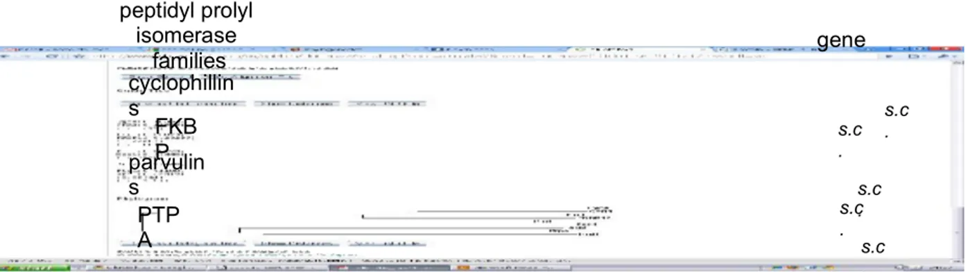
PTPA Rrd1 PP2A phosphotase activator PPP6C Sit4 PP2A related phosphatase Pin1 Ess1 PPIase in yeast homologue of Pin1
N.D. Rrd2 resistant to rapamycin deletion 2 paralogue of Rrd1 in yeast PPP2CA Pph21 PP2A catalytic subunit redundant with PPH22
PPP2CA Pph22 PP2A catalytic subunit redundant with PPH21
N.D. Ppg1 PP2A like phosphatase involved in glycogen metabolism PPiases
Pin1 Ess1 Parvulin family member, isomerizes phospho-prolines, involved in multiples diseases PTPA Rrd1 the new familiy of PPIases, so far known to regulate PP2A phosphatase complexes FKBP1A Fpr1 FKBP binding protein, binds to FK506 and Rapamycin , involved in protein folding CypA Cpr3 Cyclophilin family member, binds to cyclosporin, involved in protein folding
TOR TOR signaling pathway
Torc1 mTOR Tor1 PI3like kinase that controls growth in response to nutrient , is inhibited by rapamycin N.D. Tco89 part of Torc1 complex
MLST8 Lst8 part of Torc1 and 2 complex
Raptor Kog1 controler of growth protein 1 part of the Torc1 complex
Torc2 mTOR Tor2 PI3like kinase that controls spatial growth is not inhibited by rapamycin hSin1 Avo1 Torc2 associated factor
N.D. Avo2 Torc2 associated factor rictor Avo3 Torc2 associated factor mLST8 Lst8 part of Torc1 and 2 complex N.D. Bit61 binding partner of Tor2
Npr1 N.D. Npr1 kinase that prevents nitrogen permease degradation Mep2 N.D. Mep2 ammonium permease induced by NCR
TFs Transcription factors
N.D. Msn2/Msn4 Stress response transcription factors , binds to STRE elements N.D. Rtg1/2 retrograde signaling transcription factors
N.D. Gal4 Galactose regulation transcription factor N.D. Gln3 Nitrogen discrimination transcription factor
N.D. Fhl1 binds to ribosomal genes and activates transcription N.D. Ifh1 transcriptional activator for ribosomal gene expression N.D. Crf1 transcriptional repressor for ribosomal gene expression
1.1.2 The transcriptional machinery
Transcription is the process by which RNA is synthesized in a DNA-dependent manner. It is similar to the replication of DNA, as it makes use of a polymerase, synthesis is unidirectional and DNA is used as a template. The process can be divided into three distinct phases: initiation, elongation and termination. Unlike DNA replication, the polymerase does not need a primer but binds to specific sequences called promoters. These promoters will then drive the transcription of the gene downstream of the promoter [1].
The RNA polymerase dissociates the DNA duplex and forms a transcription bubble in which one of the separated DNA strands becomes the template for the RNA strand, forming a short DNA-RNA hybrid. This hybrid dissociates as the polymerase advances and the DNA strands reassociate, closing the bubble. In eukaryotes three different RNA polymerases (RNAP) have been identified and were named RNAPI, RNAPII and RNAPIII. Each RNAP produces specific types of RNAs: RNAPI drives the expression of only rRNA, namely 5.8S, 28S and 18S rRNA, which is required for the synthesis of the large and small subunit of the ribosome [1]. The eukaryotic rRNA genes are arranged in tandem repeats and form a few clusters within the genome. rRNA transcription accounts for about 50% of the total RNA produced in a cell [2]. RNAPIII transcribes tRNA and 5S rRNA. tRNAs are cruciform transfer RNAs which become covalently linked to a specific amino acid. They enter the ribosome and add this amino acid to a growing chain of polypeptides in a sequence specific manner during the translation of mRNA. The 5S rRNA is another part of the ribosomal complex [1]. RNAPII transcribes mRNA as well as different small RNAs including spliceosomal small nuclear RNAs (snRNA), small nucleolar RNAs (snoRNA), microRNA precursors and cryptic unstable transcripts (CUT) [3]. mRNAs are
processed and driven to a ribosome to be translated into a protein, whereas snRNAs have multiple functions in mRNA maturation, formation of heterogenous nuclear ribonucleoproteins (hnRNP), ribozymal activity, transcriptional regulation and telomere maintenance [1].
1.1.2.1 RNAPII structure
Eukaryotic RNAPII is a holoenzyme consisting of 12 individual proteins. It is highly conserved among eukaryotes and the crystallization of the yeast RNAPII has provided deep insights into the structure and function of this enzyme [4-6].
The largest subunit in yeast is called Rpb1, is homologous to the bacterial RNA polymerase and with Rpb2, the second largest unit, forms the central core of RNAPII. Within this core is located the active center, surrounded by a mobile clamp and the inner structure termed the cleft. In addition, a pore provides access from the outside to the active center [4-6].
During transcription initiation, the double stranded DNA is separated and the template strand is inserted into the cleft of RNAPII. In the active center the ribonucleotide complementary to the open DNA strand is then linked to the growing mRNA chain [4-6]. The resulting DNA-RNA hybrid leaves the active center through the pore, where it is then separated. The two loose DNA strands reform a duplex and the mRNA strand will be processed by additional factors [4-6]. Additionally, Rpb1 contains a mobile structure called the jaw that is required for efficient binding to the DNA strands. Finally, Rpb1 also contains a unique C-terminal domain (CTD) which consists of a highly conserved heptapeptide (YSPTSPS) that is repeated 26 times in yeast and up to 52 times in mammalian cells [4, 7-9]. This CTD has multiple roles throughout the transcriptional process which will be discussed in detail in section 1.1.6.
It is noteworthy that, overall, the three different RNAPs have a similar structure within their catalytic center as was revealed by structural experiments [10, 11]. However, they acquire different transcriptional properties because they associate with different subunits attached to their core enzyme. Notably, RNAPIII and RNAPI have additional subunits that are required for their transcriptional initiation and recruitment to the specific promoters [10, 11]. These different compositions also allows for the specificity of the type of RNA transcribed by each RNAP. The transcription factors (specific for each type of RNA) recruit only the RNAP which is associated with its unique subunits that recognize these transcription factors [10, 11].
1.1.2.2 Transcription factors
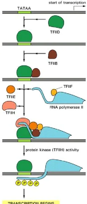
RNAPII itself does not recognize promoters and is instead recruited by a series of accessory proteins called transcription factors during transcription initiation [12]. Transcription factors are proteins that modulate the transcriptional process. One can distinguish between general transcription factors (GTFs), positive transcription factors (activators) and negative transcription factors (repressors). GTFs are required for the assembly and recruitment of RNAPII at the promoter as well as for transcription initiation. Stemming from 30 years of in vitro transcriptional studies, a model for the stepwise recruitment of the RNAPII machinery has been established. The first step is the formation of the pre-initiation complex (PIC), in which a GTF called the TATA box binding protein (TBP) binds to the promoter (Figure 1). TBP specifically recognizes the TATA box, a sequence rich in thymine and adenine upstream of the transcriptional start site (TSS) of the gene. Around 20 % of genes contain a TATA box, but the position of this box with respect to the TSS varies. The consensus sequence of the TATA box in yeast is TATA(A/T)A(A/T)(A/G) [13]. However, TBP also binds to promoters which do not contain a TATA box. TBP is part of a multiprotein complex composed of TBP associated factors (TAFs) collectively termed TFIID. This DNA-protein complex recruits TFIIA, which stabilizes the complex and recruits TFIIB, which is required for the recognition of the TSS. TFIIF, TFIIE and TFIIH are then recruited [12, 14] (Figure 1). TFIIE reorganizes the structure of RNAPII by modifying the jaw-like structure from a closed to an open position. TFIIH has three important functions: (i) its helicase activity unwinds and separates the DNA duplex; (ii) it ensures that the correct DNA strand is transcribed, and (iii) it phosphorylates the CTD of RNAPII on serine 5 of heptapeptide repeats. This forms the complete PIC, which is ready to initiate transcription [12, 14] (Figure 1).
Figure 1 Pre-initiation complex formation at the promoter
(Molecular Biology of the Cell)
Activators and repressors regulate the recruitment of the PIC complex. Most activators are DNA binding proteins that recognize specific sequences within the promoter or within upstream activation sequences (UAS), which are located distantly from the promoter. These activators recruit large co-activator complexes as well as ATP dependent nucleosome remodeling complexes, both of which remodel and render the local DNA accessible. This can be done by two mechanisms, one by weakening the nucleosome-DNA interactions through acetylation of the
histone tails, and second by actively removing the histones from the DNA. As a consequence the TATA box and the transcriptional start site become more accessible to GTFs, allowing assembly of the PIC [15]. Activators can also directly bind to GTFs to promote PIC assembly, although both mechanisms of GTF recruitment are probably interrelated [12, 14].
Transcriptional repressors can inhibit the PIC formation in different ways, by competing with activators for the same sequence, by directly inhibiting GTF recruitment, or by recruiting chromatin modifying enzymes such as histone deacetylases (HDAC) to the promoter region. These HDAC complexes deacetylate histones, resulting in a more compact and less accessible form of chromatin.
In addition, the mediator, a large complex of 20 polypeptides, mediates the interaction of activators with RNAPII. It bridges distant activator sites with the PIC at the promoter and stimulates transcription. More precisely, it interacts directly with the CTD of RNAPII and can therefore mediate signaling from the activator directly to RNAPII. It is composed of several modules, which can vary depending on the cellular conditions and can provide an interface for integration with additional signaling pathways. Thus, the mediator provides an additional target for transcriptional regulation [12, 14]. The balance between activators and repressors allows for the integration of different signaling pathways towards the decision of PIC assembly. Once the PIC has been assembled successfully, transcription initation can take place [12, 14].
1.1.3 Transcription initiation
Once the PIC is assembled, TFIIH, with the help of TFIIE, recruits RNAPII onto the DNA template. This conformation is termed the open complex and once it is formed, RNAPII becomes ready to initiate transcription. Upon phosphorylation of serine 5, RNAPII clears the promoter. During transcription of the first 2-15 nucleotides, the process is often abortive and RNAPII restarts a new round of initiation. However, once the first 15 nucleotides are transcribed, RNAPII escapes the promoter and enters the processive phase of transcription elongation.
In addition, RNAPII dissociates from the GTFs TFIID, TFIIA, TFIIB and the mediator, which remain bound to the promoter. These GTFs then allow for formation of a new PIC, leading to rapid subsequent rounds of transcription [12, 14]. Elongation factors (EFs) then associate with RNAPII to regulate transcription elongation [12, 14].
1.2.4 Transcription elongation
The elongation process is complex and involves multiple factors, which are exchanged during elongation. For simplicity, the role of each elongation factor will be described separately. 1.1.3.1 SAGA
SAGA stands for Spt–Ada–Gcn5 acetyltransferase and is a multiprotein complex which has multiple roles during transcription. First, like TFIID, it can bind to TBP and be recruited to promoters [16-18]. Also, like TFIID it contains histone acetyl transferase activity and is able to recruit RNAPII and initiate transcription. Both complexes contain shared subunits necessary to associate with TBP, the TAFs [17]. TFIID and SAGA together are essential for gene expression of RNAPII but each of them is required for different gene sets. Notably, TFIID expresses house keeping genes and dominates up to 90% of all genes expressed whereas SAGA is required for stress regulated genes and is important for only 10% of the genes [16]. Interestingly, SAGA has additional roles which include regulating transcriptional elongation and linking transcription to mRNA nuclear export. It is thought that SAGA associates through the serine 5 phosphorylated CTD of RNAPII, remains associated during elongation and enhances this step through different mechanisms, notably by acetylating histones and deubiquitinating histone H2B [18]. Furthermore, SAGA is thought to assist in mRNA export through some of its subunits that recruit mRNA export factors and bring the transcription site close to nuclear pores, favoring a rapid export [18].
1.1.3.2 TFIIS
TFIIS is the first elongation factor that was found to interact with elongating RNAPII and is thought to promote elongation. TFIIS co-localizes with elongating RNAPII throughout the gene and can stimulate the intrinsic mRNA cleavage activity of RNAPII by reaching into its active center and altering its structure [19-21]. This activity is important when RNAPII stalls. RNAPII may arrest at each step of nucleotide addition. The time of arrest is variable and depends on nucleotide availability and the sequence of the DNA template. Drugs that diminish the overall nucleotide pool, such as mycophenolic acid (MPA) and 6-Aza uracil (6AU) (both of which inhibit GTP synthesis,) increase RNAPII arrests. If GTP synthesis is inhibited, the nucleotides UTP and GTP are depleted and RNAPII cannot insert the complementary nucleotides and
therefore stops transcribing the genes [22, 23]. In addition, the sequence context influences RNAPII arrest by affecting the stability of the RNA-DNA hybrid. AT-rich sequences are particularly weak and cause RNAPII arrest[22]. Upon arrest, RNAPII may backtrack 2-4 nucleotides. When RNAPII backtracks more then 7-15 nucleotides, it typically stalls irreversibly, unless TFIIS stimulates the restart of RNAPII by cleaving the mRNA [20-22]
1.1.3.3 TFIIF, Elongin, Ell and Csb
TFIIF, Elongin, Ell and CSb are additional factors that also influence the rate of transcription elongation by regulating the pausing of RNAPII during elongation [12]. These factors will be discussed in more detail in section 1.1.7.
1.1.3.4 PTEFb
PTEFb is a cyclin dependent kinase (named Ctk1 in yeast). It is a positive elongation factor that phosphorylates RNAPII during elongation on serine 2 of the CTD. PTEFb phosphorylates serine 2 after RNAPII enters the processive phase of elongation and, serine 2 remains phosphorylated until termination [7-9]. Serine 2 phosphorylation has several consequences which will be described in more detail in section 1.1.6.
1.1.3.5 The Paf1 complex
The Paf1 complex is another positive elongation complex associated with RNAPII that was initially found as being essential for the expression of some genes. It is present in yeast and higher eukaryotes including mammalians. Paf1 physically and genetically interacts with other elongation factors such as FACT and DSIF, and has multiple roles including chromatin modifications during elongation and mRNA processing [24]. It was proposed that Paf1 acts as a platform for the recruitment of other elongation factors to RNAPII [25].
1.1.3.6 DSIF and NELF
In higher eukaryotes, DSIF consists of a heterodimer of Spt4 and Spt5, and is thought to negatively regulate elongation. NELF is a multiprotein complex that interacts with DSIF and is also required for DSIF function. Together, they slow down the elongation rate in vitro, counteracting the positive elongation effect of PTEFb [26]. In addition, they inhibit the mRNA cleavage activity of TFIIS when RNAPII stalls. In yeast, DSIF is named and composed of Spt4 and Spt5 and they positively influence transcription. However, NELF is not found in yeast suggesting that it has evolved later during evolution [26].
1.1.3.7 Elongator and FACT
DNA is wrapped around nucleosomes and this inhibits transcription elongation, since it acts as a physical barrier for RNAPII processivity. To overcome this nucleosome barrier, the evolutionarily conserved complexes Elongator and FACT promote elongation by remodeling the chromatin as RNAPII slides along the DNA. Elongator contains a histone acetyltransferase that co-transcriptionally acetylates histones H3 and H4, which in turn diminishes the histone-DNA interaction and opens the chromatin [27].
FACT is a chromatin remodeling complex which removes the H2A-H2B dimers of the nucleosomes ahead of RNAPII in order to facilitate RNAPII movement. After passage of RNAPII, the nucleosomes are then restored by FACT [28].
1.1.3.8 CTD-phosphatases
During transcriptional initiation at the promoter, the CTD of RNAPII is highly phosphorylated on serine 5 by Kin28 which is part of TFIIH (see section 1.1.2.2), but serine 5 becomes progressively dephosphorylated in the body and end of the gene. In contrast, PTEFb phosphorylates serine 2 progressively after RNAPII leaves the promoter and until it reaches the end of the gene (see section 1.1.4.4). After the round of transcription RNAPII becomes dephosphorylated on serine 2. Several phosphatases mediate these dephosphorylations. The Ssu72 phosphatase is a component of the mRNA processing machinery that dephosphorylates serine 5 once RNAPII is processively elongating [7-9, 26]. More recently, another phosphatase, Rtr1, was shown to be required to dephosphosphorylate the serine 5-phosphorylated form and thus favor the serine 2-phosphorylated form of the CTD.
Finally, to restore the unphosphorylated form of the CTD at the end of the gene, Fcp1 phosphatase dephosphorylates serine 2 during transcription termination. This allows RNAPII to reinitiate a new round of transcription [7-9].
It was long thought that transcription elongation was a simple processive step regulated only by PIC assembly. However, during the last decade, research in this field has clearly shown that multiple factors regulate the transcription elongation process at multiple levels. Most studies have been performed in vitro, and only recently has chromatin immunopreciptiation (ChIP) allowed gene-specific analysis in vivo. The precise mechanisms underlying how these factors interact together in vivo remains to be elucidated [29]. However, it seems that their concerted action regulates at least two crucial events during elongation:
a) mRNA processing
The processing and maturation of nascent mRNA occurs cotranscriptionally. For example, the capping enzyme is recruited to the CTD upon serine 5 phosphorylation and mediates the cotranscriptional capping of the 5’ end of the mRNA chain [30]. 3’ mRNA processing also takes place during transcription and is regulated by proteins which are recruited to the CTD, such as the PAF complex [24]. In addition, the spliceosome, a ribonucleoprotein complex, is recruited during the elongation process to mediate alternative splicing of the mRNA [30]. Finally, it has even been shown that mRNA export from the nucleus and translation are regulated during elongation [31, 32]. Taken together, it seems that all known mechanisms of mRNA processing are linked to active transcription.
b) Cotranscriptional chromatin modification
As discussed above, the positive elongation factors FACT and Elongator facilitate the passage of RNAPII through the chromatin. However, once RNAPII has moved through the gene, the initial chromatin state must be restored; otherwise, the transcriptional machinery can be inappropriately recruited to “open chromatin” within the body of the gene. This is referred to as cryptic initiation [33]. To restore the initial chromatin state and to protect against cryptic initiation, an HDAC complex is recruited to de-acetylate the nucleosomes after passage of RNAPII. To accomplish this, the histone methyltransferase Set2 is recruited by phosphorylated serine 2 on the CTD and methylates histone H3 on lysine 36. This methylation allows the recruitment of the Rpd3 HDAC complex, which then deacetylates histones to restore a closed chromatin state [34, 35].
Chromatin modifications during transcription are also important for the recruitment of mRNA processing factors during transcriptional initiation. At every actively transcribed gene, lysine 4 of histone H3 is heavily methylated at the promoter by the histone methyltransferase Set1, which is recruited by phosphorylated serine 5 on the CTD. This mark is thought to recruit mRNA processing factors to the initiating RNAPII. [34].
1.1.4 Transcription termination and polyadenylation
The end of a gene is marked by a transcription termination site (TTS), which is specifically recognized by the ribosomal machinery [1]. However, at this site, RNAPII continues
transcribing the template strand and since there is no conserved signal for transcription termination, it can occur variably anywhere ranging from a mere few to thousands of nucleotides after the 3’ end of the mature mRNA [3, 36]. As well, shortly after the TTS lies a specific polyadenylation element (AAUAAA). This and a second (GU rich) element, determine where the mRNA is cleaved off from RNAPII and where the poly(A) tail will be added [37]. For this, the cleavage specificity and polyadenylation factor (CSPF) travels along with elongating RNAPII. At the end of the gene a large polyadenylation complex is recruited, that contains the poly(A) polymerase (PAP), the poly(A) binding protein (PABP) as well as additional factors required for efficient cleavage (CstF, CFI and CFII) [37, 38]. This complex mediates the cleavage of the pre-mRNA from the still transcribing RNAPII. PAP, then, extends the poly(A) tail by adding adenines to it. PABP binds to this elongating poly(A) tail. This poly(A) tail and the associated factors have the important function of regulating mRNA stability by inhibiting 3’ exonucleases that chew off the tail in the cytoplasm [37-39].
The RNAPII continues transcribing after the mRNA has been cleaved off and needs to be released from the DNA. For this, two models of termination have been proposed and current evidence suggests that termination might occur through a combination of both. First, the anti-terminator or allosteric model postulates that transcription of the polyadenylation signal induces a structural change in RNAPII and the elongation complex, causing them to dissociate and recruit termination factors. The second model is called the torpedo model and is based on the observation that the 3’ mRNA is rapidly degraded after cleavage of the polyadenylation site from the mRNA. The cleavage recruits a 5’-3’ exonuclease which degrades the 5’ end of the uncapped mRNA associated with RNAPII, and when it rejoins the elongating RNAPII, this exonuclease dissociates the complex from the DNA and terminates transcription [3, 36].
A different termination mechanism occurs during snoRNA and snRNA transcription, since these RNAs are not polyadenylated. This mechanism involves Nrd1, a protein complex that binds to the serine 5-phosphorylated form of the CTD. Nrd1 regulates recruitment of termination factors as well as the exosome, an mRNA processing complex [3].
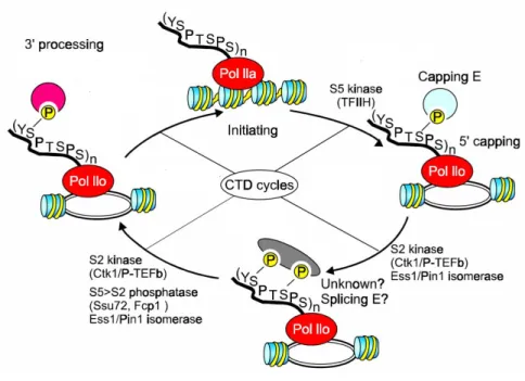
1.1.5 A central role of the RNAPII CTD during transcription
As discussed previously, the CTD is phosphorylated differentially along the transcription cycle. The CTD phosphorylation pattern distinguishes between the different states of transcription. A hypophosphorylated form of RNAPII is recruited to the promoter and is found during PIC assembly (figure 2). Then, during open complex formation, the CTD becomes hyperphosphorylated on serine 5 and RNAPII enters initiation. Once processive elongation is underway, serine 2 becomes phosphorylated and serine 5 becomes dephosphorylated. Finally, when termination occurs, serine 2 is then also dephosphorylated to regenerate the hypophosphorylated form and prepare for a new round of transcription (figure 2) [7-9, 14, 26]. More recently, it was shown that the CTD is also phosphorylated on serine 7. This phosphorylation was found to be enriched within the promoter region as well as towards the 3’ end of the gene, following a pattern similar to that of serine 2 [40, 41]. Recent publications suggest that this phosphorylation is mediated by TFIIH and by components of the mediator complex [42, 43]. A precise role has not yet been characterized; however, enrichment towards the end of the gene suggests a role in 3’ mRNA processing. Additional research will be required to understand the precise function of this phosphorylation.
The CTD has also been shown to be isomerized by peptidyl prolyl isomerases (PPIases). These enzymes catalyze the cis-trans isomerization of proline residues (see section 1.2.5 and figure 4). Since the CTD is rich in prolines, their isomerization might affect the binding of proteins to the CTD and alter the ability to phosphorylate or dephosphorylate the CTD [7-9, 44, 45]. As discussed above, numerous proteins have been shown to bind to the CTD and any of these interactions might be regulated by PPIases. Since the heptapeptide is repeated multiple times, this allows for a high number of different structural possibilities. Therefore it has been postulated that the RNAPII regulates transcription through a ‘CTD-code’ that is dynamic and changes as elongation goes on. This also allows for tight regulation of the transcription elongation process as well as integration of the multiple events, such as mRNA processing and chromatin remodelling, taking place during mRNA production [7-9].
Figure 2 Transcription cycle of RNAPII
Modified from [8]. RNAPII is recruited as an unphosphorylated form during initation, becomes phosphorylated on serine 5 during the early elongation phase, then phosphorylated on serine 2 and dephosphorylated on serine 5 during the late elongation phase. The last step is the transcription termination where RNAPII is dephosphorylated on serine 2.
1.1.6 RNAPII arrest during transcription
As RNAPII transcribes the gene, various obstacles can slow down or arrest it. Several phenomena may cause RNAPII blockage: the intrinsic DNA sequence can cause slippage of RNAPII, which then backtracks and ultimately arrests [22]. Also, if the chromatin structure in front of elongating RNAPII is in a repressive state, RNAPII progression can be slowed or even blocked [28]. Finally a variety of DNA lesions can cause an irreversible block of RNAPII progression, such as cyclo butane pyrimide dimers or 6-4 photoproducts generated by UV light (see also section 1.2.1) [46].
A blocked polymerase on a crucial gene can have deleterious effects, if this gene product has vital cellular roles. Cells have therefore developed multiple mechanisms to assist RNAPII in overcoming these blocks. To counter those blockages and continue transcription, RNAPII recruits different factors, depending on the specific situation [47]. As mentioned earlier, the elongation factor TFIIS is very important for restarting backtracked and stalled RNAPII by
stimulating its intrinsic mRNA cleavage activity. Once this mRNA end is clipped off, RNAPII can restart elongation where it stopped. The importance of TFIIS is emphasized when cells are challenged with the drug 6-azauracil (6AU). This uracil analog inhibits GTP and UTP synthesis, decreases their cellular concentration and causes RNAPII to slow down during elongation [48, 49]. TFIIS mutants are highly sensitive to 6AU because TFIIS is crucial for RNAPII to restart after stalling. In fact, 6AU was used to discover a variety of elongation factors that are required for RNAPII progression, as mutants of these factors are hypersensitive to this drug [22, 23]. Besides TFIIS, there are other elongation factors such as Elongin, TFIIF and ELL that stimulate the restart of paused RNAPII as shown by in vitro transcriptional systems [12, 47].
1.1.6.1 Transcriptional blocks caused by DNA lesions
DNA integrity is constantly challenged by lesions caused by endogenous and exogenous factors, including reactive oxygen species (ROS) generated during normal cellular metabolism, UV light from the sun, radiation or genotoxic agents. Besides causing gene mutations, deletions and single strand or double strand breaks that are a major threat to cellular survival, certain types of lesions also block the progression of RNAPII during transcription. Throughout evolution, cells have developed mechanisms to protect themselves against these deleterious lesions. These include a variety of DNA repair pathways as well as induction of apoptosis when the damage is not reparable [46, 50]. As mentioned earlier, several lesions can cause a RNAPII blockage, for example, UV can create cyclobutane pyrimidine dimers (CPD) as well as 6-4 photoproducts (6-4PP) in DNA, which block RNAPII progression [51]. Also, bulky adducts in DNA can cause RNAPII arrest as they block the active center of RNAPII. Even byproducts of ROS: for example, malondialdehyde generated through lipid peroxidation can form guanine adducts that block elongation [46, 50]. In addition, abasic sites are known to block transcription. Abasic sites are produced during a step of the base excision repair pathway [52]. Some lesions that cause RNAPII arrest may simply be bypassed by RNAPII, although this can lead to transcriptional errors [46, 50]. To protect against lesions that cause the stalling of RNAPII during transcription, a specific DNA repair pathway is activated. This pathway is called transcription coupled repair (TCR), and is a sub-pathway of nucleotide excision repair (NER). TCR is one way to recruit the NER, which alternatively can be recruited by global genomic repair (GGR). In fact GGR and TCR are two separate mechanisms of lesion recognition which then use the NER pathway to repair the lesion [46, 50]. TCR is initiated when CSB (Rad26 in yeast) recognizes and binds to
stalled RNAPII at the lesion. CSB is loosely associated with elongating RNAPII, and upon stalling, this association becomes tighter and a second TCR factor, CSA, is recruited. Then, the NER factors that excise the lesion, XPF, XPG and XPA, are recruited. More precisely, these factors incise the lesion at the 5’end, then the intact DNA strand is replicated by a DNA polymerase while the 3’ end of the DNA strand containing the lesion is cleaved off. Finally, the newly synthesized strand is ligated and the intact double stranded DNA is repaired [46, 50]. 1.1.6.2 RNAPII ubiquitylation during transcriptional arrest
If RNAPII restart fails, an ultimate mechanism is activated that consists of ubiquitylation and subsequent proteasomal degradation of RNAPII. Ubiquitin (Ub) is a highly conserved polypeptide of 76 amino acids that is covalently linked to lysine residues of the target protein. The process of ubiquitylation is highly regulated via three factors, the Ub-activating enzyme that associates to Ub (E1), the Ub-conjugating enzyme E2 that recieves Ub from E1 and adds the Ub to the substrate, and finally, the Ub ligase E3, which provides substrate specificity and ligates Ub to the substrate [53]. The substrate often becomes polyubiquitylated, meaning that additional Ubs are linked to the first, eventually forming an Ub chain. This Ub chain is then recognized by the proteasome, a large complex responsible for protein degradation. RNAPII contains two lysine residues in its major subunit Rpb1 that can be ubiquitylated: K330 and K695 [54]. The ubiquitylation process originally observed in response to DNA damage seems to be a general mechanism in response to RNAPII stalling [53]. For example, mutant cells lacking the TFIIS gene accumulate high levels of ubiquitylated RNAPII when treated with 6AU [53]. One key factor required for efficient RNAPII ubiquitylation is Def1 [55]. The double deletion of DEF1 and TFIIS is lethal, suggesting that both mechanisms-- RNAPII-Ubiquitylation and TFIIS-mediated mRNA cleavage-- are essential for clearing RNAPII arrest. This is further confirmed by the fact that the K330R mutation of RPB1 is also synthetic lethal with TFIIS, and indeed strains containing the single deletion of DEF1 or the K330R mutation of RPB1 are hypersensitive to 6AU [47].
During TCR, RNAPII is also ubiquitylated by Def1. Interestingly, CSB inhibits ubiquitylation when engaging the NER pathway. RNAPII becomes ubiquitylated and degraded only if this repair fails [55]. Taken together, data suggests that RNAPII ubiquitylation and degradation is a “last chance” mechanism that is only used when other RNAPII release mechanisms fail [46, 47].
1.1.7 Transcriptional regulation of RNAPII
Cells constantly need to sense and adapt to environmental changes so that they can modify their transcriptional program accordingly. Multiple signaling pathways sense the external conditions and signal the nucleus to induce transcription of the appropriate genes for each condition. To achieve this, specific transcription factors (TFs) are required to regulate gene expression. Genes belonging to a similar pathway are generally regulated by the same TF, and these groups of genes are called regulons. For example, the galactose regulon consists of multiple genes regulated by the Gal4 TF in response to galactose. This regulon is then expressed and allows the cell to produce proteins required for galactose metabolism [56].
1.1.7.1 The target of rapamycin (TOR) signaling pathway
Cells must constantly adapt to the availability of nutrients in the environment. Nutrient levels are critical for the decision to grow and multiply or to limit consumption and metabolism. A signaling pathway conserved from yeast to human senses nutrients availability via the TOR protein kinases. When nutrients are readily available, TOR becomes active and stimulates transcription of genes involved in anabolic processes, including translation, ribosome biogenesis and gene transcription. Additionally, catabolic processes such as protein degradation and autophagy are inhibited. When nutrients are limited, TOR becomes inactive and catabolic processes, stress response genes, and G1 cell cycle arrest is activated whereas anabolic processes are repressed [57-59]. The TOR kinases are PI3K like kinases and two isoforms (Tor1 and Tor2) have been identified in yeast. Tor1 was originally discovered as being inactivated by the immunosuppressant rapamycin, a macrocyclic lactone that was isolated from the bacteria
Streptomyces hygrocopicus on the Rapa Nui islands. Tor1 is associated with several cofactors
including Kog1, Tco89, and Lst8, and together they form the rapamycin-sensitive TORC1 complex. Tor2 is found in another complex, where it is associated with additional factors Avo1, Avo2, Avo3, Bit61 and Bit2. Together they form the rapamycin-insensitive TORC2 complex [57, 59]. It is thought that the TORC1 complex regulates growth temporally (i.e. it makes the decision of whether the cell should grow and divide), whereas TORC2 regulates growth in a spatial context, meaning that it decides in which direction the cell will grow [57, 59].
In yeast, TOR signaling is controlled by amino acid levels, such as the nitrogen rich amino acid glutamine. In multicellular organisms, additional upstream signals include the insulin signaling
pathway and the platelet derived growth factor [58]. There are several downstream targets of Tor1, which will be discussed in detail below. In yeast, Tor1 signaling, like most signaling pathways, results in translocation of TFs into the nucleus to alter gene expression.
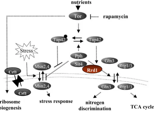
1.1.7.1.1 TORC1 regulates transcription of specific regulons
The first pathway, downstream of TORC1 signaling that was characterized includes the PP2A-like phosphatase Sit4 and its regulator Tap42. Under high nutrient conditions, TORC1 maintains Tap42 in a phosphorylated state. Upon amino acid depletion or rapamycin treatment, TORC1 becomes inactivated and Tap42 is dephosphorylated by PP2A, activating several classes of genes including stress-regulated (STRE) genes, nitrogen catabolite repressed (NCR) genes and retrograde signaling (RTG) genes [59, 60] (see figure 3). For example, Tap42 dissociates from the Sit4 phosphatase, which in turn becomes activated and dephosphorylates the cytoplasmic TF Gln3. Gln3 is retained in the cytoplasm in a phosphorylated state when TOR is active. When Gln3 becomes de-phosphorylated, it moves into the nucleus and activates NCR genes, which generate nitrogen from proline or urea [61] (see figure 3).
Similarly, the TFs Rtg1 and Rtg3 are retained in the cytoplasm in a phosphorylated form and upon activation of the Sit4 phosphatase; they are dephosphorylated and move into the nucleus to activate the RTG genes (see figure 3) [59, 60]. RTG genes are important for the TCA (tricarboxylic acid) cycle, which in turn is crucial for respiration and to mediate the conversion of nutrients into energy in the mitochondria. In addition, it provides molecules that are important for biosynthetic pathways. For example, α-ketoglutarate is a precursor of glutamate and glutamine, which in turn are used for nucleotide biosynthesis as well as for nitrogen containing molecules including NAD+ [62].
Finally, Tap42 is also implicated in the transcription of the STRE genes by regulating the translocation of the TFs Msn2 and Msn4. When Tap42 is active, it inhibits Sit4 and the Msn2/4 TFs are exported from the nucleus. When Tap42 is inactivated, the Sit4 phosphatase can dephosphorylate Msn2/4, causing them to stay in the nucleus [60] (see figure 3).
Msn2/4 binds to specific DNA sequences called stress response elements (STRE). STRE-containing genes are divided of several sub-classes including carbohydrate metabolism, genes required to scavenge reactive oxygen species, protein chaperones such as heat shock proteins and DNA repair proteins. Additionally in response to stress, genes are induced that regulate the transcriptional stress response with negative or positive feedback loops. For example, upon
induction of stress, transcripton of the Msn4 gene is activated, thus contributing to a faster and stronger stress response [63].
Tor1 is implicated through its regulation of Tap42 in the nuclear retention of Msn2/4, which keeps STRE gene transcription active. However, Tor1 does not regulate the translocation of Msn2/4 from the cytoplasm to the nucleus, as has been shown for other transcription factors. Therefore, additional upstream signaling pathways are required to first mediate the translocation of Msn2/4 from the cytoplasm to the nucleus in order to activate the STRE response induction [60]. Next, Tor1 may keep this STRE response active by retaining Msn2/4 in the nucleus.
Figure 3 regulation of transcription by Tor1 signaling
Transcription factors are regulated by translocation from the cytoplasm to the nucleus in yeast.
1.1.7.1.2 TORC1 regulates ribosome biogenesis
Ribosome biogenesis is crucial for cellular anabolism and growth, which is in turn a prerequisite for cell cycle progression. At least 100 protein-coding genes are required as well as the combined action of the three RNA polymerases. This requires tight regulation, as the process
Modified from Duevel et al. 2003
Crf1
Rrd1
Crf1
ribosome biogenesis
stress response nitrogen
discrimination TCA cycle nutrients
expends a large amount of energy [57, 59]. Upon TORC1 inactivation, via rapamycin treatment or in response to nutrient limitation, Tor1 moves into the nucleus and inhibits RNAPI and RNAPIII by binding to the promoter of the rDNA genes [64]. To regulate RNAPII ribosomal gene expression, Tor1 controls the nuclear translocation of Ifh1, a transcriptional activator, and Crf1, a transcriptional repressor [65-67]. When Tor1 is active, Ifh1 is associated to Fhl1, a ribosomal gene TF, and together they are bound to ribosomal gene promoters and stimulate their transcription. When Tor1 becomes inactive, Crf1 is dephosphorylated and enters the nucleus to compete with Ifh1 for binding to Fhl1 (see figure 3). Once Crf1 binds to Fhl1, it represses transcription of the ribosomal genes [67]. This mechanism seems to be specific to some strain backgrounds since it has been demonstrated that in a crf1Δ mutant from a W303 background, ribosomal genes are still repressed upon rapamycin treatment, suggesting that alternative mechanisms of ribosomal repression via the Tor signaling pathway must exist [68].
1.1.7.1.3 Differences between mammalian and yeast cells
In mammalian cells, anabolism is regulated through translation initiation rather than transcription. The S6K kinase and 4EBP-1 are regulated by TORC1 and are responsible for the regulation of translation initiation and mRNA production [58]. In yeast, there is no homologue of 4EBP-1 and the majority of Tor signaling is regulated through transcription [60]. Only recently has Sch9, the homologue of the S6K kinase, been identified [69]. Sch9 is required for optimal ribosomal gene expression and translation of mRNA [70].
1.1.7.2 The environmental stress response (ESR)
Yeast cells must constantly adapt to various extracellular conditions, which can cause cellular stress. Such conditions include heatshock, pH variations, changes in osmolarity, an increase in reactive oxygen species as well as toxins. To adapt to these conditions, yeast have developed a rapid response termed the ESR [63]. Within this ESR, various sensors signal changes to an intracellular signaling pathway, resulting in changes to the transcriptional program. Around 300 genes are upregulated in response to stress and 600 are repressed [71]. There is a correlation between the ESR and the severity of the stress, indicating that the ESR is tightly regulated [63, 71]. Downregulated genes include those associated with cellular anabolism and cell cycle progression as well as genes required for ribosome biogenesis. In contrast, upregulated genes include the STRE genes induced by the Msn2/4 transcription factors. Interestingly, not every condition induces exactly the same response, and not every branch of the signaling
pathway is required for every condition [63, 71]. For example, Sko1 is only activated during osmotic changes via the Hog1 signaling branch, and induces genes required for osmotic regulation. Alternatively, upon oxidative stress, the Yap1 TF is activated to regulate oxidative stress genes [63, 71]. For DNA damage response, the Mec1 kinase is activated which in turn activates Dun1 to trigger the ESR [63]. Together this suggests a network of overlapping signaling pathways that allow for a precise response to each condition [63, 71].
1.2 Rrd1 and its biological role
1.2.1 Introduction
Our laboratory is interested in the cellular responses to exogenous stresses caused by drugs used in clinical therapies. These drugs include various DNA damaging agents as well as agents causing oxidative stress or the starvation mimicking drug rapamycin. It is crucial to understand how cells respond to drug treatment in order to understand drug resistance mechanisms of cancer cells. In addition, a better understanding of cellular responses to drug treatment might provide insights into new drug targets and prevent inappropriate treatments. To study these cellular responses, yeast is an ideal model as genes can be easily deleted, allowing the analysis of their functions. Through yeast genetics, major cell activities such as transcription, cell cycle regulation, replication and DNA repair have been studied [72]. Throughout evolution, these pathways have been conserved from yeast to man, which validates the use of yeast as a model system in current molecular biology research. The yeast strain library harbouring each viable gene deletion (approx. 4800 genes of a total of 6125 genes) which is now available for the research community, allows for genome wide identification of genes important for cellular responses to stresses [73-76]. In order to study these responses, several ‘model’ drugs are commonly used and will be briefly described in this section:
4-nitroquinoline 1-oxide (4NQO)
This carcinogen is not used as a chemotherapeutic agent in clinics but widely in research. It is thought to cause cellular damage through two different modes of action. After entering the cell, 4NQO is activated via chemical modifications and becomes 4-hydroxyaminoquinoline, which reacts with purines and forms stable bulky adducts [77]. These bulky adducts resemble lesions caused by 254 nm UV radiation, namely cyclobutane pyrimidine dimers and 6-4 photoproducts [51, 74, 77]. Similarly to UV lesions, 4NQO-dependent bulky adducts are also recognized and repaired via the NER pathway.
The second mode of action by which 4NQO causes cellular damage is thought to be through the generation of reactive oxygen species (ROS), thereby causing intracellular oxidative stress [74,
77]. ROS cause cellular damage by altering DNA as well as through lipid peroxidation and protein oxidation [78].
Ultraviolet radiations
UV radiations have wavelengths ranging from 400 nm to 100 nm. They are divided into UVA (400-315 nm), UVB (315-280 nm) and UVC (280-100 nm); their toxicity differing depending on the wavelength. UVC radiation is mostly used at a wavelength of 254 nm for research purposes as a standard and it mainly induces cyclobutane pyrimidine dimers as well as 6-4 photoproducts, whereas UVA generates reactive oxygen species. UVB causes both types of stress, ROS becoming more prominent as the wavelength increases [51].
Gamma rays
Gamma irradiation is used in clinical oncology to treat several types of cancer. It is also used in molecular biology as it is known to induce double-strand breaks (DSB) as well as oxidative stress [79]. DSB are of high interest in research as they are very toxic to the cells and their repair is highly regulated and complex [79, 80].
Hydrogen peroxide
Hydrogen peroxide (H2O2) reacts with metal ions and generates ROS in vivo, such as superoxide
anions, which are known to react with lipids, proteins and DNA [81]. It is widely used in research to induce oxidative stress in cells.
1.2.2 Discovery of the RRD1 gene in yeast
RRD1 (rapamycin resistant deletion 1) was originally identified in a genome-wide screen
for mutants hypersensitive to 4NQO but not to UVC, suggesting that this gene product might be important for the response to increased ROS but not to lesions repaired by the NER pathway. This hypothesis was further confirmed since rrd1Δ mutants showed sensitivity towards UVA and diamide, which both cause oxidative stress but showed no increased sensitivity to γ-radiations, MMS, UVB or UVC [74]. Taken together, these phenotypes suggest that the function of Rrd1 would be in the cellular response to oxidative stress but not to other stresses such as those caused by DNA lesions [74]. Subsequently, rrd1Δ mutants were shown to be highly resistant to rapamycin as well as to caffeine but sensitive to vanadate, Ca2+
, ketokonazole and cycloheximide. Both caffeine and vanadate are also thought to influence cellular oxidative stress,
![Figure 4 Model of proline cis- trans isomerisation by PPIases [96]](https://thumb-eu.123doks.com/thumbv2/123doknet/2167265.9944/39.918.171.731.553.926/figure-model-proline-cis-trans-isomerisation-ppiases.webp)