Workshop CSTC – KULeuven 11 Décembre 2002 La microscopie appliquée aux matériaux de construction
Prof. Eric PIRARDUniversité de Liège GeomaC – MICA – Sart Tilman B52 / 3 – 4000 LIEGE eric.pirard@ulg.ac.be http://www.ulg.ac.be/geomac
Q
Qu
ua
an
nt
ti
it
ta
at
ti
iv
ve
e
M
Mi
ic
cr
ro
o-
-I
Im
ma
ag
g
in
i
ng
g
o
of
f
B
B
ui
u
il
ld
di
in
ng
g
M
Ma
a
te
t
er
ri
ia
al
ls
s
:
:
f
fr
ro
o
m
m
r
re
ea
al
li
it
ty
y
t
to
o
d
dr
re
ea
am
m.
.
Eric PIRARD Introduction
Digital imaging techniques are now commonplace in many labs. It is for most material scientists particularly evident to rely on imaging and image measurement capabilities when dealing with microscopical observations. Obviously, image analysis gives access to properties such as shape or texture that where simply inaccessible to quantification up to now. However, much improvement is still required to move from sophisticated user-driven measures to fully automatic and statistically representative results on materials. Too often, relying on the exceptional capabilities of the human brain, scientist are expecting unrealistic achievements from quantitative digital image analysis.
Quantitative Micro-Imaging
Quantitative imaging is not a recent field of investigation. A quick review of the very first attempts to quantify visual information under the microscope will reveal that almost everything had been said and conceptually formalised before 1930. Typical examples include the volumic phase ratio estimation from sections (first principle of stereology, Delesse 1848), the 3-D size distribution from sections (second principle of stereology, Wicksell 1922) and shape descriptors for particle abrasion (Wadell, 1933). At that time, investigators were lacking the intensive computing tools available nowadays but they relied on their visual appreciation to recognise complex patterns and special mineral phases. On the other hand, nowadays, image analysis still faces the problem of reaching full automation capabilities in terms of mineral identification, image segmentation and adequate quantification of parameters such as : phase ratios, size distributions, shape indices, orientation and dispersion patterns.
Mineralogical imaging
Image analysis of mineral materials should start from a reliable imaging mode able to contrast the individual mineral components of a material. Among the possible candidates for imaging Back-Scattered Electron (BSE) Imaging; Energy Dispersive X-Ray (EDX) Mapping and Microspectrophotometric (MSP) Imaging have their own advantages in terms of speed, accuracy, flexibility and discrimination between species. MSP is available from an optical microscope by taking care to correctly calibrate the spectral response of a video camera. It has proven to be particularly useful in opaque ore minerals identification with reflectances above 10 %.
Workshop CSTC – KULeuven 11 Décembre 2002 La microscopie appliquée aux matériaux de construction
Prof. Eric PIRARDUniversité de Liège GeomaC – MICA – Sart Tilman B52 / 3 – 4000 LIEGE eric.pirard@ulg.ac.be http://www.ulg.ac.be/geomac
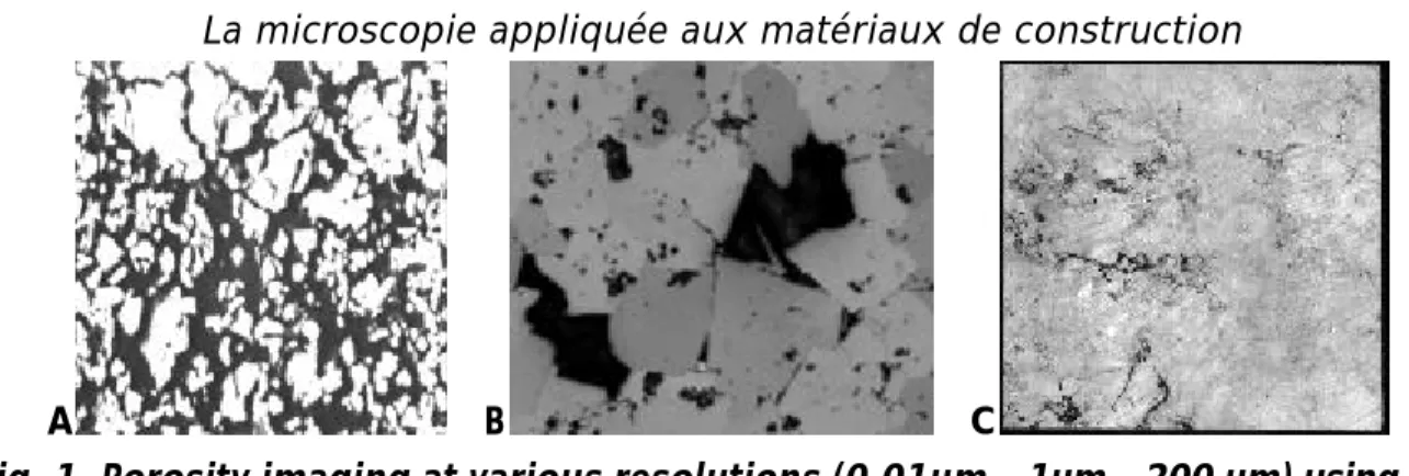
A B C
Fig. 1. Porosity imaging at various resolutions (0,01µm – 1µm – 200 µm) using BSE imaging of a resin impregnated chalk (A), reflected light microscopy of dolomite (B),
45° reflection imaging macroscopy of a marble tile (C)
A major but very peculiar component of many building materials is porosity. Porosity imaging remains problematic because in most imaging modes (BSE, EDX) it is not adequately revealed unless specific resins are injected into the material. Reflected light imaging both in microscopy and in macroscopy is probably the most straightforward method, provided polishing is perfectly achieved and scratches are kept to a minimum.
Size distribution estimation
Probably one of the most convincing application of image analysis is size distribution analysis of granular materials such as sands, abrasives and other industrial minerals. Several ISO norms are in progress in order to make recommendations on the correct use of such technologies both in a static and dynamic way. Validation tests based on a European Bureau of Certified Reference Sand (BCR 68) has given very accurate results on a 6g sample whereas traditional sieving relies on a minimum of 100g. Moreover, the image analysis device (http://www.occhio.be) is fully automatic, stores and displays results in real-time and requires no previous calibration. The quality of the correlation with sieve analysis has been achieved thanks to collimated light
imaging of particles resting in a controlled position combined with the accurate measurement of a sieving diameter. Sand BCR68 µm Sieve Alpaga (100g) (6g) 160 4.2 3.89 250 22.9 20.68 320 44.9 39.80 400 68.9 67.95
Workshop CSTC – KULeuven 11 Décembre 2002 La microscopie appliquée aux matériaux de construction
Prof. Eric PIRARDUniversité de Liège GeomaC – MICA – Sart Tilman B52 / 3 – 4000 LIEGE eric.pirard@ulg.ac.be http://www.ulg.ac.be/geomac Fig. 2. Sample image from BCR 68. Analysis in terms of relative weight fractions and plot against five reference sieving labs.
Other unrivalled applications of micro-imaging for size distribution analysis include contextual size distributions were particles are retained for analysis in function of being included or not within a given phase. Even more exceptional is the unique capacity of opening functions as defined in mathematical morphology to provide a rigorous granulometric concept for describing a continuous network such as the porosity of the chalk sample in fig. 1.
Shape analysis
Shape description is responsible for an incredible amount of literature among which most concepts are of no particular interest to building materials. Adequate descriptors for sands and minerals were already suggested by early studies on the subject (Wadell, 1933; Krumbein, 1941). An adequate formalisation of these concepts by use of mathematical morphology operators proves to reveal interesting insights into the understanding of the physical behaviour of sands.
40.00% 60.00% 80.00% 100.00%
4.00% 6.00% 8.00%
Volumic Mass increment
2
21 18
22 2 0
Fig. 3. Roundness analysis using Wadell (Krumbein) concepts explains increase in compactness after tapping when moving from very rounded (2) to more angular
materials (20) with identical size distribution curves.
Orientation and dispersion
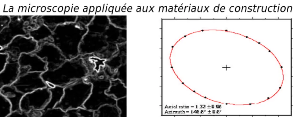
Orientation and relative position of individual grains to form the material texture are probably the most challenging concepts that are not yet fully addressed by quantitative imaging. Here again specific imaging devices using for example multiple polarisation positions (multiradial imaging) or electron backscattering pattern analysis (EBSP) could bring significant improvements in the future, in particular in terms of 3D analysis. A practical application already pioneered by geologists is the microstructural fabric analysis of sandstones whose grain delineation is favoured by the use of gradient images under different rotations of the polariser / analyser filters. Although the boundary extraction is still imperfect, this does not ruin the accuracy of the analysis as performed using numbers of intercepts in different directions.
Workshop CSTC – KULeuven 11 Décembre 2002 La microscopie appliquée aux matériaux de construction
Prof. Eric PIRARDUniversité de Liège GeomaC – MICA – Sart Tilman B52 / 3 – 4000 LIEGE eric.pirard@ulg.ac.be http://www.ulg.ac.be/geomac
Fig. 4. Example of a maximum of gradient images obtained from a series of analyser / polariser rotation positions in transmitted light. After computation and elliptical modelling of the rose of number of intercepts both azimuth and axial ratio information can be estimated.
Conclusions
Image analysis brings a unique contribution to microscopy and often appears as the only method capable of addressing subtle problems of spatial information quantification. In order for this method to become more mature, much work is still required in terms of image acquisition and image analysis particularly. It is of paramount importance to realise that only rigorous and calibrated imaging of materials will open the way to fully automatic and stand alone procedures.
On the other hand, material scientist must realise that they have to develop and standardise their own tools for image quantification. Visually similar objects may have completely different meanings from one application to the other and as such require the development of specific parameters driven by the application itself. Finally much more attention should be given to sampling and error analysis which remain major challenges in microscopy.