This is an author-deposited version published in:
http://oatao.univ-toulouse.fr/
Eprints ID: 5676
To link to this article: DOI:10.1016/J.ELECTACTA.2010.03.085
URL:
http://dx.doi.org/10.1016/J.ELECTACTA.2010.03.085
To cite this version: Cournet, Amandine and Bergé, Mathieu and Roques,
Christine and Bergel, Alain and Délia, Marie-Line (2010) Electrochemical
reduction of oxygen catalyzed by Pseudomonas aeruginosa.
Electrochimica Acta pp. 4902-4908. ISSN 0013-4686
O
pen
A
rchive
T
oulouse
A
rchive
O
uverte (
OATAO
)
OATAO is an open access repository that collects the work of Toulouse researchers and
makes it freely available over the web where possible.
Any correspondence concerning this service should be sent to the repository
administrator:
[email protected]
Electrochemical reduction of oxygen catalyzed by Pseudomonas aeruginosa
Amandine Cournet
a,b, Mathieu Bergé
a, Christine Roques
a, Alain Bergel
b, Marie-Line Délia
b,∗aUniversité de Toulouse; UPS; LU49, Adhésion bactérienne et formation de biofilms; 35 chemin des Maraîchers, 31062 Toulouse Cedex 09, France bLaboratoire de Génie Chimique CNRS UMR5503; 4 allée Emile Monso, BP 84234, 31432 Toulouse Cedex 04, France
a b s t r a c t
Pseudomonas aeruginosa has already been shown to catalyze oxidation processes in the anode compart-ment of a microbial fuel cell. The present study focuses on the reverse capacity of the bacterium, i.e. reduction catalysis. Here we show that P. aeruginosa is able to catalyze the electrochemical reduction of oxygen. The use of cyclic voltammetry showed that, for a given range of potential values, the current generated in the presence of bacteria could reach up to four times the current obtained without bacteria. The adhesion of bacteria to the working electrode was necessary for the catalysis to be observed but was not sufficient. The electron transfer between the working electrode and the bacteria did not involve mediator metabolites like phenazines. The transfer was by direct contact. The catalysis required a certain contact duration between electrodes and live bacteria but after this delay, the metabolic activity of cells was no longer necessary. Membrane-bound proteins, like catalase, may be involved. Various strains of P. aeruginosa, including clinical isolates, were tested and all of them, even catalase-defective mutants, presented the same catalytic property. P. aeruginosa offers a new model for the analysis of reduction catalysis and the protocol designed here may provide a basis for developing an interesting tool in the field of bacterial adhesion.
1. Introduction
Electroactive bacteria are able to transfer electrons to an electrode and thus produce current[1]. They have recently gen-erated considerable interest concerning bioelectricity production in Microbial Fuel Cells (MFCs), in which they offer the possibil-ity of converting chemical energy from a wide range of organic wastes into electrical energy[2]. Various bacteria have already been shown to be electroactive[3]and a lot of work is being done to try to decipher the cellular mechanisms by which such an electron transfer is possible. Many studies are devoted to oxidation pro-cesses that occur in the anode compartment of MFCs in an attempt to understand how bacteria transfer electrons to electrodes. Three different processes of transfer have been described. Some bacteria produce electrochemically active mediator metabolites that serve as electron shuttles between cells and electrodes[4]. Other bacteria have been shown to transfer electrons directly to electrodes with-out the implication of metabolites but through membrane-bound redox compounds such as C-cytochromes[5]. Finally, in some thick biofilms, involvement of conductive nanowires produced by the cells has been demonstrated with Shewanella and Geobacter species
[6,7]. These various studies have been mostly performed for anodic processes.
∗ Corresponding author. Tel.: +33 534323615; fax: +33 534323700. E-mail address:[email protected](M.-L. Délia).
Microbially catalyzed reductions also exist[8–10]but have not been so largely studied. The catalysis of oxygen reduction by micro-bial biofilms was first identified in the field of micromicro-bial corrosion
[11]. Biofilms formed in seawater have been shown to be efficient in catalyzing the reduction of oxygen on stainless steels and other materials but, for now, the mechanisms remain a subject of debate. Several hypotheses have been proposed, including direct cataly-sis by adsorbed enzymes like catalase[12,13]or indirect catalysis via the production of hydrogen peroxide[14]or the production of manganese oxides/hydroxides by manganese oxidizing bacte-ria[15]. In recent years, attempts have been made to exploit the electrocatalytic properties of aerobic seawater biofilms to design biofilm-cathodes in MFCs[10,16,17]. Marine electroactive biofilms are composed of natural microbial consortia, in which microbial diversity is very large. It is suspected that mechanisms similar to those identified in corrosion studies may be involved but these mechanisms alone cannot explain the high current density val-ues that are observed with natural seawater biofilms operating at controlled potential.
Several studies have been performed on the electroactivity of Pseudomonas species but most of them have focused on the ability of these microorganisms to give electrons to electrodes using their own electron shuttles. In 1996, pyocyanin, a blue pigment produced by several strains of P. aeruginosa, was studied by adsorptive stripping voltammetry [18]. This work revealed that pyocyanin had a redox activity of around −0.20 V vs Ag/AgCl. Some work on Pseudomonas chlororaphis assessed the necessity
of phenazine-1-carboxamide (PCN) production for this strain to keep its ability to reduce Fe(III)[19]. More recently, P. aeruginosa was isolated from the anodic compartment of an MFC[20]. This bacterium was shown to produce soluble components called phenazines that served as electron shuttles between the bacteria and the electrode [4]. P. aeruginosa has already been used to improve electricity generation in the anodes of MFCs[21]but, to our knowledge, this bacterium has never been shown to catalyze a cathodic reaction. However, previous data have been published showing the increment of cathodic currents by an unidentified specie of Pseudomonas, correlated to its catalase activity[22].
P. aeruginosa is an aerobic bacterium, so its usual electron accep-tor is oxygen. For this reason, we focused on the ability of P. aeruginosa to catalyze the electrochemical reduction of oxygen. This catalysis cannot be used in cathodic compartments of MFCs since the potential of the oxygen reduction catalyzed by bacteria is still too low for such an application. However, a specific exper-imental protocol was designed to study the reaction using cyclic voltammetry (CV). This method has already been shown to be effi-cient for rapid detection of the electroactivity of bacterial cells
[20,23]. The objectives of the present study were (i) to see whether P. aeruginosa was able to catalyze the electrochemical reduction of oxygen (ii) to progress in understanding the mechanisms of the electron transfer, and (iii) to discuss the potential applications of such a property.
2. Experimental
2.1. Bacterial strains, culture conditions and chemicals
Experiments were performed on the reference strain PA01 of P. aeruginosa. Clinical isolates from patients with cystic fibrosis obtained from the Hospital Bacteriology and Hygiene Laboratory in Toulouse (France) were also tested (Table 1). Four P. aerugi-nosa PA14 catalase mutants were provided by Sogang University, Korea: PA14 KatA−, PA14 KatB−, PA14 KatE− and PA14 KatABE−
[24]. Catalase activity was assessed with the ID color Catalase test (Biomérieux, France). All the strains were stored at −80◦C in Eugon medium (10% glycerol). They were plated on Trypcase Soy Agar (Biomérieux, France) under aerobic conditions. Before each experiment, the strains were grown overnight in 20 mL Trypcase Soy Broth (Biomerieux, France) at 37◦C under gentle stir-ring (200 rpm). The bacterial suspensions were then centrifuged (10 min, room temperature, 3600 × g), washed twice in phosphate buffer (K2HPO4/KH2PO4, 0.1 M, pH 7), and resuspended in the same buffer. All experiments were performed in the same buffer at room temperature.
Table 1
Phenotypic characterization of various electroactive strains of P. aeruginosa.
Name Origin Phenotype
PA01 Pasteur institute (France) Green pigmentation
7844M Clinical isolates Mucoid
7844S Clinical isolates Non-mucoid revertant
7645M Clinical isolates Mucoid
A01 Clinical isolates No pigmentation
A02 Clinical isolates Brown pigmentation
A03 Clinical isolates No pigmentation
A04 Clinical isolates No pigmentation
A05 Clinical isolates Blue pigmentation
A06 Clinical isolates Brown pigmentation
PA14 Sogang University (Korea) Catalase +
PA14 Kat A− Sogang University (Korea) Catalase −/+ PA14 Kat B− Sogang University (Korea) Catalase + PA14 Kat E− Sogang University (Korea) Catalase + PA14 Kat ABE− Sogang University (Korea) Catalase −
2.2. Cyclic voltammetry method
Cyclic voltammetry consists in performing a potential scan on a solution while recording the current obtained. Currents due to oxi-dation processes are conventionally noted as positive and those due to reduction as negative. Experiments were monitored by a Prince-ton Applied Research potentiostat and the Power Suite software. Working electrodes were 0.07 cm2glassy carbon rods inserted in an inert resin cylinder of approximately 1 cm diameter. Prior to use, the rods were polished with abrasive silicon carbide paper and cleaned in distilled water. The counter-electrode was a platinum wire and a saturated calomel electrode (Radiometer Analytical) was used as a reference (SCE).
2.3. Protocol conditions
Experiments were conducted in a 40 mL cell. A first voltammo-gram was run with 20 mL buffer to check the quality of the working electrode surface. A second was performed immediately after the addition of bacterial cells (resuspended culture in buffer) at the final concentration of 108bacteria/mL. Then the suspension was stirred for 15 min, 30 min or 1 h to maintain a homogeneous oxygen con-centration and the CV scan was performed again. Measurements under anaerobic conditions were carried out after removing oxy-gen from the solution by 20 min of nitrooxy-gen bubbling. There was no stirring or gas bubbling during current recording.
Similar CV experiments were done on the filtrate of P. aeruginosa PA01. After 1 h of stirring, 20 mL of bacterial suspension was filtered through a 0.45 mm sterile filter then through a 0.20 mm one. The resulting filtrate was tested by CV as described above.
CVs were also performed on dead cells of PA01. After an overnight culture, bacteria were washed as described above and resuspended in pure methanol (Aldrich, France) for 30 min. The dead cells were then washed again in phosphate buffer and tested in CV in the same way as live cells. In another experiment, cells were killed after the 1-h contact time between electrodes and bacteria directly on the surface of the working electrode: the glassy carbon rod was dipped into pure methanol (Aldrich, France) for 30 min. CV was also performed on this electrode with adhered dead cells.
Each experiment was independently performed at least three times.
2.4. Scanning electron microscopy (SEM)
Scanning electron microscopy was used on small pieces of working electrodes to analyze the coverage of the surface by microorganisms. Prior to observations, the samples were fixed in 2.5% glutaraldehyde at 4◦C for 1 h and dehydrated in a graded series of aqueous ethanol solutions (30, 50, 80 and 99%). Finally, samples were sputtered with gold and examined in a LEO 435 VP microscope (Carl Zeiss SMT).
3. Results
3.1. Preliminary measurements
Before the measurements, voltammograms of P. aeruginosa PA01 in buffer were analyzed with different scan rates: 1000, 750, 500, 200, 100, 50, 20 and 10 mV s−1. The shape of the curves was the same whatever the scan rate, the only differences being the back-ground signals, the time the measurement lasted, and the intensity of the response. The scan rate of 100 mV s−1was chosen as the best compromise between quality, intensity and rapidity of the response obtained. Voltammograms starting at 0.1 V/SCE, continuing with three cycles from 0.7 V/SCE to −1 V/SCE and ending at the initial
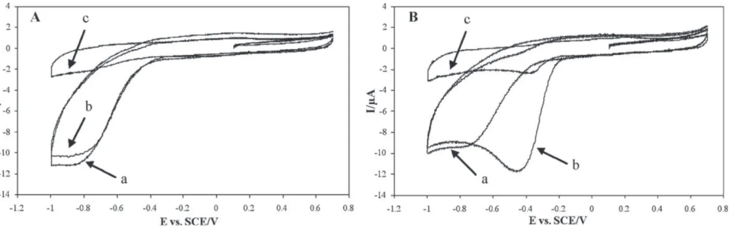
Fig. 1. Cyclic voltammograms of (A) phosphate buffer alone and (B) P. aeruginosa PA01 in phosphate buffer (line a), after 1 h of gentle stirring (line b) and after 20 min of nitrogen bubbling (line c).
potential were reproducible and allowed easy observation of oxy-gen reduction. In the figures, only the first cycle is shown, to make the curves clearer.
3.2. Control voltammograms on phosphate buffer
The voltammograms performed on phosphate buffer under aerobic conditions presented a reduction wave (Fig. 1A, line a). This wave was confirmed to correspond to the electrochemical reduction of the dissolved oxygen (O2+ 2H++ 2e−→ H2O2(1) and H2O2+ 2H++ 2e−→ 2H2O (2)) since it disappeared after oxygen was removed by nitrogen bubbling (Fig. 1A, line c). It has pre-viously been shown that the balance between the two reactions depends on the nature of the carbon electrode and of its pre-treatment. The balance between reactions (1) and (2) controls the general shape of the current–potential curve obtained by voltam-metry. Numerical modelling has also indicated that, at high scan rate (as used here in voltammetry experiments), hydrogen per-oxide is almost completely reduced by reaction (2) before it is able to move far from the electrode surface by diffusion[12]. So, in a first approach, an overall equation can be used to explain the phenomenon (O2+ 4H++ 4e−→ 2H2O). The voltammogram in phosphate buffer under anaerobic conditions showed neither a current peak nor a current wave (Fig. 1A, line c). The flat curve corresponded to the capacitive (non-Faradic) current induced by the charging and discharging of the electrochemical double layer, but no electron exchange (oxidation–reduction phenomenon) took place between the electrode and dissolved compounds. Under aer-obic conditions, oxygen reduction started at −0.37 ± 0.01 V/SCE, and a steady state current was established from −0.81 ± 0.02 V/SCE due to mass transfer limitation of oxygen to the electrode surface (diffusion-limited current). The average values and standard devi-ations were the result of 7 independent experiments. Moreover, oxygen bubbling led to an augmentation of the curve amplitude (data not shown). After 1 h of stirring, the voltammogram remained the same (Fig. 1A, line b). Stirring ensured that the solution was regularly re-oxygenated and led to reproducible voltammograms. 3.3. Electrochemical reduction of oxygen by P. aeruginosa PA01
The voltammogram recorded immediately after the addition of the suspension of P. aeruginosa PA01 (Fig. 1B, line a) was iden-tical to the first voltammogram performed in phosphate buffer alone (Fig. 1A, line a). In contrast, after 1 h of stirring, a displace-ment of the oxygen reduction wave was observed (Fig. 1B, line b). The presence of bacteria clearly modified the shape of the current–potential curve. In these new conditions, the wave started
at −0.18 ± 0.01 V/SCE, reached its maximum at −0.44 ± 0.03 V/SCE (average values and standard deviations from 7 independent exper-iments) and finally returned to the same diffusion-limited current. For a given value of the potential, the current generated in presence of P. aeruginosa PA01 after 1 h of stirring was higher than immedi-ately after bacteria addition. It was observed to rise to as much as four times the current obtained without bacteria: −11.4 ± 0.4 mA (−162 ± 5 mA/cm2) compared to −2.4 ± 0.5 mA (−34 ± 7 mA/cm2) for the same potential value (7 independent experiments). After 1 h of stirring in presence of bacteria and 20 min of nitrogen bub-bling, very weak catalysis was observed (Fig. 1B, line c), probably due to traces of oxygen remaining in the solution.
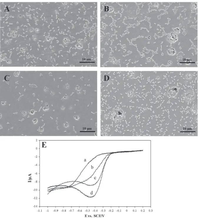
Different durations of stirring were tested. As shown inFig. 2E (only the part of the voltammogram that is of interest is shown in order to make the phenomenon more clearly visible) the amplitude of the oxygen reduction wave increased with the contact duration. Weak catalysis was observed after 15 min (Fig. 2E, line b) but was maximal after 1 h of contact (Fig. 2E, line d). Beyond 1 h (3 h, 6 h, and 24 h), the wave amplitude did not increase further and it appeared that a steady state had been reached. Scanning microscopy was used to assess the working electrode surface coverage by bacteria. Observations were made after 15 s, 15 min, 30 min and 1 h of con-tact between electrodes and bacteria in phosphate buffer under gentle stirring (Fig. 2A–D). There were not more adhered bacteria after 1 h of contact between electrodes and the bacterial suspen-sion than after 15 s (Fig. 2A and D). These pictures show that the coverage did not significantly increase with the contact duration (Fig. 2A–D). The catalytic effect observed on the voltammogram was consequently not directly correlated to the bacterial surface coverage.
3.4. Characterization of the electrochemical activity
Three other tests were performed on P. aeruginosa PA01 to understand the phenomenon further. The bacterial suspension was stirred for 1 h without any contact with the electrodes. After this delay, when the electrodes were plunged into the suspension, the voltammogram recorded was identical to the control curves recorded in buffer alone; no catalytic effect could be observed (data not shown). Thus, a certain time of contact between microor-ganisms and electrodes was absolutely necessary. In a second set of experiments, the bacteria were filtered out of the suspension, and the electrodes were plunged for 1 h in the filtrate under stir-ring. Voltammograms revealed only a very weak catalytic effect compared with that of the bacterial suspension (Fig. 3). This demon-strated that a mediator alone was not responsible for the catalysis and that the presence of bacteria was necessary. The final test
eval-Fig. 2. Scanning electron micrographs of the working electrode surface after 15 s (A), 15 min (B), 30 min (C) and 1 h (D) of contact with bacteria, magnification 5000, and corresponding cyclic voltammograms of P. aeruginosa PA01 in phosphate buffer (E) after 15 s (line a), 15 min (line b), 30 min (line c), and 1 h (line d) of contact. Beyond 1 h (3 h, 6 h and 24 h), the wave amplitude does not increase further.
uated the activity of the matter adhering to the working electrode. After 1 h of contact with bacteria under stirring, the electrodes were removed from the suspension, rinsed in distilled water and tested again in 20 mL fresh phosphate buffer (Fig. 4). The curve in the fresh buffer presented the same catalysis of oxygen reduction. Bacte-ria, proteins or other molecules they produced, and which became attached to the working electrode surface, were consequently fully responsible for the catalytic effect.
Tests were performed to assess the necessity for metabolic activity of the cell during the transfer of electrons. Voltammo-grams performed on dead cells even after 1 h of contact between electrodes and dead bacteria revealed no catalysis of the oxygen reduction (Fig. 5A, line c). The catalysis was, however, observed
when bacteria were killed on the electrode surface after 1 h of con-tact (Fig. 5B, line c).
3.5. Electrochemical reduction of oxygen by other strains of P. aeruginosa
All the strains of P. aeruginosa tested in this study by the cyclic voltammetry method (Table 1) presented the same behavior as PA01. The difference between the maximal intensity of the oxy-gen reduction peak in presence of PA01 and other strains of P. aeruginosa was lower than 5% (3 independent experiments on each strain). The clinical strains were chosen because of their different phenotypes so as to assess a possible implication of alginate and
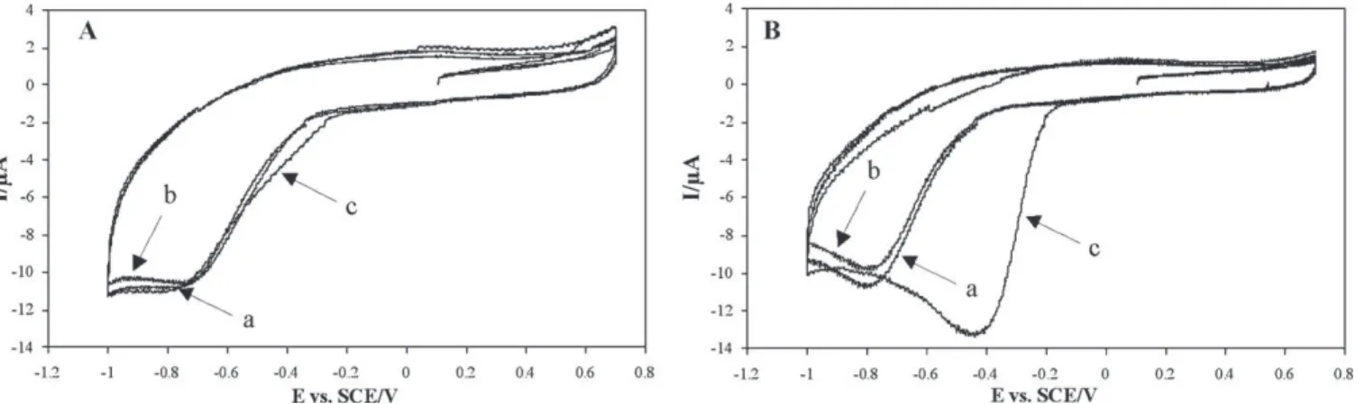
Fig. 3. Cyclic voltammograms in phosphate buffer (line a), with P. aeruginosa PA01 filtrate (line b) and P. aeruginosa PA01 filtrate after 1 h of stirring (line c). After 1 h of stirring, the bacterial suspension was filtered through a 0.45 mm sterile filter then through a 0.20 mm one. CV scans were performed on this filtrate (lines b and c).
Fig. 4. Cyclic voltammograms of P. aeruginosa PA01 in phosphate buffer (line a), after 1 h of stirring (line b) and after the electrodes were removed and plunged into a fresh phosphate buffer (line c).
pigments in the catalytic effect. The strain 7844M is mucoid; it produces a lot of alginate. The 7844S is a non-mucoid revertant obtained after plating 7844M several times. Both presented the same restriction card and differed only by the production of algi-nate. These two strains showed similar positive activity, suggesting that alginate production was not involved in the electrochemical effect. Four strains of P. aeruginosa were tested because of their
differences in pigmentation: green (pyoverdin), blue (pyocyanin), brown (pyomelanin and pyorubin) and three with no pigmenta-tion at all. All of them presented the same electrochemical activity. Finally, no significant electrochemical differences were observed between the PA14 catalase-defective mutants and the wild type strain. The catalase activity of the mutants was tested and revealed that PA14 KatB− and PA14 KatE− presented a catalase activity sim-ilar to the wild type one, PA14 KatA− presented a weak catalase activity, and the triple mutant PA14 KatABE− no catalase activity at all (Table 1).
4. Discussion
In the presence of bacteria, the reduction of oxygen started for lower potentials and higher currents were obtained (Fig. 1). For the first time, P. aeruginosa has been shown to catalyze the elec-trochemical reduction of oxygen. Previous studies on P. aeruginosa have been devoted to anodic processes only, where the bacterial cell transfers electrons to the electrodes. In the present work, the reverse phenomenon has been demonstrated (Fig. 1B).
A certain time of contact between the bacterial suspension and electrodes is necessary for the full catalytic effect to be observed (Fig. 1B). A weak effect was detected in the first few minutes and was complete only after 1 h (Fig. 2E). Microscopic images showed that a few seconds were enough to get a bacterial coverage of the electrode surface similar to that observed after 1 h (Fig. 2A and D). Nevertheless, it was necessary to wait for 1 h to obtain the full catalytic effect even though there were not more bacteria on the electrode surface after this delay. On the other hand, experiments performed in fresh buffer after adhesion indicated that bacteria, proteins or other produced molecules adhering to the electrode surface were responsible for the phenomenon (Fig. 4). Adhesion is necessary but not sufficient; a prolonged contact time is also required.
The attachment of bacteria to a solid surface implies two steps: reversible and irreversible attachment[25]. In the first step, the bacteria are transported close enough to the surface, involving van der Waals forces, electrostatic forces and hydrophobic interactions. The second stage of adhesion is the locking phase and employs molecularly mediated binding between the surface and specific adhesins, like exopolysaccharides (biofilm matrix), pili, fimbriae, etc. On the SEM images, no difference was observed between bac-teria adsorbed on the electrode surface after 15 s or after 1 h of contact. The 1-h contact time did not lead to a visual modification of the adsorption pattern, which would have indicated biofilm devel-opment. Moreover, the tests on mucoid and non-mucoid strains indicated that alginate, which is the main component of the biofilm
Fig. 5. Effects of different methanol treatments on the catalysis of oxygen reduction by P. aeruginosa PA01. (A) CV on phosphate buffer alone (line a), after addition of PA01 cells previously treated with methanol (line b), and after 1 h of stirring (line c). (B) CV on phosphate buffer alone (line a), after the addition of PA01 cells (line b), and after the working electrode was removed, treated with methanol, and then plunged into a fresh phosphate buffer (line c).
matrix formed by P. aeruginosa[26], had no effect on the catalysis (Table 1). Preliminary work on a P. aeruginosa pilA mutant (defi-cient in biogenesis of type IV pili) did not show any difference with respect to the wild type strain in cyclic voltammetry (data not shown). Like alginate, these pili are crucial for irreversible attach-ment and maturation of biofilm[25,27]. In addition, beyond 1 h of contact (3 h, 6 h or 24 h), no increase in the catalysis was observed. All these data suggest that the pure process of adhesion and biofilm formation is not entirely responsible for the transfer of electrons. Something more than adhesion is needed.
A real contact between bacterial cells and the electrode surface was required since, after 1 h of stirring of the bacterial suspen-sion without any contact with the electrode, no catalytic effect was observed on the voltammogram. So the catalysis does not depend on compounds that bacteria produce and release into the medium, like phenazines. The negative result on the filtrate test confirmed this hypothesis (Fig. 3). Previous studies that have demonstrated the involvement of phenazines in anodic processes related to P. aeruginosa showed a strong activity of the filtrate[4]. Then, what-ever the pigmentation of the strain, due to the production of phenazines[28], detection became possible, even for strains with no pigmentation at all (Table 1). Phenazines are definitively not involved in the transfer of electrons described here. Hence, unlike in anodic catalysis, P. aeruginosa does not use released electron shuttles.
Another explanation could be that bacteria do produce elec-troactive molecules but that they are bound into the membrane or are diffusible only for a short distance from the bacteria. This may explain why contact with the electrodes is required during the delay time if the catalytic effect is to be observed. The weak effect obtained with the filtrate would be explained by a small release of this compound from the cell membrane to the solution. For instance, type IV pili produced at the surface of Geobacter sul-furreducens were responsible for the transfer of electrons[7]. As presented above, P. aeruginosa mutants deficient in type IV pili biogenesis were able to catalyze the electrochemical reduction of oxygen. These pili were consequently not involved in the elec-tronic transfer observed here. Experiments performed on dead cells showed that bacteria had to be alive during the 1 h contact time. After this delay, probably necessary for the production or relocation of proteins or other bacterial compounds on the surface of cells, the bacteria could be killed without their death influencing the catalysis. This observation supports the hypothesis of the involve-ment of membrane-bound proteins having no direct link with the metabolic activity of the cells. Several biological compounds adsorbed alone on an electrode surface are known to catalyze the electrochemical reduction of oxygen without any involvement of bacterial cells. The most studied are iron-based proteins: haeme proteins[29], cytochrome[30], cytochrome c oxidase[31], cata-lase[12,32]. Here, we focused on catalase, which had already been demonstrated to induce the catalysis of electrochemical reduction of oxygen on glassy carbon electrodes[13]and which activity had been shown to be correlated to the increment of cathodic currents for Pseudomonas sp.[22]. Catalase is an enzyme located on the outer membrane of the majority of negative and on several Gram-positive bacteria[33]. It catalyzes hydrogen peroxide dismutation. Catalase adsorbed on glassy carbon electrodes has been tested with cyclic voltammetry under conditions similar to those in the work presented here. The voltammograms obtained had the same gen-eral shape as those plotted here with P. aeruginosa. The oxygen reduction wave was displaced and started around −0.20 V/SCE in presence of catalase. We consequently suspected that this protein was responsible for the phenomenon observed here. In P. aerugi-nosa, there are three differentially evolved monofunctional catalase genes, katA, katB, and katE[34]. PA14 KatABE− strain does not present any catalase activity[24], but this strain is able to catalyze
the electrochemical reduction of oxygen like the wild type strain, PA14 (Table 1). Therefore catalase is not entirely responsible for the electrochemical catalysis of oxygen reduction.
The concept of microbial electro-catalysis of oxygen reduction on metallic surfaces was first identified in the field of micro-bial corrosion with biofilms formed in fresh seawater. The work presented here, which was carried out with pure cultures of different P. aeruginosa strains, opens a new route for investiga-tion. Actually, the different assumptions that have been evoked to explain the electro-catalysis of oxygen reduction by natural biofilms: production of hydrogen peroxide, involvement of man-ganese oxides/hydroxides, direct catalysis by adsorbed catalase, can no longer explain the electro-catalytic effect observed here with pure strains. Data obtained in the present work suggest that electro-catalysis is sustained by the production of a compound that remains linked to the cells or in the cell vicinity, with only weak release and with no direct link with the cell’s metabolism after the 1 h contact time. Catalase cannot be responsible but other small iron-binding proteins, such as ferredoxin, cytochrome, rubredoxin, etc., could play this role. The common denominator among all these proteins is their active site called porphyrin, and such haeme pro-teins are known to catalyze oxygen reduction on metallic cathodes
[35]. Involvement of porphyrin molecules in the catalytic effect observed here would explain the similarity of the effect for all the P. aeruginosa strains tested. Moreover, recent work devoted to the identification of the microbial composition of the seawa-ter biofilms that catalyze oxygen reduction has evidenced the large microbial diversity of these biofilms (Vandecandelaere, personal communication, to be published). The involvement of such ubiq-uitous molecules as porphyrins would also explain the fact that a large number of various species are able to catalyze the electro-chemical reduction of oxygen. The present study will be completed by focusing on possible production of haeme compounds during the contact period.
In a more technical approach, although the catalysis of oxygen reduction demonstrated here is not interesting for MFC applica-tions (potential too low), the protocol allows the detection of the early stages of P. aeruginosa biofilm formation, i.e. the first step of adhesion. All the tested strains of P. aeruginosa, including clin-ical isolates, were able to catalyze the electrochemclin-ical reduction of oxygen. This property seems to be universal among P. aerugi-nosa strains. The catalytic effect allowed a detection of the bacterial adhesion to the electrode after a few minutes only (1 h for the full catalytic effect). The signal obtained was easily identifiable and highly reproducible. Therefore this property may provide an oppor-tunity for early detection of bacterial adhesion. Indeed, the majority of the electrochemical methods already proposed to detect biofilm colonization has focused on biofilm growth and can only detect a deposit on the electrode surface, without indication of its biotic or abiotic character[36,37]. In contrast, the technique presented here detects the presence of bacteria on the surface. The method does not only measure surface fouling; it requires a real interaction between the bacteria and the electrode. Further work is now in progress to assess whether the cyclic voltammetry protocol highlights a prop-erty present in most aerobic bacteria or a specificity of P. aeruginosa. Preliminary results have shown that other opportunistic bacte-ria, like Escherichia coli, Enterobacter cloacae and Shigella sp., are detectable using the CV method in the same way as P. aeruginosa.
5. Conclusions
It has been demonstrated here that P. aeruginosa is able to cat-alyze the electrochemical reduction of oxygen. This phenomenon implies a prolonged contact between bacteria and the working electrode. The transfer of electrons is not due to electron
shut-tles released into the medium by bacteria and phenazines are not involved. Type IV pili are not involved in the process either. The metabolic activity of the cell is necessary during the 1-h con-tact time but, after this delay, the catalysis is possible even if the bacteria are killed. Finally, the activity of membrane-bound redox compounds having no direct link with the cell’s metabolism seems the most probable explanation because of the similarity of results reported in the literature with adsorbed proteins. Tests per-formed on catalase mutants showed that this enzyme was not necessary for the catalysis described. P. aeruginosa now offers a new, easy-to-handle model to advance in deciphering the mecha-nism of microbial electro-catalysis of oxygen reduction. Moreover, the procedure designed here may become a suitable technique for detecting the first steps of bacterial adhesion.
Acknowledgments
We are grateful to Sandrine Parot (Laboratoire de Génie Chim-ique, Toulouse) for her expert advice and assistance in the cyclic voltammetry method and to Marie-Line de Solan (Laboratoire de Génie Chimique, Toulouse) for her precious help with SEM. Thanks to You-Hee Cho (Sogang University, Korea) for having kindly pro-vided the catalase mutant strains.
We also thank the federative structure FERMAT for its support. References
[1] K. Rabaey, J. Rodriguez, L.L. Blackall, J. Keller, P. Gross, D. Batstone, W. Verstraete, K.H. Nealson, ISME J. 1 (2007) 9.
[2] D.R. Lovley, Curr. Opin. Biotechnol. 19 (2008) 564. [3] B.E. Logan, Nat. Rev. Microbiol. 7 (2009) 375.
[4] K. Rabaey, N. Boon, M. Hofte, W. Verstraete, Environ. Sci. Technol. 39 (2005) 3401.
[5] D.E. Holmes, S.K. Chaudhuri, K.P. Nevin, T. Mehta, B.A. Methe, A. Liu, J.E. Ward, T.L. Woodard, J. Webster, D.R. Lovley, Environ. Microbiol. 8 (2006) 1805.
[6] Y.A. Gorby, S. Yanina, J.S. McLean, K.M. Rosso, D. Moyles, A. Dohnalkova, T.J. Beveridge, I.S. Chang, B.H. Kim, K.S. Kim, D.E. Culley, S.B. Reed, M.F. Romine, D.A. Saffarini, E.A. Hill, L. Shi, D.A. Elias, D.W. Kennedy, G. Pinchuk, K. Watanabe, S. Ishii, B. Logan, K.H. Nealson, J.K. Fredrickson, Proc. Natl. Acad. Sci. U.S.A. 103 (2006) 11358.
[7] G. Reguera, K.D. McCarthy, T. Mehta, J.S. Nicoll, M.T. Tuominen, D.R. Lovley, Nature 435 (2005) 1098.
[8] A. Bergel, D. Feron, A. Mollica, Electrochem. Commun. 7 (2005) 900. [9] Z. He, L.T. Angenent, Electroanalysis 18 (2006) 2009.
[10] K. Rabaey, S.T. Read, P. Clauwaert, S. Freguia, P.L. Bond, L.L. Blackall, J. Keller, ISME J. 2 (2008) 519.
[11] V. Scotto, R. Dicintio, G. Marcenaro, Corros. Sci. 25 (1985) 185. [12] M.E. Lai, A. Bergel, J. Electroanal. Chem. 494 (2000) 30. [13] M.E. Lai, A. Bergel, Bioelectrochemistry 55 (2002) 157.
[14] I. Dupont, D. Feron, G. Novel, Int. Biodeterior. Biodegrad. 41 (1998) 13. [15] W.H. Dickinson, Z. Lewandowski, Biofouling 10 (1996) 79.
[16] C. Dumas, A. Mollica, D. Feron, R. Basseguy, L. Etcheverry, A. Bergel, Electrochim. Acta 53 (2007) 468.
[17] C. Dumas, A. Mollica, D. Feron, R. Basseguy, L. Etcheverry, A. Bergel, Bioresour. Technol. 99 (2008) 8887.
[18] D.V. Vukomanovic, D.E. Zoutman, G.S. Marks, J.F. Brien, G.W. vanLoon, K. Nakatsu, J. Pharmacol. Toxicol. Methods 36 (1996) 97.
[19] M.E. Hernandez, A. Kappler, D.K. Newman, Appl. Environ. Microbiol. 70 (2004) 921.
[20] K. Rabaey, N. Boon, S.D. Siciliano, M. Verhaege, W. Verstraete, Appl. Environ. Microbiol. 70 (2004) 5373.
[21] T.H. Pham, N. Boon, K. De Maeyer, M. Hofte, K. Rabaey, W. Verstraete, Appl. Microbiol. Biotechnol. 80 (2008) 985.
[22] J.P. Busalmen, M. Vazquez, S.R. de Sanchez, Electrochim. Acta 47 (2002) 1857.
[23] H.J. Kim, H.S. Park, M.S. Hyun, I.S. Chang, M. Kim, B.H. Kim, Enzyme Microb. Technol. 30 (2002) 145.
[24] J.S. Lee, Y.J. Heo, J.K. Lee, Y.H. Cho, Infect. Immun. 73 (2005) 4399. [25] J. Palmer, S. Flint, J. Brooks, J. Ind. Microbiol. Biotechnol. 34 (2007) 577. [26] C. Ryder, M. Byrd, D.J. Wozniak, Curr. Opin. Microbiol. 10 (2007) 644. [27] M.R. Parsek, T. Tolker-Nielsen, Curr. Opin. Microbiol. 11 (2008) 560. [28] M.O. Husson, M. Hamze, A. Fruchart, D. Izard, in: J. Freyney, F. Renaud, C. Bollet,
R. Leclercq (Eds.), Actualités permanentes en bactériologie clinique, Editions ESKA & Editions Alexandre Lacassagne, Paris, 2002.
[29] L. Shen, N. Hu, Biochim. Biophys. Acta 1608 (2004) 23.
[30] V.V. Shumyantseva, T.V. Bulko, Y.O. Rudakov, G.P. Kuznetsova, N.F. Samenkova, A.V. Lisitsa, I.I. Karuzina, A.I. Archakov, J. Inorg. Biochem. 101 (2007) 859. [31] L. Su, F.M. Hawkridge, M.C. Rhoten, Chem. Biodivers. 1 (2004) 1281. [32] A. Salimi, A. Noorbakhsh, M. Ghadermarz, Anal. Biochem. 344 (2005) 16. [33] P. Chelikani, I. Fita, P.C. Loewen, Cell. Mol. Life Sci. 61 (2004) 192.
[34] C.K. Stover, X.Q. Pham, A.L. Erwin, S.D. Mizoguchi, P. Warrener, M.J. Hickey, F.S. Brinkman, W.O. Hufnagle, D.J. Kowalik, M. Lagrou, R.L. Garber, L. Goltry, E. Tolentino, S. Westbrock-Wadman, Y. Yuan, L.L. Brody, S.N. Coulter, K.R. Folger, A. Kas, K. Larbig, R. Lim, K. Smith, D. Spencer, G.K. Wong, Z. Wu, I.T. Paulsen, J. Reizer, M.H. Saier, R.E. Hancock, S. Lory, M.V. Olson, Nature 406 (2000) 959. [35] H. Iken, L. Etcheverry, A. Bergel, R. Basseguy, Electrochim. Acta 54 (2008) 60. [36] J. Gamby, A. Pailleret, C.B. Clodic, C.M. Pradier, B. Tribollet, Electrochim. Acta 54
(2008) 66.