Université du Québec
Institut National de la Recherche Scientifique Institut Armand-Frappier
TURNIP MOSAIC VIRUS MEMBRANE-ASSOCIATED REPLICATION
COMPLEXES- ULTRASTRUCTURE AND ROLE IN VIRUS MOVEMENT
Par Juan Wan
Thèse présentée pour l’obtention du grade de Philosophiae doctor (Ph.D.)
en Immunologie et Virologie
Jury d’évaluation
Président du jury et Prof. Peter Tijssen
examinateur interne INRS – Institut Armand-Frappier Examinateur externe Prof. Peter Moffett
Université de Sherbrooke
Examinateur externe Prof. Hugo Germain
Université du Québec à Trois-Rivières Directeur de recherche Prof. Jean-François Laliberté
INRS – Institut Armand-Frappier
RÉSUMÉ
Les virus à ARN positifs [ARN(+)] remanient les membranes cellulaires pour faciliter la réplication de leur ARN viral (ARNv). Les complexes ribonucléoprotéiques viraux (RNPv) formés d'ARNv et de protéines du virus et de l’hôte sont associés aux membranes et se déplacent au travers des plasmodesmes (PD) pour atteindre les cellules avoisinantes. Une fois arrivé dans la nouvelle cellule, un nouveau cycle de synthèse d'ARNv est initié. Ces complexes RNPv peuvent à leur tour atteindre les tissus vasculaires et entreprendre leur déplacement sur une longue distance. Un nouveau concept émerge dans la communauté scientifique. Il consiste à considérer que la réplication de l'ARNv et le mouvement viral sont deux processus étroitement liés. Cependant, la nature du complexe RNPv demande encore à être définie.
La présente thèse se concentre sur l'étude des complexes associés aux membranes induits par le virus de la mosaïque du navet (Turnip mosaic virus, TuMV). Le TuMV est un virus à ARN (+) appartenant au genre viral Potyvirus. Ce dernier représente le plus grand genre des virus de plante. Ils sont responsables de plus de la moitié des pertes agricoles imputées aux virus.
La protéine virale 6K2 est membranaire et est responsable de la réorganisation du système endomembranaire pour former des vésicules mobiles. Ces dernières contiennent les complexes de réplication virale. Les complexes du TuMV associés aux membranes ont été largement étudiés par microscopie confocale essentiellement dans les cellules épidermiques.
Les deux objectifs de cette thèse ont été d'une part d'analyser la présence de complexes de réplication du TuMV associés aux membranes dans les tissus vasculaires et d'autre part de découvrir en détails l’ultrastructure de ces complexes dans plusieurs types cellulaires.
Le premier objectif a consisté à observer la présence de complexes de réplication associés aux membranes dans tous les types cellulaires présents dans les feuilles et les tiges de la plante. En faisant des coupes cryohistologiques, suivies
d'immuno-histolocalisations par microscopie confocale, il a pu être montré que les complexes de réplication associés aux membranes et induits par la protéine 6K2 étaient présents dans tous les types cellulaires y compris les tissus vasculaires tels que les tubes criblés du phloème et dans les vaisseaux du xylème. Des observations par microscopie électronique à transmission (MET) ont permis de montrer la présence de vésicules contenant l'ARNv en association avec des particules virales dans les vaisseaux du xylème. En menant des expériences de "girdling" sur tiges, où toutes les cellules meurent tout en maintenant l'intégrité des vaisseaux du xylème, il a été confirmé que le TuMV utilise les vaisseaux du xylème pour son infection systémique. Finalement, il a été démontré que des complexes RNPv du Potato virus X (PVX) associés à des membranes étaient aussi présents dans le phloème et le xylème de plantes infectées par ce virus.
Ces études indiquent que les usines de réplication virale peuvent se retrouver dans le phloème et le xylème, suggérant ainsi que la réplication virale et le mouvement à longue distance sont deux évènements étroitement liés.
Le second objectif a été dedéfinir par MET l'ultrastructure des réorganisations cellulaires induites par le TuMV. Une cinétique a été effectuée pour analyser le remodelage membranaire induit par le TuMV. À des étapes précoces de l'infection, il a été observé des amas de membranes ["convoluted membranes", (CM)] dérivant du réticulum endoplasmique rugueux (RER). À des étapes intermédiaires de l'infection, des vésicules à simple membrane (SMV) étaient observables. À des étapes plus tardives de l'infection, en plus d'agrégats de SMVs, des vésicules à double membrane (DMV) au cœur dense aux électrons étaient observables à proximité de corps denses aux électrons. Ensuite il a été montré par immuno-MET que les vésicules induites par le TuMV contenaient la protéine virale 6K2 et que seules les SMV constituaient les sites de réplication de l'ARNv. À des étapes tardives de l'infection, il a été aussi observé des corps dense aux électrons à proximité d’agglomérats de particules virales du TuMV qui
Ces résultats contribuent d'une part à une meilleure compréhension de la nature des complexes RNPv impliqués dans le mouvement viral sur une longue distance et d'autre part, à avoir une vision plus fine de ce que sont les sites de réplication de l'ARNv du TuMV, ainsi que des sites d'assemblage et d'accumulation des particules virales.
--- --- Juan Wan Jean-François Laliberté Étudiante Directeur de recherche
ABSTRACT
Positive-strand RNA [(+) RNA] viruses remodel cellular membranes to facilitate viral RNA (vRNA) replication. Membrane-bound progeny viral ribonucleoprotein (vRNP) complexes then move through plasmodesmata (PDs) to adjacent cells to initiate new rounds of vRNA synthesis. These complexes ultimately reach the vascular tissues and are loaded into the vascular conducting tubes for long-distance trafficking. An emerging concept is that vRNA replication and movement are tightly linked processes. The moving vRNP complex, however, has yet to be defined.
This thesis focuses on Turnip mosaic virus (TuMV) -induced membrane-associated complexes. TuMV is a (+) RNA virus belonging to the genus Potyvirus, which is the largest genus of plant viruses, and is responsible for more than half of the viral crop damage in the world. The viral membrane protein 6K2 is responsible for membrane reorganization that leads to the formation of motile vesicles. These vesicles contain virus replication complexes. TuMV membrane-associated replication complexes dynamics have been widely studied by confocal microscopy in epidermal cells.
The two aims of this thesis were to investigate the presence of TuMV membrane-associated replication complexes in vascular tissues, and then to uncover the ultrastructure of the TuMV-induced membrane reorganization.
The first research objective consisted of looking at the presence of TuMV membrane-associated replication complexes in all types of plant leaf and stem cells. By using cryohistological preparation, immunohistolocalization and confocal microscopy, it was shown that 6K2-tagged membrane-associated replication complexes were present in all cell types, including the vascular conducting tubes, which were phloem sieve elements and xylem vessels. Transmission electron microcopy (TEM) observation results showed the presence of vRNA-containing vesicles that were associated with
Collectively, these studies indicate that viral replication factories could end up in the phloem and the xylem, suggesting that virus replication and long-distance trafficking are linked events.
The second objective was to define the ultrastructure of TuMV-induced cellular reorganization by TEM. A time course analysis of TuMV-induced membrane remodeling was initially performed.
Early during infection, rough endoplasmic reticulum (ER)-connected convoluted membrane (CM) structures were observed. Single-membrane vesicles (SMVs) were found at the mid stage of infection, while mixed aggregates containing SMVs, double-membrane vesicles (DMVs) with an electron-dense core, and electron-dense bodies were detected at a late stage of infection. Then the immuno-EM experiments showed that the vesicles are tagged with 6K2, and that only SMVs are the vRNA replication sites. I also observed electron-dense bodies associated TuMV particle bundles, which probably are the particle assembly site. Finally, with electron tomography (ET), it was shown that viral particles accumulating in the central vacuole as membrane-associated complexes.
Taken together, this work contributed to a better understanding of the nature of vRNP complexes for virus long-distance trafficking, of the ultrastructure of TuMV RNA replication sites, as well as particles assembly and accumulation sites.
--- --- Juan Wan Jean-François Laliberté Student Director of Research
SOMMAIRE RÉCAPITULATIF
Les virus à ARN positifs (ARN+) remodèlent les membranes cellulaires pour faciliter la réplication de leur ARN viral (ARNv). Ces réarrangements membranaires donnent naissance à ce qui est qualifié d'usines de réplication. Il est généralement admis que ces nouveaux compartiments constituent un environnement protégeant les facteurs viraux de la dégradation par les défenses de l'hôte. De plus, ces quasi-organites permettent de concentrer localement les éléments nécessaires à la réplication de l'ARNv, ce qui augmenterait l'efficacité de ce processus. Cependant différents virus à ARN+ induisent différentes structures membranaires pour la réplication de leur ARNv. Notamment, le nodavirus Flock house virus (FHV) qui en remodelant la membrane externe de la mitochondrie induit la formation de sphérules. Ces dernières n'étant pas totalement closes, elles laissent un canal d'environ 10 nm qui permet la connexion entre leur intérieur et le cytoplasme. Le flavivirus Dengue virus (DENV) quant à lui remodèle le réticulum endoplasmique (RE) pour former des amats de vésicules (vesicle packets, VPs) constituées de simples membranes (single-membrane vesicles, SMVs) dans le lumen de ce dernier. Les poliovirus remodèlent aussi le RE et/ou l'appareil de Golgi pour former des agrégats de SMVs. Le syndrome respiratoire aigu sévère (SRAS)- coronavirus remodèle aussi le RE mais en induisant la formation de vésicules doubles membranes.
Ces dernières années, quelques modèles en trois dimensions (3D) ont été générés pour des virus a ARN+ en faisant de la microscopie électronique a transmission couplée à de la tomographie électronique (MET-TE). La complexité des structures dévoilées a grandement amélioré notre compréhension sur la biogenèse et la fonction de ces structures membranaires. Les modèles 3D ont révélé que les virus à ARN+ induisent différentes structures membranaires pouvant coordonner divers processus de leur cycle infectieux. Par exemple, le SRAS-coronavirus induit la
Il existe très peu d'études par MET pour les virus de plante à ARN+. Les structures membranaires générées par ces derniers sont similaires à celles induites par les virus animaux à ARN+. Par exemple, le Turnip yellow mosaic virus (TYMV) induit la formation de sphérules à partir de la membrane externe des chloroplastes et leur intérieur est connecté au cytoplasme via un canal ouvert. Selon l'organisme hôte le Brome mosaic virus (BMV) induit soit la formation de sphérulesà partir des membranes du RE dans la levure et la formation de VPs dans le lumen du RE sous la forme d'une série de SMVs dans les cellules végétales.
Deux représentations 3D d'usines virales de virus de plantes ont été générées. Le Melon necrotic spot virus (MNSV) modifie les mitochondries pour former de larges dilatations internes interconnectées et semblablement connectées au cytoplasme par des pores et/ou d'autres structures plus complexes. Le Beet black scorch virus (BBSV) induit la formation de sphérules dans le lumen du RE.
En revanche nous avons peu de connaissances sur les détails architectureaux des usines virales induites par le virus de la mosaïque du navet (Turnip Mosaic Virus, TuMV). Le TuMV est un virus à ARN+ appartenant au genre Potyvirus dans la famille des Potyviridae. Les potivirus constituent le genre viral le plus important parmi les virus de plante et sont responsables de plus de la moitié des pertes agricoles induites par des virus dans le monde. Les particules virales sont flexibles et filamenteuses avec une longueur de 680 à 900 nm et un diamètre d'environ 11-15 nm. Ils ont un génome à ARN simple brin d'environ 10 kb. L'extrémité 5' du génome est liée de façon covalente à une protéine VPg (viral protein genome linked) et l'extrémité 3' comporte une queue poly(A). L'ARNv code pour une polyprotéine clivée par 3 protéinases virales pour générer au moins 12 protéines. La protéine membranaire 6K2 est responsable de la formation des usines de réplication virale du TuMV.
Les observations par microscopie optique ont montré que lors de l'infection par le TuMV de nombreuses vésicules induites par 6K2 prennent naissance à partir du RE. Les vésicules contiennent l'ARNv, et des protéines virales et de l'hôte impliquées dans la réplication virale. Ainsi, ces dernières sont considérées comme étant le lieu de la réplication de l'ARNv. Les analyses ultrastructurelles les plus récentes du
réarrangement cellulaire par les potyvirus se sont principalement concentrées sur des descriptions d'inclusions nucléaires et cytoplasmiques. Des images en MET de vésicules induites par le TuMV et le Potato virus Y (PVY) à partir de cellules infectées ont dévoilé d'une part la présence de nombreuses SMVs et parfois de SMVs directement connectés au RE rugueux (RER). Cependant aucune étude n'a été faite d'une part sur la biogenèse des usines virales au cours du temps durant l'infection et d'autre part sur la relation entre ces dernières et l'assemblage des particules virales.
La propagation d'un virus dans une plante implique des mouvements viraux de courte distance, de cellule à cellule au travers des plasmodesmes (PD), et de longue distance au travers des vaisseaux vasculaires notamment au travers des tubes criblés du phloème et des vaisseaux du xylème. Il est généralement entendu que les entités capables de se propager sont soit des particules virales soit des complexes ribonucléoprotéiques (RNP). Cependant, la nature exacte de ces complexes RNP n'a pas été définie. Un concept commence à émerger en mettant en avant le fait que la réplication de l'ARNv et le mouvement viral sont deux processus étroitement liés. Par exemple, il été proposé que le Tobacco mosaic virus (TMV) se déplace d'une cellule à l'autre en tant que complexes de réplication. La réplication et le trafic du Potato virus X (PVX) ont aussi été montrés comme étant couplés à l'entrée des PDs. Dans le cas du TuMV, il a été démontré par de l'imagerie cellulaire en temps réel que les complexes de réplication induits par 6K2 pouvaient se déplacer d'une cellule à l'autre. Cependant il est totalement inconnu si ces complexes de réplication avaient la capacité d'atteindre les tissus vasculaires et se déplacer sur une longue distance.
Les objectifs de ma recherche ont été:
1. de savoir si les usines de réplication induites par 6K2 pouvaient être impliquées dans le mouvement viral sur une longue distance ;
ont été agroinfiltrées avec le clone infectieux TuMV/6k2:GFP qui permet d'avoir les usines de réplication étiquetées avec une étiquette fluorescente, ici la GFP. Des coupes transversales de feuilles systémiques au niveau de la nervure centrale, ainsi que des coupes longitudinales de tiges juste au dessus des feuilles inoculées (premier internode) ont été observées par microscopie confocale. Il a été observé que 6K2:GFP était présente dans tous les types cellulaires dans les tiges et feuilles de plantes infectées, notamment dans les tubes criblés du phloème et les vaisseaux du xylème. Ce dernier point suggère que le mouvement systémique du TuMV implique tous les vaisseaux vasculaires. Suite à une coloration au DioC6(3) lipophile il a été mis en évidence que les agrégats présentant la 6K2 étiquetée dans les vaisseaux vasculaires contiennaient des lipides. Il a été trouvé que ces agrégats contenaient de l'ARNv, l'ARN polymérase ARN dépendante virale (RdRpv). Ceci suggère que ces agrégats contiennent les complexes de réplication. Il a été aussi détecté la protéine de capside (CP) en association avec les agrégats contenant la 6K2 étiquetée, suggérant ainsi que des particules virales du TuMV ou de la CP associée aux complexes RNP sont aussi présentes.
Par ailleurs il a aussi été mené des analyses par des techniques conventionnelles de MET pour mieux caractériser l'ultrastructure des agrégats contenant la 6K2 étiquetée dans les vaisseaux vasculaires. Ces analyses ont permis de montrer la présence de vésicules contenant l'ARNv dans les vaisseaux du xylème en association avec des particules virales. Des analyses de sève xylémienne provenant de plantes infectées par microscopie confocale et Western blot ont montré qu'elle contenait des vésicules induites par 6K2, des éléments essentiels à la réplication du TuMV et l'assemblage de ses particules virales tels que la 6K2, la CP, la RdRpv ainsi qu'une protéine de l'hôte eIF(iso)4e. La sève xylémienne provenant de plantes infectées est infectieuse. Des expérimentations visant à détruire toute cellule vivante autour du xylème sur une portion de tige (stem girdling) a confirmé que le TuMV peut établir une infection systémique en exploitant les vaisseaux du xylème.
Les résultats des immunohistolocalisations ont montré que les tubes criblés du phloème et du xylème provenant de plantes infectées par le Potato virus X (PVX)
que la présence de complexes RNP associés aux membranes dans les vaisseaux vasculaires n'est pas une caractéristique limitée seulement au TuMV.
Pris ensembles, ces études indiquent que les usines virales peuvent aboutir dans le phloème et le xylème. D'après toutes ces observations, il a été proposé un modèle permettant d'expliquer le mouvement du TuMV dans les tubes criblés du phloème et les vaisseaux du xylème. Dans ce schéma, les vésicules induites par la 6K2 étiquetée et contenant l'ARNv et la RdRpv se déplacent depuis les cellules de l'épiderme vers les cellules compagnes en passant par les cellules du mésophile, de la gaine et du parenchyme vasculaire via les PDs. Une fois arrivées aux cellules compagnes les vésicules sont chargées dans les tubes criblés du phloème via des plasmodesmes particuliers appelés pore plasmodesmal unit (PPUs). Ces dernières pourraient soit voyager individuellement soit s'agglutiner pour former un support membranaire à l'assemblage des particules virales dans les tubes criblés du phloème suivant ainsi le flux de sève phloémienne à longue distance. Également, les usines virales du TuMV pourraient aussi entrer dans les cellules non différentiées du xylème, assurant ainsi la réplication virale avant que la mort cellulaire ne libère ces structures dans les vaisseaux matures du xylème.
Le second objectif a été d'obtenir l'ultrastructure du remodelage cellulaire induit par le TuMV. Différentes étapes de la préparation des échantillons ont été optimisé pour des analyses par MET, notamment afin d'améliorer le contraste des structures membranaires. Tout d'abord, en comparant des cellules provenant de feuilles agroinfiltrées et des feuilles systémiquement infectées par le TuMV, ces dernières ont été choisies pour faire mes analyses par MET du fait qu'il était bien plus facile de trouver les structures induites par le TuMV, ainsi que les inclusions cytoplasmiques. La zone autour de la nervure centrale de ces feuilles a été collectée à différents jours après l'infection (j.p.i) avec le clone TuMV/6K2:GFP. L'infection des tissus vasculaires a été décelée à 5 j.p.i et la colonisation des autres tissus à partir de 6 j.p.i.
à des stades intermédiaires de l'infection. La présence de SMVs et de vésicules double membranes (double-membrane vesicles, (DMVs)) avec un cœur dense aux électrons a été constatée en MET à des stades tardifs de l'infection. Le marquage par immunogold a révélé que les agrégats de vésicules contenaient la protéine 6K2 et que les SMV sont les sites de la réplication de l'ARNv.
Le remodelage cellulaire induit par le TuMV a été observé au niveau des cellules du mésophile par MET. Deux méthodes de traitement des échantillons ont été comparées l'une par fixation chimique et l'autre par congélation à haute pression et substitution par congélation (High-pressure freezing/freeze substitution (HPF/FS)). En utilisant ces deux méthodes des structures membranaires similaires ont été observées dans les tissus infectés. Cependant quelques différences ont pu être observées. Notamment, lors des observations menées dans les échantillons ayant subit le HPF/FS que les particules virales du TuMV étaient empaquetées en association avec des corps dense aux électron à proximité des vésicules induites par le TuMV. Ces structures et leur proximité suggèrent qu'elles seraient impliquées dans l'assemblage des particules virales. Une autre différence réside dans la taille, la forme et la densité aux électrons au cœur des SMV et des DMV. En effet, lors du traitement par HPF/FS les vésicules étaient plus petites, plus rondes et contenaient un cœur plus dense aux électrons. Par ailleurs, il a été détecté la présence de points denses aux électrons le long de la face cytoplasmique du tonoplaste. Ces mêmes structures étaient observables au centre de la vacuole. Par la suite, des analyses par MET-TE ont révélées que ces points alignés n'étaient autre que les particules virales du TuMV emprisonnées dans la partie centrale de la vacuole en étroite association avec le tonoplaste. Par un autre moyen, il a été confirmé que cette apparente présence de particules virales dans la vacuole avait bien lieu et non à côté de ce compartiment.
Pris ensemble, tous ces résultats ont permis de découvrir l'ultrastructure, la biogenèse et les fonctions de structures membranaires issues du remodelage induit par le TuMV au niveau cellulaire. En se basant sur ces observations, je propose un modèle qui lie les structures membranaires induites par le TuMV, la réplication de l'ARNv du TuMV et le stockage des particules virales au cœur de la vacuole. Ce modèle consiste
système endomembranaire conduisant à une accumulation de CM connectées au RE. Ces CM pourraient être impliquées dans la maturation de la polyprotéine virale. Deuxièmement, à partir de ces CM des SMVs sont formées. Ces SMVs sont impliquées dans la réplication de l'ARNv. Troisièmement, une fois que suffisamment d'ARNv et de CP ont été produits l'assemblage des particules virales prendrait place à proximité des DMVs et des corps denses aux électrons. Quatrièmement, les particules virales seraient chargées dans la vacuole en étant protégées par le tonoplaste.
dcvPour conclure le travail effectué a permis de montrer que:
1. Le TuMv utilise à la fois le xylème et le phloème pour sont mouvement à longue distance dans la plante.
2. L'entité virale du TuMV se déplaçant à longue distance est un complexe de réplication associé aux membranes. Ceci souligne le lien étroit entre réplication et mouvement à longue distance.
3. Le TuMV induit différents types de structures membranaires dont une partie serait impliquée dans la réplication de l'ARNv, une autre dans l'assemblage des particules virales et une dernière pour le stockage de ces dernières.
Le travail réalisé ouvre de nouvelles perspectives de travail :
1. La collecte des usines virales induites 6K2 à partir de sève xylémienne de plantes infectées par le TuMV permettrait de faire des analyses protéomiques par spectrométrie de masse. Ceci permettrait de dévoiler une multitude de facteurs de l'hôte dans le complexe de réplication du TuMV.
2. Investiguer comment les usines virales arrivent dans les vaisseaux du xylème serait intéressant. La sève xylémienne étant connectée avec l'apoplaste dans la plante, il se pourrait que des complexes de réplication du TuMV y soient aussi présents. Il serait intéressant de vérifier si des exosomes pourraient être présent
avec le HPF/FS. Ceci permettrait de mieux comprendre comment les particules virales du TuMV sont assemblées et comment elles s'accumulent dans la vacuole.
ACKNOWLEDGEMENTS
First and foremost, I want to thank my supervisor Professor Jean-François Laliberté. It was an honor to be his Ph.D student. I really appreciated all his time and ideas he devoted to me during my Ph.D. He provided unreserved support throughout the past four years, and guided me on the way of becoming a good research scientist. Without him, this Ph.D project would not have been possible. When my experiments did not work at the beginning, he kept on telling me “Don’t worry, you just need to practice and practice, the same as learning how to play the guitar.”
I would like to thank Professor Hugo Zheng from Mcgill University for his support and useful suggestions on my project.
I want to thank all the research group of the Facility for Electron Microscopy Research (FEMR) at Mcgill University, where I learned all the techniques and did all the experiments about transmission electron microscopy. I would like to thank especially the group leader Professor Hojatollah Vali for his support and suggestion about adding HA-tag for the immunogold labeling experiment, the EM coordinator Jeannie Mui who taught me all the techniques about conventional TEM sample preparation and electron microscope operation, the staff scientist Kaustuv Basu for helping me with HPF/FS sample preparation, the staff Scientist Mihnea Bostina for helping me with the electron tomography data collection and teaching me use the Linux operating system and the associated software for electron tomography data analyzing.
Many thanks to Jessy Tremblay who taught me how to operate the Zeiss LSM 780 confocal microscope, and who provided useful suggestions to decrease the background signal for the different dye staining and immunohistolocalization experiments, which resulted in the beautiful confocal images shown in my manuscripts. I also want to thank the different laboratory that shared equipments for my experiments,
Garcia Cabanillas, who contributed the French translation of my thesis. He is also my co-author and we worked together on the xylem sap experiments. We also spend a lot of time together out of the laboratory. I very much appreciated his enthusiasm, wisdom, attention and amazing patience.
I would like to thank my family for their unconditional love and encouragement: my parents who raised me with freedom and supported me in all of my undertakings. Finally, my last thanks goes to my former classmate, current colleague and loving husband, Jun Jiang, for his patience and support through all these years.
TABLE OF CONTENTS
RÉSUMÉ ... i
ABSTRACT ... iv
SOMMAIRE RÉCAPITULATIF ... vi
ACKNOWLEDGEMENTS ... xiv
TABLE OF CONTENTS ... xvi
LIST OF FIGURES ... xx
LIST OF TABLES ... xxii
LIST OF ABBREVIATIONS ... xxiii
CHAPTER 1: INTRODUCTION ... 1
1. The genus Potyvirus ... 2
1.1 Taxonomy and generality ... 2
1.2 Particles ... 2
1.3 Genome organization ... 5
1.4 Function and localization of potyviral proteins ... 7
2. Cellular remodeling during animal (+) RNA virus infection ... 12
2.1 Ultrastructure of animal (+) RNA viruses vRNA replication sites ... 12
2.2 3D reconstitution of viral replication factories ... 15
3. Comparison between animal and plant cells ... 23
3.1 Special structures and organelles in plant cells ... 23
3.2 Secretory pathway ... 27
5.2 Host proteins hijacked for replication factories formation ... 35
6. Plant virus movement ... 37
6.1 Cell-to-cell movement ... 39
6.2 Long-distance movement ... 42
7. Cellular remodeling during potyvirus infection ... 49
7.1 TuMV replication factories ... 49
7.2 Ultrastructure of TuMV induced cellular remodeling ... 53
8. Problematic and research objectives ... 54
CHAPTER 2: PUBLICATION NO. 1 ... 56
Contribution of student ... 58
Résumé ... 59
Abstract ... 60
Introduction ... 61
Materials and Methods ... 65
Plasmid DNA and Plant Inoculation ... 65
Antibodies ... 65
Cryohistological Preparation and Immunohistolocalization ... 66
Fluorescent Brightener 28, Aniline Blue, and DiOC6(3) Staining ... 66
Confocal Microscopy ... 67
TEM ... 67
Immunogold Labeling ... 67
Xylem Sap Collection and Western-Blot Analysis ... 68
Surface Application of CFDA ... 68
Results ... 69
TuMV Factories Are Present in All Types of N. benthamiana Leaf and Stem Cells ... 69
6K2-Associated Membrane Complexes Are Present in Sieve Elements ... 72
6K2-Associated Membrane Complexes Are Present in Xylem Vessels ... 78
Xylem Sap from TuMV-Infected Plants Contains Eukaryotic Initiation Factor (iso)4E and Is Infectious ... 85
Stem Girdling Confirms That TuMV Can Spread Systemically through the Xylem ... 87
Membrane-Associated vRNA of PVX Is Present in Both Phloem and Xylem ... 92
Acknowledgments ... 99
References ... 100
CHAPTER 3: PUBLICATION NO. 2 ... 110
Contribution of student ... 112
Résumé ... 113
Abstract ... 115
Introduction ... 116
Materials and Methods ... 119
Plasmid DNA and plant inoculation ... 119
Histological preparation and confocal microscopy ... 120
Chemical fixation ... 120
Immunogold labeling ... 121
High-pressure freezing and freeze substitution (HPF/FS) ... 122
Electron tomography ... 123
Vacuole isolation ... 123
Results ... 125
TEM protocol for improved membrane contrast ... 125
Time course analysis of TuMV-induced cellular reorganization ... 127
SMVL structures are RNA replication sites of TuMV ... 132
Comparison of TuMV-induced cellular reorganization in chemically fixed and HPF/FS prepared samples ... 136
Three-dimensional (3-D) architecture of TuMV-induced membrane reorganization ... 138
Viral particle bundles associated with electron-dense bodies ... 143
TuMV particles accumulate in vacuoles ... 145
Discussion ... 148
Acknowledgments ... 153
4. Distinct organellar membranes are modified during TuMV infection ... 171
5. Several cellular pathways are hijacked during TuMV infection ... 175
5.1 Secretory pathway ... 175
5.2 Lipid synthesis pathway ... 176
5.3 Other cellular pathways ... 179
6. Perspectives and future direction ... 181
6.1 Identifying host factors present in 6K2 vesicles ... 181
6.2 Understanding how 6K2 vesicles cross PDs and end up in the vascular conducting tubes ... 181
6.3 Locating viral and host proteins in TuMV-induced membrane structures ... 184
7. Conclusion ... 186
REFERENCES ... 187
CHAPTER 5: OTHER CONTRIBUTIONS ... 205
Publication no. 3 ... 206
Contribution ... 207
Résumé ... 208
Abstract ... 209
Introduction ... 210
Brief summary of the recently published manuscript ... 211
Change of paradigm? ... 213
Virus components located in the apoplast and their relationship with plant innate immune responses ... 217
Acknowledgements ... 217
References ... 218
Publication no. 4 ... 222
Contribution ... 222
LIST OF FIGURES
CHAPTER 1: INTRODUCTIONFigure 1. Ultrastructure of potyvirus particles and CP. ... 4 Figure 2. Potyvirus genome organization and functions of encoded proteins. ... 6 Figure 3. Transmission electron microscopy (TEM) images showing animal (+) RNA virus induced
membrane structures for vRNA replication. ... 14 Figure 4. The principle of ET. ... 17 Figure 5. 3D models of animal (+) RNA virus induced membrane structures. ... 22 Figure 6. Schematic representation of a plant cell. ... 26 Figure 7. TEM images of plant (+) RNA virus replication factories. ... 33 Figure 8. Plant virus movement. ... 38 Figure 9. Plant viruses and PD. ... 40 Figure 10. Differentiation and structure of plant vascular tissue. ... 43 Figure 11. Plant virus particles are associated with vascular conducting tubes. ... 47 Figure 12. Potyvirus induced cellular remodeling. ... 52
CHAPTER 2: PUBLICATION NO. 1
Figure 1. Distribution of TuMV 6K2:GFP in N. benthamiana leaf and stem tissues. ... 71
Figure 2. Phloem membrane aggregates contain viral replication complexes. ... 75 Figure 3. CP distribution in TuMV-infected cells. ... 77 Figure 4. TuMV replication complexes in xylem vessel. ... 79 Figure 5. Ultrastructure of TuMV-induced membrane alterations in xylem vessels. ... 82 Figure 6. Immunoelectron microscopy localization of dsRNA in TuMV-infected xylem vessels. .... 84 Figure 7. Western-blot protein analysis of xylem sap. ... 86 Figure 8. TuMV infection following the stem-girdling experiment. ... 90 Figure 9. Membrane-associated vRNA in PVX-infected sieve elements and xylem vessels. ... 93 Figure 10. Model for TuMV long-distance movement. ... 97
Figure 2. Time course analysis of TuMV-induced membranous aggregates in N.benthamiana leaf midrib. ... 129 Figure 3. Time course analysis of TuMV-induced membranous aggregates in mesophyll cells. ... 131 Figure 4. Subcellular localization of TuMV RNA replication sites ... 135 Figure 5. Comparison of TuMV-induced membranous aggregates in HPF/FS prepared and
chemically fixed samples ... 137 Figure 6. 3-D reconstruction of TuMV-induced membrane rearrangement at mid stage of infection
... 139 Figure 7. 3-D architecture of TuMV-induced complex membrane structures at late stage of
infection ... 142 Figure 8. TuMV particles are associated with electron-dense bodies ... 144 Figure 9. TuMV acquires an envelope by hijacking the tonoplast ... 147 Figure 10. Model for TuMV-induced membrane structures formation. ... 152
CHAPTER 4: DISCUSSION
Figure 1. A model for exosome-like vesicle movement in the paramural space for bypassing the cell wall. ... 167 Figure 2. Exosome-like vesicles are present in the paramural space of TuMV-infected leaves. .... 169 Figure 3. 6K2 vesicles are present in apoplastic fluid. ... 170
Figure 4. Possible ultrastructure of 6K2:GFP-tagged globular structure. ... 174
Figure 5. Lipid accumulation during TuMV infection. ... 178 Figure 6. 6K2 vesicles reach the PDs and PPUs. ... 183
CHAPTER 5: OTHER CONTRIBUTIONS Publication no. 3
Figure 1. TuMV membrane-bound complexes are present in phloem sieve elements, xylem vessels and xylem sap. ... 212 Figure 2. A model for exosome-like vesicle movement in the paramural space for bypassing the
cell wall. ... 214 Figure 3. Exosome-like vesicles are present in the paramural space of TuMV-infected leaves. .... 216
LIST OF TABLES
CHAPTER 2: PUBLICATION NO. 1LIST OF ABBREVIATIONS
(+) RNA positive-strand RNA 2D two-dimensional 3D three-dimensional Arf1 ADP ribosylation factor 1
AMV Alfalfa mosaic virus
BBSV Beet black scorch virus
BMV Brome mosaic virus
BSA bovine serum albumin BSMV Barley stripe mosaic virus
CC companion cells
CFDA 5(6)-carboxyfluorescein diacetate CGMMV Cucumber green mottle mosaic virus
CI cylindrical inclusion
CIRV Carnation Italian ringspot virus
CLEM correlative light electron microscopy CM convoluted membranes
CMV Cucumber mosaic virus
COPI coat protein complex I COPII coat protein complex II CP coat protein
CPMV Cowpea mosaic virus
CRLV Carrot red leaf virus
CVB3 Coxsackievirus B3
DiOC6(3) 3,3′-dihexyloxacarbocyanine iodide
DMVs double-membrane vesicles
DPBS Dulbecco’s phosphate-buffered saline dsRNA double-stranded RNA
dpi days post infection
EAV Equine arteritis virus
EE early endosome
eEF1A eukaryotic elongation factor 1A
eIF(iso)4E eukaryotic translation initiation factor iso4E eIF4E eukaryotic translation initiation factor 4E EPTA ethanolic phosphotungstic acid
ER endoplasmic reticulum ERES ER exit sites
ERGIC ER-Golgi intermediate compartment
ESCRT endosomal sorting complexes required for transport ET electron tomography
EXPO exocyst positive organelle
FHV Flock house virus
FS freeze-substitution GFLV Grapevine fanleaf virus
HAV Hepatitis A virus
HC helper component
Hsc70-3 heat shock cognate 70-3 protein HSP70 heat shock protein 70
LE late endosomes
LMV Lettuce mosaic virus
LVs lytic vacuoles
MAMPs/DAMPs microbe/danger-associated molecular patterns METTEM Metal-Tagging TEM
miniSOG mini singlet oxygen generator MMVs multi-membrane vesicles MNSV Melon necrotic spot virus
MOI multiplicity of cellular infection MPs movement proteins
MTOC microtubule organization center MVB multi vesicular bodies
MVB-PM multivesiclular body-plasma membrane MW membranous web
NI nuclear inclusions
NIa nuclear inclusion protein a
NIa-pro nuclear inclusion protein a protease NIb nuclear inclusion protein b
NLS nuclear localization signals NS nonstructural
O-GlcNAc O-linked N-acetylglucosamine
P3N-PIPO N-terminal half of P3 fused to the pretty interesting potyviridae ORF
PBS phosphate-buffered saline PD plasmodesma
PDLP1 plasmodesmata located protein 1 PDs plasmodesmata
PKR protein kinase PM plasma membrane PMMV Pepper mild mottle virus
PMTV Potato mop-top virus
pMVBs peroxisomal multivesicular bodies PPUs pore plasmodesmal units
PPV Plum pox virus
Pro Proteinase
PRRs pattern recognition receptors PSbMV Pea seed-borne mosaic virus
PSVs protein storage vacuoles
PTB3 polypyrimidine tract-binding protein3
PVA Potato virus A
PVBV Pepper vein banding virus
PVC prevacuolar compartments
PVX Potato virus X
PVY Potato virus Y
RCNMV Red clover necrotic mosaic virus
RNP ribonucleoprotein
RubisCO-LSU ribulose-l,5-biphosphate carboxylase/oxygenase large subunit RYMV Rice yellow mottle virus
SARS Severe acute respiratory syndrome
SBWMV Soilborne wheat mosaic virus
SE sieve elements SEL size-exclusion limit SMTs single-membrane tubules
SMV Soybean mosaic virus
SMVs single-membrane vesicles
SNARE soluble N-ethylmaleimide-sensitive-factor attachment protein receptors
SUMO small ubiquitin-like modifier TAV Tomato aspermy virus
TBSV Tomato bushy stunt virus
TEM transmission electron microscopy
TEM-ET transmission electron microscopy coupled to electron tomography
TEV Tobacco etch virus
TGBp triple gene block protein TGN trans-Golgi network
TMV Tobacco mosaic virus
ToMV Tomato mosaic virus
TRSV Tobacco ringspot virus
TRV Tobacco rattle virus
TuMV Turnip mosaic virus
VPg viral genome-linked protein VPs vesicle packets
vRdRp viral RdRp vRNA viral RNA
vRNP viral ribonucleoprotein ZYMV Zucchini yellow mosaic virus
1. The genus Potyvirus
1.1 Taxonomy and generality
The potyviruses [named after the type species Potato virus Y (PVY)] belong to the family Potyviridae in the picorna-like superfamily. Potyviruses are the largest genera of plant viruses (Hull 2002), and are responsible for more than half of the viral crop damage in the world (Shukla, Ward et al. 1994). Potyviruses affect the plant at different stages of its development: vegetative growth, flowering, fruiting and seedling stages. The disease symptoms that are observed on leaves are mosaics that are seen as yellow areas surrounded by normal green, healthy regions. The whole plants often show lighter coloring, stunting, leaf curling and ultimately decay.
Potyviruses are transmitted from plant to plant by aphids in a non-persistent manner. Each potyvirus may be transmitted by many different aphid species and each aphid species may transmit many different potyviruses (Gibbs, Ohshima et al. 2008). This increases the difficulty of preventing and controlling potyvirus infections by pesticides in crop plants. Some species are also transmitted through seeds, such as Pea seed-borne mosaic virus (PSbMV) and Lettuce mosaic virus (LMV).
1.2 Particles
Potyviruses are non-enveloped flexuous rod-shaped particles, of approximately 750 nm in length and 12 nm in diameter (Fig. 1A) (Varma, Gibbs et al. 1968). The three-dimensional (3D) structure of purified Soybean mosaic virus (SMV) particles has been determined (Kendall, McDonald et al. 2008) (Fig. 1B-D). The diameter of SMV is 14 nm, the helical symmetry is 8.8 subunits per turn, and the central hole has a radius of 1.5 nm. A potyvirus particle contains approximately 2000 copies of the coat protein (CP) monomer (Shukla, Ward et al. 1994). A model of the 3D structure of Potato virus A (PVA) CP has been determined (Baratova, Efimov et al. 2001). This model predicts
bundle of four long α-helices (α2, α3, α4, and α6) in a fold similar to that found in Tobacco mosaic virus (TMV) CP and a short α5-helix (Fig. 1E) (Baratova, Efimov et al. 2001). The mechanism of particle assembly has been studied in vitro for PVY (McDonald and Bancroft 1977) and Pepper vein banding virus (PVBV) (Anindya and Savithri 2003), both of which show that the CP first forms ring-like intermediates, and then assemble into a helically constructed nucleoprotein. For PVBV, the formation of ring-like intermediates has been shown to be based on the interaction of N- and C-termini of CP subunits.
Figure 1. Ultrastructure of potyvirus particles and CP.
(A) Electron micrograph of PVY particles mounted in uranyl formate. (B to D) 3D representation shows, the outside surface view (B), a longitudinal cross-section (C) and a transverse cross-section (D) of SMV. (E) Schematic representation of the overall fold of the PVA CP. α-Helices are shown as cylinders, and
β-1.3 Genome organization
Potyviruses have a monopartite positive-strand RNA [(+) RNA] genome of approximately 10 kb in length. The 5’ end of the viral RNA (vRNA) is covalently linked to a viral genome-linked protein (VPg) (Siaw, Shahabuddin et al. 1985) and the 3’ end has a poly (A) tail (Puurand, Makinen et al. 1992) (Fig. 2). Once a potyviral particle enters into a plant cell, the genome is released into the cytoplasm and translated to produce a polyprotein, which is proteolytically processed into at least eleven mature proteins by three viral proteinases (Jiang and Laliberté 2011) (Fig. 2). The proteinases P1 and helper component proteinase (HCpro) are produced from the amino-terminus of the polyprotein, and they cleave themselves at their C-terminus (Carrington, Freed et al. 1989, Verchot, Koonin et al. 1991). The polyprotein from P3 to CP is processed by the proteinase “nuclear inclusion protein a (NIa)” (Adams, Antoniw et al. 2005).
Figure 2. Potyvirus genome organization and functions of encoded proteins.
The viral genome-encoded polyprotein is represented as a rectangle in which individual proteins are divided by vertical lines. The overlapping PIPO ORF is indicated as a striped area below the P3 region. The terminal protein (VPg) is represented as a black ellipse. Arrows starting from the three proteases (P1, HCpro, and NIa) above the box indicate the cleavage sites in the polyprotein. All the known functions of each protein indicated with a blue dot are given at the end of the dotted lines starting from the given protein. P1, P1 proteinase; HCpro, helper component proteinase; P3, P3 protein; PIPO, pretty interesting potyviridae ORF; CI, cylindrical inclusion protein; NIa, nuclear inclusion protein a; VPg, viral genome-linked protein; Pro, Proteinase; NIb, nuclear inclusion protein b; CP, capsid protein. Taken from (Revers and Garcia 2015).
1.4 Function and localization of potyviral proteins
The eleven fully processed potyviral proteins are P1, HCpro, P3, N-terminal half of P3 fused to the pretty interesting potyviridae ORF (P3N-PIPO), 6K1, cylindrical inclusion (CI) protein, 6K2, VPg, nuclear inclusion protein a protease (NIa-pro), nuclear inclusion protein b (NIb)/RNA-dependent RNA polymerase (RdRp) and CP. Interactions between these potyviral proteins (including self-interactions), potyviral and plant proteins have been depicted as an elaborate protein-protein interaction network (Elena and Rodrigo 2012). Most of these mature potyviral proteins are multifunctional and their functions are summarized in Figure 2 (Revers and Garcia 2015). The detailed information (function and localization) of each potyviral proteins have been reviewed in (Revers and Garcia 2015) and (Sochor, Babula et al. 2012). Here, I will give detailed information concerning the proteins that were involved in this study.
1) HCpro
The name of the multifunctional protein HCpro (Maia, Haenni et al. 1996) is derived from its first discovered function of being an helper component (HC) for aphid transmission (Govier, Kassanis et al. 1977) and being a cysteine protease (pro) (Carrington, Freed et al. 1989). HCpro is also involved in suppression of RNA silencing (Llave, Kasschau et al. 2000), cell-to-cell (Rojas, Zerbini et al. 1997) and long-distance (Saenz, Salvador et al. 2002) movement, synergism between co-infecting viruses (Pruss, Ge et al. 1997) and symptom development (Redondo, Krause-Sakate et al. 2001). HCpro has also recently been shown to enhance CP stability, yield of viral particles and progeny infectivity (Valli, Gallo et al. 2014). HCpro has three structural domains: the N-terminal domain is essential for aphid transmission, the C-terminal domain is responsible for the proteinase activity, and the central region is involved in all the other functions (Plisson, Drucker et al. 2003). HCpro has been shown to interact with a large number of viral proteins: P1 (Merits, Guo et al. 1999), CI (Choi, Stenger et al. 2000), VPg (Yambao, Masuta et al. 2003), CP (Roudet-Tavert, German-Retana et al. 2002); and host proteins [See Table 1 of review (Revers and Garcia 2015)].
HCpro is located both in the cytoplasm and nucleus (Sahana, Kaur et al. 2014). HCpro has been shown to be a soluble protein in Plum pox virus (PPV) infected plants
(Ravelonandro, Peyruchaud et al. 1993), but also aggregates as cytoplasmic amorphous inclusions in some potyvirus-infected plants (de Mejia, Hiebert et al. 1985). The mutant HCproN182L is absent from the nucleus, and the amino acid N182 may regulate RNA silencing mechanisms (Sahana, Kaur et al. 2014). Moreover, HCpro has been detected at one end of the potyvirus particles, which may be associated with aphid transmission (Torrance, Andreev et al. 2006).
2) CI
The name of CI (Sorel, Garcia et al. 2014) derives from the ultrastructural observation that this protein forms cylindrical inclusions of different morphology (pinwheel, bundles and short curved laminated aggregates or scrolls) in the cytoplasm of potyvirus-infected cells (Edwardson, Christie et al. 1984). CI is an RNA helicase (Eagles, Balmori-Melian et al. 1994), and this activity plays an essential role in virus RNA replication (Fernandez, Guo et al. 1997). Mutation analysis results show that CI is involved in both cell-to-cell and long-distance movement (Carrington, Jensen et al. 1998). CI has been shown facilitating virus cell-to-cell movement by associating with P3N–PIPO (Wei, Zhang et al. 2010). Moreover, CI protein acts as an avirulence factor in gene-for-gene interactions with dominant-resistance host genes and as a recessive-resistance overcoming factor (Sorel, Garcia et al. 2014). Beside its self-interaction (Lopez, Urzainqui et al. 2001), CI has been shown to interact with three potyviral proteins: P1 and HCpro (Guo, Rajamaki et al. 2001), as well as P3N-PIPO (Wei, Zhang et al. 2010). CI has also been shown to interact with three host proteins: the plant ortholog of a dsRNA-activated protein kinase (PKR) inhibitor P58IPK that is involved in viral pathogenesis (Bilgin, Liu et al. 2003), the chloroplastic photosystem I PSI-K protein that may be involved in antiviral defense (Jimenez, Lopez et al. 2006), the eukaryotic translation initiation factor 4E (eIF4E) that may be associated with overcoming the eIF4E-based resistance (Tavert-Roudet, Abdul-Razzak et al. 2012).
3) 6K2
6K2 is a 6 kDa membrane protein. It induces endoplasmic reticulum (ER) -derived membrane structures that contain virus replication complexes (Schaad, Jensen et al. 1997). 6K2-induced vesicles contain virus replication complexes that move intracellularly (Cotton, Grangeon et al. 2009) and intercellularly (Grangeon, Jiang et al. 2013). 6K2 protein is an avirulence determinant of systemic infection (Rajamaki and Valkonen 1999). It also affects viral long-distance movement and symptom induction independently and in a host-specific manner (Spetz and Valkonen 2004).
6K2 is a very dynamic protein and localizes along the secretory pathway. It colocalizes with ER exit sites (ERES) markers Sar1, Sec23, and Sec24, and it thus has been suggested that biogenesis of 6K2 vesicles occurs at ERES in a coat protein complex I (COPI)- and coat protein complex II (COPII)- dependent manner (Wei and Wang 2008). 6K2 indirectly interacts with the SNARE (soluble N-ethylmaleimide-sensitive-factor attachment protein receptors) protein Syp71 via a third protein Vap27-1, which is essential for the fusing of 6K2 vesicles with chloroplasts during Turnip mosaic virus (TuMV) infection (Wei, Zhang et al. 2013).
The N-terminal soluble domain of 6K2 is required for ER export of the protein and for the formation of vesicles. ER export is not absolutely required for vRNA replication, but is necessary for virus cell-to-cell movement. Furthermore, 6K2 physically interacts with the COPII coatomer Sec24a and an Arabidopsis thaliana mutant line with a defective Sec24a shows a delay in the systemic infection by TuMV (Jiang, Patarroyo et al. 2015) .
4) NIb/RdRp
Certain potyviruses induce the accumulation of nuclear inclusions (NI) containing two proteins, designated as NIa and NIb (Knuhtsen, Hiebert et al. 1974, Domier, Shaw et al. 1987). The reason for the nuclear localization of NIb is not known. NIb contains two independent nuclear localization signals (NLS I and NLS II). Mutations within these NLS regions disrupt the nuclear translocation activity of NIb and also abolish vRNA amplification (Li, Valdez et al. 1997). In addition, NIb interacts with the small
ubiquitin-like modifier (SUMO) -conjugating enzyme SCE1 both in the nucleus and in the cytoplasm, and NIb SUMOylation is required for viral infection (Xiong and Wang 2013).
NIb is the RdRp for potyviral genome replication (Hong and Hunt 1996). RdRp uridylylates VPg, and the uridylylated VPg acts as primer for progeny RNA synthesis (Anindya, Chittori et al. 2005). It has been shown that RdRp directly interacts with three host proteins, the translation factor poly-A binding protein (PABP), the cellular chaperone heat shock cognate 70-3 protein (Hsc70-3), and the eukaryotic elongation factor 1A (eEF1A), which contribute to the formation of functional replication complexes (Beauchemin and Laliberté 2007, Dufresne, Thivierge et al. 2008, Thivierge, Cotton et al. 2008). RdRp is a soluble protein located in the nucleus (excluding nucleolus) and cytoplasm (Dufresne, Thivierge et al. 2008), and it is targeted to the membrane-bound replication complexes by interacting with VPg (Fellers, Wan et al. 1998) and Pro (Li, Valdez et al. 1997), domains of the 6K2-VPg-Pro intermediate (Dufresne, Thivierge et al. 2008).
5) CP
The main function of CP is the encapsidation of the viral genome. CP can be divided into three domains: the variable N- and C- terminal domains that are exposed on the surface of the potyvirus particle, and the conserved central core domain that is located inside of the potyvirus particle (Shukla, Ward et al. 1994). The N- and C-terminal domains are essential for CP inter-subunit interactions that are involved in the initiation of particle assembly (Anindya and Savithri 2003, Kang, Lim et al. 2006). The mutation studies of CP have shown that the central core domain of Tobacco etch virus (TEV) is essential for virus cell-to-cell movement, the N- and C-terminal domains being necessary for long-distance movement (Dolja, Haldeman et al. 1994, Dolja, Haldeman-Cahill et al. 1995). CP has also been shown to increase the size-exclusion limit (SEL) of PDs and to facilitate cell-to-cell movement of vRNA (Rojas, Zerbini et al. 1997). On the
Some posttranslational modifications, such as phosphorylation and O-GlcNAcylated, of CP have been described. CP was not phosphorylated when packaged into particles, and phosphorylated CP has reduced affinity for vRNA, suggesting CP phosphorylation may be involved in regulating formation and/or stability of viral ribonucleoproteins (Ivanov, Puustinen et al. 2001). The N-terminal domain of the PPV CP has been shown to be O-linked N-acetylglucosamine (O-GlcNAc) modified (Chen, Juarez et al. 2005). O-GlcNAcylation of PPV CP showed enhanced viral infection (Perez Jde, Udeshi et al. 2013). CP has been reported to interact with heat shock protein 70 (HSP70) and its cochaperone CPIP to regulate CP degradation (Hafren, Hofius et al. 2010). CP has also been reported to interact with tobacco ribulose-1,5-biphosphate carboxylase/oxygenase large subunit (RubisCO-LSU), which may be involved in the production of mosaic and yellowing symptoms of the plant (Feki, Loukili et al. 2005).
Immunohistolocalization result in TuMV-infected protoplast showed that some CP signal was located in close proximity to dsRNA punctate structures (a marker for replicating vRNA), suggesting that replication and encapsidation of vRNA are spatially separated (Cotton, Grangeon et al. 2009). During TuMV infection, CP was observed in the cytoplasm in close proximity to TuMV 6K2-tagged replication complexes or chloroplasts, and was associated with PD-localized structures of CI in the presence of P3N-PIPO (Wei, Zhang et al. 2010). This suggests that CP has a role in cell-to-cell movement of the virus.
2. Cellular remodeling during animal (+) RNA virus infection
(+) RNA viruses remodel intracellular membranes to generate organelle-like structures that are called replication factories. It is believed that these membrane organelles create an environment protecting viral components from a hostile degradative environment, and this environment also increases the local concentration of all necessary factors for vRNA replication thus ensuring high efficiency (Paul and Bartenschlager 2013). The ultrastructure and biogenesis of some animal (+) RNA virus replication factories have been studied by transmission electron microscopy coupled to electron tomography (TEM-ET). These rearranged sites contain distinct membrane structures that coordinate the different steps of the virus life cycle (polyprotein translation and processing, vRNA replication, virions assembly and release) in space and time to achieve efficient replication.
2.1 Ultrastructure of animal (+) RNA viruses vRNA replication sites
Different animal (+) RNA viruses generate distinct membrane structures as vRNA replication sites.
1) Spherules
The nodavirus Flock house virus (FHV) induces approximately 50-nm spherule formation by invagination of the outer mitochondrial membrane. The FHV RNA replication factor protein A and newly synthesized vRNA are localized in these spherules. The spherules have a neck region of approximately 10 nm in diameter connecting the interior of the spherules to the cytoplasm. This enables the import of ribonucleotides and export of progeny vRNA (Fig. 3A) (Kopek, Perkins et al. 2007). 2) Vesicle packets (VPs)
single-Poliovirus remodels ER to form SMVs aggregate (Fig. 3C). Polioviral P2 proteins, which are nonstructural proteins associated with replication, induce rough ER (rER) membrane protrusions that ultimately form vesicles. These contain the replication complex that move away from the rER and that form a continuously growing vesiculated area in the center of the infected cell (Bienz, Egger et al. 1987).
4) Double-membrane vesicles (DMVs) aggregate
Severe acute respiratory syndrome (SARS)-coronavirus also remodels the ER to form a cluster of DMVs (Miller and Krijnse-Locker 2008) (Fig. 3D). SARS-coronavirus induced DMVs are labeled with replicase-specific antibodies, suggesting that they contain the viral replication complex (Snijder, van der Meer et al. 2006).
Figure 3. Transmission electron microscopy (TEM) images showing animal (+) RNA virus induced membrane structures for vRNA replication.
(A) FHV-induced spherules on mitochondria outer membrane. (B) DENV-induced VPs in ER lumen. (C)
Poliovirus-indcued SMVs from ER membrane. (D) SARS-coronavirus-induced DMVs from ER membrane.
(A) is taken from (Kopek, Perkins et al. 2007), (B) is taken from (Welsch, Miller et al. 2009), (C) and (D) are taken from (Miller and Krijnse-Locker 2008).
300 nm
A
B
2.2 3D reconstitution of viral replication factories
In recent years, several 3D models of animal (+) RNA virus replication factories have been generated by TEM-ET. The sophisticated generated structural profile largely improves our understanding about the biogenesis and functions of these membrane structures.
2.2.1 Principle of TEM-ET
To reveal the high-resolution 3D structure of biological samples, electron tomography (ET) is the method of choice. The resolution of the 3D structures generated by ET reaches approximately 3 nm (Hoenger and McIntosh 2009), which allows the analyzing of their spatial distribution and their interactions in the native cellular context approaching the molecular level (Nickell, Kofler et al. 2006). Thus ET helps to bridge the gap between the cellular and molecular worlds.
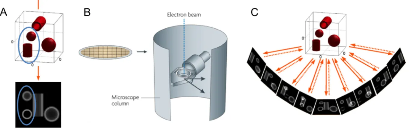
The principle of ET has been fully described in two reviews (McIntosh, Nicastro et al. 2005, Barcena and Koster 2009). Briefly, TEM images give misleading information about the structural properties of biological samples. For example, in the 0° projection image of a sample, a small vesicle and a small tubule have a similar profile (Fig. 4A, blue ovals). One way to solve this problem is to collect a series of two-dimensional (2D) TEM images of these samples from different directions, which is the basic principle of ET.
There are several steps involved in ET: sample preparation, data collection, image alignment, tomographic reconstruction, post-processing and interpretation of the tomograms. Both chemical-fixation and high-pressure freezing coupled with freeze substitution [HPF/FS, including a fast-freezing (20–50 msec) step under high-pressure, a water substitution step (dehydration at −80°C to −90°C with organic solvent), and a subsequent resin embedding step.] prepared samples have been used for ET data collection. The thickness of the sections up to 300 nm that can be performed depends on the accelerating voltage of the electron microscope. To collect the raw data, the sections were put on a grid and placed into the tilt holder of an electron microscope (Fig. 4B). The holder is gradually tilted inside the microscope around an axis perpendicular to the electron beam, and the 2D images are recorded at different angles
(single axis tilt series) under the control of an automated data collection software (Fig. 4C, continuous arrows). The range of tilt angles that can be performed is limited (normally up to ±60° or ±70° with a 1-2° interval), because of the limited tilt available in most electron microscopes and the possible holder shadowing at high tilt. The missing image part of the sample can be partially compensated by a dual-axis tilt series sample collection (the second tilt series is taken by rotating the sample in 90°). Then, the recorded raw data is computationally aligned and reconstructed into a series of tomographic slices of the 3D structure, which are shown as serial “virtual sections” (each image shows a slice about 2–10 nm thick) of the sample. Finally, the 3D models are generated with special software. The key step is the segmentation, in which the different structures of interest are separated and masked in different colored surfaces.
Figure 4. The principle of ET.
(A) The 0° projection of a sample results in a 2D image shown below. (B) The grid for the sections supporting, and the tilt holder in the column of an electron microscope. (C) For raw data collection, a series of 2D images are collected at different orientations of a sample (continuous arrows). For the back-projection, each recorded image is used to contribute to a 3D representation (discontinuous arrows). (A) and (C) are taken form (Barcena and Koster 2009), and (B) is taken from (Milne and Subramaniam 2009).
2.2.2 Distinct membrane structures support different steps in virus replication cycle
Distinct membrane structures can be simultaneously observed in one cell that is infected with some (+) RNA viruses, or they can be observed during the time course analysis and showed successive development. TEM-ET shows that these distinct membrane structures may contribute to different infectious steps, such as polyprotein translation and processing, vRNA replication, particles assembly, maturation and release.
1) DENV induced membrane structures.
Distinct membrane structures, including convoluted membranes (CM) and VPs can be found in DENV-infected cells. Immuno-EM studies indicated that CM may represent the site of DENV RNA translation/polyprotein processing (Mackenzie, Jones et al. 1996). The 3D architecture of VPs generated by ET shows a possible topological link between replication and assembly in infected cells (Welsch, Miller et al. 2009). As mentioned before the immuno-EM studies showed that the SMVs in the VPs are vRNA replication sites. The 3D model shows that the inter SMV membranes are continuous with the ER membrane (Fig. 5A, index of left panel), and there are pore-like openings that could enable release of newly synthesized vRNA (Fig. 5A, right panel). The viral particles are located on the nuclear envelope membrane just opposite of the pores, and this area might be for virus budding (Fig. 5A, index of left panel). The particles may travel in the ER and via secretory vesicles to the Golgi apparatus for maturation, since they can also be found in the ER lumen and Golgi apparatus (Fig. 5A, left panel) (Welsch, Miller et al. 2009).
2) SARS-coronavirus induced membrane structures.
The 3D model of SARS-coronavirus induced membrane structures is a unique reticulovesicular network of modified ER that integrates CM, numerous interconnected DMVs, and VPs (Fig. 5B) (Knoops, Kikkert et al. 2008). SARS-coronavirus replicase
vRNA replication, and VPs for virus assembly and budding (Knoops, Kikkert et al. 2008).
3) Hepatitis C virus (HCV) induced membrane structures
HCV is a Hepacivirus in the family Flaviridae. Expression of the entire HCV polyprotein induces the accumulation of vesicles that called membranous web (MW) (Egger, Wolk et al. 2002). Early in HCV infection, the main constituents of MW are SMVs and DMVs; the DMVs are predominating and are the vRNA replication sites, the role of SMVs is unknown (Fig. 5C, left panel) (Romero-Brey, Merz et al. 2012). Late in HCV infection, multi-membrane vesicles (MMVs) become more abundant. MMVs might be a result of a stress-induced reaction (Fig. 5C, right panel) (Romero-Brey, Merz et al. 2012). The 3D reconstructions show that the DMVs seem to be formed as ER protrusions connected to ER membranes via neck-like structures (Fig. 5C, index of the left panel). MMVs are likely to be formed by the extensive enwrapping and curling of membranes (Fig. 5C, index of the right panel) (Romero-Brey, Merz et al. 2012).
4) Coxsackievirus B3 (CVB3) induced membrane structures
CVB3 is an Enterovirus in the family Picornaviridae. Conventional TEM images showed that enterovirus induces either heterogeneous SMVs (Bienz, Egger et al. 1983, Bienz, Egger et al. 1987) or DMVs (Schlegel, Giddings et al. 1996, Wong, Zhang et al. 2008, Kemball, Alirezaei et al. 2010) clustering in the perinuclear region and eventually occupying most of the cytoplasm. However, 3D models of enterovirus CVB3-induced membrane structures that were generated by ET showed different results (Limpens, van der Schaar et al. 2011). For example, the 3D architecture generated on HPF/FS-prepared CVB3-infected cells showed closed single-membrane tubules (SMTs) early in infection (Fig. 5D, left panel), DMVs and multilamellar structures at late stage (Fig. 5D, right panel). The SMTs are the sites of vRNA replication, but the functions of DMVs and multilamellar structures are not clear.
The transformation process of these different membrane structures has been proposed as follows. The membranes of the SMTs become more tightly apposed, resulting in flattened cisternae. Via an enwrapping mechanism, the cisterna curves into a vase-like configuration, and their membrane ends fuse, producing a closed, spherical