Updated standards and processes for accreditation
of echocardiographic laboratories from
The European Association of Cardiovascular
Imaging: an executive summary
Bogdan A. Popescu
1*
, Alexandros Stefanidis
2*
, Petros Nihoyannopoulos
3, Kevin F. Fox
4,
Simon Ray
5, Nuno Cardim
6, Fausto Rigo
7, Luigi P. Badano
8, Alan G. Fraser
9,
Fausto Pinto
10, Jose Luis Zamorano
11, Gilbert Habib
12,13, Gerald Maurer
14,
and Patrizio Lancellotti
151
Department of Cardiology, University of Medicine and Pharmacy ‘Carol Davila’, Euroecolab, Emergency Institute of Cadiovascular Diseases ‘Prof. Dr. C. C. Iliescu’, Sos. Fundeni 258, sector 2, 022328 Bucharest, Romania;2
1st Department of Cardiology, General Hospital of Nicea, 3 D. Mantouvalou Street, 185 54 Piraeus, Greece;3
Hammersmith Hospital, NHLI, Imperial College, London, UK;4
Imperial College, London, UK;5
Department of Cardiology, University Hospitals of South Manchester, Manchester, UK;6
Hospital da Luz, Nova Medical School, Lisbon, Portugal;7
Dell’Angelo Hospital, Mestre, Venice, Italy;8
Department of Cardiac, Thoracic and Vascular Sciences, University of Padova, Padova, Italy;9
Wales Heart Research Institute, Cardiff University, Cardiff, UK;10
Department of Cardiology, Lisbon Academic Medical Centre, University Hospital Santa Maria, CCUL, University of Lisbon, Lisbon, Portugal; 11
University Hospital Ramon y Cajal, Madrid, Spain;12
Aix-Marseille Universite´, Marseille, France;13
Cardiology Department, APHM, La Timone Hospital, Marseille, France;14 Division of Cardiology, Department of Internal Medicine II, Medical University of Vienna, Vienna, Austria; and15
GIGA Cardiovascular Sciences, Department of Cardiology, University of Lie`ge Hospital, Valvular Disease Clinic, CHU Sart Tilman, Lie`ge, Belgium
Received 8 March 2014; accepted after revision 10 March 2014
Standards for echocardiographic laboratories were proposed by the European Association of Echocardiography (now the European Association of Cardiovascular Imaging) 7 years ago, to raise standards of practice and improve the quality of care. Criteria and requirements were published at that time for transthoracic, transoesophageal, and stress echocardiography. This paper reassesses and updates the quality standards to take account of experience and the technical developments of modern echocardiographic practice. It also discusses quality control, the incentives for laboratories to apply for accreditation, the re-accreditation criteria, and the current status and future prospects of the laboratory accreditation process.
-Keywords Accreditation † Re-accreditation † Echocardiography † Echocardiography laboratory † Quality standards
Introduction
The mission of the European Association of Cardiovascular Imaging (EACVI) is ‘to promote excellence in clinical diagnosis, research, technical development, and education in cardiovascular imaging in Europe and worldwide’. The goals of certification and accreditation under the ‘aegis’ of the EACVI are to protect patients from undergo-ing cardiovascular imagundergo-ing examinations performed by unqualified persons and/or in an inappropriate environment, and to set European standards for competence and excellence in this field. The present document is an executive summary focusing specifically on labora-tory accreditation in echocardiography.
A more detailed version of this document can be found on the
Journal website.1
The EACVI provides a voluntary service of lab accreditation and institutions need to submit their applications in order to be accre-dited. Laboratories fulfilling or wishing to fulfil the European stan-dards on echocardiography will also have a strong argument to use when they request appropriate resources to improve their services.
Aims
This paper appraises the published minimum standards2for
accredit-ation of echocardiographic facilities, but also intends to spread the philosophy of homogenizing the echocardiographic practice and equipment across Europe. Laboratory accreditation is designed to apply to all countries whatever their model of provision of
*Corresponding author. Tel:+40 213175227 (B.A.P.)/+30 2132077306 (A.S.); Fax: +40 213175227 (B.A.P.)/+30 2104924472 (A.S.), Email: bogdan.a.popescu@gmail.com (B.A.P.)/ plato203@yahoo.com (A.S.)
echocardiography in order to improve the patients’ care in a way similar across Europe.
This update is needed because cardiovascular ultrasound is an ever-growing and developing technology, with an increasing list of clinical indications and state of the art practices.
The document also discusses: the policy of the EACVI concerning incentives and benefits for accredited laboratories, the change in the review process involving members of the National Societies (NS), criteria for reaccreditation, quality-control measures, and the current status and future aspects of laboratory accreditation.
The basic structure of laboratory
standards
Certification of individual echocardiographers alone cannot guaran-tee a high-quality department. It is also necessary to have adequate
equipment, management, and organization.2,3
The establishment of laboratory accreditation will enable: (i) The development of local autonomy in echocardiography, e.g.
the ability to train doctors and sonographers in echocardiog-raphy and to encourage trainees to sit the individual certifica-tion examinacertifica-tions. Accredited laboratories will also have to assure continuing professional development for already certi-fied individuals.
(ii) A secured quality of basic or advanced echocardiography for the patient. To satisfy the progressively increasing subspecialization in echocardiography, laboratory standards are available in three modules
(a) transthoracic echocardiography (TTE); (b) transoesophageal echocardiography (TOE); (c) stress echocardiography.
Throughout this document, the term ‘echocardiographer’ is used to include any person who is nationally authorized to perform echo-cardiography. We acknowledge that in some countries within the
EACVI, non-medical qualified echocardiographers perform
cardiac ultrasound. Throughout this document, the term ‘sonogra-pher’ is used to mean a non-medical echocardiographer and subsumes the terms clinical physiologist, nurse, cardiac or echocar-diography technician, and/or radiographer. It is also recognized that while the vast majority of echo laboratories will need to provide only a routine clinical service to their institution, a number of laboratories will have also academic endeavours with commitments to teaching and research. To this end, there will be two levels of minimal standards leading to respective laboratory accreditations.
(i) The basic standard: this will aim to fulfil ‘mandatory’ requirements offering an adequate basic clinical service. It is postulated that the majority of echocardiographic services in each country will fulfil these basic requirements.
(ii) The advanced standard: this will aim to fulfil requirements and offer an advanced service with state-of-the-art equipment and is also accredited for training and research. For this level, it will be necessary for the laboratories to have a history of re-search as well as teaching with state-of-the-art equipment
performing all echo modalities such as Doppler tissue imaging, contrast, three-dimensional, and speckle tracking echocardiography.
Grading process of applications
Laboratories eligible for the EACVI Laboratory Accreditation pro-gramme have to meet the following requirements: (i) being estab-lished for at least 3 years and (ii) at least one of the senior physician members of the echo-lab staff should hold a valid
individ-ual TTE certification as described in Tables1–3as a pre-requisite for
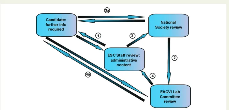
the standard level of the TTE module of lab accreditation. Labora-tory applications are submitted through an online platform enclos-ing several forms and certifications and are first checked for their administrative content. If the data are incomplete the applicants will be contacted and asked to supply missing information within 1 month. After this initial step, the EACVI adopted a revised strategy in terms of the grading process of new applications. This novel policy creates a direct chain of communication between the NS/EACVI
reviewers and the applying laboratory (Figure1). The involvement
of NS will make the process more efficient while also reinforcing the partnership between the NS and the EACVI. In the beginning, senior NS-EACVI members will be involved in the accreditation process taking action as ‘local NS representatives’ and after this initial phase members of the EACVI Lab Committee will review the application.
Upon review by the NS Representative, the EACVI Laboratory Ac-creditation SubCommittee will review the index application, discuss the initial recommendation and finalizes the investigation. A possible rejection of an application does not preclude a second revision by the committee after the applicant lab has adopted a number of recom-mended changes/improvements.
Quality-control measures
As the applications are online and the reviewing of applications is largely a paper-based one with reviewers assessing self-reported documents and data submitted by the laboratories, it was felt neces-sary to implement an additional level of quality-control check. Thus, starting with 2012, every year a number of accredited laboratories, randomly selected, are visited onsite by a team of two EACVI and one NS representative. The visit is announced in advance and the lab prepares a number of documents and information for this purpose. The aim of such visits is to assure the accuracy of the infor-mation submitted and to provide support for improvements. In case of discrepancies, corrective measures are suggested within a certain timeframe. The EACVI Board and laboratory accreditation commit-tee considered this as an important ‘post-accreditation’ addition to the existing ‘pre-accreditation’ quality-control measures (http:// www.escardio.org/communities/EACVI/accreditation/lab/Pages/ process.aspx).
Apart from these external measures/verifications, this writing committee reinforces the internal laboratory quality-control recom-mendations contained in the document on training, competence, and
Benefits for the accredited
laboratories
The current strategy of the EACVI regarding accredited laboratories is not only promoting excellence and encouraging quality improve-ment but also giving prospects for rewards
(i) Educational
† preference to be hosting centres for EACVI grants (i.e. pres-tige and reimbursement)
† preference in participation in specific EACVI educational pro-jects (e.g. e-learning)
† preference to be hosting centres for fellowships
† preference in the selection of location for educational courses and meetings
(ii) Scientific
† preference in participation in specific EACVI multicentric
sci-entific projects4
(iii) Research
† strong preference for research programmes5
(iv) Economical
† preference to be selected as sites for trials involving echo sub-studies with clear quality-control requirements.
Re-accreditation process
The aims of reaccreditation are to maintain quality standards for the labs already accredited by the EACVI, while also keep on meeting the standards of clinical competence. The process for re-accreditation should be more flexible, accessible, and faster
. . . . Table 1 Summary of criteria for rating transthoracic echocardiography
Basic standard Advanced standard
Staff
Both clinical and technical heads of Echocardiography Clinical head performs at least one session including transthoracic studies each week
The clinical head holds a valid EACVI or recognized NS individual adult TTE certification or a Level III training (American Society of Echocardiography) plus a valid NBE (American National Board of Echocardiography) certification (Examination of Special Competence in Adult Echocardiography-ASCeXAMw)
with an EACVI TTE individual certification practical e-logbook
Both technical and clinical heads possess individual TTE certifications as described for the standard level
Technical head spends six or more sessions in echocardiography activities (including management or quality control)
At least two echocardiographers hold EACVI or recognized NS adult TTE certification
Organization/equipment
Studies archived. Written reports of routine studies issued within 24 h at the latest
Digital archiving of both reports and images for all studies (scheduled and emergency). Written reports issued on the day of the examination
System of review for echocardiograms in place Formal and systematic quality control Standardized examination protocol and list of indications for echocardiographic
studies
Agreed minimum standards, standardized examination protocol and list of indications for echocardiographic studies
Provision for continuing education System of liaison with other departments to advise about timing of or results of studies
All machines have second harmonic imaging and full quantitation package Less than 1500 studies per echocardiographer per annum All machines have colour and spectral Doppler All machines have stand-alone continuous wave Doppler probes At least one machine has stand-alone continuous wave Doppler probe Weekly departmental meetings
No machine in regular use upgraded .7 years ago Available standard operating procedures
Maintenance and scheduled service programme of echo machines A core library with echo and general cardiology textbooks and preferably access to cardiology journals and updated books electronically
30 – 40 min allocated per standard study and up to 1 h for a complex study There should be regular teaching to junior doctors, fellows and sonographers with appropriate provision of teaching material (videos, CDs, books, etc)
Compliance with appropriate European and national personal data’s protection legislation
Advanced quantitation (tissue imaging, 3D, contrast, regurgitant volumes) when needed
Rooms uncluttered and of adequate size Evidence of scientific work produced by the department Appropriate provision of patient facilities and information History of success in training for EACVI/national accreditation
than the initial. The intention of the committee is to encourage a straightforward, less complex, and less expensive process for reac-creditation. The criteria and requirements for reaccreditation are identical with the initial ones. Applicants for re-accreditation have to fill in a new application form containing appropriate fields for
description of changes that have possibly occurred since the first ac-creditation was issued. Documentation of the reacac-creditation process must be kept on file and available for inspection upon request. The Accreditation committee might still conduct site visits after successful reaccreditation.
. . . . Table 3 Summary of criteria for rating stress echocardiography
Basic standard Advanced standard
Staff
Designated head of stress echocardiography Head maintains continuous medical education for stress echo Performing a minimum of 100 studies/year per laboratory More than 300 studies/year per laboratory
Studies performed by at least two people, one of whom is a clinician. At least one must have advanced life support or equivalent
Head has a substantial experience of TTE and stress echo Organization/equipment
List of indications, provision of information to the patient and written informed consent Machine capable of changing mechanical index and have full digital stress echo package
ECG and BP monitoring capabilities (see text for details) Audit of results against angiography or other independent standard
Established appropriate protocols Advanced software dedicated to contrast imaging Machine with second harmonic imaging and TDI software Capability for both pharmacological and exercise stress Resuscitation facilities readily available and record of complications Additional quantification package should be available Lockable drug cupboard Standard operating procedures should be available Contrast agent for left ventricle opacification available A history of training junior doctors
Provisions for continue educational activities
. . . . Table 2 Summary of criteria for rating transoesophageal echocardiography
Basic standard Advanced standard
Staff
Designated head of TOE Head of TOE performs/supervises .50 studies each year Designated head should be performing or supervising at least 50 TOE
annually
Head of TOE has the EACVI/recognized NS certification in TOE Designated person, usually a nurse, to manage airway and recover the
patient Organization/equipment
Established protocols and list of indications for TOE agreed internally Recovery area
Room typically 20 m2in area Minimum standards for studies established
Written informed consent Quality control of results, e.g. against surgery, pathology, other imaging Provision for continuing education Regular audits
Resuscitation equipment Written standard operating procedures Provision for quality control History of success in training students Thorough and precise report Digital storage and retrieval Routine use of Provision of intra-operative services
Patient preparation including letter and pre-procedural checklist 3D imaging is recommended Multiplane probe
Suction, oxygen and pulse oximeter, BP monitoring Sedation used according to published guidelines Lockable drug cupboard
Facilities for cleaning/sterilizing the probe Electrical safety testing for TOE probe
Current status of accredited
laboratories and future aspects
To date, 46 laboratories received European accreditation. Details about the accredited labs can be found on the relevant EACVI ac-creditation webpage.
Applications from laboratories in countries outside Europe/ESC countries are allowed, provided they strictly follow the same process and meet all the required recommendations.
The ongoing development of the new ESCel platform, apart from an educational and collaborative tool between the ESC and NS will also assist training and accreditation in several modules and subspe-cialties. The purpose of this platform is the provision of a user-friendly, flexible, modular software tool while reducing the costs
related to education, training, and certification/accreditation in Europe. After a period of repeated testing, demonstrations, and simu-lation, the ESCel will be implemented for the needs of the EACVI, both for individual certification and lab accreditation. This implemen-tation is supposed to make the whole accrediimplemen-tation process easier for both the applicant laboratories and the assessors.
Conclusions
In this document, we raised the standards for echocardiography
la-boratories as initially set in the former document.2These updated
recommendations were reassessed to recognize facilities of either standard or advanced level of laboratory accreditation. Important new topics included in this update are related to quality control,
Figure 1: Review of the grading process. Applications are sent initially to the ESC staff. Double arrows: when data are missing the reviewer requests further information. The candidate sends the corrected/completed information. This exchange could happen a number of times if neces-sary.
Figure 2: Framework for assessing quality in echocardiography and its influence on patient management. The proposed model consists of four main domains which may influence clinical outcome. Laboratory infrastructure and organization support the whole echocardiography process. Reproduced with permission from Ref.3.
re-accreditation criteria, and possible incentives/benefits for the accredited laboratories to encourage applications.
Acknowledgements
The following individuals constitute the current Echocardiography Laboratory Accreditation Committee (2012-2014). We are deeply grateful for all their hard work. Dr Alexandros Stefanidis (Greece, Accreditation Chair), Dr Nuno Cardim (Portugal, Co-chair), Dr Fausto Rigo (Italy, Co-chair), Dr George Athanassopoulos (Greece), Dr Luigi Badano (Italy), Dr Roland Brandt (Germany), Dr Ole Breithardt (Germany), Mr David Dawson (UK), Dr Kevin Fox (UK), Dr Piotr Hoffman (Poland), Dr Patrizio Lancellotti (Belgium), Dr Aleksandar Neskovic (Serbia), Dr Hans Joachim Nesser
(Austria), Dr Petros Nihoyannopoulos (UK), Dr Bogdan
A. Popescu (Romania), Dr Simon Ray (UK), Dr Rick Steeds (UK), Dr Heikki Ukkonen (Finland).
Conflict of interest: none declared.
References
1. Popescu BA, Stefanidis A, Nihoyannopoulos P, Fox KF, Ray S, Cardim N et al. Updated standards and processes for accreditation of echocardiographic laboratories from The European Association of Cardiovascular Imaging. Eur Heart J Cardiovasc Imaging 2014;15:717 – 27.
2. Nihoyannopoulos P, Fox K, Fraser A, Pinto F, on behalf of the Laboratory Accredit-ation Committee of the EAE. EAE laboratory standards and accreditAccredit-ation. Eur J Echo-cardiogr 2007;8:80 – 7.
3. Popescu BA, Andrade MJ, Badano LP, Fox KF, Flachskampf FA, Lancellotti P et al. Euro-pean Association of Echocardiography recommendations for training, competence and quality improvement in echocardiography. Eur J Echocardiogr 2009;10:893 – 905. 4. Lancellotti P, Badano LP, Lang RM, Akhaladze N, Athanassopoulos GD, Barone D et al. Normal Reference Ranges for Echocardiography: rationale, study design, and meth-odology (NORRE Study). Eur Heart J Cardiovasc Imaging 2013;14:303 – 8. 5. Donal E, Badano L, Habib G, Maurer G, Lancellotti P. Research and Innovations
Com-mittee a new outlook for the European Association of Cardiovascular Imaging (EACVI). Eur Heart J Cardiovasc Imaging 2013;14:400. doi:10.1093/ehjci/jes322.
IMAGE FOCUS
. . . .
doi:10.1093/ehjci/jeu072
Online publish-ahead-of-print 17 June 2014
It’s just a coronary milking-like effect . . . or maybe not: posterolateral artery
compression caused by a ventricle pseudoaneurysm
Victor Alfonso Jime´nez*, Jorge Sepu´lveda, Josue´ Ponce, Jose´ Antonio Baz, and Andre´s I´n˜iguez
Hemodynamics and Interventional Cardiology Department, Hospital Meixoeiro, University Hospital of Vigo, Meixoeiro s/n, 36200 Vigo, Pontevedra, Spain
*Corresponding author. Tel:+34 622 323 046; Fax: +34 986 811 727. Email: victor.alfonso.jimenez.diaz@sergas.es, sooner_79@hotmail.com
A 57-year-old man was referred to our catheteriza-tion laboratory due to angina complaints on exer-tion and an inconclusive treadmill stress test. He had a history of stent implantations in the right cor-onary artery (RCA) and left anterior descending (LAD) artery 2 years before. Current coronary angi-ography (see Supplementary data online, Video S1) showed neither de novo coronary stenosis nor stent restenosis. Instead, a large posterolateral branch (PLB) of the RCA with complete compres-sion throughout its middle segment during systole
(Panel A), but with unimpaired diastolic blood flow (Panel B), was noticed. Left ventriculography (see Supplementary data online, Video S2) showed a large pseudoaneurysm in the inferolateral wall with the PLB crossing below it (Panels C and D), subsequently confirmed by computed tomography (Panels E and F ) and cardiac magnetic resonance imaging (Panels G and H ). Because of the patient symptoms and the high risk of rupture of the pseudoaneurysm over time, surgical repair was performed, achieving successful outcome and uneventful recovery. The patient remains asymptomatic after 6-month follow-up. Coronary artery constriction by a pseudoaneurysm mimicking an angiographically milking effect is an exceptional finding that resembles the milking effect of myocardial bridging, the latter being almost exclusively demonstrated in the LAD.
Coronary angiography images showing the PLB of the RCA compressed on its middle segment during systole (Panel A) but with pre-served blood flow during diastole (Panel B). Left ventriculogram images showing the large pseudoaneurysm in the inferolateral wall of the LV during systole (Panel C) and during diastole (Panel D). Computed tomography scan images with reconstruction of the RCA (Panels E and F ) showing the previously implanted stents (black arrows), and the course of the PLB below the pseudoaneurysm with the absence of contrast along its middle segment due to extrinsic compression by the pseudoaneurysm (white arrow). Cardiac magnetic resonance images (Panels G and H ) showing the pseudoaneurysm (arrow) with a large thrombus inside.
Conflict of interest: none declared.
Supplementary data are available at European Heart Journal – Cardiovascular Imaging online.