Université de Montréal
CXCR3 biased signaling, heteromerization and decoy properties
Par François Guité-Vinet
Département de microbiologie, infectiologie et immunologie Faculté de Médecine
Mémoire présenté à la Faculté de Médecine en vue de l’obtention du grade de Maîtrise en microbiologie et immunologie
Juin 2015
Membres du jury :
Dr. Nikolaus Heveker – Directeur de recherche Dre. Petronela Ancuta – Présidente Dr. Hugo Soudeyns – Membre du jury
i
Résumé
Le récepteur de chimiokine CXCR3 est un récepteur couplé à la protéine G (RCPG) exprimé, entre autre, sur les cellules T activées lors d’une réponse immune. CXCR3 est activé par trois ligands inductibles par l’interféron-γ (CXCL9, 10, 11) et, plus récemment, il a été découvert que CXCL4 liait CXCR3. Nous savons que CXCR3 joue un rôle dans la chimiotaxie des leucocytes, mais une attention limitée a été portée sur la signalisation biaisée induite par ces quatre ligands. Alors que l’homodimérisation entre récepteurs de chimiokine est un concept grandement observé, l’hétéromérisation entre deux récepteurs reste un domaine de recherche active. Enfin, certains récepteurs de chimiokine (decoy) jouent sur le gradient de chimiokines en les dégradant. À ce jour, aucune donnée n’a présenté CXCR3 en tant que récepteur decoy. La signalisation biaisée et l’hétéromérisation ont été testées grâce à la technique de bioluminescene resonance energy transfer (BRET) dans des cellules HEK293E. L’activité decoy de CXCR3 a été investiguée grâce à un essai de dégradation de chimiokines radio-marquées à l’iode 125. Nous présentons une caractérisation pharmacologique des quatre ligands de CXCR3 et démontrons l’hétéromérisation de CXCR3 avec CXCR4 et avec CXCR7. Nous démontrons que CXCR3 peut agir comme decoy en dégradant CXCL11 radio-marqué. Nos résultats suggèrent que les ligands de CXCR3 n’agissent pas de manière redondante. De plus, nos résultats de dégradation suggèrent l’absence de compétition entre les ligands de CXCR3. Enfin, nous montrons que CXCL12 n’affecte pas la dégradation de CXCL11 par CXCR3 ni par l’hétéromère CXCR3/CXCR4.
Mots clés
CXCR3, RCPG, Signalisation biaisée, Hétéromérisation, BRET, Récepteur decoy, CXCL11 radio-marquée.
ii
Abstract
The chemokine receptor CXCR3 is a G-protein-coupled receptor (GPCR) rapidly induced on naïve T cells upon activation. CXCR3 is activated by three interferon-γ inducible ligands (CXCL9, 10, 11) and, more recently, CXCL4 has been discovered as a functional ligand for CXCR3. It is known that CXCR3 acts as a chemotactic receptor, but limited attention has been directed to the biased signaling induced by all four ligands. Chemokine receptor homodimerization is now a widely accepted concept, but the extent to which heterodimerization is prevalent remain matter of active research. Some chemokine (decoy) receptors have been reported to maintain/re-shape specific chemokine gradients by degrading chemokines. To date, CXCR3 has never been described as a decoy receptor.
In this work, biased signalling and heterodimerization were assessed with bioluminescence resonance energy transfer (BRET) in HEK293E cells. CXCR3 decoy properties were investigated by radiolabelled chemokine degradation assays. We present pharmacological characterization of all four ligands of CXCR3 and heterodimerization of CXCR3 with CXCR4 or CXCR7. Finally, we present CXCR3 decoy activities on radiolabelled CXCL11. Our results suggest that CXCR3 ligands are not redundant and that CXCR3 heterodimerizes with CXCR4 and with CXCR7. Our results also suggest that CXCR3 is able of CXCL11 scavenging. Our degradation assays demonstrated the absence of competition between ligands. Finally, CXCL12 did not affect CXCL11 scavenging neither by CXCR3 nor by CXCR3/4 heterodimer.
Key words
CXCR3, GPCR, Biased signalling, Heteromerization, BRET, Decoy receptor, Radiolabelled CXCL11.
iii
Table of Contents
Résumé ... i
Abstract ... ii
Table of Tables ... v
Table of Figures ... vi
Abbreviations ... viii
Acknowledgements ... xi
1.
Introduction ... 1
1.1 Chemokines and chemokine receptors ... 1
1.1.1.Nomenclature of chemokines and chemokine receptors ... 1
1.1.2 Biological functions of chemokines ... 1
1.2 Pathophysiological relevance of CXCR3 and its ligands ... 3
1.2.1 Role of CXCR3 and its ligands in atherosclerosis ... 3
1.2.2. Role of CXCR3 and its ligands in multiple sclerosis ... 4
1.2.3 Role of CXCR3 and its ligands in asthma ... 5
1.2.4 An example of a clinical trial ... 6
1.3 Challenges and misconceptions in chemokine receptor drug design ... 7
1.3.1 Inappropriate target selection ... 7
1.3.2 Inappropriate animal models ... 8
1.3.3 The misconception of chemokine redundancy ... 9
1.4 Molecular basis of the chemokine system ... 11
1.4.1 Chemokine receptor signaling through G proteins and desensitization by β-arrestin ... 11
1.4.2 Some principles of pharmacology ... 12
1.4.3 The concept of functional selectivity (or biased agonism) ... 14
1.4.4 Chemokine receptor heteromerization ... 15
1.4.5 CXCR3 splicing ... 16
1.4.6 CXCL4 as a novel ligand for CXCR3 ... 17
1.4.7 Chemokine gradient remodelling ... 18
1.5 Objectives ... 20
iv
2.1 Plasmids ... 22
2.2 Reagents ... 22
2.3 Cell culture and transfection ... 22
2.4 BRET measurements ... 23
2.5 Gαi activity assays ... 23
2.6 Arrestin recruitment assays ... 23
2.7 BRET saturation assays ... 24
2.8 Degradation assays ... 24
2.9 Flow cytometry ... 25
2.10 Data analysis ... 25
3.
Results ... 26
3.1 CXCR3A biased signaling ... 26
3.2 CXCR3A Heterodimerization... 33
3.3 CXCR3A Decoy properties ... 41
3.4 Effects of CXCL4 on CXCR3A ... 52
4.
Discussion ... 57
4.1 CXCR3A biased signaling ... 57
4.2 CXCR3 homo- and heteromerization ... 61
4.3 CXCR3 decoy activities ... 65
4.4 Characterization of CXCL4, a novel ligand of CXCR3 ... 69
5. Conclusion ... 71
v
Table of Tables
Table 1. Distinction between BRET1 and BRET2 assays ... 27 Table 2. Pharmalogical parameters of CXCR3A biased signalling ... 32
vi
Table of Figures
Figure 1. Schematic view of the possible interactions between different ligands and
CXC-chemokine receptors. ... 10
Figure 2. Schematic view of chemokine receptor signalisation, phosphorylation by GRK, desensitization and subsequent internalization by β-arrestin. ... 12
Figure 3. The principles of efficacy and potency in pharmacology. ... 13
Figure 4. Overall overview of the chemokine system studied in this present work. ... 20
Figure 5. Schematic representation of BRET1 and BRET2 assays ... 27
Figure 6. Schematic representation of EPAC BRET2 assays. ... 28
Figure 7. Inhibition of Adenylate cyclase activity followed by stimulation of CXCR3A by chemokine CXCL11, CXCL10 or CXCL9. ... 30
Figure 8. β-Arrestin-2 recruitment to CXCR3A followed by chemokine CXCL11 , CXCL10 or CXCL9 stimulation. ... 31
Figure 9. Interpretation of BRET saturation curves ... 34
Figure 10. CXCR3A forms heterodimer with CXCR7 and with CXCR4 in HEK293E cells. . 36
Figure 11. CXCR3 has no preference for heterodimerization with CXCR4 or CXCR7. ... 37
Figure 12. CXCR7 forms homodimers and heterodimers with CXCR3A in HEK293E cells. 38 Figure 13. CXCR3 forms homodimers in HEK293E cells. ... 39
Figure 14. CXCR7 tendency for homodimerization. ... 40
Figure 15. The typical chemokine receptor CXCR3 acts as scavenger for CXCL11. ... 43
Figure 16. CXCL11 is degraded by CXCR3 and CXCR7. ... 44
Figure 17. Membrane expression of CXCR3A and CXCR7. ... 45
Figure 18. Decoy properties of chemokine receptors CXCR3A in the presence or absence of other ligands. ... 46
Figure 19. CXCR3A ligands have allotopic properties. ... 47
Figure 20. Membrane expression of CXCR3A... 48
Figure 21. Decoy properties of chemokine receptors CXCR3A, CXCR4 and the CXCR3A/CXCR4 heterodimer in the presence or absence of CXCL12. ... 49
Figure 22. CXCL12 does not affect CXCL11 scavenging neither by CXCR3A nor CXCR3A/4. ... 50
Figure 23. Membrane expression of CXCR3A, CXCR4 and CXCR3A/4. ... 51
Figure 24. Inhibition of adenylate cyclase following stimulation of CXCR3A by chemokine CXCL11 or CXCL4. ... 53
Figure 25. β-Arrestin-2 recruitment to CXCR3A followed by chemokine CXCL11 or CXCL4 stimulation... 54
Figure 26. Decoy properties of chemokine receptor CXCR3A in the presence or absence CXCL4. ... 55 Figure 27. High concentration of CXCL4 does not block CXCL11 scavenging by CXCR3A.56
vii Figure 28. Probabilities of homo- and heterodimers formation in co-transfected cells... 67
viii
Abbreviations
AC Adenylate cyclase
ACKR Atypical chemokine receptor
AIDS Acquired immunodeficiency syndrome
ATP Adenosine triphosphate
ANOVA Analysis of variance
ASM Airway smooth muscle
BRET Bioluminescence resonance energy transfer
BRET50 Half maximal bioluminescence resonance energy transfer value
BSA Bovine serum albumin
C-terminal Carboxy-terminal
Ca2+ Calcium ions
cAMP Cyclic adenosine monophosphate
cDNA Complementary deoxyribonucleic acid
CHU Centre hospitalier universitaire
COPSE Comité d'organisation du programme des stages d'été CD4 Cluster of differentiation 4
CD8 Cluster of differentiation 8
CNS Central nervous system
DNA Deoxyribonucleic acid
DMEM Dulbecco's modified Eagle’s medium
ix EC50 Half maximal effective concentration
EPAC Exchange protein directly activated by cAMP ERK Extracellular signal regulated kinase
F Forskolin
FRET Fluorescence resonance energy transfer
GABA γ-aminobutyric acid
GFP10 Green fluorescent protein 10
GPCR G protein-coupled receptor
GRK G protein-coupled receptor kinase
GDP Guanosine diphosphate
GTP Guanosine triphosphate
GTPγS Guanosine 5'-O-[gamma-thio] triphosphate
HEK Human embryonic kidney
HLMC Human lung mast cells
HIV Human immunodeficiency virus
HMVEC Human microvascular endothelial cells
IFNγ Interferon-gamma
IP-10 CXCL10, Interferon-gamma inducible protein 10
I-TAC CXCL11, Interferon–inducible T cell alpha chemoattractant
L Ligand
MAPK Mitogen-activated protein kinase
MIG CXCL9, Monokine induced by interferon-gamma
mRNA Messenger ribonucleic acid
N-terminal Amino-terminal
x
MS Multiple sclerosis
PBS Phosphate buffered saline
PF-4 CXCL4, Platelet factor-4
PKC Protein kinase C
PLC Phospholipase C
PTX Pertussis toxin
RA Rheumatoid arthritis
Rluc Renilla luciferase
Rluc3 Renilla luciferase 3
RNA Ribonucleic acid
SDF-1α CXCL12, Stromal cell-derived factor 1 alpha
SDS Sodium dodecyl sulfate
SEM Standard error of the mean
TCA Trichloroacetic acid
TNFα Tumor necrosis factor α
TREG Regulatory T cell
wt Wildtype
YFP Yellow fluorescent protein
xi
Acknowledgements
What would we be, students, without motivated and motivating mentors? I had the chance and the huge opportunity to work with Dr. Nikolaus Heveker, who really introduced me to research and, most importantly, who transmitted to me his contagious passion for science. A special thank to you Nikolaus.
Who says student, says student’s life. I shared specials moments will all my laboratory team. It surely would have been harder without them. In order of appearance, from my first internship to my last days on the bench: Stéphanie Gravel, Nicolas Montpas, Nassr Nama, Geneviève St-Onge, Marilou Lefrançois, Julien Bonneterre and Mathias Plourde. Thank you again for your support. We had a good time. But, before I forget, a rainbow thank for Nassr, my constant source of motivation.
I also want to thank my family for constant support. My grandparents, Raymond and Fernande, my parents, Luce and Claude, my aunts Michelle and Renée and my brothers Julien and Vincent.
Finally, an ultimate thank for the financial support from COPSE, CHU Sainte-Justine, the Faculty of Medicine and the Canadian Institutes of Health Research.
1
1. Introduction
1.1 Chemokines and chemokine receptors
Chemokines form a large family of proteins that can be divided into sub-families based on their structures and on their functions.
1.1.1. Nomenclature of chemokines and chemokine receptors
Originally, chemokines have been named randomly without a clear system of nomenclature. Some chemokines were included in the large family of interleukins (e.g. IL-8), while others were given a name suggesting their function (e.g. I-TAC for Interferon-inducible T-cell Alpha Chemoattractant)[1].
More recently, members of the chemokine family were classified into four different groups according to their NH2-terminal cysteine-motifs. There are now C chemokines, CC chemokines, CXC chemokines and finally, CX3C chemokines [1]. For example, the first chemokine of a group having one residue between its two cysteines would be named CXCL1. Here, L stands for ligand.
Chemokine receptors are classified according to their ability to respond to a certain chemokine family. For instance, a chemokine receptor capable to interact with CXCL1 would be called a CXC Receptor, or CXCR.
The present master’s thesis deals mainly with the chemokine receptor CXCR3 and with its interactions with the chemokine receptors CXCR4 and CXCR7.
1.1.2 Biological functions of chemokines
Chemokine are chemotactic cytokines involved in the directed migration of cells; a process called chemotaxis. Chemotaxis needs the formation of a concentration gradient of
2 chemokines. Cells that are attracted by chemokines move through the gradient from lower to higher concentration.
It has been established that some chemokines play essential roles in hematopoiesis and organ development [2]. For instance, CXCL12 is a constitutively expressed chemokine from bone marrow stromal cells. This homeostatic chemokine promotes proliferation of B cell progenitors[3]. CXCL12 also induces the migration of hematopoietic precursors to the bone marrow during embryogenesis [4, 5]. Of note, CXCL12 binds two different chemokine receptors: CXCR4 and CXCR7.
Chemokines are chemotactic, homeostatic, but also immunoregulatory proteins. In fact, chemokine were originally discovered as mediators of relocalization processes during inflammatory and immune responses. For example, some chemokines guide immune cells from tissues to the lymph nodes so they can act as antigen-presenting cells [2]. When microorganisms are phagocytosed by dendritic cells in peripheral tissues, these dendritic cells mature [2]. Then, the mature dendritic cells start to express the chemokine receptor CCR7. This chemokine receptor allows the dendritic cells to migrate in response to CCR7 ligands into the lymph nodes in order to present processed antigen to T cells[2].
During an immune response, different subsets of T cells are generated following the interaction of naïve T cells with antigen-presenting cells in the lymphoid compartment [6]. Once activated, these T cell subsets upregulate chemokine receptors, which guide them out of the lymphoid compartment towards sites of injury[6]. For instance, CXCR3 is upregulated on T cells following activation and is implicated in their migration.
3
1.2 Pathophysiological relevance of CXCR3 and its ligands
The expression of CXCR3 is associated with CD4+ type-1 helper and CD8+ cytotoxic lymphocytes[7-10] but is also expressed on various other cells types like B cells, NK cells, smooth muscle cells and endothelial cells[2].
CXCR3 is activated by three ligands: CXCL9 (Monokine induced by IFN-γ or MIG), CXCL10 (IFN-γ inducible Protein 10 or IP-10) and CXCL11 (Interferon-inducible T-cell Alpha Chemoattractant or I-TAC). In contrast to the constitutive chemokine CXCL12 secreted by the bone marrow stromal cells, the three ligands of CXCR3 are not constitutively expressed, but are up-regulated in an IFN-γ cytokine environment [11]. Neutrophils, monocytes/macrophages, dendritic cells, CD4+, CD8+, NK and NK-T cells, in response to IFN-γ, can secrete CXCL9, CXCL10 or CXCL11[11-14].
Consequently, the role of CXCL9-10-11 is to recruit immune cells to inflammation sites [6, 15]. Therefore, a prevailing function in inflammatory and immune diseases has been suggested for CXCR3 and its ligands. For instance, it has been proposed that CXCR3 pathways are involved in local amplification loops of inflammation in targeted regions [6, 15].
We will briefly discuss the pathophysiological relevance of CXCR3 and its ligands in auto-immune/inflammatory diseases such as atherosclerosis, multiple sclerosis and asthma.
1.2.1 Role of CXCR3 and its ligands in atherosclerosis
The early phase of atherosclerosis, atherogenesis, is characterized by the adherence of blood circulating monocytes to the surface of arterial walls and by their migration to the sub-endothelial space where they undergo transformation into macrophages and causes fibrotic plaques [16, 17]. These plaques, called atheroma, are accumulated fatty substance and cells in
4 the inner layer of arterial walls. The worsening accumulation of cells and debris causes atherosclerosis.
These plaques contain the cytokine IFN-γ, considered as the master regulator of atherosclerosis [18]. The differential expression of the three IFN-γ–inducible chemokines CXCL9, CXCL10 and CXCL11 by atheroma-associated cells was reported [19]. These findings suggest that the expression of CXCR3 ligands by atheroma-associated cells act as recruiters and retainers of activated T lymphocytes within the vascular wall lesions in the course of atherogenesis [19]. Moreover, the expression CXCR3 by all T lymphocytes within human atherosclerotic lesions in situ was observed[19]. Since these cells persist at the site of the lesion, it has been suggested that they may play an important role in the development of atherosclerosis.
Combined treatment with two drugs, simvastatin with niacin, provided clinical benefits in patients suffering from atherosclerosis [20]. Simvastatin is an inhibitor of HMG-CoA reductase, it reduces low-density lipoproteins also called the "bad" cholesterol [21]. Niacin, also known as vitamin B3, appears to reduce the risk of cardiovascular disease[22]. However, since the complications of advanced atherosclerosis are chronic, there is an emerging need for alternative or complementary therapeutic interventions. For instance, these alternatives could target molecular mechanisms underlying the initiation of cell recruitment into the arterial wall’s plaques [17]. It was reported that a CXCR3 antagonist attenuated atherosclerotic lesion formation by blocking direct migration of CXCR3+ effector cells from the circulation into the atherosclerotic plaque in mice[23]. Therefore, CXCR3 antagonists may be a possible therapy to inhibit inflammation-induced leukocyte migration and to subsequently reduce atherogenesis [23]. A better understanding of CXCR3 signalling is mandatory for better drug development in atherosclerosis.
5 Multiple sclerosis (MS) is an inflammatory disorder of the human central nervous system (CNS) [24]. This inflammatory disease largely involves mononuclear phagocytes and T cells [25]. These cells enter into the brain through a permissive brain-blood barrier[26] and attack myelin because they recognize it as an antigen [27]. Thus, it was argued that chemokine and chemokine receptors expressed on these cells may play a role in MS. However, other immunomodulatory cytokines, like tumor necrosis factor alpha (TNFα) or interferons-β (IFN-β), can also be implicated in MS [28].
It was reported that CXCL10 and CXCR3 co-localized in the inflamed CNS [29]. In addition, elevated levels of three chemokines, CXCL9, CXCL10 and CCL5 (the ligands of the chemokine receptors CCR1, CCR3 and CCR5) were observed in the cerebrospinal fluid of patients[25]. Compared with the circulation, the cerebrospinal fluid was significantly enriched in CXCR3+CCR5+ cells [25, 30]. These findings imply that specific chemokine–chemokine receptor interactions, and more precisely CXCR3 and CCR5 with their ligands, play important pathogenic roles in MS.
Current MS treatments include interferon immunosuppressive agents and antibodies [31]. However, these treatments often have unsatisfactory outcomes [31][32]. For example, the neutralizing antibodies persist in the body and reduce the biologic activity of IFN-β therapies. This persistence of neutralizing antibodies is also associated with a reduced treatment efficacy [32]. One promising avenue to overcome this issue is the CXCR3/CXCL10 axis. A better comprehension of CXCR3+ cells chemotaxis towards the CNS will surely gives us precious insight into how to block their migration. This could lead to an alternative treatment of MS patients that are now treated with antibodies and immunosuppressive agents.
1.2.3 Role of CXCR3 and its ligands in asthma
Asthma is an inflammatory lung disease characterized by airflow obstruction, airway contraction and hyper-responsiveness. The infiltration of airway smooth muscle (ASM) by mastocytes, also called mast cells, is a major determinant of the asthmatic phenotype [33].
6 The localization of mast cells within the airway structures is important in the pathophysiology of inflammatory lung disease[34]. Cytokine expression is also observed in airway pathology [35, 36]. Chemokines are likely candidates mediating mast cells migration into lung tissues.
Compared to mast cells localized elsewhere, human lung mast cells (HLMC) highly express CXCR3[34]. In addition to CXCR3, it has been found that more than 10 % of ex vivo HLMC were expressing other chemokine receptors such as CCR3 and CXCR4 [34]. A fundamental question is how mast cells accumulate in the ASM in asthma. This is a key question because if this accumulation can be repressed, the symptoms can be attenuated [37]. It has been suggested that the interaction between CXCL10 derived from ASM and CXCR3+ mast cell may be the dominant pathway facilitating the migration of mast cells into the ASM bundles [37].
It has been demonstrated that the neutralization of CXCL10 strongly reduced allergic airway inflammation in a mouse model of asthma[38]. However, to date, no approved drug targeting CXCR3 is available. In fact, inhaled corticosteroids are the preferred treatment for long-term control of symptoms in asthmatic patients[39].
1.2.4 An example of a clinical trial
Even though CXCR3 is considered as a promising drug target [40], the outcome of clinical trials were disappointing. For instance, AMG487, a CXCR3 antagonist, has been tested for the treatment of psoriasis, a skin inflammatory disease in which CXCR3 plays an important role in the development of the pathology [41-43]. In preclinical studies, AMG-487 blocked immune-cell migration and showed excellent potency, selectivity and bioavailability[44]. In a phase 1 trial, the safety and tolerability of AMG487 was confirmed [45]. However, the phase 2 trial was discontinued due to a lack of clinical efficacy [45, 46]. This failure of using a small synthetic ligand of CXCR3 is one example among others that suggests that the CXCR3 mode of operation remains still insufficiently understood. Further investigations are needed to
7 develop new small molecule CXCR3 antagonists for the treatment of autoimmune diseases, including rheumatoid arthritis (RA) and MS.
1.3 Challenges and misconceptions in chemokine receptor drug design
Chemokine receptors are attractive therapeutic drug targets because they are central in many pathophysiological processes. To date, drugs inhibiting chemokine receptors (antagonists) are approved for the treatment of HIV infection and for stem cell mobilization [47]. However, no drugs have been approved yet for the treatment of inflammatory and autoimmune diseases [41, 47]. Why is that so? We will here briefly discuss the challenges of developing compounds antagonizing chemokine receptors.
The easiest way to explain our incapacity to successfully target these receptors in inflammatory diseases would be that we do not comprehend enough the chemokine receptor biology. Although this is true, this generality only superficially seizes the problem. It has been proposed that inappropriate target selection and animal models – and not the chemokine redundancy (see below) – are among the main hurdles to the use of chemokine receptor antagonists as anti-inflammatory treatments [41, 47].
1.3.1 Inappropriate target selection
Inappropriate target selection is a simple way to explain a complex reality: several chemokine receptor targets can exist for a given clinical condition but only some receptors can lead to clinical benefits. In addition, some animal models are irrelevant mirrors of human diseases in particular cases. We will use the case MS to explain these barriers to the design of drugs antagonizing chemokine receptors.
CCR1, CCR2, CCR3, CCR4, CXCR2 and CXCR3 have all been reported to be expressed in inflammatory diseases [47-51]. Although the presence of these receptors has been
8 demonstrated, the functions of cells expressing such receptors are not always crystal clear. Therefore, inhibiting one of these receptors might not necessarily lead to clinical benefits in a specific disease.
For instance, a CCR1 antagonist has been used in a phase II trial in patients suffering from MS without good clinical outcomes [41, 47]. The clinical study was stopped after the failure to show a reduction in the number of new inflammatory CNS lesions [52]. Was CCR1 a good target selection for MS? Some authors do believe it is [41, 52, 53]. However, even if evidence tends to demonstrate the implication of CCR1 in MS [41, 54, 55], there is no indication whether the expression of CCR1 is pathological, homeostatic or circumstantial [47]. In fact, the cells expressing CCR1 could either be aggressive, passive or regulatory cells. For instance, it has been shown that TREG cells can express CCR1 [56]. Ultimately, the blockade of CCR1 expressing TREGS bya CCR1 antagonist might worsen the condition instead of improving it.
1.3.2 Inappropriate animal models
In the case of animal disease models, some of them are not always predictive of human disease or even representative of human biology [57, 58]. There are several examples illustrating the unrecognized pitfalls of using an animal model to screen for potential treatments. This is especially true for the experimental autoimmune encephalomyelitis (EAE) model, ananimal model of brain inflammation used to represent MS [41, 58].
First, tumour-necrosis factor inhibitors ameliorated the symptoms in animal models of MS but actually worsened the disease in patients [59, 60]. Second, CCR1 is constitutively expressed on neutrophils in rodents but is mainly expressed on monocytes and activated T cells in humans [41, 61]. According to these observations, it is possible that the inhibition of CCR1+ neutrophils could account for the beneficial effect of CCR1 antagonist in the EAE model [41]. However, CCR1-expressing neutrophils are not considered as a driving force of MS in humans [41]. This is in line with the inappropriate drug target choice mentioned earlier. In fact, both
9 arguments (inappropriate target and model) could account for the clinical failure of this chemokine receptor antagonist.
Finally, we see that the presence of multiple receptors can lead researchers to think they are all involved in a specific disease. This chemokine receptor overlap may reflect a certain redundancy of targets and surely causes problems in drug development. Therefore, the generation of drugs that can target and inhibit multiple receptors might represent a way to overcome this issue[41]. Also, animal models can mimic symptoms of a disease but only to a certain extent. In some cases, animal models can even be inappropriate.
We will now introduce another form of redundancy that misled researchers investigating the chemokine system.
1.3.3 The misconception of chemokine redundancy
The notion of redundancy, also referred to as ligand promiscuity, implies that a single receptor binds multiple ligands, and conversely, a single ligand can bind several receptors. The chemokine system displays considerable promiscuity with multiple ligands and chemokine receptors shared in common. This promiscuity led to the belief that several chemokines or chemokine receptors can carry out the same functions in vivo [47]. This perception of redundancy in the chemokine system may have developed for different reasons.
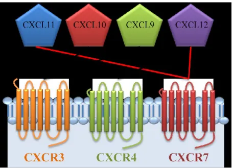
The redundancy model seems to be supported by the number of possible interactions between chemokines and their receptors. For instance, CXCR3 has three potential ligands (CXCL9, CXCL10, and CXCL11). However, CXCL11 is also a ligand for CXCR7. In addition, CXCR7 shares CXCL12 with CXCR4 (Figure 1).
10
Figure 1. Schematic view of the possible interactions between different ligands and CXC-chemokine receptors.
The chemokine system is large and complex: about 50 chemokines for only 20 receptors[62]. The disparity between the number of chemokines and the limited number of receptors may have led researchers to oversimplify the biological functions of the chemokine system. This was especially true for early in vitro experiments describing chemokine activities [47, 63]. Indeed, in the early phase of chemokine discovery, many assessments of chemokines reported their ability to induce migration of given cell types in vitro. This led to the oversimplified conclusion that, because many chemokines could induce migration of the same cell type, they were redundant.
However, several reports indicate that different chemokines do not necessarily result in the same biological response on a given receptor or vice versa [47, 64, 65]. For instance, CCL5 is the ligand of three receptors: CCR1, CCR3 and CCR5. CCL5 induces very different patterns of receptor recycling to the cell surface on the different receptors. Specifically, CCR5 recycles to the cell surface[66]. CCR3 is partially restored to the cell surface and partially degraded in lysosomes[67]. Finally, CCR1 does not recycle at all[68].
11 In fact, several other examples demonstrated that redundancy does not exist at the molecular level of chemokine receptors (see also the section 1.4.3). Obviously, this includes CXCR3 [64, 69, 70]. Therefore, the failure of targeting chemokine receptors has nothing to do with the chemokine redundancy. The notion of redundancy rather suggests a fine-tuning of the chemokine system.
That being said, we will now elaborate on the molecular level of chemokine receptor functions. More precisely, we will focus on CXCR3.
1.4 Molecular basis of the chemokine system
1.4.1 Chemokine receptor signaling through G proteins and desensitization by β-arrestin
Chemokines are ligands that bind to members of the super-family of heptahelical G-protein-coupled receptors (GPCRs). As their name suggests, the signalization of GPCRs is through heterotrimeric G protein subunits (αβγ). Ligand binging on a GPCR causes subsequent conformational change in the receptor. This conformational change then activates the G protein by exchanging a GDP for a GTP. The Gα protein subunit, now bound to a GTP, dissociate from βγ subunits to further activate downstream signaling pathways (Figure 2, step 1). Most commonly, chemokine receptors are coupled to the Gαi subunit, which has as major function to modulate cAMP production by inhibiting adenylate cyclase (Figure 2, step 1). However, there is considerable evidence for alternative Gαq coupling that activates phospholipase C (Figure 2, step 1) [71, 72]. Finally, it has never been reported that a chemokine receptor stimulates adenylate cyclase through Gαs coupling (Figure 2, step 1).
One process that regulates GPCRs is desensitization when a receptor is exposed to its ligand for a prolonged time. Following receptor activation by a ligand, GPCR kinase (GRK) phosphorylates the cytoplasmic C-terminal of the agonist-bound receptor (Figure 2, step 2). This phosphorylation initiates impairment of the signaling and allows desensitization by the the subsequent recruitment of β-arrestin, which uncouples the receptor from further G protein
12 activation (Figure 2, step 3). In addition, β-arrestin recruitment may lead to internalization or other signalisation pathways (Figure 2, step 4).
Figure 2. Schematic view of chemokine receptor signalisation, phosphorylation by GRK, desensitization and subsequent internalization by β-arrestin.
Inspired from Ma et al. Journal of Cell Science, 2007 [73].
1.4.2 Some principles of pharmacology
The present work mainly deals with CXCR3 at a pharmacogical level. Therefore, it is important to recall certain essential concepts in pharmacology such as affinity, efficacy, potency, agonist, partial agonist. These concepts will be especially useful in the section treating of functional selectivity, also called biased agonism (see section 1.4.3.).
Affinity, efficacy and potency are essential referential quantities used in drug discovery and in fundamental studies on receptors. Agonist affinity can be explained in terms of the
13 dissociation constant (Kd) for agonist binding to a receptor using in vitro techniques such as ligand binding assays [74]. The lower the dissociation constant is, the higher is the affinity of an agonist to its receptor.
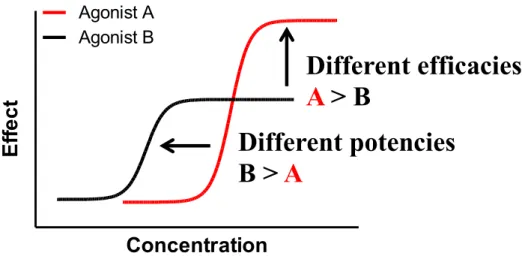
Efficacy is associated to the maximal activity in a specific assay using a range of concentrations of the agonist (Figure 3). Therefore, the assay can also be referred to as a dose response experiment. In contrast to efficacy, potency is associated with the concentration of drug required to produce an effect of given intensity (Figure 3). The higher the potency is, the lower is the amount of drug required to produce a response. Figure 3 describes the difference between potency and efficacy.
Figure 3. The principles of efficacy and potency in pharmacology.
Agonist A has a higher efficacy then agonist B, while agonist B has a higher potency then agonist A.
A full agonist is a ligand or a drug with high efficacy (defining maximal response). Generally, full agonists are defined as a reference ligand, most commonly associated to the endogenous ligand with the highest efficacy. For instance, CCR2 has been reported to bind seven natural ligands. However, CCL2 is the only ligand to elicit maximal response in the different
Agonist A Agonist B
Concentration
Ef
fe
c
t
Different efficacies
A > B
Different potencies
B > A
14 pathways [75]. Therefore, it is considered as the reference for the determination of relative agonist efficacy on CCR2. A partial agonist is a ligand or a drug that activates a given receptor but only has partial efficacy in comparison to a full agonist.
Noteworthy, the determination of a full agonist is pathway dependent. This implies that a full agonist in one assay can turn into a partial agonist in a different assay, assessing a different pathway. This is explained by the concept of functional selectivity.
1.4.3 The concept of functional selectivity (or biased agonism)
Over the past 20 years, several articles reported different agonists with different potencies and efficacies on their respective receptor [76]. In some cases, the different ligands of a same receptor can all be agonists for one pathway but with different potencies and efficacies. For example, CCL2 acts as a full agonist for the recruitment of β-arrestin on CCR2 [75]. Compared with CCL2, other CCR2 ligands were described as partial agonists with reduced efficacy and potency on the same pathway [75]. Moreover, the different ligands of a same receptor can be agonists for some pathways and antagonists for others. For instance, angiotensin II acts as an agonist of β-arrestin pathways but as an antagonist of G protein pathways on the angiotensin II type 1A receptor [77]. This phenomenon is named biased agonism and the ligands that display such behavior are called biased agonists[78]. These biased agonists have been classified as being functionally selective or biased toward certain response pathways compared with the other agonists[76].
This new concept provoked a paradigm shift in GPCR pharmacology, in that different ligands targeting the same receptor do not necessarily induce qualitatively similar intracellular signals. Different signalling axes, such as G-protein signalling versus β-arrestin signalling, are activated independently from one another. The promiscuous ligand-receptor relations in the chemokine system make chemokines one of the ideal systems to study “ligand-biased signalling”.
15 Biased agonism has been described for other chemokine receptors such as CCR2 [75]. Few articles reported the functional selectivity for CXCR3 and its ligands[64, 79]. For instance, it has been shown that CXCL11 was the most potent and efficacious inducer of CXCR3 internalization and chemotaxis in a leukemia cell line [79]. At the time the present work started, no data reported the biased agonism of CXCR3 and its ligands for Gαi and β-arrestin pathways. However, two groups now reported the biased agonism of CXCR3 [64, 80].
1.4.4 Chemokine receptor heteromerization
A factor further complicating the picture in the chemokine system is GPCR homo-and heteromerization. Before the introduction of the concept of receptor homo- or heteromerization, the results of GPCR signaling studies were attributed to the interaction of agonists with receptor monomers[81]. Now, GPCRs homo- and heteromerization have been assessed by a broad range of techniques: from co-immunoprecipitation, complementation, bioluminescence resonance energy transfer (BRET), fluorescence resonance energy transfer (FRET), to crystallography [81, 82].
While chemokine receptor homomerization is now widely accepted, the extent to which heteromerization is prevalent, and its functional consequences, remain matter of active research. One striking example of heteromerization effects is the negative modulation of CXCR4 signaling by heteromerization with CXCR7 [83]. Of note, these receptors share the common ligand CXCL12 (see Figure 1). On the functional side, CXCR7 attenuates CXCR4-mediated signals, and this may occur via CXCR4/7 heteromerization.
Potential heteromerization of CXCR7 with CXCR3 (with which it shares the CXCL11 ligand), and possible effects of CXCR7 on CXCR3 signalling have not been reported and remain to be tested. The same holds true for potential heteromerization of CXCR3 and CXCR4 (but see reference 56).
16
1.4.5 CXCR3 splicing
Alternative splicing is a crucial mechanism for gene regulation. It results from a single gene coding for multiple proteins. Chemokine receptor splicing affects the chemokine system by increasing its diversity.
For example, the ccr2 gene encodes for two alternatively spliced transcript variants of the receptor: CCR2A and CCR2B. These two variants share common functionsand differ only in their terminal carboxyl tails [84]. Both CCR2A and CCR2B mediate inhibition of adenylyl cyclase via coupling to Gαi [85]. Finally, both receptors are present on monocytes and macrophages but CCR2B is the predominant isoform[86].
Another example of splicing is CXCR3. Northern blot analysis revealed that CXCR3 mRNA was alternatively spliced to generate two variants: CXCR3A and CXCR3B [87]. The translation of CXCR3B mRNA generates a receptor containing a longer N-terminal extracellular domain, different from the CXCR3A sequence [87].
The two splice variants of CXCR3 are reported to have opposite functions. CXCR3A promotes cell growth and plays a major role in IFN-γ-inducible immune responses, whereas CXCR3B mediates apoptosis and inhibits cellular proliferation [87, 88]. In addition, the two variants are expressed on different cell types. CXCR3A is mainly expressed on T cells, B cells and NK cells, whereas CXCR3B is mainly expressed on endothelial cells [89]. The expression of variants is not exclusive to one cell type since both CXCR3 isoforms were detected in activated T lymphocytes [87]; CXCR3A being the predominant one [87, 90].
17 Another variant of CXCR3, named CXCR3-alt, results from alternative splicing via exon skipping [91]. CXCR3-alt has severe structural changes comparatively to CXCR3A but still localizes to the cell surface [91]. CXCR3-alt responds exclusively to CXCL11 [91].
The existence of CXCR3 splice variants is matter of debate. Campanella et al. suggested that CXCR3B does not even exist in mice[92]. They argued that an in-frame stop codon would terminate the CXCR3B splice variant in mice. This master’s thesis focuses on CXCR3A and its ligands.
1.4.6 CXCL4 as a novel ligand for CXCR3
Platelets are cells involved in the process of blood clotting and act as reservoirs of some chemokines with inflammatory properties. For example, once activated, platelets release CXCL4 (or Platelet factor-4) in micromolar concentrations [93, 94]. For long, CXCL4 was considered as an orphan ligand since it acted as a chemoattractant for leukocytes via an unidentified receptor.
CXCR3 was described as a receptor for CXCL4 [87, 95]. For instance, it has been reported that CXCL4 acts as a chemoattractant on a murine cell line transiently expressing CXCR3A or CXCR3B. It also has been shown that CXCL4 can induce intracellular calcium release and the migration of activated human T lymphocytes [95].
The study of the CXCL4/CXCR3 axis is of a particular importance since it has been suggested that it was involved in T lymphocyte recruitment and the subsequent amplification of inflammation reported in diseases such as atherosclerosis [95]. In addition, studies suggested that platelets are involved in inflammatory phenomena, like bronchial asthma [96]. For instance, the plasma level of CXCL4 of patients suffering from asthma attacks is significantly higher than those of controls[97].
Although the discovery of CXCL4 as a ligand of CXCR3 was a step forward in elucidating the role of CXCL4, pharmacological characterizations of CXCL4 on CXCR3 are still missing. For
18 example, no publication assessed yet the recruitment of β-arrestin or the activation of Gαi on CXCR3 upon CXCL4 stimulation.
1.4.7 Chemokine gradient remodelling
During chemotaxis, cells that are attracted by chemokines move through the extracellular chemokine concentration gradients. During in vitro experiments, the gradient is diffused from a lower well, containing high concentrations of chemokines, to higher wells containing the cells. Therefore, cells are attracted by diffusion of chemokines. However, the chemokine gradient in vivo is much more complex.
In vivo maintenance and remodelling of these gradients require chemokine inactivation by extracellular proteases. However, cleavage of chemokine by proteases is another of layer of complexity since cleaved chemokine can bind to unexpected receptors. For instance, ligation of CXCR3 by proteolytically-processed CXCL12 has been implicated especially in brain diseases such as neuro-AIDS[98].
The maintenance of gradients in vivo can also happen through chemokine degrading receptors which internalize and, subsequently, degrade chemokines. These receptors, such as CXCR7, are commonly called decoy or atypical chemokine receptors (ACKRs) [99]. CXCR7 is atypical in that it does not mediate chemotaxis[100]. Also, unlike the well described CXCR3 and CXCR4 signalling (Gαi signalling followed by β-arrestin recruitment), CXCR7 does not mediate classical G-protein responses. No proximal intracellular CXCR7 signalling was known at all until our group discovered that CXCR7 does recruit β-arrestin in response to CXCL12[101].
Atypical receptors like CXCR7 reshape gradients by constantly recycling to the cell surface. Until recently, this gradient remodelling was considered exclusive to ACKRs. To date, CCR2 is the only known typical receptor to have decoy properties [102]. However, it has been
19 reported that signalling chemokine receptors (typical), including CXCR3, could play a role in the clearance of chemokines from circulation and tissues [103].
20
1.5 Objectives
There are several lines of evidence suggesting a prevalent role of CXCR3 in different diseases. However, clinical trials investigating CXCR3 antagonists were often terminated due to a lack of efficacy [41, 45].This suggests that the mode of operation of CXCR3 and its ligands is not sufficiently understood and warrants further exploration. There are thus unmet needs for better understanding in many aspects of CXCR3 biology. Theses aspects are illustrated in Figure 4.
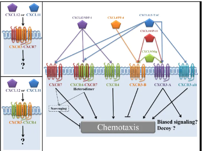
Figure 4. Overall overview of the chemokine system studied in this present work.
Biased agonist, decoy activity of CXCR3 and its heteromerization with CXCR4 or CXCR7 warrant further investigation. Figure inspired from Singh et al. [104].
21 In this present work, we propose to better delineate CXCR3A functions taking new developments such as new CXCR3 ligands, biased agonism, chemokine receptor heteromerization and decoy properties of classical (typical) chemokine receptor into account. We will achieve our goal by pursuing the following specific aims:
i) To test for agonist bias of CXCR3A ligands, including CXCL4 ;
ii) To demonstrate heteromerization of CXCR3A with CXCR4 and CXCR7;
iii) To address the effects of CXCR3A/CXCR4 or CXCR3A/CXCR7 heteromerization on CXCR3 biased signaling ;
iv) To demonstrate the decoy properties of the typical receptor CXCR3A ;
v) To address the effects of CXCR3A/CXCR4 or CXCR3A/CXCR7 heteromerization on CXCR3 decoy properties.
Taken together, this work adds new aspects to our knowledge on the molecular mechanism of CXCR3A function, a recognized potential drug target. Furthermore, this work provides fundamental insight into the organization of chemokine receptor heterodimers and potential mechanism underlying chemokine gradient shaping in vivo.
22
2. Materials and methods
2.1 Plasmids
The expression vectors containing CXCR3, CXCR4 and CXCR7 were obtained from the Missouri S&T cDNA Resource Center (www.cdna.org). CXCR3, CXCR4 and CXCR7 receptor sequences were subcloned to yield -YFP and -RLuc fusion proteins as described in Berchiche et al and in Kalatskaya et al.[101, 105] β-arrestin-RLuc was a generous gift from Dr. Michel Bouvier and the GFP10-EPAC-RLucIII was obtained as described in Leduc et al.[106]
2.2 Reagents
CXCR3 ligands (CXCL4, CXCL9, CXCL10 and CXCL11) were purchased from PeproTech (USA). They were dissolved as 100 μM stocks in phosphate buffered saline (PBS) 1 % bovine serum albumin (BSA) and used freshly diluted at the concentration indicated. Radiolabelled ligand (125I-CXCL11) was purchased from Perkin Elmer (USA).
2.3 Cell culture and transfection
Human embryonic kidney (HEK) 293E (passage number 10 to 30) were maintained in Dulbecco’s modified Eagle’s medium (DMEM) supplemented with 10% fetal bovine serum (WisEnt), 100 units/mL penicillin/streptomycin (Invitrogen) and 200 μg/mL G418 and incubated at 37 ºC, 5% CO2. Twenty-four hours before transfection, cells were plated at a density of 800,000 cells per well in 6-wells plates. Transient transfections were performed in 6-wells plates using the polyethylenimine method [107].
23
2.4 BRET measurements
Twenty-four hours post-transfection, transfected cells were plated in 96 wells plates pretreated with poly-D-lysine. Forty-eight hours post-transfection, DMEM media was changed for BRET buffer, containing PBS, 0.1% fetal bovine serum and 0.5 mM MgCl2, at room temperature. Then, coelenterazine H (BRET1) or coelenterazine 400a (BRET2) were added to reach a final concentration of 10 uM. Total fluorescence and luminescence were measured with a Mithras LB940 luminometer (Berthold technologies) as described in Berchiche et al [105].
2.5 Gαi activity assays
Inhibition of forskolin-induced cAMP production was measured as previously in Leduc et al.[106]. Briefly, HEK293E cells were cotransfected with 1 μg of CXCR3 and 0.04 μg of an intramolecular GFP10-EPAC-RLucIII BRET2 biosensor. Each condition was adjusted to 2 ug of DNA per well with the empty vector pcDNA3. Ten minutes after addition of the BRET2 substrate coelenterazine 400a (Biotium, Hayward, CA), cells were stimulated with the indicated chemokine concentration in the presence or absence of 10 μM forskolin. Then, BRET measurements were performed. Experiments were carried out in triplicate and presented as mean ± SEM.
2.6 Arrestin recruitment assays
β2-Arrestin recruitment assays were conducted as BRET1 experiments and protocols were previously described in Kalatskaya et al.[108] Briefly, HEK293E cells were cotransfected with 1 μg of CXCR3A-YFP and 0.05 μg of β2-Arrestin-RLuc. Each condition was adjusted to 2 μg of DNA per well with the empty vector pcDNA3. Cells were stimulated with the indicated chemokine concentration and then incubated for 5 min at 37ºC, 5% CO2. Ten minutes after addition of the BRET1 substrate coelenterazine H, BRET measurements were performed. BRET values were corrected to Net BRET by subtracting the background BRET signal detected when the β2-Arrestin-RLuc construct was expressed alone. Total fluorescence
24 and luminescence were used as a relative measure of total expression of the acceptor and donor proteins, respectively. Experiments were carried out in triplicate and presented as mean ± SEM.
2.7 BRET saturation assays
BRET titration experiments were done as described in Mercier et al.[109] Briefly, HEK293E cells were cotransfected with a constant amount of receptor-RLuc (0.03 ug) and increasing concentration of plasmids encoding YFP-tagged receptors, from 0 ug to 1.9 ug. BRET signal was determined by calculating the ratio of the receptor-YFP over the receptor-RLuc emission without ligand stimulation 10 minutes after coelenterazine H addition. Values were corrected to Net BRET by subtracting the background BRET signal detected when the receptor-RLuc construct was expressed alone. Saturation curves were obtained by plotting Net BRET values as a function of the [acceptor]/[donor] ratio. Total fluorescence and luminescence were used as a relative measure of total expression of the acceptor and donor proteins, respectively. Experiments were carried out in triplicate and presented as mean ± SEM. Each plotted point represents a different transfection.
2.8 Degradation assays
CXCR3 decoy properties have be assessed with radio-labelled 125I-CXCL11 degradation assays. HEK293E cells were incubated with DMEM 0.1% BSA 50 pM 125I-CXCL11 (Perkin Elmer) followed by 150 minutes incubation at 37 ºC, 5% CO2. Then, supernatants were collected and cell surface–bound chemokines were removed by the addition of a 3M glycine solution (pH 2.7). Trichloroacetic acid (TCA) precipitation was used to distinguish between the radioactivity associated with intact chemokine (TCA-precipitable fraction) and degraded chemokine (TCA-non precipitable fraction). The TCA-precipitable fraction was dissolved in 250 μL PBS and collected. The cell layers (cell uptake) were harvested with PBS 1 % SDS. The radioactivity associated with all fractions was measured with a Cobra II gamma counter. Figures are represented either as in percent of the total input of radioactivity, or as the absolute
25 counts of degraded chemokines. Unlabelled ligand competitions (CXCL4-9-10-11) of 125 I-CXCL11 degradation have been tested by adding the indicating unlabelled chemokines. As controls of membrane expression of chemokine receptors, cell membrane staining assays were performed with antibodies coupled to fluorochromes. Data acquisition and analyses by flow cytometry were done on a BD FACSCalibur (BD biosciences).
2.9 Flow cytometry
HEK293E cells were washed once with PBS than stained (30 minutes, 4ºC) with the following antibodies: human (h) CXCR3-Phycoerythrin, hCXCR4-allophycocyanin, hCXCR7-allophycocyanin, murine (m) IgG2A-hCXCR7-allophycocyanin, mIgG1-Phycoerythrin (R&D systems). Cells were washed twice and fixed with fixation buffer (PBS 2 % formaldehyde).
2.10 Data analysis
Data from BRET and degradation assays are the mean of independent experiments performed in triplicate and duplicate, respectively. Curve-fitting and statistical analyses were done with GraphPad Prism 5 software (GraphPad Software Inc., San Diego, CA). Statistical significance of the differences between more than two groups was calculated by one-way ANOVA, followed by Bonferroni’s post-test.
26
3. Results
3.1 CXCR3A biased signaling
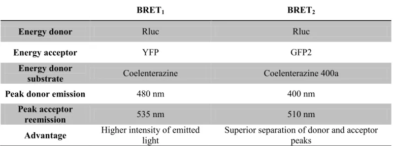
Although CXCR3A functions seem to be thoroughly described in the literature, few pharmacological characterizations have been done on its three endogenous ligands - CXCL9, CXCL10 and CXCL11. Studying the role of CXCR3A and its ligands on a pharmacological level will undoubtedly provide new insights on the regulation of T cell function [6], but also on every subsets of cells expressing CXCR3A [110-113]. Therefore, we investigated downstream CXCR3A signaling induced by its three ligands using bioluminescence resonance energy transfer (BRET). These assays investigate the activation of the Gαi subunit, and β-Arrestin recruitment to the receptor. Here, β-arrestin recruitment is assessed with BRET1 and Gαi activation is assessed with BRET2. The differences between both assays are explained in Figure 5 and Table 2.
To monitor the Gαi-dependent pathway in living cells, we used a BRET2 version of the EPAC-based BRET sensor, for which the inactive cytosolic mutant form of human EPAC was inserted between GFP10 and RLucIII. This cAMP response assay has been previously described in Jiang et al and in Leduc et al [106, 114]. Briefly, cells were stimulated with a final concentration of 20 μM forskolin, which activates adenylate cyclase (AC) and leads to an increased concentration of cAMP. Bound to GFP10-EPAC-RLucIII, cAMP induces a conformational change that increases the distance between RLucIII and GFP, leading to a decrease in BRET signal. When stimulated with ligand, the Gα/i-CXCR3A complex inhibits AC. By inhibiting AC, Gα/i-CXCR3A signaling reduces cAMP production. This leads to a reduction of the forskolin-dependent decrease of the BRET signal (increase of BRET signal) (see Figure 6). Increase in BRET signal reflects inhibition of AC (and thus Gai activation), as represented in figure 2. Raw BRET2 ratios have been normalized by using forskolin stimulated results as 0 % of inhibition of AC activity, and by using the non-stimulated (no ligand, no forskolin) results as 100 % of inhibition of AC.
27
Figure 5. Schematic representation of BRET1 and BRET2 assays
Figure inspired from Institut Cochin website
Table 1. Distinction between BRET1 and BRET2 assays
BRET1 BRET2
Energy donor Rluc Rluc
Energy acceptor YFP GFP2
Energy donor
substrate Coelenterazine Coelenterazine 400a
Peak donor emission 480 nm 400 nm
Peak acceptor
reemission 535 nm 510 nm
28
Figure 6. Schematic representation of EPAC BRET2 assays.
After stimulation with forskolin, cAMP level increases in the cells. This leads to a conformational reorganisation of RLuc-EPAC-YFP biosensor. This reorganisation leads to a decrease of the BRET signal due to the increased distance and unfavorable orientation between the energy donor RLuc and the energy acceptor YFP. After ligand stimulation of Gα/i-CXCR3A, cAMP concentration decreases in the cells, leading to a conformational change in RLuc-EPAC-YFP biosensor. This reorganisation leads to an increase in BRET signal due to the reduced distance and favorable conformation between the energy donor RLuc and the energy acceptor YFP. Figures adapted from Salahpour et al., Front Endocrinology, 2012. [115] (F, Forskolin. AC, Adenylate cyclase. L, Ligand.)
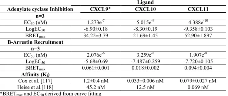
Stimulation of HEK293E cells coexpressing this EPAC biosensor and CXCR3A receptor with its three ligands lead to different concentration-dependent cAMP responses (Figure 7 and Table 2). Out of the three ligands tested, CXCL11 was the most efficient and potent with an EC50 of 0.43 nM, leading to 53±2% inhibition of AC activity. Although CXCL10 shows
29 efficacy, with an EC50 of 5 nM, and it shows a smaller efficacy than CXCL11 with 22±1% inhibition of AC activity. As of CXCL9, it shows very little potency with an EC50 of 122nM, but reaches an efficacy of 34±4 % inhibition of AC activity at a concentration of 1uM. Of note, CXCL9 BRETmax and EC50 were derived from curved fitting. Therefore, the EC50 must be interpreted carefully.
To test β-arrestin recruitment to CXCR3A, we used a BRET-based proximity assay system in HEK293E cells transiently coexpressing the BRET donor β-arrestin-2-RLuc and CXCR3A fused to the BRET acceptor YFP. If both proteins, RLuc and YFP, are brought close enough for resonance energy transfer to occur, the bioluminescence energy generated by RLuc can be transferred to YFP, which then emits yellow light [116]. This light is detected as the BRET signal. Background BRET, which is the signal obtained from cells only expressing β-arrestin-2-RLuc, has been deduced from raw BRET. This leads to the plotted Net BRET. Dose-response experiments revealed the following potency rank order of the chemokines: CXCL11 > CXCL10 > CXCL9 (EC50 of 19 nM for CXCL11, 32 nM for CXCL10 and 207 nM for CXCL9) (Figure 8 and Table 2). As observed for AC inhibition assays, CXCL9 induced β-arrestin responses but did not reach saturation even at highest chemokine doses, leaving some uncertainty concerning the EC50 values, which had to be determined by curve fitting. CXCL11 showed the strongest efficacy and potency compared with CXCL10 and CXCL9. Here, we observed the same pattern of potency as in the cAMP assays; CXCL11 being the most potent and efficient of all three ligands of CXCR3A. Ligands with high affinities for a receptor can sometimes have low efficacies and/or potencies in different pathways. However, it is interesting to mention that our results are congruent with the different affinities of CXCR3 ligands described in Cox et al. [117]. The ligand with the highest affinity for CXCR3A (CXCL11) is also the ligand with the highest efficacy and potency[117].
30 -13 -12 -11 -10 -9 -8 -7 -6 -5 0 10 20 30 40 50 60 70 CXCL11 CXCL9 CXCL10 Log[Chemokine] % I n h ib it io n o f A C a ct iv it y
Figure 7. Inhibition of Adenylate cyclase activity followed by stimulation of CXCR3A by chemokine CXCL11, CXCL10 or CXCL9.
HEK293E cells coexpressing CXCR3A and EPAC reporter were incubated with indicated concentrations of ligand and resulting BRET was measured after 10 min at room temperature. (Blue) ○ CXCL11, (Green) □ CXCL10, (Red) ∆ CXCL9. Data are means of three independent experiments performed in triplicate and presented as mean ± S.E.M.
31 -12 -11 -10 -9 -8 -7 -6 -5 0.00 0.05 0.10 CXCL11 CXCL10 CXCL9 Log[Chemokine] NE T BR E T
Figure 8. β-Arrestin-2 recruitment to CXCR3A followed by chemokine CXCL11 , CXCL10 or CXCL9 stimulation.
HEK293E cells transiently coexpressing CXCR3A-YFP and β-arrestin2-RLuc were incubated with indicated concentrations of ligand and resulting BRET was measured after 10 min at room temperature. (Blue) ○ CXCL11, (Green) □ CXCL10, (Red) ∆ CXCL9. Data are means of three independent experiments performed in triplicate and presented as mean ± S.E.M.
32
Table 2. Pharmalogical parameters of CXCR3A biased signalling
Ligand
Adenylate cyclase Inhibition CXCL9* CXCL10 CXCL11
n=3
EC50 (nM) 1.273e-7 5.015e-9 4.388e-10
LogEC50 -6.90±0.18 -8.30±0.19 -9.358±0.103
BRETmax 34.22±3.79 21.69±1.45 52.90±1.897
Β-Arrestin Recruitment n=3
EC50 (nM) 2.076e-6 3.259e-8 1.907e-8
LogEC50 -5.68±0.69 -7.487±0.259 -7.720±0.105
BRETmax 0.061±0.001 0.018±0.002 0.094±0.004
Affinity (Ki)
Cox et al.[117] 1.2±0.4 nM 0.033±0.006 nM 0.079±0.027 nM
Heise et al.[118] 45.2 nM 12.5 nM 0.069 nM
33
3.2 CXCR3A Heterodimerization
CXCR3, CXCR4 and CXCR7 are implicated in many cancers and inflammatory and auto-immune diseases [119]. Previously, CXCR4 has been shown to form heterodimer complexes with CXCR7 [83]. Also, these receptors and their ligands are expressed in tumour microenvironment and on various immune and cancer cells [104]. Finally, because this receptor trio is an attractive target for therapeutic uses, we investigated whether CXCR3 could form heterodimer complexes with CXCR4 or with CXCR7.
We have characterized the relative propensities of CXCR3A to heterodimerize with CXCR4 and CXCR7 using a BRET-based saturation assay as described in Mercier et al. [109]. Constant (low) quantities of CXCR3A, CXCR4 or CXCR7 fused to RLuc were cotransfected with increasing quantities of CXCR3A or CXCR7 fused to YFP. The level of energy transfer detected for a given concentration of the RLuc (energy donor) rises with increasing concentration of the YFP (energy acceptor), until all RLuc fused molecules are engaged by a YFP fused molecule [109]. The level of energy transfer rises because more YFP acceptor molecules are expressed in the cells. Background BRET, which is the signal obtained from cells only expressing RLuc-fused receptor, has been deduced from raw BRET. This leads to the plotted Net BRET. The concentration of acceptor yielding 50% of the maximal energy transfer (BRET50) can be interpreted as a measure of the relative propensity of two proteins to interact (Figure 9) [109].
34
Figure 9. Interpretation of BRET saturation curves
The two proteins interacting in curve 1 (black) have a higher affinity towards each other comparatively to the two proteins interacting in curve 2 (grey). The straight line (red) represents a non specific interaction between two proteins.
In cells expressing RLuc-fused CXCR4 (Figure 10), CXCR3A (Figure 12) or CXCR7 (Figure 10 and 12), increasing the concentration of CXCR3A receptor tagged with YFP resulted in BRET signals that increased hyperbolically, reaching an asymptote when all RLuc-tagged receptors are associated with those fused to YFP (BRET max). In contrast, we observed non-specific interactions between CXCR3A-RLuc and CXCR7-YFP that led to a linear BRET signal increasing with YFP/RLuc ratios in cells coexpressing the YFP and RLuc-tagged receptors (Figure 9-13). Interestingly, an exchange between the two fusion proteins in the two receptors (CXCR3A and CXCR7) led to an unfavorable proximity or orientation for energy
35 transfer to occur. These arrangements (CXCR3A-RLuc/CXCR7-YFP) led to a straight linear BRET signal increasing with YFP/RLuc ratios. Constructions known to lead to straight linear BRET signal are often used as negative controls for BRET saturation curves since they represent non specific interaction (Figure 9). That being said, the two first saturation curves presented here lack negative controls. For example, experiments could be reproduced with RLuc-tagged receptor with increasing concentration of YFP alone or any unrelated receptor (for instance, GABA receptor) tagged with YFP.
The BRET50, which is an instrument-dependent relative value of the YFP/RLuc ratio when half of the BRET max is reached, represents the propensity of two proteins to interact. A smaller BRET50 reflects a greater propensity of two proteins to interact (Figure 9). BRET50 values were not significantly different for both heterodimers CXCR3A-CXCR4 and CXCR3A-CXCR7 (Figure 10). However, BRET50 values for the CXCR7 homodimer were lower than for the CXCR3A-CXCR7 heterodimer, even though this difference did not reach statistical significance (Figure 14). It is tempting to suggest that this difference reflects the preference of CXCR7 to form homodimers, instead of heteromers with CXCR3A. As for the CXCR3A homodimer, the BRET50 values cannot be compared to those of CXCR3A-CXCR7 heterodimer since saturation curves values did not produce a hyperbole (Figure 13). By comparing the calculated BRET50, we cannot speculate that CXCR3A also has a propensity for homomerization over heteromerization with CXCR7 (Figure 14). Intriguingly, Watts et al. found comparable BRET50 values for all CXCR3 and CXCR4 homo- and heterodimers, suggesting that CXCR3 and CXCR4 have comparable propensities to form homo- and heterodimer complexes [80]. In the same order of idea, Levoye et al. found comparable BRET50 for CXCR4 and CXCR7 homodimers and CXCR4/7 heterodimer [83]. This suggests that heterodimerization of CXCR4 and CXCR7 occurs with the same efficiency as receptor homodimerization. Of note, Levoye et al. performed the saturation curves for CXCR4/7 heterodimers with two distinct constructions (CXCR4-RLuc-CXCR7-YFP and CXCR7-RLuc-CXCR4-YFP) [83]. This might explain the difference observed for the published BRET50 of CXCR4/7 heterodimers[83]. This will be further discussed in the discussion.
36 0 100 200 300 0.00 0.05 0.10 0.15 0.20 CXCR4-Rluc+CXCR3-YFP CXCR7-Rluc+CXCR3-YFP Ratio YFP/Rluc Ne t BR E T
Figure 10. CXCR3A forms heterodimer with CXCR7 and with CXCR4 in HEK293E cells.
Saturation curves were generated in cells expressing a constant amount of CXCR7/CXCR4-RLuc and an increasing amount of CXCR3A-YFP without stimulation. BRETmax and BRET50 were calculated by nonlinear regression curve fit base on the model of one site total. Saturation curves represent pool data of 3 experiments performed in triplicate and presented as mean ± SEM. Each point represents a different transfection. BRET50 of CXCR3/4 is 77±9. B BRET50 of CXCR3/7 is 47±9. BRET50 of the two heterodimers are not significatively different. (Black) ■ CXCR3A-YFP + CXCR7-RLuc, (Green) ● CXCR3A-YFP + CXCR4-RLuc.
37 CXCR3/7 CXCR3/4 1 10 100 1000
ns
BR E T50Figure 11. CXCR3 has no preference for heterodimerization with CXCR4 or CXCR7. BRET50 values of chemokine receptor dimerization were extracted from three curves. Saturation curves were generated in cells expressing a constant amount of CXCR7-RLuc or CXCR4-RLuc and an increasing amount of CXCR3A-YFP without ligand stimulation. BRET50s were calculated by nonlinear regression curve fit (based on the model of one site total binding). Data are from three to six experiments, each performed in triplicate and presented as mean ±SEM. Unpaired T test was performed and the difference between the BRET50 values did not reach significance (p value of 0.6772).