Published Ahead of Print 29 February 2012.
10.1128/JCM.05057-11.
2012, 50(5):1673. DOI:
J. Clin. Microbiol.
Moulin-Schouleur
Jacques Mainil, Jorge Blanco and Maryvonne
Azucena Mora, Ghizlane Dahbi, François Biet, Eric Oswald,
Catherine Schouler, Brigitte Schaeffer, Annie Brée,
Patterns of Virulence Genes
Pathogenic Escherichia coli Based on Four
Diagnostic Strategy for Identifying Avian
http://jcm.asm.org/content/50/5/1673
Updated information and services can be found at:
These include:
SUPPLEMENTAL MATERIALml
http://jcm.asm.org/content/suppl/2012/04/09/50.5.1673.DC1.ht
REFERENCEShttp://jcm.asm.org/content/50/5/1673#ref-list-1
at:
This article cites 57 articles, 34 of which can be accessed free
CONTENT ALERTS
more»
articles cite this article),
Receive: RSS Feeds, eTOCs, free email alerts (when new
http://journals.asm.org/site/misc/reprints.xhtml Information about commercial reprint orders:
http://journals.asm.org/site/subscriptions/ To subscribe to to another ASM Journal go to:
on May 24, 2012 by UNIVERSITE DE LIEGE
http://jcm.asm.org/
Based on Four Patterns of Virulence Genes
Catherine Schouler,aBrigitte Schaeffer,bAnnie Brée,aAzucena Mora,cGhizlane Dahbi,cFrançois Biet,dEric Oswald,eJacques Mainil,f
Jorge Blanco,cand Maryvonne Moulin-Schouleura
INRA, UR1282 Infectiologie Animale et Santé Publique, Nouzilly, Francea
; INRA, UR0341 Mathématiques et Informatique Appliquées, Jouy-en-Josas, Franceb
; Laboratorio de Referencia de Escherichia coli, Departamento de Microbioloxía e Parasitoloxía, Facultade de Veterinaria, Universidade de Santiago de Compostela, Lugo, Spainc
; Sanders Aliments, Laboratoires Vétérinaires, Laval, Franced
; INRA, USC1043, Toulouse, Francee
; and Bacteriology, Department of Infectious Diseases, Faculty of Veterinary Medicine, University of Liège, Liège, Belgiumf
In order to improve the identification of avian pathogenic Escherichia coli (APEC) strains, an extensive characterization of 1,491 E. coli isolates was conducted, based on serotyping, virulence genotyping, and experimental pathogenicity for chickens. The iso-lates originated from lesions of avian colibacillosis (nⴝ 1,307) or from the intestines of healthy animals (n ⴝ 184) from France, Spain, and Belgium. A subset (460 isolates) of this collection was defined according to their virulence for chicks. Six serogroups (O1, O2, O5, O8, O18, and O78) accounted for 56.5% of the APEC isolates and 22.5% of the nonpathogenic isolates. Thirteen virulence genes were more frequently present in APEC isolates than in nonpathogenic isolates but, individually, none of them could allow the identification of an isolate as an APEC strain. In order to take into account the diversity of APEC strains, a statis-tical analysis based on a tree-modeling method was therefore conducted on the sample of 460 pathogenic and nonpathogenic isolates. This resulted in the identification of four different associations of virulence genes that enables the identification of 70.2% of the pathogenic strains. Pathogenic strains were identified with an error margin of 4.3%. The reliability of the link be-tween these four virulence patterns and pathogenicity for chickens was validated on a sample of 395 E. coli isolates from the col-lection. The genotyping method described here allowed the identification of more APEC isolates with greater reliability than the classical serotyping methods currently used in veterinary laboratories.
C
olibacillosis is the major cause of morbidity and mortality in poultry and is responsible for significant economic losses worldwide. Avian pathogenic Escherichia coli (APEC) induces dif-ferent syndromes in poultry, including systemic and localized in-fections, such as respiratory colibacillosis, acute colisepticemia, salpingitis, yolk sac infection, and swollen-head syndrome. The most common form of colibacillosis is characterized by an initial respiratory disease in 3- to 12-week-old broiler chickens and tur-keys, which is usually followed by a systemic infection with char-acteristic fibrinous lesions (airsacculitis, perihepatitis, and peri-carditis) and fatal septicemia. The infection is generally initiated or enhanced by predisposing agents, such as mycoplasmal or viral infections, and environmental factors (3, 18).Two major issues currently make it difficult to control avian colibacillosis, namely, the lack of a reliable method to identify the causative strains of E. coli and the fact that currently available vaccines are not totally effective, despite the recent identification of protective vaccine antigens (42). These factors are due to the diverse characteristics of APEC strains preventing the identifica-tion of common properties, which could be used as a basis for diagnostic methods and vaccination.
Although APEC strains clearly belong to the phylogenetic group of extraintestinal pathogenic E. coli, numerous studies have shown wide serological diversity (7, 43). Predominant serogroups are O1, O2, and O78, but they account for only 15 to 60% of isolates depending on the study (18, 26, 36, 47).
Several virulence genes have been demonstrated to be impli-cated in avian colibacillosis, including those encoding for adhesins (F1, P, and Stg fimbriae, curli, and EA/I), anti-host defense factors (OmpA, Iss, lipopolysaccharide, and K1), iron acquisition systems (aerobactin, Iro proteins, yersiniabactin, and the Sit iron
acquisi-tion locus), autotransporters (Tsh, Vat, and AatA), the phosphate transport system, sugar metabolism, and the IbeA protein (1, 12, 13, 24, 35, 38, 48).
However, numerous studies have demonstrated that these vir-ulence factors (VFs) are rarely all present in the same isolate, showing that APEC strains constitute a heterogeneous group. Dif-ferent isolates may harbor difDif-ferent associations of virulence fac-tors, each one able to induce avian colibacillosis.
This diversity currently hinders clear identification of an avian
E. coli isolate as a pathogenic or nonpathogenic strain. However,
there is a significant difference in gene prevalence between E. coli strains isolated from chickens with colibacillosis and those iso-lated from the feces of apparently healthy birds (47). Individually, virulence factors, such as adhesins, are not reliable tools for diag-nosis because of their low incidence in avian pathogenic isolates (56).
Serotyping remains the most frequently used diagnostic method in laboratories, but it only allows the identification of a limited number of APEC strains. This method cannot therefore be used as an effective diagnostic tool, particularly since serotype does not reflect the virulence trait. Various studies have
high-Received 10 July 2011 Returned for modification 27 August 2011 Accepted 21 February 2012
Published ahead of print 29 February 2012
Address correspondence to Catherine Schouler, catherine.schouler@tours.inra.fr. Supplemental material for this article may be found athttp://jcm.asm.org/. Copyright © 2012, American Society for Microbiology. All Rights Reserved.
doi:10.1128/JCM.05057-11
on May 24, 2012 by UNIVERSITE DE LIEGE
http://jcm.asm.org/
lighted the possibility of using some virulence factors to identify APEC strains. They have attempted to determine a common iden-tification scheme, allowing better ideniden-tification of APEC strains than serotyping. These methods are mostly based on the detection of virulence factors carried on colicin V (ColV) plasmids using multiplex PCR. One study described a multiplex PCR protocol to detect the presence of iss, tsh, iucC, and cvi (55). Another described a minimum number of genes that could be used to identify an APEC strain, namely, iutA, hlyF, iss, iroN, and ompT (33). In a third study, a multiplex PCR protocol was designed to detect the presence of eight virulence genes (toxin genes astA and vat, the increased serum survival protein gene iss, iron acquisition genes
irp2 and iucD, adhesin genes papC and tsh, and the ColV plasmid
operon genes cva-cvi) (27).
The present study involved extensive characterization of a large collection of European APEC isolates, based on serotyping, iden-tification of virulence factors, and experimental pathogenicity for chickens. Statistical analysis, based on supervised learning meth-odology, allowed us to define several sets of virulence factors ac-counting for the majority of pathogenic strains and which could be used for diagnosis.
MATERIALS AND METHODS
Bacterial strains. A total of 1,491 E. coli isolates were collected over an
8-year period (1992 to 2000) in France (n⫽ 920), Spain (n ⫽ 486), and Belgium (n⫽ 85), from chickens (n ⫽ 938), turkeys (n ⫽ 399), and ducks (n⫽ 154) (Table 1). Most isolates (n ⫽ 1,307) were from characteristic lesions of animals exhibiting clinical symptoms of different forms of coli-bacillosis (L strains), and 184 isolates were collected during the same period from the intestinal content of healthy animals with no character-istic symptom or lesion of colibacillosis at necropsy (I strains).
Isolates were collected by authorized diagnostic laboratories in each country and sent to a single laboratory. Each isolate was checked for purity and identified as E. coli using API 20E (bioMérieux, Inc.). Samples of each isolate were prepared from a single culture and used to inoculate conser-vation agar tubes (Bio-Rad) to be dispatched to the various collaborators. The same culture was also used to inoculate four tubes with brain heart infusion broth (Difco Laboratories) containing 20% glycerol, which were kept at⫺80°C.
Serogrouping. Determination of O antigens was carried out by
agglu-tination according to the method described by Blanco et al. (5) and using
all available O (O1 to O181) antisera. The antisera were obtained and adsorbed with the corresponding cross-reacting antigens to remove the nonspecific agglutinins. The O antisera were produced in the Laboratorio de Referencia de Escherichia coli (Lugo, Spain).
Virulence genotyping. The presence of genes encoding virulence
fac-tors was detected using PCR amplification. Crude DNA extracts were prepared by a rapid boiling method (51). A single PCR assay was used to detect sfa-focDE (operons encoding S fimbriae and F1C fimbriae), and nine multiplex PCR assays were performed to detect simultaneously: (i)
fimA (with consensus primers), fimA variant (fimAvMT78), and fimH; (ii)
neuC, felA, and papC; (iii) papGJ96class I, papGIA2class II, and prsGJ96 class III; (iv) tsh and iutA, (v) cdt (variants 1 to 4); (vi) cnf1 and cnf2; (vii) VT1 and VT2 encoding genes; (viii) LT encoding gene; and (ix) STa and STb encoding genes. Strains were noted as F1 positive when both fimA (or
fimAvMT78variant) and fimH genes were present and as P(F11) positive when the felA, papC, and a variant of papG genes were present. Gene iutA accounted for the aerobactin iron acquisition system and neuC for K1 antigen. The corresponding primers are listed in Table S1 in the supple-mental material. DNA fragments were amplified in a 25-l PCR mix con-taining 1 U of Taq DNA polymerase (Promega), 25 pmol of the forward and reverse primers, and 5 nmol of each deoxynucleoside triphosphate (Promega) in 1⫻ buffer. The PCR conditions were as follows: 94°C for 3 min, followed by 30 cycles of 94°C for 1 min, annealing for 1 min at 72°C for at least 30 s according to the size of the amplified fragment (1 min/ kbp), and then a final extension at 72°C for 10 min. The E. coli strains used as positive controls in PCR assays were as follows: BEN 2908 (17, 52) for
fimA, fimAvMT78, fimH, neuC, and iutA; MT189 (19) for felA and papC; 7122 (21) for tsh; KH576 (61) for iutA; 536 (29) for sfa-focDE; E6468/62 (53) for cdt; JJ48 (papGJ96), HB101/pDC1 (papGIA2), and P678-54/ pJFK102 (PrsGJ96) for papG class I, ppaG class II, and papG class III, respectively (31); MR199 and B26a for cnf1 and cnf2 (8); 933 for vt1 and
vt2; PD32b for the STa, STb, and VT2e encoding genes; and IP102a for the
LT and STb encoding genes (9). The negative control strains were E. coli MG1655 (10) and the nonpathogenic E. coli avian strain EC79 (16).
Screening for the F17, afa, and eae encoding genes was performed using colony hybridization. The corresponding probes were obtained, purified, and labeled, and DNA colony hybridization was performed as previously described (56). The positive control strains were 25KH9 (F17⫹) (39), A30 (Afa⫹) (37), and E2348/69 (Eae⫹) (30), and the neg-ative control strain was the HS strain (44). In addition, labeled cloned fragments of the APEC strain BEN2908 (aec26 [A9], sitA [A12], tkt1 [D1],
frzorf4[D7], and aec4 [D10]) were prepared and hybridized with dot blots prepared from crude extracts of genomic DNA as previously described (52). The positive and negative control strains were BEN2908 and EC79, respectively.
Detection of cytotoxic activity of bacterial lysates and supernatants.
Detection of toxins was also conducted using phenotypic assays. For de-tection of LT, VT1, VT2, CNF1, and CNF2, filtrates of cultures treated with mitomycin were inoculated into Vero and HeLa cells. For STa en-terotoxin detection, extracellular fluids were assayed by the infant-mouse test, and sonic extracts were tested for necrosis using the rabbit skin test (6). Morphological changes of the HeLa cells characteristic of cytolethal distending toxin (CDT) were determined on nonconfluent HeLa cell monolayers in 96-well plates incubated with culture supernatants and with sonic lysates obtained from 48-h bacterial cultures as previously de-scribed (59). E. coli strain DH5␣ (51) was used as a negative control, and all experiments were conducted in triplicate.
Virulence for chicks. Groups of five 1-day-old specific-pathogen-free
chicks were inoculated subcutaneously with 0.5 ml of an overnight culture in LB-Miller broth without agitation (the inoculum in stationary phase was⬃108CFU) as previously described (16), and the mortality was re-corded at 4 days postinoculation. Strains were classified as pathogenic when at least one chick died (21). The nonpathogenic E. coli avian strain EC79 (serogroup O2) (16) was used as a control (no chicks died). The housing, husbandry, and slaughtering conditions conformed to European
TABLE 1 Origins of E. coli isolatesa
Isolate type and origin
No. of isolates
Chicken Turkey Duck Total
L isolates France 293 330 153 776 Spain 427 19 1 447 Belgium 84 0 0 84 Total 804 349 154 1,307 I isolates France 94 50 0 144 Spain 39 0 0 39 Belgium 1 0 0 1 Total 134 50 0 184 Total 938 399 154 1,491
aE. coli isolates were collected from the internal organs or blood of animals showing typical symptoms and/or macroscopic lesions of avian colibacillosis (L isolates) or from the intestinal content of healthy animals without lesions at necropsy (I isolates). Schouler et al.
1674 jcm.asm.org Journal of Clinical Microbiology
on May 24, 2012 by UNIVERSITE DE LIEGE
http://jcm.asm.org/
Union guidelines for the care and use of laboratory animals. The animal experimentation authorization number for people directing these studies was 37-041.
Statistical analysis. The prevalence of virulence factors was compared
using a chi-square test. P values of⬍0.05 were considered significant. The association of virulence factors with pathogenicity was deter-mined by a tree-based modeling method. A classification tree (11) was constructed using the R package rpart, version 3 (58). This method pre-pares a binary tree using a divisive algorithm from a top node (called the root) to terminal nodes (called the leaves). The top node contains all strains. At each step of the algorithm, a node is partitioned into two sub-nodes corresponding to the presence or absence of a virulence factor. The splitting factor is determined to obtain the purest subnodes according to pathogenicity.
RESULTS
Four associations of virulence genes are correlated with patho-genicity. A statistical analysis was performed to identify groups of virulence genes preferentially associated with APEC strains. This required accurately identifying E. coli isolates as pathogenic or nonpathogenic. Thus, among the collection of 1,491 strains, a sample of 460 isolates was tested for pathogenicity using the 1-day-old chick lethality test. Strains were classified as pathogenic (P; of five chicks inoculated, one to five died) or nonpathogenic (NP; of five chicks inoculated, none died).
Based on the lethality test, 352 isolates were identified as patho-genic (310 strains isolated from lesions of diseased birds [L iso-lates] and 42 strains isolated from the intestine of healthy animals [I isolates]) and 108 isolates (47 L isolates and 61 I isolates) as nonpathogenic. Among the pathogenic strains, 196 killed 5 chicks, and 41, 33, 38, and 44 killed 4, 3, 2, or 1 chick, respectively. Patho-genic I isolates did not differ from pathoPatho-genic L isolates when considering the number of dead chicks. It is not surprising to observe that some I isolates were virulent, since it is well estab-lished that the chicken intestine is the main reservoir of patho-genic strains (4, 25). Notably, 47 strains isolated from lesions were nonpathogenic. They were of various serogroups and scattered in the classification tree. This observation confirms the fact that iso-lation of an E. coli strain from a pathological lesion is not a suffi-cient criteria to classify it as a pathogen.
Among the 460 isolates, 84 serogroup O isolates were observed. Six serogroups accounted for 56.5% of pathogenic strains: O78 (17.5%), O2 (17.3%), O8 (2.0%), O18 (9.0%), O5 (4.5%), and O1 (6.0%) (Table 2). A total of 22.5% of nonpathogenic isolates be-longed to these six serogroups (Table 2). These six most common serogroups were the same whatever the geographical origin (France, Spain, or Belgium) or animal species (chicken, turkey, or duck) (data not shown). Nontypeable isolates were less frequently observed among P strains (6.5%) than among NP strains (13.9%) (data not shown).
The presence of 19 associated virulence genes on the 460 iso-lates was detected using PCR assays and hybridization (see Table S2 in the supplemental material). The predominant virulence-associated genes on pathogenic strains were sitA (96.3%), F1 fim-briae encoding genes (86.4%), and iutA aerobactin gene (82.7%) (Table 2). Other genes were present in 20 to 60% of the pathogenic strains: tsh, frzorf4, tkt1, aec4, P(F11) fimbriae encoding genes,
aec26, and neuC (K1 antigen). Highly significant differences (P⬍
1‰) were observed in the frequency of these virulence genes be-tween P and NP isolates (Table 2). Five virulence genes were pres-ent in fewer than 10% of the P isolates: f17, sfa-focDE, afa-draBC,
cdt, and eae. Production of CDT toxin was confirmed by
pheno-typic assays. Excluding afa-draBC and f17, significant differences were observed in the frequency of the virulence genes between P and NP isolates: P⬍ 1.5% for sfa-focDE, eae, and cdt (Table 2).
Genes encoding for enterotoxins and verotoxins usually asso-ciated with intestinal pathologies in mammals, such as CNF1, LT, STa, STb, VT1, and VT2, were not detected, and no exotoxin could be detected in phenotypic assays.
First, a statistical analysis was performed using this set of 460 isolates. The statistical analysis revealed four patterns of virulence factors, each including more than 90% of the pathogenic strains:
iutA⫹, P(F11)⫹; iutA⫹, P(F11)⫺, frzorf4⫹; iutA⫹, P(F11)⫺,
frzorf4⫺, O78⫹; and iutA⫺, aec26⫹, sitA⫹ (Table 3, analysis 1).
Among the 258 isolates belonging to one of these four patterns, 247 (95.7%) were pathogenic strains. Pathogenic strains were identified with an error margin of 4.3%. Since strains that killed only one chick in the lethality test are probably less virulent strains than those that killed two to five chicks, we checked whether re-moving them from the analysis could influence the resulting pat-terns.
Thus, a second statistical analysis (analysis 2) was performed using a set of 416 strains issued from the initial set of 460 strains after removal of the 44 strains that had killed only one chick. This
TABLE 2 Most frequently occurring serogroups and virulence genes
among the 460 pathogenic and nonpathogenic isolates used for statistical analysis Serogroup or virulence genea % Total isolatesb Pathogenic (n⫽ 352) Nonpathogenic (n ⫽ 108) Serogroup O78* 17.6 0.9 O2* 17.3 4.6 O8 2.0 4.6 O18 9.0 5.6 O5 4.5 2.8 O1 6 3.7 Total 56.5 22.2 Virulence genes sitA* 96.3 59.3 F1* 86.4 71.3 iutA* 82.7 26.9 tsh* 52.8 16.7 frzorf4* 53.4 16.7 tkt1* 50.6 16.7 aec4* 46.9 13.9 P(F11)* 30.4 7.4 aec26* 34.4 6.5 neuC* 27.0 5.6 F17 5.7 4.6 sfa-focDE† 9.1 4.6 afa-draBC 4.0 6.5 cdt† 9.1 1.9 eae† 2.0 12.0
a*, Significant differences (P⬍ 0.001) were observed between pathogenic and nonpathogenic strains; †, significant differences (P⬍ 0.015) were observed between pathogenic and nonpathogenic strains. Strains were noted as F1 positive when both fimA (or the fimAvMT78variant) and fimH genes were present and P(F11) positive when
the three genes—felA, papC, and a variant of papG—were present. b
For pathogenic strains, of five chicks inoculated subcutaneously, one to five died; for nonpathogenic strains, none of five chicks inoculated subcutaneously died.
on May 24, 2012 by UNIVERSITE DE LIEGE
http://jcm.asm.org/
second analysis revealed four patterns of virulence factors each including more than 90% of the pathogenic strains. Three corre-sponded to the first three patterns identified above, and the fourth one regrouped strains that were iutA⫺, sitA⫹, aec26⫹, aec4⫹ (Table 3, analysis 2). Of the 241 strains belonging to one of the four patterns, 230 (95.4%) were pathogenic strains (error margin of 4.6%).
Thus, among the 352 pathogenic isolates of the sample used in the statistical analysis, 70.2% (247/352) were identified using the four genetic patterns defined by the first analysis, and 69.3% (244/ 352) were identified using the four genetic patterns defined by the second analysis. Three isolates were responsible for the difference observed between both analyses since they were excluded from the fourth genetic pattern of the second analysis because they were negative for the gene aec4. All other isolates were identical in both analyses. In sum, both analyses yielded very similar results, and the addition of gene aec4 in the fourth pattern did not improve the identification of pathogenic isolates.
Consequently, we decided to keep the four patterns of viru-lence factors identified in the first statistical analysis, which we named genetic patterns A [iutA⫹, P(F11)⫹), B (iutA⫹, P(F11)⫺,
frzorf4⫹], C [iutA⫹, P(F11)⫺, frzorf4⫺, O78⫹), and D (iutA⫺,
sitA⫹, aec26⫹). However, when a strain did not belong to one of
the defined genetic patterns (A, B, C, or D), it was not possible to conclude whether such a strain was pathogenic or not since the other genetic patterns contained both pathogenic and nonpatho-genic strains in proportions that were not discriminatory (see Fig. S1 in the supplemental material).
Validation of virulence patterns. The reliability of the corre-lation of these four virulence patterns with the pathogenicity of avian E. coli strains was checked. First, the presence of the 19 associated virulence genes on the remaining 1,031 isolates of the collection was detected by using PCR assays and hybridization. E.
coli isolates belonging to genetic pattern A, B, C, or D were then
selected and tested for pathogenicity using the 1-day-old chick lethality test.
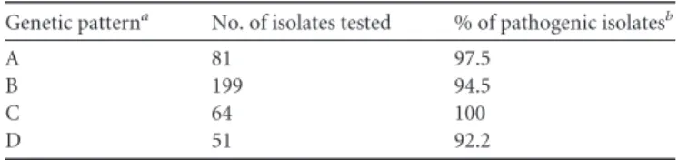
The results showed that 97.5% of the E. coli isolates belonging to the pattern A were pathogenic strains, as well as 94.5, 100, and 92.1%, of the isolates belonging to patterns B, C, and D, respec-tively (Table 4). These four virulence patterns allow the identifi-cation of pathogenic avian E. coli, whatever the animal origin of the isolates (see Table S3 in the supplemental material). Moreover, well-studied APEC strains belong to these virulence patterns. In-deed, the APEC strains TK3 (22), BEN2908 (12), 789 (2), and BEN687 (43) belong to patterns A, B, C, and D, respectively.
DISCUSSION
It has been reported that virulence factors (VFs) could be used as detection markers for APEC. In the present study, we defined a validated PCR diagnostic assay based on four different associa-tions of VFs that enables the identification of 70.2% of APEC. Pathogenic strains were identified with a 4.3% error margin.
The prediction of the pathogenicity of E. coli strains isolated from diseased animals is crucial. Routine diagnosis of avian E. coli isolates in veterinary laboratories is based on serotyping using commercially available serogrouping assays, which allow identifi-cation of the O1, O2, and O78 serogroups. Almost half of the strains studied here belonged to six different serogroups: O1, O2, O5, O8, O18, and O78. In the sample of 460 isolates used for statistical analysis, 223 isolates (48.5%) belonged to one of these six major serogroups, among which 199 were pathogenic strains. A diagnostic assay based on determining the major serogroups would thus only identify 56.5% (199/352) of the pathogenic strains (Table 2). However, the error margin would be 10.8% (24/ 223), considerably impairing the reliability of the method. Our data clearly show that the diagnostic scheme described here, which is based on identifying VFs, allows more APEC strains to be iden-tified with a lower error margin than when using serogrouping. This is probably due to the fact that these six serogroups are shared between APEC strains and avian fecal isolates of E. coli, as demon-strated by Rodriguez-Siek et al. (47).
The statistical analysis leading to the PCR diagnostic protocol described here is based on a decision-tree algorithm. Decision-tree algorithms are promising tools that could improve diagnosis. For example, they have been used to predict the diagnosis and outcome of dengue fever (57). To date, several PCR multiplex assays have been described to diagnose APEC (27, 33, 55). How-ever, each assay only described one association of VFs. Consider-ing the extreme diversity encountered in E. coli species, a sConsider-ingle set of VFs is insufficient to diagnose the majority of APEC strains. Ewers et al. developed a multiplex PCR system to detect the pres-ence of eight VFs, revealing various combinations of five to eight VFs (27). Moreover, diagnostic tests were not validated in most of
TABLE 3 Genetic patterns associated with E. coli pathogenicity in
chickens
Analysis and identified genetic patterna No. of isolates % of pathogenic isolatesb Analysis 1 iutA⫹, P(F11)⫹ 105 98.1 iutA⫹, P(F11)⫺, frzorf4⫹ 107 93.5
iutA⫹, P(F11)⫺, frzorf4⫺, O78⫹ 32 96.9
iutA⫺, sitA⫹, aec26⫹ 14 92.9
Analysis 2
iutA⫹, P(F11)⫹ 99 98.0
iutA⫹, P(F11)⫺, frzorf4⫹ 99 92.9
iutA⫹, P(F11)⫺, frzorf4⫺, O78⫹ 32 96.9
iutA⫺, sitA⫹, aec26⫹, aec4⫹ 11 90.9
aFor analysis 1, a statistical analysis was conducted on a sample of 460 isolates, including 352 pathogenic strains (of five chicks inoculated subcutaneously, one to five died) and 108 nonpathogenic isolates (of five chicks inoculated subcutaneously, none died). For analysis 2, a statistical analysis was conducted on 416 isolates from the analysis 1 sample excluding the isolates that killed only one chicken. This group comprised 308 pathogenic strains (of five chicks inoculated subcutaneously, two to five died) and 108 nonpathogenic isolates (of five chicks inoculated subcutaneously none died).
bFor analysis 1, “pathogenic isolates” refers to isolates where, of five chicks inoculated subcutaneously, one to five died. For analysis 2, “pathogenic isolates” refers to isolates where, of five chicks inoculated subcutaneously, two to five died.
TABLE 4 Validation of the genetic patterns A, B, C, and D
Genetic patterna No. of isolates tested % of pathogenic isolatesb
A 81 97.5
B 199 94.5
C 64 100
D 51 92.2
aGenetic patterns: A, iutA⫹, P(F11)⫹; B, iutA⫹, P(F11)⫺, frz
orf4⫹; C, iutA⫹, P(F11)⫺, frzorf4⫺, O78⫹; D, iutA⫺, sitA⫹, aec26⫹.
bThat is, of five chicks inoculated subcutaneously, one to five died. Schouler et al.
1676 jcm.asm.org Journal of Clinical Microbiology
on May 24, 2012 by UNIVERSITE DE LIEGE
http://jcm.asm.org/
the published studies. The only exception is a study conducted to identify the minimum number of VFs that could be used to dis-tinguish an APEC strain from an avian fecal isolate, which showed that a subset of five genes enabled the identification of an APEC strain, although the error margin was 14.6% (33).
The panel of genes implemented in the diagnostic schemes described here includes various VFs, some of which have been well characterized. They are mainly involved in the colonization of the internal organs of chickens, via different strategies. These include two iron acquisition systems. Gene iutA, one of the five genes of the aerobactin operon, encodes an outer membrane protein in-volved in the high-affinity binding of Fe3⫹-aerobactin and is plas-mid located (32, 41) or could be also chromosomally encoded in some APEC strains (unpublished data). The aerobactin system plays a role in the persistence and generation of lesions in APEC-infected chickens (20). Gene sitA encodes a periplasmic binding protein of the SitABCD transport system, which is involved in iron and manganese transport and could be both chromosomally and plasmid located (41, 50, 52). SitABCD has been shown to play a role in virulence (49).
P fimbriae are known to mediate the binding of bacteria to ␣-D-galactopyranosil--D-galactopyranoside receptors. Strains expressing P fimbriae have a mannose-resistant hemagglutination phenotype (23). Genes encoding fimbriae are located on a chro-mosomal pathogenicity island (34). It has been shown that P fim-briae are expressed in vivo in the air sacs, lungs, and kidneys and in blood and pericardial fluid. No expression has been observed in the liver, spleen, or trachea. This observation in line with in vitro studies demonstrating that P fimbriae are not involved in the ad-herence of APEC to tracheal and pharyngeal epithelial cells (46, 60). It has recently been shown in an APEC strain of serogroup O1 that P fimbriae are involved in the virulence of that strain in chick-ens (35). Gene frzorf4is chromosomally located and belongs to the
frz operon, which encodes a phosphoenolpyruvate carbohydrate
phosphotransferase system transporter and enzymes involved in sugar metabolism (48). A link between the expression of this locus and E. coli pathogenic abilities was confirmed by experiments showing its role in promoting bacterial fitness under stressful con-ditions, such as oxygen restriction or the late stationary phase of growth, and in promoting growth in chicken serum or the intes-tinal tract during in vivo competition assays. The frz operon also seems to be involved in the cell surface expression of F1 fimbriae, which are major adhesins of E. coli, via a mechanism yet to be characterized (48). Gene aec26 is chromosomally located and en-codes a putative membrane protein component of a type VI secre-tion system (T6SS) (52, 54). In an APEC strain, this T6SS has recently been shown to contribute to the adhesion and invasion of epithelial cells (HeLa) by influencing the expression of F1 fimbriae and to improve the virulence of the strain for 1-day-old chicks (15). O78 lipopolysaccharide contributes to serum resistance and colonization of internal organs of inoculated chicken with an APEC strain of serogroup O78 (40).
The fact that the VFs described here have been demonstrated to be involved in the pathogenic process of avian colibacillosis prob-ably accounts for the reliability of the diagnostic method that has been developed.
Moreover, the four associations of VFs identified by the statis-tical analysis suggest that APEC strains use different strategies to invade the host, according to their genetic equipment, all resulting in the same pathogenic process known as avian colibacillosis.
Nevertheless, almost 30% of APEC isolates in our sample were not identified as pathogenic strains, possibly due to the existence of other APEC VFs that have not been examined here and which could improve strain discrimination. The use of newly identified VFs, for example, a novel fimbrial adhesin called EA/I (1) and the autotransporter adhesin AatA (14), or VFs not included in the present study, such as IbeA (28) and Vat (45), would help to im-prove the present diagnostic method.
In sum, we propose a diagnostic tool based on four associations of virulence genes that enables the identification of more APEC strains than previously described methods.
ACKNOWLEDGMENTS
We thank Maryline Répérant-Ferter, Sandrine Perera-Melo, Katia Cour-voisier-Guyader, Valérie Ullman, and Anne-Soizic Guillon de Princé for their invaluable assistance in the practical aspects of the work and the staff of the INRA experimental platform of Nouzilly for care of the animals.
This study was funded by the European Community (contract FAIR6-CT-98-4093, coordinator, Maryvonne Dho-Moulin-Schouleur), the Na-tional Institute for Agronomic Research (France), the University of San-tiago de Compostela (Spain), the University of Liège (Belgium), and by Sanders Aliments, Laboratoires Vétérinaires (France). It was also partially supported by grants 09TAL007261PR, 10MRU261023PR, and 2007/ 000044-0 to J.B. (Xunta de Galicia [Spain] and The European Regional Development Fund [ERDF]) and grants 1363 and 1642 to J.M. (Fonds National de la Recherche Scientifique [Belgium]).
REFERENCES
1. Antao EM, et al. 2009. Signature-tagged mutagenesis in a chicken infec-tion model leads to the identificainfec-tion of a novel avian pathogenic
Esche-richia coli fimbrial adhesin. PLoS One 4:e7796.
2. Babai R, Blum-Oehler G, Stern BE, Hacker J, Ron EZ. 1997. Virulence patterns from septicemic Escherichia coli O78 strains. FEMS Microbiol. Lett. 149:99 –105.
3. Barnes HJ, Vaillancourt J-P, Gross WB. 2003. Colibacillosis, p 631– 652.
In Saif YM, et al. (ed), Diseases of poultry, 11th ed. Iowa State University
Press, Ames, IA.
4. Belanger L, et al. 2011. Escherichia coli from animal reservoirs as a poten-tial source of human extraintestinal pathogenic E. coli. FEMS Immunol. Med. Microbiol. 62:1–10.
5. Blanco J, et al. 1992. Serogroups of Escherichia coli strains producing cytotoxic necrotizing factors CNF1 and CNF2. FEMS Microbiol. Lett.
75:155–159.
6. Blanco JE, Blanco M, Mora A, Blanco J. 1997. Production of toxins (enterotoxins, verotoxins, and necrotoxins) and colicins by Escherichia
coli strains isolated from septicemic and healthy chickens: relationship
with in vivo pathogenicity. J. Clin. Microbiol. 35:2953–2957.
7. Blanco JE, et al. 1998. Serotypes of Escherichia coli isolated from septice-mic chickens in Galicia (northwest Spain). Vet. Microbiol. 61:229 –235. 8. Blanco M, et al. 1996. Polymerase chain reaction for detection of
Esche-richia coli strains producing cytotoxic necrotizing factor type 1 and type 2
(CNF1 and CNF2). J. Microbiol. Methods 26:95–101.
9. Blanco M, et al. 1997. Genes coding for enterotoxins and verotoxins in porcine Escherichia coli strains belonging to different O:K:H serotypes: relationship with toxic phenotypes. J. Clin. Microbiol. 35:2958 –2963. 10. Blattner FR, et al. 1997. The complete genome sequence of Escherichia
coli K-12. Science 277:1453–1474.
11. Breiman L, Freidman J, Olsen R, Stone C. 1984. Classification and regression trees. Chapman and Hall, New-York, NY.
12. Chouikha I, et al. 2006. A selC-associated genomic island of the extraint-estinal avian pathogenic Escherichia coli strain BEN2908 is involved in carbohydrate uptake and virulence. J. Bacteriol. 188:977–987.
13. Cortes MA, et al. 2008. Inactivation of ibeA and ibeT results in decreased expression of type 1 fimbriae in extraintestinal pathogenic Escherichia coli strain BEN2908. Infect. Immun. 76:4129 – 4136.
14. Dai J, et al. 2010. Suppression subtractive hybridization identifies an autotransporter adhesin gene of Escherichia coli IMT5155 specifically as-sociated with avian pathogenic E. coli (APEC). BMC Microbiol. 10:236.
on May 24, 2012 by UNIVERSITE DE LIEGE
http://jcm.asm.org/
15. de Pace F, et al. 2010. The type VI secretion system plays a role in type 1 fimbria expression and pathogenesis of an avian pathogenic Escherichia
coli strain. Infect. Immun. 78:4990 – 4998.
16. Dho M, Lafont JP. 1984. Adhesive properties and iron uptake ability in
Escherichia coli lethal and nonlethal for chicks. Avian Dis. 28:1016 –1025.
17. Dho M, Lafont JP. 1982. Escherichia coli colonization of the trachea in poultry: comparison of virulent and avirulent strains in gnotoxenic chick-ens. Avian Dis. 26:787–797.
18. Dho-Moulin M, Fairbrother JM. 1999. Avian pathogenic Escherichia coli (APEC). Vet. Res. 30:299 –316.
19. Dho-Moulin M, et al. 1990. Surface antigens from Escherichia coli O2 and O78 strains of avian origin. Infect. Immun. 58:740 –745.
20. Dozois CM, Daigle F, Curtiss R III. 2003. Identification of pathogen-specific and conserved genes expressed in vivo by an avian pathogenic
Escherichia coli strain. Proc. Natl. Acad. Sci. U. S. A. 100:247–252.
21. Dozois CM, et al. 2000. Relationship between the Tsh autotransporter and pathogenicity of avian Escherichia coli and localization and analysis of the Tsh genetic region. Infect. Immun. 68:4145– 4154.
22. Dozois CM, Fairbrother JM, Harel J, Bosse M. 1992. pap- and pil-related DNA sequences and other virulence determinants associated with
Esche-richia coli isolated from septicemic chickens and turkeys. Infect. Immun.
60:2648 –2656.
23. Dozois CM, Pourbakhsh SA, Fairbrother JM. 1995. Expression of P and type 1 (F1) fimbriae in pathogenic Escherichia coli from poultry. Vet. Mi-crobiol. 45:297–309.
24. Dziva F, Stevens MP. 2008. Colibacillosis in poultry: unraveling the molecular basis of virulence of avian pathogenic Escherichia coli in their natural hosts. Avian Pathol. 37:355–366.
25. Ewers C, Antao EM, Diehl I, Philipp HC, Wieler LH. 2009. Intestine and environment of the chicken as reservoirs for extraintestinal pathogenic
Escherichia coli strains with zoonotic potential. Appl. Environ. Microbiol.
75:184 –192.
26. Ewers C, Janssen T, Kiessling S, Philipp HC, Wieler LH. 2004. Molec-ular epidemiology of avian pathogenic Escherichia coli (APEC) isolated from colisepticemia in poultry. Vet. Microbiol. 104:91–101.
27. Ewers C, Janssen T, Kiessling S, Philipp HC, Wieler LH. 2005. Rapid detection of virulence-associated genes in avian pathogenic Escherichia
coli by multiplex polymerase chain reaction. Avian Dis. 49:269 –273.
28. Germon P, et al. 2005. ibeA, a virulence factor of avian pathogenic
Esch-erichia coli. Microbiology 151:1179 –1186.
29. Hacker J, Knapp S, Goebel W. 1983. Spontaneous deletions and flanking regions of the chromosomally inherited hemolysin determinant of an
Escherichia coli O6 strain. J. Bacteriol. 154:1145–1152.
30. Jerse AE, Martin WC, Galen JE, Kaper JB. 1990. Oligonucleotide probe for detection of the enteropathogenic Escherichia coli (EPEC) adherence factor of localized adherent EPEC. J. Clin. Microbiol. 28:2842–2844. 31. Johnson JR, Brown JJ. 1996. A novel multiply primed polymerase chain
reaction assay for identification of variant papG genes encoding the Gal(␣1-4)Gal-binding PapG adhesins of Escherichia coli. J. Infect. Dis.
173:920 –926.
32. Johnson TJ, Siek KE, Johnson SJ, Nolan LK. 2006. DNA sequence of a ColV plasmid and prevalence of selected plasmid-encoded virulence genes among avian Escherichia coli strains. J. Bacteriol. 188:745–758.
33. Johnson TJ, et al. 2008. Identification of minimal predictors of avian pathogenic Escherichia coli virulence for use as a rapid diagnostic tool. J. Clin. Microbiol. 46:3987–3996.
34. Kariyawasam S, Johnson TJ, Nolan LK. 2006. The pap operon of avian pathogenic Escherichia coli strain O1:K1 is located on a novel pathogenic-ity island. Infect. Immun. 74:744 –749.
35. Kariyawasam S, Nolan LK. 2009. Pap mutant of avian pathogenic
Esch-erichia coli O1, an O1:K1:H7 strain, is attenuated in vivo. Avian Dis. 53:
255–260.
36. La Ragione RM, Woodward MJ. 2002. Virulence factors of Escherichia
coli serotypes associated with avian colisepticemia. Res. Vet. Sci. 73:27–35.
37. Le Bouguenec C, Archambaud M, Labigne A. 1992. Rapid and specific detection of the pap, afa, and sfa adhesin-encoding operons in uropatho-genic Escherichia coli strains by polymerase chain reaction. J. Clin. Micro-biol. 30:1189 –1193.
38. Li G, et al. 2010. AatA is a novel autotransporter and virulence factor of avian pathogenic Escherichia coli. Infect. Immun. 78:898 –906.
39. Lintermans P, et al. 1988. Isolation and nucleotide sequence of the F17-A gene encoding the structural protein of the F17 fimbriae in bovine entero-toxigenic Escherichia coli. Infect. Immun. 56:1475–1484.
40. Mellata M, et al. 2003. Role of virulence factors in resistance of avian pathogenic Escherichia coli to serum and in pathogenicity. Infect. Immun.
71:536 –540.
41. Mellata M, Touchman JW, Curtiss R. 2009. Full sequence and compar-ative analysis of the plasmid pAPEC-1 of avian pathogenic E. coli chi7122 (O78:K80:H9). PLoS One 4:e4232.
42. Moriel DG, et al. 2010. Identification of protective and broadly conserved vaccine antigens from the genome of extraintestinal pathogenic
Esche-richia coli. Proc. Natl. Acad. Sci. U. S. A. 107:9072–9077.
43. Moulin-Schouleur M, et al. 2007. Extraintestinal pathogenic Escherichia
coli strains of avian and human origin: link between phylogenetic
relation-ships and common virulence patterns. J. Clin. Microbiol. 45:3366 –3376. 44. O’Brien AD, LaVeck GD, Thompson MR, Formal SB. 1982. Production of Shigella dysenteriae type 1-like cytotoxin by Escherichia coli. J. Infect. Dis. 146:763–769.
45. Parreira VR, Gyles CL. 2003. A novel pathogenicity island integrated adjacent to the thrW tRNA gene of avian pathogenic Escherichia coli en-codes a vacuolating autotransporter toxin. Infect. Immun. 71:5087–5096. 46. Pourbakhsh SA, et al. 1997. Dynamics of Escherichia coil infection in
experimentally inoculated chickens. Avian Dis. 41:221–233.
47. Rodriguez-Siek KE, Giddings CW, Doetkott C, Johnson TJ, Nolan LK. 2005. Characterizing the APEC pathotype. Vet. Res. 36:241–256. 48. Rouquet G, et al. 2009. A metabolic operon in extraintestinal pathogenic
Escherichia coli promotes fitness under stressful conditions and invasion of
eukaryotic cells. J. Bacteriol. 191:4427– 4440.
49. Sabri M, et al. 2008. Contribution of the SitABCD, MntH, and FeoB metal transporters to the virulence of avian pathogenic Escherichia coli O78 strain chi7122. Infect. Immun. 76:601– 611.
50. Sabri M, Leveille S, Dozois CM. 2006. A SitABCD homologue from an avian pathogenic Escherichia coli strain mediates transport of iron and manganese and resistance to hydrogen peroxide. Microbiology 152:745– 758.
51. Sambrook J, Fritsch EF, Maniatis T. 1989. Molecular cloning: a labora-tory manual, 2nd ed. Cold Spring Harbor Laboralabora-tory, Cold Spring Har-bor, NY.
52. Schouler C, Koffmann F, Amory C, Leroy-Setrin S, Moulin-Schouleur
M. 2004. Genomic subtraction for the identification of putative new
vir-ulence factors of an avian pathogenic Escherichia coli strain of O2 sero-group. Microbiology 150:2973–2984.
53. Scott DA, Kaper JB. 1994. Cloning and sequencing of the genes encoding
Escherichia coli cytolethal distending toxin. Infect. Immun. 62:244 –251.
54. Shrivastava S, Mande SS. 2008. Identification and functional character-ization of gene components of type VI secretion system in bacterial ge-nomes. PLoS One 3:e2955.
55. Skyberg JA, et al. 2003. Characterizing avian Escherichia coli isolates with multiplex polymerase chain reaction. Avian Dis. 47:1441–1447. 56. Stordeur P, et al. 2002. Examination of Escherichia coli from poultry for
selected adhesin genes important in disease caused by mammalian patho-genic E. coli. Vet. Microbiol. 84:231–241.
57. Tanner L, et al. 2008. Decision tree algorithms predict the diagnosis and outcome of dengue fever in the early phase of illness. PLoS Negl. Trop. Dis.
2:e196.
58. Therneau TM, Atkinson B, Ripley BR. 2009. rpart: recursive partitioning, R package, version 3.1– 42.http://CRAN.R-project.org/package⫽rpart. 59. Toth I, Herault F, Beutin L, Oswald E. 2003. Production of cytolethal
distending toxins by pathogenic Escherichia coli strains isolated from hu-man and animal sources: establishment of the existence of a new cdt vari-ant (type IV). J. Clin. Microbiol. 41:4285– 4291.
60. van den Bosch JF, et al. 1993. Identification of F11 fimbriae on chicken
Escherichia coli strains. Infect. Immun. 61:800 – 806.
61. Williams PH. 1979. Novel iron uptake system specified by ColV plasmids: an important component in the virulence of invasive strains of Escherichia
coli. Infect. Immun. 26:925–932.
Schouler et al.
1678 jcm.asm.org Journal of Clinical Microbiology