HAL Id: inserm-00332755
https://www.hal.inserm.fr/inserm-00332755
Submitted on 21 Oct 2008
HAL is a multi-disciplinary open access archive for the deposit and dissemination of sci-entific research documents, whether they are pub-lished or not. The documents may come from teaching and research institutions in France or abroad, or from public or private research centers.
L’archive ouverte pluridisciplinaire HAL, est destinée au dépôt et à la diffusion de documents scientifiques de niveau recherche, publiés ou non, émanant des établissements d’enseignement et de recherche français ou étrangers, des laboratoires publics ou privés.
ElePhant–an anatomical electronic phantom as
simulation-system for otologic surgery.
Ronny Grunert, Gero Strauss, Hendrick Moeckel, Mathias Hofer, Antje
Poessneck, Ulrich Fickweiler, Mario Thalheim, Ronny Schmiedel, Pierre
Jannin, Thomas Schulz, et al.
To cite this version:
Ronny Grunert, Gero Strauss, Hendrick Moeckel, Mathias Hofer, Antje Poessneck, et al.. ElePhant–an anatomical electronic phantom as simulation-system for otologic surgery.. Conference proceedings : .. Annual International Conference of the IEEE Engineering in Medicine and Biology Society. IEEE En-gineering in Medicine and Biology Society. Annual Conference, Institute of Electrical and Electronics Engineers (IEEE), 2006, 1, pp.4408-11. �10.1109/IEMBS.2006.260542�. �inserm-00332755�
Abstract—This paper describes the ElePhant (Electronic
Phantom) - an anatomical correct simulation system based on 3D Rapid Prototyping models for the oto-rhino-laryngoscopic intervention “Mastoidectomy”. The anatomical structures of the head are created with plaster as base material using 3D-printing as rapid prototyping technology (RPT). Structures at risk, represented by electrically conductible material and fiber optics, are realized as an electric circuit and can be detected during the simulation of the surgical procedure. An accuracy study of 15 identical RPT-models compared to the 3D recon-structed CT-dataset of the patient showed that the mean accu-racy is (0.101 ± 0.077) mm, with the maximum and minimum values of +0.380 mm and -0.272 mm. An evaluation study of the ElePhant-system for “Mastoidectomy” was performed by 7 ENT-surgeons. The mean value of the study questionnaire (evaluation range from -2 (not at all) to +2 (very good)) was +1.2. The results showed that the ElePhant can simulate “Mas-toidectomy” realistically. It is especially suitable for the simula-tion of the correct representasimula-tion and posisimula-tion of the anatomi-cal structures, realistic operation setting, and realistic milling properties of the bone structure. Furthermore it is applicable for training of surgeons.
Keywords—simulation system, Rapid Prototyping,
ENT-surgery, evaluation
I. INTRODUCTION
For the evaluation of different computer assisted surgery (CAS) systems and surgical techniques, a standardization is necessary. The aim of this study is the development of an anatomically correct simulation system for the evaluation of CAS-systems, surgical interventions and instruments as well as surgical training of physicians.
Until now, simulation systems did not provide an objective evaluation and important system properties e.g. realistic milling properties of the bone, the detection of damaging structures at risk during the simulation, different pathology, the realistic operating room environment and the use of the surgical instrument being also used during the real surgery with a single system [1], [2], [3]. Virtual simulators need complex computer models to simulate the above mentioned
properties [2]. It is easier to implement this with physical phantoms, since polyurethane infiltrated plaster has realistic properties for milling applications on bone structures.
Interventions at the Mastoid are a special field of ENT-surgery with a high number of cases (ca. 5800 per year in Germany). During “Mastoidectomy” inflamed bone struc-tures of the Mastoid are removed with a milling cutter. Near the bone to be removed, structures at risk (facial nerve, sinus sigmoideus and semicircular canals) are located, which have to be prevented from injuring.
In this paper we present the ElePhant-system, which was developed to simulate realistically “Mastoidectomy”. It can simulate, i.e. the correct representation and position of the anatomical structures, realistic operation setting as well as realistic milling properties of the bone structure. Moreover, the ElePhant has an adequate anatomical accuracy.
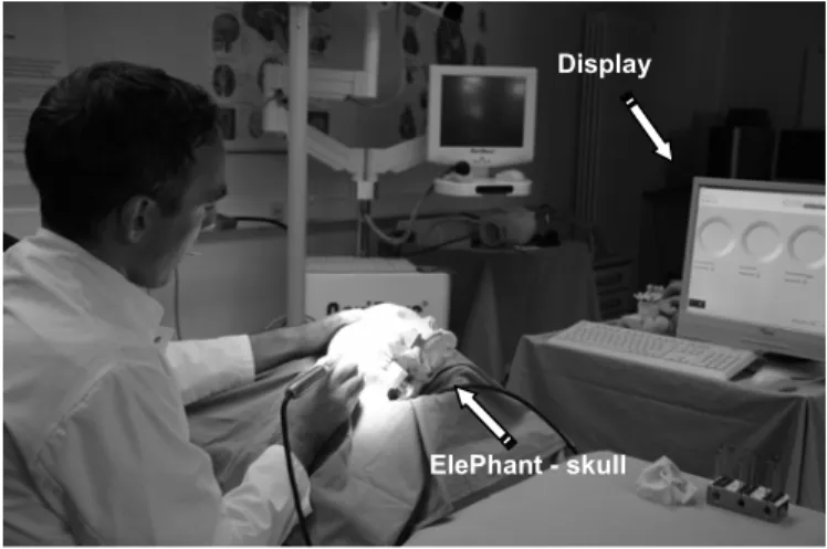
A study was performed with 7ENT-surgeons who evaluated the anatomical simulation system with the help of a ques-tionnaire after performing the milling procedure (Fig. 1).
Fig. 1. Simulation of the surgical intervention "Mastoidectomy" with a realistic operation setting, using the ElePhant-system. If the surgical instru-ment damages the structure at risk, a signal appears on the display.
ElePhant - An anatomical Electronic Phantom as simulation-system
for oto-rhino-laryngoscopic-surgery
R. Grunert a, G. Strauss a, b, H. Moeckel a, M. Hofer a, b, A. Poessneck a, U. Fickweiler b, M. Thalheim a, R. Schmiedel c, P. Jannin d, T. Schulz e, J. Oeken f, A. Dietz a, b, W. Korb a
a Innovation Center Computer Assisted Surgery (ICCAS), University of Leipzig, Germany b Department of Otorhinolaryngology/Plastic Surgery, University of Leipzig, Germany
c Department of Biomedical Technology, Heart Center, Leipzig, Germany
d Visages U746, Inserm INRIA, Faculté de médecine, Université de Rennes 1, Rennes, France e Department of Diagnostic Radiology, University of Leipzig, Germany
f Department of Otorhinolaryngology, Hospital of Chemnitz, Germany
Display
II. MATERIALS AND METHODS
A. Segmentation and Rapid Prototyping (Anatomical Structures)
The CT of a human skull preparation was acquired with the CT Siemens Somatom Volume Zoom, with the recon-structed layer thickness 0.5 mm, and a 512 x 512 matrix. The segmentation of the anatomical structures was based on the acquired CT dataset using the software Mimics 9.0 (Ma-terialise, Leuven, Belgium).
After segmentation, the dataset was stored as STL-file and sent to the 3D printer Z™510 (4D Concepts, Gross-Gerau, Germany). The building material was based on plaster. Sub-sequently the printed model was infiltrated with polyure-thane and acetone to influence the material to achieve nearly human bone properties.
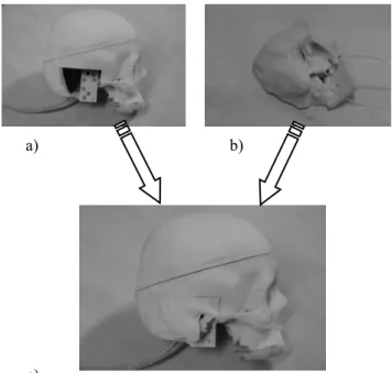
The anatomical simulation system is designed as a modu-lar structure, i.e. the petrous bone including the mastoid and structures at risk constitutes one module, while the rest of the skull is another module (Fig. 2). To create the modules with a pin-hole-connection, the software Polyworks 9.0 (InnovMetric Software Inc, Sainte-Foy, Canada) was used. The pin-hole-connection allows a positionally correct re-placement of a new petrous bone module (the structure of resection). The dimensions of the pin were 4.9 mm in diame-ter and 3.9 mm in length, and of the hole 5.0 mm in diamediame-ter and 4.0 mm in depth. The pin-hole-connection could be created using a special cutting function provided by the software to define the dimensions of the pin-hole-connection. For each experiment a new petrous bone mod-ule was positioned into the master simulator. This is more cost-effective for multiple experiments than printing the whole skull.
For the assessment of the intervention quality, the contact of the functional structure (e.g. the structure at risk) with the surgical instrument can be detected, processed, and analyzed with the help of an electrically respective optoelectronic system.
B. Mechatronic System
The disruption of the blood flow by damage of the vascu-lar structures as well as the neurological trauma through the damage of the nerve structures are simulated with the Ele-Phant. The facial nerve, sinus sigmoideus, and semicircular canals were realized, since they are critical for “Mastoidec-tomy”.
An important parameter during surgery is the detection of contacts between the surgical instrument and structures at risk. Two technical principles are used to give feedback about the contacts to the user of the ElePhant-system:
• structures at risk represented by electrically con-ductible materials,
• structures at risk represented by fiber optics
1. Structures at risk represented by electrically conductible materials
The petrous bone model includes the structures at risk such as sinus sigmoideus and the semicircular canals. In the RPT process, these structures are printed as hollow channels. The hollow channels are filled with an alloy (lead, bismuth, tin) having the melting point at 96°C. After the alloy had been melted, it was filled into the cavities. The tips of the metal filled sinus sigmoideus and the semicircular canals are connected with the analogue output and input channel of a data acquisition card PCI 6221 (DAQ-card) (National In-struments, Austin, Texas, USA). In the skull module, a con-necting appliance with sprung metal pins is located. If the petrous bone module is positioned into the skull module, the tips of the electrically conductible structures at risk are pressed against the metal pins and connected with the elec-tric circuit. The voltage of the analogue output has a value of 0.5 V. For the "Mastoidectomy", the milling cutter is used as surgical instrument.
Fig. 2. Modular structure of the ElePhant-system a) skull module, b) petrous bone module, c) bonded modules.
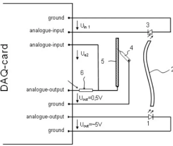
The milling cutter, being electrically conductible, was con-nected with the ground of the DAQ-card (Fig. 3).
If the milling cutter damages the sinus sigmoideus or the semicircular canals, the current through the structures at risk will not flow to their input channels but over the milling cutter to the ground. The current maximum value is 50 µA, which is beyond the human perception current of ca. 0.5 mA. The moment of the contact between surgical instrument and structure at risk is registered with a LabView program (National Instruments, Austin, Texas, USA).
a) b)
Fig. 3. Electric circuit of the simulation system: 1- LED, 2- fiber optic (structure at risk – facial nerve), 3- Photodiode, 4- surgical instrument, 5-electrically conductible material (structure at risk- sinus sigmoideus, semi-circular canals), 6- resistance.
2. Structures at risk represented by fiber optics
In contrast to the electrically conductible representation of the structures at risk, a fiber optic is positioned in the chan-nel of the facial nerve in the petrous bone module. The light source is a LED providing the light through the fiber optic. At the other end of the fiber optic, a photodiode detects the intensity of the light. To evaluate the signal of the photodi-ode, it is connected with an input channel of the DAQ-card. If the milling cutter damages the fiber optic (the facial nerve), the illuminance level of the photodiode will be re-duced. In addition to counting and measuring the contact between the milling cutter and fiber optic, it is possible to determine the percentage of damage in the cross-sectional area of the fiber optic.
To realize the above-mentioned option, a calibration was performed when the program was developed. A calibration-function was generated giving information about the per-centage of the damaged cross-sectional area in the fiber optic and the appendant illuminance level.
3. Software for simulation of physiological structures
The software LabView 7.1 (National Instruments, Austin, Texas, USA) was used to develop a program for the simula-tion system. The program controls the DAQ-card, which received the data resulting from the simulation process and to evaluate the data. Through the output channel, a voltage of 0.5 V is provided to the simulation system (LED and structure at risk based on the electrical principle). The DAQ-card distinguishes between the structures at risk. Each struc-ture at risk is connected with a separate input channel. Be-fore running the simulation process, a new software user profile is created or an already existing one is selected. Dur-ing the simulation procedure the program is able to count the contacts between the milling cutter and the structure at risk as well as to determine the damaged percentage of the
cross-sectional area of the structures at risk represented by fiber optics. If there is a contact during an experiment, a graphical and acoustic signal is triggered, so that the surgeon gets feedback about an accidental contact between the milling cutter and structure at risk. This option can be turned off for background logging of errors for already trained test per-sons. The duration of the simulation, number of contacts according the damaged structures at risk, and moment of the structure at risk damage are measured during the simulation process.
A learning curve is created for every user profile (contacts over number of simulations; time over number of simula-tions).
C. Accuracy study
Based on the CT-scan of an ENT-patient, the regions of interests (i.e. the petrous bone, facial nerve, and artery) were segmented using the software Mimics 9.0 (Materialise, Leu-ven, Belgium). To determine the accuracy of the 3D RPT-models, 15 3D RPT-models based on the same segmented original patient dataset were created. After had been printed out, the 15 models were scanned with the CT (Siemens Somatom Volume Zoom) with a reconstructed layer thick-ness of 0.5 mm. The software Polyworks 9.0 (InnovMetric Software Inc, Sainte-Foy, Canada) was used for the surface comparison of the triangulated models. The accuracy was quantified by the distance between the corresponding surface points of the triangulated model of the patient CT dataset and the surface points of the CT dataset of the 15 printed RPT-models based on the patient dataset.
D. Evaluation study of the ElePhant-system
To show that the simulation system based on 3D RPT-models is realistic for surgical ENT-intervention “Mastoi-dectomy” and that it is helpful for training of surgeons, 7 surgeons performed “Mastoidectomy” using the ElePhant simulation system (Fig. 4).
Fig. 4. ElePhant – skull after the simulated milling procedure “Mastoidec-tomy” was performed.
After the milling procedure, a questionnaire with 10 ques-tions was filled out. The first question was the number of already performed “Mastoidectomies”. The range of the further 9 questions’ score was from +2 (very good) to -2 (not at all). The questions can be subclassified into three catego-ries:
1. anatomical correctness 2. milling properties, handling
3. correct response of the electronic system
III. RESULTS
A. Mechatronic System
An anatomically correct simulation system based on 3D RPT-models generated by the 3D-Printing Rapid Prototyp-ing technology was developed. The contact between the surgical instrument and structures at risk during the realistic surgical simulation using the ElePhant-system can be de-tected. Electrically conductible materials such as fiber optics are used for the representation of structures at risk. Due to the modular structure, it is possible to position the petrous bone module easily in the skull module and thereby auto-matically connect it to the electric circuit.
B. Accuracy study
A previous work [4] showed that inside the region of inter-est for the surgical intervention the mean accuracy between corresponding surface points of the triangulated model of the patient CT dataset and the surface points of the CT dataset of 15 printed RPT-models based on the patient dataset was (0.101±0.077) mm, with the maximum and minimum values of +0.380 mm and -0.272 mm considering 380,146 data-points.
C. Evaluation study of the ElePhant-system
The mean value of the whole questionnaire was +1.2 and focused on the three categories:
1. anatomical correctness +1.6 2. milling properties, handling +0.2 3. correct response of the electronic system +1.9
IV. DISCUSSION
The results of the accuracy study show that the anatomical accuracy of the 3D RPT-models is suitable to represent the position of the anatomical structures realistically.
As far as the results of the questionnaire are concerned, it could be seen that especially the anatomical correctness
(+1.6) and the electronic system response (+1.9) could be realized very well. The surgeons declared that the milling properties are comparable to human bone. The cavities in-side the Mastoid structure could not be realized realistically (+0.2). The reason for that is based on the method of the 3D printing technology. The unbound plaster powder cannot be removed from the closed structures. The anatomical correct simulation system ElePhant, based on 3D RPT-models, can be used to simulate a surgical intervention for “Mastoidec-tomy” realistically, i.e. correct representation and position of the anatomical structures and different pathology, realistic operation setting as well as realistic milling properties of the bone structure. If the surgical instrument damages structures at risk, an objective evaluation is possible with the help of the detection. The ElePhant can also be used as a simulation system for training of surgeons.
A similar system setup is also suitable for other surgical disciplines such as milling procedures at the spine.
Compared to other rapid prototyping technologies, anatomi-cal 3D models based on plaster can be produced cost-effectively. The generated 3D RPT-models enable the sur-geon to apply the same surgical instrument which will also be used during the operation on the patient. Milling and drilling procedures can be performed at the 3D models by the surgeons.
During the next research phase a micro-controller-circuit positioned inside the skull will be developed instead of the PCI DAQ-card. The skull will be easily connected to the USB-interface with a cable. This system-configuration will allow a mobile and well-applicable solution to perform sur-gical simulations using the same system in different institu-tions. All collected data will be used for an objective evalua-tion of CAS-systems, other surgical devices, and training of surgeons.
Methodologies for the evaluation of CAS-systems, de-scribed in [5], [6] and [7], regarding different impact factors e.g. technical capacity, diagnostic accuracy, therapeutic impact, patient outcome and social impact will be further investigated. The ElePhant may support this evaluation.
REFERENCES
[1] U. v. Lukas and E. Carrasco, "The Nasal Endoscopy Simulator
(NES)," Medizinische Technologie und Anwendung. pp. 25, 3, 2003.
[2] C. Trantakis, J. Meixensberger, U. Kühnapfel et al, ''IOMaster 7D' - a
new device for virtual neuroendoscopy,” Computer Assisted
Radiol-ogy and Surgery 2004, Chicago, pp. 707-712, 2004.
[3] L. B. Tuason, “Laparoscopic surgery simulator,” U. S. Pat. No. 5,403,191. 04.04.1995.
[4] R. Grunert, M. Hofer, W. Korb et al, “Accuracy of anatomical Rapid
Prototyping Models for preoperative surgical planning,” Computer
Assisted Radiology and Surgery, 2006, to be published.
[5] C. S. Goodman, “HTA 101 Introduction to health technology
assess-ment,” U.S. National Library of Medicine, 2004.
[6] D. G. Fryback and J. R. Thornbury, "The Efficacy of Diagnostic
Imaging," Medical Decision Making, vol. 11, pp. 88-94, 1991.
[7] P. Jannin, J. M. Fitzpatrick, M. W. Vannier et al, "Validation of
Medical Image Processing in Image-guided Therapy," IEEE