Amniotic Membrane Transplantation and Fibrin Glue in
the Management of Corneal Ulcers and Perforations
A Review of 33 Cases
Sandrine Hick, MD,*†‡ Pierre E. Demers, MD, FRCSC,†§ Isabelle Brunette, MD, FRCSC,‡§
Ce´line La, MD,* Miche`le Mabon, MD,‡ and Bernard Duchesne, MD*
Purpose:To evaluate the efficacy of amniotic membrane in corneal ulcers refractive to conventional treatment and amniotic membrane with fibrin glue in corneal perforations.
Methods: Amniotic membrane transplantation (AMT) was per-formed in 33 eyes from 32 patients for corneal ulcers refractive to conventional treatment. Fourteen ulcers were perforated and received fibrin glue and amniotic membrane. Ulcers were divided into 3 groups: neurotrophic or exposure, autoimmune, and other etiology.
Results:Overall success was observed in 80% (27/33 eyes) of the cases, with success rates of 87.5% (14/16 eyes), 70% (7/10 eyes), 85.7% (6/7 eyes) in groups 1, 2, and 3, respectively. The ulcers healed in a mean time of 3.6 6 1.6 weeks and the follow-up was 14.8 6 9.9 months. Failure was noted in 6 eyes with severe neurotrophic keratitis, Stevens-Johnson syndrome, ocular cicatricial pemphigoid, and Acanthamoeba keratitis. Grafts with fibrin sealant showed a success rate of 92.9 % (13/14 eyes) compared to 73.7% (14/19 eyes) for amniotic grafts alone. In patients with severe limbal damage, a success rate of only 20% (1/5) was observed.
Conclusions:AMT is a viable option in the treatment of nonhealing corneal ulcers of various depth and etiologies. Perforations up to 3 mm can be safely managed by fibrin glue and AMT. These techniques lead to rapid reconstruction of the corneal surface and can give a good final functional result or allow keratoplasty to be done in more favor-able conditions.
Key Words: amniotic membrane, corneal ulcer, corneal perforation, fibrin glue
(Cornea 2005;24:369–377)
C
orneal epithelial defects usually heal without complica-tions. However, many factors can interfere with wound healing, including dry eye, lid malfunction, inflammation, in-fection, or damage to corneal nerves. These factors are found in primary ocular surface disorders, such as neurotrophic keratitis and keratoconjunctivitis sicca, or in systemic diseases, such as rheumatoid arthritis, ocular cicatricial pemphigoid, and Stevens-Johnson syndrome. When corneal stem cells, epithe-lial cells, and epitheepithe-lial basement membrane are damaged by persistent inflammation, epithelial defects, corneal scarring and neovascularization may develop.1Moreover, matrix metalloproteinases produced by keratocytes,2
epithelial cells, and neutrophils3
can cause progressive stromal ulceration, which can lead to corneal perforation.
Current treatments include artificial tears, lubricants, patching, therapeutic bandage contact lenses,4
and scleral lenses.5
Experimental trials with fibronectin,6
insulin growth factor, substance P,7
and nerve growth factor8
have also been attempted, with limited success. If the defect progresses, surgical treatments such as tissue adhesives,9
tarsorrhaphy, conjunctival flaps,10
and penetrating or lamellar keratoplasty11
if corneal perforation ensues may become necessary. However, these surgical modalities are associated with a large number of problems including tissue inflammation from glue toxicity, diminished vision, and poor cosmesis with a conjunctival flap and tarsorrhaphy and graft failure in the setting of an inflamed or infected eye.11
In 1997, Lee and Tseng12
were the first to propose the use of human amniotic membrane (AM) for the treatment of corneal epithelial defects. Subsequently, AM transplantation (AMT) was applied to patients with cicatricial eye diseases,13
pte-rygium,14
and ocular surface reconstruction after removal of large conjunctival lesions15
or following chemical or thermal burns.16
AMT has been shown to promote epithelial healing and to reduce vascularization, inflammation, and scarring. Kruse et al17
showed rapid epithelial healing and stromal thickening after multilayer AMT in patients with deep corneal ulcers. In 2000, Chen et al18
reported AMT as an effective alternative for treating severe neurotrophic ulcers, and recently they described the favorable outcome of 34 eyes with AMT for corneal ulcers and perforations of less than 0.5 mm in diameter.19
This study evaluates the efficacy of AM in the man-agement of a variety of corneal ulcers, central and paracentral, and reports the outcome of perforations up to 3 mm in dia-meter treated by fibrin glue and AMT.
Received January 8, 2004; revision received August 26, 2004; accepted August 27, 2004.
From the *Department of Ophthalmology, University of Lie`ge, Lie`ge, Belgium; †Department of Ophthalmology, Centre Hospitalier Universi-taire de Montre´al, Notre-Dame Hospital, Montreal, Quebec, Canada; ‡Department of Ophthalmology, Maisonneuve-Rosemont Hospital, Montreal, Quebec, Canada; §Department of Ophthalmology, University of Montreal, Montreal, Quebec, Canada.
This study was supported by the Fondation Le´on-Fre´de´ricq, University of Lie`ge and a Mandate for Clinical Research from the Centre Hospitalier Universitaire de Lie`ge, Lie`ge, Belgium.
Reprints: Bernard Duchesne, MD, Department of Ophthalmology, CHU Sart-Tilman, 4000 Lie`ge, Belgium (e-mail: b.duchesne@chu.ulg.ac.be). CopyrightÓ 2005 by Lippincott Williams & Wilkins
PATIENTS AND METHODS
We studied 33 cases of AMT (32 patients) for corneal ulcers performed between 1997 and 2002. They were managed at 3 different sites (Centre Hospitalier Universitaire de Lie`ge, Belgium [n = 20] and Centre Hospitalier Universitaire de Montre´al and Maisonneuve-Rosemont Hospital, Canada [n = 13]). This retrospective review conformed to the requirements of the ethics committee of each of the 3 centers. Criteria for inclusion were refractory corneal ulcers, including corneal ulcers with perforations smaller than 3 mm in diameter. Patients were referred by different ophthalmologists after failure of conventional treatment of the ulcer for a minimum of 4 weeks. In all cases, toxic topical medications were removed and lubrication, punctal occlusion if appropriate, and bandage contact lenses were tried without success. Clinical data including patient demography, etiology, surgical procedure, associated therapies, visual acuity, complications, and final outcome were retrieved retrospectively. Patients were divided in 3 groups according to etiology of corneal disease: group 1 for neurotrophic keratitis, group 2 for autoimmune diseases, and group 3 for others. Patients with corneal ulcer received AMT alone, and patients with corneal perforations were managed by fibrin glue and AMT. Surgical success was de-fined as cessation of aqueous leakage and formation of a deep anterior chamber in cases with perforation, complete epithelialization of ocular surface, and the presence of a visible stromal thickness at the operated site by the second month following AMT. Failure was defined as persistence or re-currence of aqueous leakage, lack of or incomplete epithe-lialization, or recurrence of corneal ulceration at the same site at 2 months.
Preparation of Preserved AM
Human AM was prepared and preserved using a method previously described by Lee and Tseng.12
AMs for Montreal were obtained from Bio-Institute (Miami, FL) and AMs for Lie`ge were acquired and preserved at the eye bank of Lie`ge (Belgium) following the same protocol. Human immunode-ficiency virus, human hepatitis types B and C, and syphilis had been excluded by serologic tests.
Amniotic Membrane Transplantation
Procedures were performed by 3 surgeons (P.E.D, B.D., and M.M.) using topical (n = 16), peribulbar (n = 8), or general (n = 9) anesthesia. The base of the ulcer was debrided with fine forceps and a microsponge, and the poorly adherent epithelium adjacent to the edge of the ulcer was removed. One or 2 pieces of AM were removed from the nitrocellulose paper filter and transferred onto the ulcer with the basement membrane facing up. Interrupted sutures or a running 10-0 nylon suture secured the AM beyond the edge of the defect. For patients 16 and 24, 3 and 4 pieces of AM, respectively, were used. In these cases, small pieces were stacked one above the other to fill the cavity of the ulcer, and a larger piece was used to cover the ulcer and the deepithelialized zone. This membrane was then secured with interrupted sutures.
Corneal perforations were managed using a fibrin glue (Tissucol, Baxter, Belgium or Tisseel, Baxter, Deerfield, IL) and AMT. A small volume of sodium hyaluronate viscoelastic
(Healon, Pharmacia, Brussels, Belgium or Pharmacia, Montreal, Canada) was used to restore an adequate anterior chamber depth. Debridement and epithelium removal were performed with a blunt blade. After cleaning and drying of the ulcer, the fibrin glue was used to fill up the perforation site creating a fibrin plug. An AM (basement membrane side up) was then used to cover and secure the fibrin plug using interrupted sutures.
A bandage contact lens was applied in all cases until the epithelial defect completely healed. Patients received a com-bination of corticosteroid-antibiotic drops 3 to 4 times daily. They were examined on postoperative day 1, then weekly until complete epithelialization occurred, monthly for 3 months, and every 3 to 6 months thereafter.
Statistical Analysis
The qualitative data were analyzed by x2
exact test and the quantitative postoperative data were analyzed by t tests and ANOVA (SAS version 8.2). A P value of less than 0.05 was considered to be statistically significant.
RESULTS
Thirty-two patients were involved, 16 men and 16 women (Table 1). The average age of our population was 62 6 17.8 years (range, 16–85). The average follow-up was 14.8 6 9.9 months (range, 1–36). Patients were divided in 3 groups according to the ulcers’ etiology. Group 1 (16 eyes) included neurotrophic and exposure ulcers. Group 2 (10 eyes) included ulcers associated with autoimmune diseases such as Stevens-Johnson syndrome (n = 2), Sjo¨gren syndrome (n = 3), ocular cicatricial pemphigoid (n = 1), rheumatoid polyarthritis (n = 3), and Lyell disease (n = 1). Group 3 (7 eyes) was composed of ulcers of different etiologies.
When considering all ulcers, perforated or not, 30 of 33 eyes (90%) epithelialized within 3.6 6 1.6 weeks (Table 2). The ulcer reoccurred within 2 months in 4 eyes (13%), with a mean recurrence time of 6.5 6 1.9 weeks. The AMT was successful in ulcer reconstruction in 27 of 33 eyes (81%), after 1 (26 patients) or 2 (1 patient) procedures. Patient 32 was counted as a success but died 1 month later. In patients with successful AMT, the anterior chamber was deep, and there was no further leakage of aqueous fluid; the site of the ulcer showed a subepithelial scar with, in some cases, a thickening of the stroma, and epithelialization was complete. Conjunc-tival inflammation and pain were reduced in all patients, although pain relief was not specifically quantified. The final visual acuity remained unchanged in 11 eyes, improved in 17 eyes, and decreased in 2 eyes. Of the 17 patients with improved visual acuity, 8 gained 2 lines or more on the Snellen chart. In 2 patients, the preoperative visual acuity was not recorded. In general, the final acuity remained weak with 20 eyes (60%) seeing 20/200 or less.
Among the 3 groups, the success rate, ie, epithelial healing with stable corneal surface within the first 2 post-operative months, was 87.5% in group 1, 70% in group 2, and 86 % in group 3. The mean epithelialization times after AMT and the mean follow-up for each group are summarized in
TABLE 1. Clinical Data and Surgical Outcome
Case/Age/Sex Eye Diagnosis Etiology Surgery
Associated Procedures Complete Reepithelialization (Weeks) Neurotrophic/exposure keratitis
1/55/M OD Ulcer Neurotrophic, post-HSK Diffuse limbal deficiency
AMT No 1
2/74/M OD Ulcer Neurotrophic AMT No 1
3/76/F OD Ulcer Neurotrophic, post-infectious (streptococcal)
AMT No No
4/16/F OD Ulcer Neurotrophic AMT No 5
Damage to V1 2nd AMT 5
5/82/M OD Ulcer Neurotrophic + exposure (polyneuropathy lagophtalmy)
AMT No 5
6/52/M OS Ulcer Exposure Keratitis Lid surgery AMT No 1 7/77/M OD Ulcer +
perforation 1 mm
Neurotrophic Diabetic mellitus AMT + fibrin glue
No 6
8/69/F OD Ulcer Exposure Keratitis Lid surgery AMT No 4 9/66/F OS Ulcer Neurotrophic + exposure
Damage to V et VII Meningioma removal
AMT SK 2
10/81/F OD Ulcer Neurotrophic Failed graft HZV
AMT No 3.5
11/28/F OS Ulcer Neurotrophic HSV AMT No 2 12/74/F OS Ulcer + perforation 2 mm Neurotrophic Failed graft AMT + fibrin glue No 2.5 13/74/M OS Ulcer + perforation 3 mm Neurotrophic AMT + fibrin glue No 2 14/78/M OD Ulcer + perforation 3 mm
Exposure Ke´ratitis Yblinking Senile dementia
AMT + fibrin glue
No 2
15/43/M OS Ulcer Neurotrophic Post-infectious AMT No 6
16/56/M OS Ulcer Neurotrophic AMT No 2
HSV 2nd AMT ML No 1
Autoimmune diseases
17/36/M OD Ulcer Stevens-Johnson Diffuse limbal deficiency
AMT No No
18/68/F OD Ulcer + perforation 2 mm
Sjo¨gren syndrome AMT + Fibrin glue
Prednisone 6
19/68/F OS Ulcer + perforation 2 mm
Sjo¨gren syndrome AMT + Fibrin glue
Prednisone 6
20/80/M OD Ulcer + perforation 1.5 mm
Ocular Pemphigoid AMT + Fibrin glue
Immuran 3
Diffuse limbal deficiency 2nd AMT + Fibrin glue
4 21/81/F OS Descemetocele +
perforation 0.5 mm
RA 2 weeks post cataract surgery AMT + Fibrin glue Plaquenil 6 22/72/M OS Ulcer + perforation 2 mm
Sjo¨gren syndrome 3 weeks post cataract surgery
AMT + Fibrin glue Cyclosporine PO Punctal occlusion 2 23/85/F OS Ulcer + perforation 2 mm
RA 2 weeks post cataract surgery
AMT + Fibrin glue
Prednisone 2 Fibrin glue
TABLE 1. (continued) Clinical Data and Surgical Outcome
Case/Age/Sex Eye Diagnosis Etiology Surgery
Associated Procedures Complete Reepithelialization (weeks) 24/82/F OD Ulcer + perforation 3 mm
RA Corneal graft AMT ML + Fibrin glue
Me´thotrexate 3
25/48/M OD Ulcer + perforation 2.5 mm Lyell disease Partial limbal deficiency AMT + Fibrin glue Punctal occlusion 2.5 26/50/F OS Ulcer Stevens-Johnson syndrome AMT No No
Diffuse limbal deficiency Other etiology
27/59/F OS Ulcer Acantamoeba keratitis AMT No 7 28/34/M OD Ulcer Staph aureus keratitis
Congenital glaucoma
AMT No 4
29/68/F Ulcer PBK AMT No 6
30/44/F OS Ulcer + perforation 1 mm Permanent CL Coma AMT + Fibrin glue
No 4
31/52/M OD Ulcer + perforation 2 mm 1 month post foreign body extraction
AMT + Fibrin glue
No 3
32/52/M OD Ulcer PBK Ca++ keratopathy Tropical anesthetics
AMT SK EDTA 3 33/78/M OD Ulcer Chronic conjunctivitis
Lacrymal obstruction
AMT DCR 5
Case/Age/Sex
Recurrence of
ED (weeks) Pre-op VA Post-op VA Result Evolution
F/up (months) Neurotrophic/exposure keratitis
1/55/M 5 FC 30 cm FC 1 m F Epithelial defect after 5 weeks Perforation at 2 months Keratoplasty 9 2/74/M No LP LP S 2 3/76/F NA LP- LP- F Recurrence at 1 month Conjunctival flap 19 4/16/F 54 20/100 20/100 S 18 No 5/82/M No 20/100 20/80 S 2 6/52/M 14 20/400 20/300 S Recurrence at 3 months Tarsorraphy 12 7/77/M No 20/200 20/80 S 10 8/69/F No 20/100 20/25 S 6 9/66/F No HM FC 50 cm S 24 10/81/F No HM HM S Vascularized cornea 18 11/28/F No 20/100 20/30 S 4 12/74/F No LP LP S 24 13/74/M No HM HM S 21 14/78/M No FC 20/200 S 21 15/43/M No 20/100 20/60 S 2 16/56/M 4 LP LP S 5 No Autoimmune diseases 17/36/M NA FC 30 cm HM F Perforation at 6 weeks Keratoplasty and tarsorraphy 18 18/68/F No NR 20/100 S 13 19/68/F No NR 20/60 S 13 20/80/M 8 FC 30 cm FC 1 m F Perforation recurred at 2 months 9
7 Repeat AMT + Tisseel
Reperforation after 2 months
Table 2. No statistically significant differences were found between the postoperative data of the 3 groups.
Failures included eyes with neurotrophic keratitis (n = 2), autoimmune disorders (n = 3), and persistent amoebic keratitis (n = 1).
Two patients received a second AMT during the 2 first months of follow-up. Repeat AMT allowed for a stable epithelium for one of them (patient 16), and this case was considered as a success. The other one suffered from ocular cicatricial pemphigoid (case 20) and was not successful in achieving a stable epithelium after repeat AMT.
Table 3 comprises the results according to the technique used: AMT alone or AMT with fibrin glue. Success rates were 74% and 93% for each group, respectively. Differences between the 2 groups were not statistically significant.
Table 4 compares the results according to limbal involvement. Patients with limbal deficiency showed a 20% success rate, whereas in patients with a normal limbus, it
reached 93%. This difference was statistically significant (P = 0.0017).
CASE REPORTS
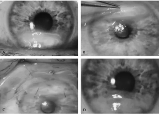
Ulcer From Exposure Keratitis: Case 8
A 69-year-old patient had undergone several lid surgeries for ptosis in the right eye. Over the last 2 years preceding his visit to our department, he had developed a large corneal ulceration resisting medical treatment (Fig. 1). The eye was sore, and the visual acuity was 20/100. An AMT was performed. After excision of necrotic material from the base of the ulcer, a conjunctival peritomy was performed inferiorly. The AM was placed on the ulcer overlapping the limbus and sutured with 10-0 nylon. The following day, the pain had disappeared, and 27 days later, the ulcer had reepithelialized. At the 6-month postoperative visit, a nonvascularized inferior nebula was noted while visual acuity had improved to 20/25. The scar remained stable after a 1-year follow-up.
TABLE 1. (continued) Clinical Data and Surgical Outcome
Case/Age/Sex
Recurrence of
ED (weeks) Pre-op VA Post-op VA Result Evolution
F/up (months) 21/81/F No FC 2 m 20/100 S 6 22/72/M No 20/200 20/40 S 25 23/85/F No FC 30 cm 20/40 S 22 24/82/F No HM FC 1 m S 18 25/48/M No HM FC 30 cm S 13 26/50/F NA HM 20/300 F No epithelialisation Surinfection at 1 month 36 Keratoplasty + limbal allograft + AMT Other etiology 27/59/F 8 HM HM F Recurrence at 2 months Keratoplasty 36 28/34/M No LP- LP- S 36 29/68/F No 6/120 HM S 10 30/44/F No 20/400 20/60 S 10 31/52/M No NR 20/20 S 6
32/52/M No LP LP S Died 1 month after surgery
1
33/78/M No 20/200 20/200 S 20
AMT, amniotic membrane transplantation; HSK, Herpes simplex keratitis; CF, counting fingers; F, failure; S, success; NA, not applicable; NR, not recorded; PED, persistent epithelial defect; ED, epithelial defect; SK, superficial keratectomy; ML, multilayer; LP, light perception; CL, contact lens; PBK, pseudophakic bullous keratitis; RA, rheumatoid arthritis; DCR, dacryocystorhinostomy.
TABLE 2. Results in Group 1,2,3 and the 3 Groups Combined
Group 1 (n = 16) Group 2 (n = 10) Group 3 (n = 7) P Value Combined (n = 33) Complete Epithelialization (No, %) 15 (93.7%) 8 (80%) 7 (100%) 0.4208 (x2exact test) 30 (90.9%)
Epithelialization time (weeks) 2.9 6 1.8 (1–6) 3.9 6 1.8 (1–7) 4.6 6 1.6 (3–6) 0.1181 (ANOVA) 3.6 6 1.8 (1–7) Recurrence of ulcer after first AMT (No, %) 4 (26.7%) 1 (12.5%) 1 (14.3%) 0.7211 (x2exact test) 6 (20%)
Success (No, %) 14 (87.5%) 7 (70%) 6 (85.7%) 0.5171 (x2exact test) 27 (81.8%) Follow-up (months) 12.3 6 8.3 (2–24) 17.3 6 8.7 (6–36) 17 6 14.2 (1–36) 0.3787 (ANOVA) 14.8 6 9.9 (1–36)
Perforated Ulcer in a Patient With Rheumatoid
Arthritis: Case 24
An 82-year-old patient suffering from long-standing rheuma-toid arthritis and poorly compliant to systemic treatment was admitted for a corneal perforation to the right eye, which had undergone corneal transplantation several years before (Fig. 2). The patient presented with a diffuse stromal thinning. The visual acuity was hand motion. The anterior chamber was shallow. The descemetocele had a diameter of 4.5 mm and the perforation was 3 mm. After injecting high viscosity hyaluronic acid in the anterior chamber, the ulcer was cleaned and the stromal bed was dried. A drop of fibrin glue was applied. A 3 3 4-mm AM was then placed epithelium side up and covered by a larger 10 3 14-mm piece was inserted under the lower conjunctiva and attached with an episcleral suture. A bandage contact lens was applied. Complete reepithelialization was observed after 20 days. The scarring remained stable after a follow-up of 18 months.
DISCUSSION
Our study demonstrates the effectiveness of the AMT in the treatment of refractory corneal ulcers with or without perforations up to 3 mm in diameter. Eighty-two percent of patients epithelialized within 3.6 weeks and had a stable corneal surface at least for 2 months after the surgery. These results are comparable to those obtained in previous studies using the AM to treat ulcers and corneal perforation.18,19,22,23
This series, which is one of the largest, is the only one involving perforations 0.5–3.0 mm in diameter.
The transplantation of an AM presents several advan-tages. The tissue, which is readily available and fully meets our needs, can be kept at 280° for many months. The AM does not express the antigens HLA-A, B, or DR and therefore poses no problem of immunological rejection.20
AMT may allow postponement of corneal grafting until the eye is less inflamed, thereby improving graft survival or even avoiding
it altogether, if the result is sufficiently functional. The amniotic membrane presents various advantages over con-junctival flaps. It is absorbed during the weeks and months following the intervention until it dissolves and is then replaced by new fibrous tissue,21
which restores a certain thickness with an imperfect transparency. In vascularized corneas, the resorption of AM is rapid due to the abundance of inflammatory cells.21
The AM causes less visual acuity loss and less corneal vascularization and allows better cosmetic results than conjunctival flaps.22
Successful epithelialization may be explained by different mechanisms. AM provides a basement layer pro-moting the adhesion, migration, and differentiation of ep-ithelial cells while preventing apoptosis.20
Also, the AM contains growth factors that may promote the epithelialization process.24,25
Nerve growth factor (NGF) seems to play an important role. It appears to promote the expansion of the progenitor limbal stem cells and appears to be present in large quantities in AMs.26
Moreover, AMT constitutes a mechanical protection to the fragile corneal epithelium, allowing sufficient oxygenation and hydration of the epithelial cells.27
In our series and according to prior studies,17–19,22
we noted a reduction in the level of conjunctival inflammation and pain after AMT. This is explained by the fact that the stroma of the AM stimulates apoptosis of inflammatory cells,28
suppresses cytokines (1a and interleukin-1b, interleukin-2, interleukin-8, interferon gamma, and tumor necrosis factor a)19 and contains various proteinase
inhibitors29
and antiinflammatory proteins.30
This local antiinflammatory activity of the AM helps in more rapid epithelialization. Resolution of epithelial defect in association with the antiinflammatory properties of AM tends to stop the progressive destruction of the stroma.27
Fourteen patients presented initially with a perforated ulcer. Fibrin glue was applied at the perforation site and AM was grafted subsequently.31
In the Salomon et al19
series in
TABLE 3. Results in Patients With AMT and AMT + Fibrin Glue
AMT (n = 19) AMT + Fibrin Glue (n = 14) P Value Complete epithelialization (No, %) 16 (84.2%) 14 (100%) 0.2443 (x2exact test)
Epithelialization time (weeks) 3.5 6 2 (1–7) 3.6 6 1.7 (2–6) 0.8388 (t-test) Recurrence of ulcer after first AMT (No, %) 6 (35.3%) 1 (7.1%) 0.1755 (x2exact test) Success (No, %) 14 (73.7%) 13 (92.9%) 0.2085 (x2exact test)
Follow-up (months) 14.6 6 12 (1–36) 15 6 6.6 (6–25) 0.9017 (t-test)
AMT, Amniotic membrane transplantation.
TABLE 4. Results in Patients With Limbal Deficiency and With Normal Limbus
Limbal Deficiency (n = 5) Normal Limbus (n = 28) P Value Complete Epithelialization (No, %) 3 (60%) 27 (96.4%) 0.0532 (x2exact test)
Epithelialization time (weeks) 2.50 6 1.5 (1–2.5) 3.69 6 1.9 (1–7) 0.3058 (t-test) Recurrence of ulcer after first AMT (No, %) 2 (66.7%) 4 (14.8%) 0.0936 (x2exact test) Success (No, %) 1 (20%) 26 (92.9%) 0.0017 (x2exact test)
Follow-up (months) 17 6 11 (9–36) 14.4 6 9.8 (1–36) 0.5995 (t-test)
2002,10 patients presented with a perforated corneal ulcer. None of these perforations exceeded 0.5 mm in diameter, and the AMT alone was successful in 8 patients. Prabhasawat et al22
reported 5 cases of perforation of less than 1 mm, with 4 of them being successfully treated with multilayer AMT. Our study looks into perforations measuring 1.9 6 1.7 (range, 0.5– 3.0) mm in diameter. At this stage, the AM by itself cannot fill the gap. The fibrin glue is used to close the perforation. It is
composed of a solution containing fibrinogen and thrombin solution that are combined at the level of the perforation site. The duplojet system, available with the product, consists of a set of 2 syringes with a common piston that allows the 2 solutions to flow at the same rate into a connection piece prior to being mixed together in the application cannula and being ejected. The resulting fibrin plug is then secured with the AM. Interesting properties are attributed to this fibrin glue: it
FIGURE 1. A, Patient with large in-ferior ulcer. B, Cleaning of the ulcer bed (surgical microscopic view). C, The amniotic membrane is anchored with interrupted sutures (surgical mi-croscopic view). D, Complete healing with stromal scar and no vasculariza-tion.
FIGURE 2. A, Perforation of 3 mm on corneal graft. B, Aspect on the first day after the operation. C and D, At 6 months, dissolution of the amniotic membrane and stable epithelium.
stimulates fibroblasts activity, provides a matrix for migration of keratocytes, and is not toxic. This results in activation of the wound healing process.30,31
All patients with AMT and fibrin glue epithelialized successfully and had a stable scar over time, except for patient 26 with severe Stevens-Johnson syndrome and diffuse limbal deficiency. The success rate (reepithelialization and stable corneal surface at 2 months) in perforated ulcers treated with AMT and fibrin glue (92%) was higher than in nonperforated ulcers treated with AMT only (74%), although this difference did not reach statistical significance. In our hands, the AMT– fibrin glue association was shown to be an efficient tool in the management of ulcers perforated up to 3 mm in diameter.
Failures occurred in 2 patients with neurotrophic keratitis, in 3 patients with autoimmune diseases and severe limbal deficiency, and in 1 patient with amoebic keratitis. These failures underline the limitations of the AM: persistent inflammation of the ocular surface; diffuse deficiency of the limbal stem cells, which prevents the regeneration of corneal epithelial cells; and damage to the sensory innervation of the cornea, which prevents epithelial regeneration and retention of epithelial integrity.
Of 5 patients presenting severe limbal deficiency, clinically diagnosed by the loss of the palisades of Vogt and encroachment by conjunctival tissue, the AM only revealed to be efficient in 1 case. The 4 failures (post-HSV neurotrophic [n = 1], Stevens-Johnson syndrome [n = 2], and ocular cicatricial pemphigoid [n = 1]) had a diffuse limbal deficiency, whereas 90 degrees of healthy limbus remained in the last case (Lyell disease). Limbal stem cells are necessary for normal corneal epithelial repopulation, preventing conjunctival epi-thelial invasion, which may result in surface inflammation, neovascularization, and recurrent erosions. Our study sug-gests, on the one hand, that preservation of a portion of healthy limbus is a prerequisite to successful AMT in patients with widespread inflammatory or neurotrophic disorders, and on the other that the AMT on its own might be sufficient to treat corneal ulcers with partial limbal deficiency. In the presence of 360-degrees limbal deficiency, stem cell transplantation be-comes necessary. These data corroborate those of Lagoutte et al32
and Anderson et al,33
who suggest that the AMT allows the restoration of the corneal epithelium surface in patients presenting partial limbal deficiency, by promoting the regen-eration and expansion of the remaining stem cells.
In our study, contrary to other reports,19,34patients with
rheumatoid arthritis were amenable to the AMT treatment. Rheumatoid arthritis ulcers are known to be difficult to treat.35,36
In these patients, rapid restoration of systemic immunosup-pression stopped the inflammation and progressive destruction of the corneal stroma and allowed the AM to fulfill its role.
In conclusion, AMT represents a viable approach for the treatment of chronic corneal ulcers resistant to conventional treatment in patients with healthy residual limbal stem cells. Moreover, this study demonstrates the advantages of the fibrin glue in association with AMT in the management of corneal perforations of up to 3 mm in diameter. This technique pro-motes a stable and rapid reconstruction of the ocular surface. AM transplantation should be considered as an initial procedure before keratoplasty or conjunctival flap. It can also
be used as a temporary measure in cases requiring penetrating keratoplasty at a later time when the eye is less inflamed and the corneal surface has reepithelialized.
REFERENCES
1. Tsai RJF, Tseng SCG. Effect of stromal inflammation on the outcome of limbal transplantation for corneal surface reconstruction. Cornea. 1995; 14:439–449.
2. Brown SI, Hook CW. Isolation of stromal collagenase in corneal inflammation. Am J Ophthalmol. 1971;72:1139–1142.
3. Macaluso DC, Feldman ST. Pathogenesis of sterile corneal erosions and ulcerations. In: Krashmer JH, Mannis MJ, Holland EJ, eds. Cornea, Fundamentals and Cornea and External Disease. St. Louis: Mosby, 1997: 204–205.
4. Donzis PB, Mondino BJ. Management of noninfectious corneal ulcers. Surv Ophthalmol. 1987;32:94–110.
5. Rosenthal P, Cotter JM, Baum J. Treatment of persistent corneal epithelial defect with extended wear of a fluid-ventilated gas-permeable scleral contact lens. Am J Ophthalmol. 2000;130:33–41.
6. Phan TM, Foster CS, Boruchoff SA, et al. Topical fibronectin in the treatment of persistent epithelial defects and trophic ulcers. Am J Oph-thalmol. 1987;104:494–501.
7. Brown SM, Lamberts DW, Reid TW, et al. Neurotrophic and anhidrotic keratopathy treated with substance P and insulinlike growth factor I. Arch Ophthalmol. 1997;115:926–927.
8. Baum J. Topical treatment with nerve growth factor for corneal neuro-trophic ulcers. Surv Ophthalmol. 1999;43:372–373.
9. Weiss JL, William P, Lindstrom RL, et al. The use of tissue adhesives in corneal perforations. Ophthalmology. 1983;90:610–615.
10. Lugo M, Arentsen JJ. Treatment of neurotrophic ulcers with conjunctival flaps. Am J Ophthalmol. 1987;103:711–712.
11. Nobe JR, Moura BT, Robin JB, et al. Results of penetrating keratoplasty for the treatment of corneal perforations. Arch Ophthalmol. 1990;108: 939–941.
12. Lee S, Tseng SCG. Amniotic membrane transplantation for persistent epithelial defects with ulceration. Am J Ophthalmol. 1997;123:303–312. 13. Prabhasawat P, Barton K, Burkett G, et al. Comparison of conjunctival autograft, amniotic membrane grafts and primary closure for pterygium excision. Ophthalmology. 1997;104:974–985.
14. Solomon A, Pires RTF, Tseng SCG. Amniotic membrane transplantation after extensive removal of primary and recurrent pterygia. Ophthalmology. 2001;108:449–460.
15. Tseng SCG, Prabhasawat P, Lee SH. Amniotic membrane transplantation for conjunctival reconstruction. Am J Ophthalmol. 1997;124:765–774. 16. Shimazaki J, Yang HY, Tsubota K. Amniotic membrane transplantation
for ocular surface reconstruction in patients with chemical and thermal burns. Ophthalmology. 1997;104:2068–2076.
17. Kruse F, Rohrschneider K, Volcker H. Multilayer amniotic membrane transplantation for reconstruction of deep corneal ulcers. Ophthalmology. 1999;106:1504–1511.
18. Chen HJ, Pires RTF, Tseng SCG. Amniotic membrane transplantation for severe neurotrophic corneal ulcers. Br J Ophthalmol. 2000;84:826–833. 19. Solomon A, Meller D, Prabhasawat P, et al. Amniotic membrane grafts for non traumatic corneal perforations, descemetoceles and deep ulcers. Ophthalmology. 2002;109:694–703.
20. Dua HS, Azuara-Blanco A. Amniotic membrane transplantation. Br J Ophthalmol. 1999;83:748–752.
21. Gris O, Wolley-Dod C, Gu¨ell JL, et al. Histologic findings after amniotic membrane graft in the human cornea. Ophthalmology. 2002;109:508–512. 22. Prabhasawat P, Tesavibul N, Komolsuradej W. Single and multilayer amniotic membrane transplantation for persistent corneal epithelial defect with and without stromal thinning and perforation. Br J Ophthalmol. 2001;85:1455–1463.
23. Letko E, Stechschulte SU, Kenyon K, et al. Amniotic membrane inlay and overlay grafting for corneal epithelial defects and stromal ulcers. Arch Ophthalmol. 2001;119:659–663.
24. Koizumi N, Inatomi T, Sotozono C, et al. Growth factor mRNA and protein in preserved human amniotic membrane. Curr Eye Res. 2000;20: 173–177.
25. Sato H, Shimazaki J, Shinozaki K, et al. Role of growth factors for ocular surface reconstruction after amniotic membrane transplantation. Invest Ophthalmol Vis Sci. 1998;39:S428.
26. Touhami A, Grueterich M, Tseng SC. The role of NGF signaling in human limbal epithelium expanded by amniotic membrane culture. Invest Ophthalmol Vis Sci. 2002;43:987–994.
27. Baum J. Amniotic membrane transplantation. Cornea. 2002;21: 339–341.
28. Wang MX, Gray T, Parks WC, et al. Reduction of corneal haze and apoptosis by amniotic membrane matrix in excimer laser photoablation in rabbits. J Cataract Refract Surg. 2001;27:310–319.
29. Na BK, Hwang JH, Shin EJ, et al. Analysis of human amniotic membrane components as proteinase inhibitors for development of therapeutic agent of recalcitrant keratitis. Invest Ophthalmol Vis Sci. 1998;39:S90. 30. Hao Y, Ma DH, Hwang DG, et al. Identification of antiangiogenic and
antiinflammatory proteins in human amniotic membrane. Cornea. 2000; 19:348–352.
31. Duchesne B, Hassan T, Galand A. Use of human fibrin glue and amniotic membrane transplant in corneal perforation. Cornea. 2001;20: 230–232.
32. Lagoutte FM, Gauthier L, Comte PR. A fibrin sealant for perforated and preperforated corneal ulcers. Br J Ophthalmol. 1989;73:757–761. 33. Anderson AF, Ellies P, Pires RTF, et al. Amniotic membrane
trans-plantation for partial limbal stem cell deficiency. Br J Ophthalmol. 2001; 85:567–575.
34. Bernauer W, Ficker LA, Watson PG, et al. The management of corneal perforations associated with rheumatoid arthritis. An analysis of 32 eyes. Ophthalmology. 1995;102:1325–1337.
35. Hick S, Duchesne B, Kaye O, et al. Corneal ulcers associated with rheumatoid arthritis. Rev Med Liege. 2002;57:228–232.
36. Kervick GN, Pflugfelder SC, Haimovici R, et al. Paracentral rheumatoid corneal ulceration. Clinical features and cyclosporine therapy. Ophthal-mology. 1992;99:842.
37. Tseng SCG, Prabhasawat P, Barton K, et al. Amniotic membrane transplantation with or without limbal allografts for corneal surface reconstruction in patients with limbal stem cell deficiency. Arch Ophthalmol. 1998;116:431–441.
38. Hanada K, Shimazaki J, Shimmura S, et al. Multilayer amniotic membrane transplantation for severe ulceration of the cornea and sclera. Am J Ophthalmol. 2001;131:324–331.