Université de Montréal
Les effets aigus de l’exercice sur la réponse cérébrovasculaire et la performance cognitive chez des personnes coronariennes stables
par Béatrice Bérubé
Faculté des arts et des sciences, Département de psychologie Université de Montréal
Projet de mémoire
Présenté en vue de l’obtention de la maîtrise (M.Sc.) Sous la direction de Louis Bherer (Ph.D.)
Le 21 août 2019 © Béatrice Bérubé, 201
Université de Montréal
Département de psychologie Faculté des arts et des sciences
Ce mémoire intitulé
Les effets aigus de l’exercice sur la réponse cérébrovasculaire et la performance cognitive chez des personnes coronariennes stables
Présentée par Béatrice Bérubé
A été évalué par un jury composé des personnes suivantes
Sven Joubert Président-rapporteur
Louis Bherer Directeur de recherche
Patrice Brassard
Résumé
Les patients coronariens (PC) sont plus à risque de présenter des déficits cognitifs et certains types démence. Les fonctions cognitives des PC ont été étudiées au repos, mais jamais au cours d’une séance d’exercice aigu. L’exercice aigu à haute intensité peut affecter négativement la performance cognitive chez des personnes saines. Bien que les PC soient plus à risque de dysfonctions cérébrales et cardiovasculaires, cette relation n’a jamais été étudiée au sein de cette population clinique et peut permettre de mieux comprendre l’axe cœur-cerveau. Il était postulé que la performance cognitive sera affectée par l’exercice à haute intensité due à une diminution de l’apport en oxygène seulement chez les patients coronariens. Trente-huit PC et 16 participants sains ont été recrutés. Les participants ont complété les mesures suivantes : (1) des tests neuropsychologiques et une familiarisation à la tâche de Stroop modifiée informatisée (2) un test mesurant la consommation d’oxygène jusqu’au pic de l’effort (VO2pic) et (3) la tâche de Stroop à 30% et 70% de leur puissance maximale atteinte au VO2pic tout en pédalant sur un ergomètre. L’oxygénation cérébrale a été mesurée grâce à la spectroscopie proche infrarouge. Les résultats ont démontré que la performance cognitive est restée stable entre les deux intensités pour les deux groupes. Chez les PC, le volume sanguin cérébral était affecté négativement par l’effort physique à haute intensité comparativement à l’intensité modérée et aux participants sains. La maladie coronarienne affecte négativement l’oxygénation cérébrale pendant un effort à haute intensité. D’autres études sont nécessaires afin de déterminer si un test cognitif administré pendant un effort physique pourrait permettre d’identifier les patients coronariens à risque de déclin cognitif.
Abstract
Coronary heart disease (CHD) patients are at higher risk for developing cognitive deficits and certain types of dementia. The cognitive functions of coronary patients have been studied at rest, but never during an acute exercise session. Acute high intensity exercise negatively affects cognitive performance in healthy people. Although coronary patients are at higher risk of cerebral and cardiovascular dysfunction, this relationship has never been studied in this clinical population and could help better understanding of the heart-brain axis. The aim of this study was to measure the effects of an acute exercise at two different intensities on cognitive performance and cerebrovascular response in CHD patients. It was hypothesized that higher exercise intensity will impair executive performance and cerebrovascular response only in CHD patients. Thirty-eight CHD patients and 16 healthy controls completed neuropsychological assessments, maximal cardiopulmonary exercise testing and two exercise bouts at 30% and 70% of their individualized maximum capacity on an ergocycle while performing a cognitive test including non-executive and executive conditions. Cerebral oxygenation and perfusion were measured during both intensities in all participants with near-infrared spectroscopy. The results demonstrated that the cognitive performance remained stable between the two intensities for both groups. In CHD patients only, cerebral blood volume was negatively affected by high intensity exercise compared with moderate intensity. Coronary heart disease negatively affects cerebral oxygenation during high intensity exercise. Further studies are needed to determine whether a cognitive test administered during physical exertion could identify coronary patients at risk of cognitive decline.
Contribution des auteurs
L’ensemble de la méthodologie concernant le protocole d’évaluation de la performance cognitive ainsi que la mesure des variations de l’oxygénation cérébrale à l’aide de la spectroscopie proche infrarouge à l’effort provient du laboratoire du Dr Louis Bherer. De plus, Dr Bherer m’a supervisé tout au long de ma maitrise en fournissant un encadrement et des révisions cruciales lors de la rédaction du présent mémoire et de l’article. Mon rôle comme première auteure concernait la rédaction de l’article, les analyses statistiques et l’entrée de données. Le co-auteur Maxime Boidin a monté le protocole du projet dans lequel s’insère le présent article et a mené du début à la fin cette étude (recrutement, passation des tests, entrée de données, etc.) en plus de participer à la correction de l’article. Dr Thomas Vincent a fourni son expertise concernant le traitement et l’analyse des données d’imagerie. Les coauteurs Dr. Mathieu Gayda et Dr. Jonathan Tremblay ont fourni leurs rétroactions sur l’article. Dr. Martin Juneau et Dr. Anil Nigam étaient en charge des évaluations médicales centrales au projet, du référencement des patients pour favoriser le recrutement et de la validation des critères d’inclusion et d’exclusion. Le projet a été subventionné par Les fonds de recherche du Québec en santé (FRQ-S) et par le Centre de médecine préventive et d’activité physique (EPIC) de l’Institut de Cardiologie de Montréal.
Table des matières
Résumé ... i
Abstract ... iii
Contribution des auteurs ... v
Table des matières ... vii
Liste des tableaux ... ix
Liste des figures ... xi
Liste des abréviations ... xiii
Remerciements ... xv
Introduction ... 1
Contexte théorique ... 3
L’exercice aigu et la cognition chez les personnes saines ... 3
Mesure des fonctions cognitives ... 3
Le niveau de la condition physique ... 4
Le délai de la passation des tâches cognitives. ... 4
L’exercice aigu et la cognition chez les patients coronariens ... 5
La spectroscopie proche infrarouge ... 6
Article ... 11 Discussion générale ... 41 Conclusion générale ... 47 Références ... 57 Annexe A ... 64 Annexe B ... 61
Liste des tableaux
Table 1. Means and standard deviations for demographics, neuropsychological, psychological and physiological data for individuals with CHD and healthy control groups ... 31 Table 2. Means and standard deviations for Stroop task performance for individuals with CHD
and healthy control group ... 32
Liste des figures
Figure 1. Means and Standard Errors of [HbO] Variations as a Function of Group, Intensity and Stroop Task Condition ... 33 Figure 2. Means and Standard Errors of [HbR] Variations as a Function of Group, Intensity and
Stroop Task Condition ... 34 Figure 3. Means and Standard Errors of [HbT] Variations as a Function of Group, Intensity and
Stroop Task Condition ... 35
Liste des abréviations
HI : Haute intensité PC : Patients coronariens
VO2pic : Consommation d’oxygène au pic de l’effort SPIR : Spectroscopie proche infrarouge
HbO : Oxyhémoglobine HbR : Déoxyhémoglobine HbT : Hémoglobine totale CO2 : Dioxyde de carbone
PaCO2 Pression artérielle partielle en CO2 HI: High intensity
CVD: Cardiovascular disease CHD: Coronary heart disease VO2peak: Peak oxygen consumption NIRS: Near infrared spectroscopy HbO: Oxyhemoglobin
HbR : Deoxyhemoglobin HbT: Total hemoglobin
MMSE: Mini-mental State Evaluation GDS: Geriatric Depression Scale CO2: Carbon dioxide
Remerciements
Tout d’abord, j’aimerais témoigner ma reconnaissance à Dr Louis Bherer pour son encadrement ainsi que pour ses judicieux conseils qui ont permis la réalisation ce projet. Dr Bherer a su me guider tout au long de ma maitrise et a fait en sorte que mon passage aux études supérieures fut une expérience enrichissante. Ainsi, Dr Bherer m’a donné la piqure de la recherche et c’est grâce à lui que je poursuis au troisième cycle. Je tiens également à adresser mes remerciements à Maxime Boidin sans qui rien de tout cela n’aurait été possible. Merci de m’avoir outillé et pour avoir pris le temps nécessaire afin de m’épauler tout au long de ce projet.
Je voudrais témoigner ma gratitude à toute l’équipe du Laboratoire d’étude de la santé cognitive des ainés (LESCA) et du Centre EPIC de l’Institut de cardiologie de Montréal pour m’avoir accueilli et pour m’avoir fait connaitre un milieu aussi stimulant et chaleureux. J’aimerais souligner tout le support que ma famille, Samuel Bérubé, Marie-Line Deslauriers et Serge Bérubé, m’a apporté. Finalement, merci, Mathieu Landry pour tout le soutien émotionnel de la dernière année. Merci à tous !
Introduction
Les personnes âgées de 65 ans et plus représenteront le quart de la population au Québec d’ici 2031 si la tendance se maintient (Institut de la statistique du Québec, 2017). Une augmentation des troubles de santé physique et cognitive liés à l’âge est donc appréhendée et pourrait devenir une problématique sociale non négligeable. La démence vasculaire et la maladie d’Alzheimer représentent deux types de maladies neurodégénératives les plus répandus lors du vieillissement (Davenport, Hogan, Eskes, Longman et Poulin, 2012). En plus de l’âge, les maladies vasculaires, comme les maladies coronariennes, constituent un facteur de risque important pour ces deux démences (Claassen, 2015). Les personnes atteintes de maladies coronariennes peuvent présenter certains déficits cognitifs au repos. Plus précisément, ces déficits sont répertoriés dans les domaines exécutifs sous-tendus par les lobes frontaux (Burkauskas et al., 2018).
La pratique régulière d’exercice physique joue un rôle de premier plan dans la prévention de nombreuses maladies chroniques (Ramos, Dalleck, Tjonna, Beetham et Coombes, 2015) et des démences (Bherer, Erickson et Liu-Ambrose, 2013). Les ainés avec une meilleure capacité cardiorespiratoire présentent généralement de meilleures fonctions exécutives, mais les explications biologiques restent encore à être étudiées (Colcombe et al., 2003). Pourtant, certaines études n’ont pas observé d’association entre un niveau élevé de la capacité cardiovasculaire et de meilleures fonctions exécutives auprès des ainés (Angevaren, Aufdemkampe, Verhaar, Aleman et Vanhees, 2008; Etnier, Nowell, Landers et Sibley, 2006). Une meilleure compréhension des mécanismes biologiques sous-jacents de la relation entre l’exercice physique et la cognition permettrait d’expliquer les différences individuelles en cognition (Bherer et al., 2013). L’effet de l’exercice sur la cognition et les fonctions cérébrales
peut être étudié lors d’une seule séance d’exercice ponctuelle. Cette approche peut aider à identifier les mécanismes physiologiques impliqués dans cette relation. Dans ce type d’étude, on compare les performances cognitives et l’activité cérébrovasculaire lors d’un exercice aigu à différentes intensités. Les études adoptant cette approche rapportent des résultats contradictoires: certaines études rapportent un effet positif alors que d’autres rapportent un impact négatif ou pas d’effet de l’exercice sur la cognition. Ces résultats divergents pourraient être associés à plusieurs différences méthodologiques, comme l’intensité de l’exercice, la condition de santé des participants, etc. (Chang, Labban, Gapin et Etnier, 2012; Lambourne et Tomporowski, 2010). Ces questions sont importantes étant donné les nouvelles tendances dans le domaine de l’exercice qui consistent à inclure des séances à haute intensité (HI) dans la programmation d’exercice, et ce, même chez les patients souffrant de maladies coronariennes. L’objectif principal de ce projet est de mieux documenter l’impact d’un exercice aigu sur la cognition en étudiant les effets aigus d’une séance d’exercice à différentes intensités sur la performance à une tâche mesurant différentes fonctions cognitives chez des participants sains et des patients coronariens (PC). Les résultats nous permettront d’améliorer la compréhension du fonctionnement de l’axe cœur-cerveau.
Contexte théorique
L’exercice aigu et la cognition chez les personnes saines
L’étude des effets de l’exercice aigu sur la performance cognitive a débuté au 20e siècle. Cette relation a d’abord été examinée d’un point de vue comportemental seulement et plusieurs chercheurs ont tenté ensuite d’expliquer l’association entre l’exercice aigu et les changements comportementaux par certains mécanismes physiologiques. Des études ont observé des bénéfices, d’autres ont observé des effets délétères alors que certaines études n’ont rapporté aucun effet de l’exercice aigu sur la cognition. La méthodologie des études diffère grandement, ce qui pourrait expliquer l’absence de consensus scientifique. Notamment, le type de tâche cognitive utilisée (exécutif et non exécutif), le niveau de condition physique du participant (faible ou élevé) et le délai temporel de la passation de la tâche cognitive (avant, après l’exercice ou de façon différée) peuvent influencer de manière importante les résultats obtenus.
Mesure des fonctions cognitives
Le type de tâche semble avoir un impact considérable sur la performance en contexte d’exercice aigu. Les tâches cognitives qui sollicitent les fonctions exécutives comme l’inhibition et la flexibilité mentale, sont davantage affectées par l’exercice aigu à HI et montrent généralement une augmentation de la performance à intensité faible et modérée comparativement aux fonctions qui ne dépendent pas directement des fonctions exécutives de ces régions, comme les tâches impliquant un temps de réaction simple (réaction à seulement un stimulus) (Dietrich et Sparling, 2004). Draper, McMorris et Parker (2010) ont observé que le temps de réaction simple n’est pas affecté par l’intensité de l’exercice aigu alors que le temps de réaction avec choix
(réaction à plusieurs stimuli) augmente de façon linéaire avec l’augmentation de l’intensité de l’effort physique.
Le niveau de la condition physique
Au niveau comportemental, Labelle et al. (2013) ont observé que les personnes avec un niveau de condition physique plus faible présentent des performances cognitives inférieures en situation d’exercice aigu et ce, peu importe l’âge. Également, l’étude de Rooks, Thom, McCully, et Dishman (2010) suggère que la réduction de la réactivité cérébrovasculaire dans le cortex préfrontal à HI est davantage marquée chez les populations inactives, et ce, indépendamment de l’âge. L’oxygénation cérébrale et les performances exécutives des individus avec un niveau élevé de condition physique seraient moins affectées par la HI que ceux ayant un niveau de condition physique plus faible (Brisswalter, Collardeau et Rene, 2002).
Le délai de la passation des tâches cognitives.
Le moment où les tests cognitifs sont effectués peut avoir un effet considérable sur la performance cognitive. Une méta-analyse (Chang et al., 2012) suggère qu’une baisse de la performance exécutive est observée lorsque les tests sont effectués pendant l’effort physique indépendamment du niveau de la condition physique. Après un délai approximatif de 0 à 11 minutes, des effets négatifs sont répertoriés. Entre 11 et 20 minutes, aucune différence n’est observée. Après 20 minutes, des effets positifs sont observés par rapport au repos.
Un phénomène similaire est rapporté lorsque les tâches cognitives sont réalisées après l’effort physique. Une diminution de la performance exécutive est observée de 0 à 11 minutes et après ce délai, les effets sont positifs. Ces résultats suggèrent qu’il pourrait y avoir un délai avant d’observer une amélioration des fonctions exécutives due à un ajustement ou à une autorégulation
physiologique (Brys, Marthol, Hilz, Brown et Franta, 2003; Fantini, Sassaroli, Tgavalekos et Kornbluth, 2016).
L’exercice aigu et la cognition chez les patients coronariens
Plusieurs personnes atteintes de maladies coronariennes présentent certains déficits cognitifs tels qu’une diminution de la performance aux tâches exécutives (Moser et al., 1999; Selnes et al., 2009). Ce déficit serait également associé à une perte du volume de matière grise dans plusieurs régions cérébrales (Longstreth et al., 2005). Les patients souffrant de ce type de maladie démontrent également une diminution de l’oxygénation cérébrale pendant un test d’effort physique sous-maximal comparativement aux personnes saines (Fu et al., 2013; Koike et al., 2008). En comparaison avec des personnes saines, les patients coronariens présentent des fonctions cognitives diminuées et une oxygénation cérébrale réduite pendant la récupération, soit après l’effort physique (Gayda et al. 2017). Par contre, l’oxygénation cérébrale des patients coronariens pendant l’exercice n’était pas différente de celle des participants sains. D’autres études ont observé un phénomène similaire (Koike et al., 2004; Koike et al., 2008) et ceci pourrait être causé par le fait que l’augmentation soudaine (overshoot) du débit cardiaque est moindre dans la phase de récupération chez des participants âgés cardiaques que chez les participants sains (Tanabe et al., 2000). Par conséquent, une quantité moins importante de sang oxygéné est éjectée du cœur vers le reste du corps diminuant la perfusion cérébrale chez les populations cardiaques. Les études précédentes ont employé la spectroscopie proche infrarouge (SPIR) comme technique d’imagerie ; elle s’est avérée efficace pour mesurer les variations de l’oxygénation cérébrale en contexte d’exercice aigu.
La spectroscopie proche infrarouge
La SPIR est une technique d’imagerie optique pour mesurer les variations relatives de concentration d’oxygène dans le sang. Cette technique permet également de localiser les régions corticales impliquées dans divers processus cognitifs. La SPIR émet des faisceaux de lumière infrarouge qui traversent les tissus organiques et le crâne. Les rayons provenant de la source lumineuse sont réfléchis et ressortent ensuite du crâne avec une trajectoire curvilinéaire. Les détecteurs disposés à 4,5 centimètres des sources lumineuses sur le crâne peuvent ainsi capter les faisceaux sortants. Une partie de ces rayons est absorbée par le système vasculaire, soit par l’oxyhémoglobine (HbO) et par la déoxyhémoglobine (HbR), puis le reste est réfléchi (Scholkmann et al., 2014). Ainsi, les fluctuations de concentration HbO et HbR peuvent être estimées avec la différence entre la lumière pénétrante et sortante. L’hémoglobine totale (HbT) est obtenue avec la somme de HbO et de HbR et représente le volume sanguin total. Une activation cérébrale est généralement représentée et estimée par une augmentation de HbO accompagnée d’une réduction de HbR (Ekkekakis, 2009; Mehagnoul-Schipper et al., 2002). En situation d’exercice aigu, une augmentation de HbO est associée à une augmentation de la performance exécutive à intensité faible et modérée (Ando, 2016). Inversement, lorsque l’exercice est pratiqué à HI, une diminution de HbO est accompagnée d’une baisse de la performance exécutive (Ando, Yamada, Tanaka, Oda et Kokubu, 2009; Mekari et al., 2015). Aucune étude à ce jour n’a permis de mettre en relation ces variations avec les performances cognitives chez les PC bien que ceux-ci soient plus à risque de dysfonctions cérébrales.
En vieillissant, les adultes présentent une diminution de l’activité cérébrale latéralisée au niveau du cortex préfrontal comparativement aux jeunes adultes. Des études utilisant la SPIR ont
(Basso et Suzuki, 2017). Une activation bilatérale au niveau du cortex préfrontal était cependant observée chez plusieurs personnes âgées. Ce phénomène, nommé la réduction de l’asymétrie hémisphérique chez les personnes âgées (Harold), permettrait de compenser pour la diminution de l’activation latérale du cortex préfrontal qui peut accompagner le vieillissement (Cabeza, 2002). La performance des personnes âgées à la tâche de Stroop était augmentée pendant une séance d’exercice aigu accompagnée d’une augmentation de l’activation au niveau du cortex préfrontal droit. Ceci suggère une augmentation du phénomène HAROLD ou une augmentation de la capacité de compensation du cerveau vieillissant comparativement aux jeunes adultes (Hyodo et al., 2012 ; Yanagisawa et al., 2010).
La relation entre les effets aigus de l’exercice et les performances cognitives peut être expliquée par la théorie de l’hypofrontalité réticulo-activatrice de l’exercice de Dietrich (Dietrich, 2006). La SPIR s’est révélée être une méthode adéquate pour évaluer cette hypothèse (Basso et Suzuki, 2017). Cette théorie suggère que pendant un exercice physique, les ressources cérébrales limitées sont accaparées par les zones cérébrales liées au mouvement moteur et à la coordination, passant ainsi des zones préfrontales responsables du traitement cognitif de haut niveau vers les aires liées au traitement moteur (ex. cortex moteur primaire, cervelet, etc.). Par conséquent, une hypoperfusion et une désactivation du cortex préfrontale seraient engendrées. En réponse à cette hypoperfusion, le sang oxygéné et l’activation cérébrale sont à nouveau augmentés dans le cortex préfrontal. Cette théorie pourrait donc expliquer pourquoi la performance aux tests mesurant les fonctions exécutives, qui sont sous-tendues par les lobes frontaux et préfrontaux, a tendance à augmenter à intensité faible et modérée conjointement avec les variations en oxygénation cérébrale. À HI, la demande des ressources (apport sanguin, oxygénation, etc.) serait possiblement trop importante pour maintenir l’élévation de la perfusion et de l’activation en
région préfrontale, expliquant ainsi la diminution des variations de l’oxygénation cérébrale accompagnée de la baisse des performances exécutives à 80 % de la capacité maximale répertoriée dans la littérature (Ando, 2016).
D’autres variables jouent un rôle dans la diminution de l’oxygénation cérébrale à HI pendant un effort physique cardiovasculaire. Notamment, la pression artérielle partielle en dioxyde de carbone (PaCO2) est un régulateur important du flux sanguin et du volume sanguin cérébral (Scholkmann et al., 2014). La PaCO2 est stable ou augmente généralement lors d’effort physique d’intensité faible à modérée et diminue lors d’effort physique intense. À intensité élevée, une augmentation soudaine de la ventilation pulmonaire est associée à une production plus grande de CO2, ce qui réduit la PaCO2, entraine une vasoconstriction des vaisseaux sanguins cérébraux et par conséquent, une diminution du flux sanguin, du volume sanguin et de l’oxygénation cérébrale (Braz et Fisher, 2016). Bhambhani et al. (2007) ont démontré que la réduction de l’oxygénation et du flux sanguin cérébral dans le cortex frontal à HI survient conjointement avec une diminution de la PaCO2. Pour résumer, la vasoconstriction résultant de la diminution de la PaCO2 pendant un effort à HI réduit le flux et le volume sanguin cérébral. Les plus grosses artères ainsi que les artérioles sont notamment plus sensibles à cette variation du PaCO2 (Poeppel et al. 2007; Szabo et al. 2011). Certaines études ont également démontré que les personnes âgées présentaient une réduction du flux sanguin cérébrale et de la PaCO2 par rapport aux jeunes adultes (Fisheret al. 2013; Fluck et al. 2014). Bien que les mécanismes responsables de la réduction du flux sanguin et de la perfusion cérébrale ne sont pas encore bien compris, certaines composantes ont été identifiées comme jouant un rôle dans la relation entre l’âge et la diminution de la perfusion cérébrale : une diminution de l’activité neuronale, une augmentation de la rigidité artérielle et une diminution de la réactivité cérébrovasculaire (Fluck et al. 2014). La
vélocité de l’artère moyenne cérébrale, une mesure estimée du flux sanguin cérébral, diminue avec l’âge autant au repos qu’à l’exercice. Jusqu’à 60 % de la capacité cardiovasculaire maximale, la vélocité de cette artère augmente, puis diminue près des valeurs au repos à plus HI ce qui diminue la perfusion cérébrale. Fluck et al. (2014a) ont ajouté du CO2 à l’air respiré par des jeunes adultes et des personnes âgées pendant un effort physique maximal sur ergocycle pour prévenir la réduction de la PaCO2 suivant l’hyperventilation qui survient lors d’un exercice physique à HI. Le supplément en CO2 a réduit de 50 % le déclin de la vélocité de l’artère moyenne cérébrale associé à l’âge suggérant ainsi que la PaCO2 joue un rôle important dans cette diminution reliée à l’âge. Il a également été démontré que chez les personnes ayant une meilleure condition physique, la perfusion cérébrale et la réactivité au CO2 pendant un effort étaient supérieures comparativement à un niveau faible de la condition physique (Ainslie et al., 2008). Ce phénomène serait aussi davantage marqué au sein des populations atteintes de maladies cardiovasculaires, comme chez les personnes souffrant insuffisance cardiaque (Brassard et Gustafsson, 2016). En ce qui a trait aux patients cardiaques ayant des dysfonctions au ventricule gauche, ils présentent un flux sanguin cérébral réduit comparativement à leurs homologues sains et une transplantation cardiaque permet d’améliorer le flux sanguin cérébral (Choi et al., 2006). Il a donc été proposé que le flux sanguin cérébral pendant un effort physique intense chez les populations cardiaques, notamment chez les personnes coronariennes, serait inférieur à une population saine considérant leur débit cardiaque inférieur pendant l’exercice et la réduction de l’activité des facteurs associés à la vasoconstriction et à la vasodilatation des vaisseaux au niveau cérébral, comme la PaCO2 (González-Alonso et al., 2004 ; Koike et al., 2008). Puisque les facteurs régulant l’oxygénation cérébrale sont mal compris, l’étude avec des PC peut aider à comprendre les mécanismes cérébrovasculaires impliqués dans la relation entre l’exercice et la
cognition et ainsi mettre en évidence le fonctionnement de l’axe cœur-cerveau. Ces résultats ont également un impact majeur pour la prescription de l’exercice chez les patients, puisque si un délai est nécessaire afin que l’oxygénation cérébrale et la cognition se stabilisent après un exercice intense, il faut en tenir compte dans la prescription d’exercice physique en ajoutant un moment de récupération plus long après chaque séance. Finalement, considérant les différences interindividuelles des profils cognitifs au sein de la population atteinte de maladies coronariennes, le stress induit par une séance d’exercice aigu pourrait permettre d’identifier précocement les PC à risque de développer un déclin cognitif.
Acute Effects of Exercise on Cerebrovascular Response and Cognitive
Performance in Individuals with Stable Coronary Heart Disease
Bérubé, B.1,2,3, Boidin, M.1,4, Gayda, M.1,2,5,Vincent, T.1,2, Tremblay, J.4, Juneau, M.1,2,5, Nigam, A.1,2,5 & Bherer, L.1,2,5
1Research Center and Preventive Medicine and Physical Activity Center (EPIC), Montreal Heart Institute, Montreal, Quebec, Canada
2Research Center, Institut Universitaire de Gériatrie de Montreal, Montreal, Quebec, Canada 3Department of Psychology, Université de Montréal, Montreal, Quebec, Canada
4School of Kinesiology and Exercise Science, Faculty of Medicine, Université of Montréal, Montreal, Quebec, Canada
5Department of Medicine, Faculty of Medicine, Université of Montréal, Montreal, Quebec, Canada Article type: Original article
Short title: Acute Effects of Exercise in Individuals with Coronary Heart Disease Funding: The Montreal Heart Institute and the EPIC Center Foundations
Corresponding author: Béatrice Bérubé
Research Center and Preventive Medicine and Physicial Activity Center (EPIC) 5055 Saint-Zotique Street
Montreal, QC, Canada, H1T 1N6
Tel (514)-374-1480 ex.4340
Abstract
Background. Individuals with coronary heart disease (CHD) exhibit cognitive deficits and cerebrovascular dysfunctions and are at higher risk of developing dementia. Cognitive function in individuals with CHD has never been studied during acute aerobic exercise. Given the increasing popularity of high-intensity training, its impact on cerebrovascular and cognitive functions in individuals with CHD should be further studied.
Objective. The aim of this study was to assess the acute effects of exercise at high intensity on cognitive performance and cerebrovascular response in individuals with CHD compared to age-matched healthy controls.
Method. Thirty-eight individuals with CHD and 16 healthy controls completed two exercise bouts at 30% and 70% of their individualized peak power output on an ergocycle while performing a cognitive test including non-executive and executive conditions. Variations of oxy- deoxy-hemoglobin, and total hemoglobin (blood volume) concentrations were measured at both intensities using near-infrared spectroscopy.
Results. Both groups did not show significant differences in cognitive performances in both intensities and higher intensity did not alter performances for individuals with CHD. Individuals with CHD exhibited larger negative variations of cerebral blood volume at 70% of peak power output compared to healthy controls.
Conclusion. CHD has negative impacts on cerebral blood volume variations during a cognitive task and while performing exercise. Other studies are needed to determine if a cognitive test administered during an exercise test could help identify individuals with CH at higher risk of developping cognitive decline. Key words: Aging, Acute Exercise, Cerebral Oxygenation, Cognitive Functions, Coronary Heart Disease, Near-infrared Spectroscopy
Introduction
Cardiovascular disease (CVD) is the leading cause of death worldwide and is expected to be for the next 20 years1. Coronary heart disease (CHD) is the most common type of CVD2. Individuals with CHD frequently show deficits in cognition at rest (executive and non-executive performances)3 and are at higher risk of developing Alzheimer’s type and vascular dementia4. The impact of CHD on resting cognitive functions supports the direct link between cardiovascular and cerebral health.
Daily life activities not only require cognitive functions at rest but also during a physical effort such as home repairs, housekeeping, or simply walking on the street. Direct effects of acute aerobic exercise on cognition could be used as a stress test and could reflect critical situations of daily life (e.g. running after the bus when accidents or falls can occur if executive functions are impaired). Moreover, studying the acute effects of exercise could provide a better understanding of physiological mechanisms involved in the relationship between exercise and cognition. Several meta-analyses concluded that acute aerobic exercise has a positive effect on cognition, especially in prefrontal cortex-dependent functions5, 6. Intensity of exercise is considered as an important factor influencing the cognitive effects of acute aerobic exercise. In healthy individuals, moderate intensity exercise (40 – 65 % of peak oxygen uptake (𝑉O2peak)) seems to have positive effects on cognition, whereas high intensity exercise (80 % 𝑉O2peak) seems not to. In previous studies, it was shown that acute high intensity exercise negatively impacts executive performances in both higher fit and lower fit healthy subjects independently of age5. However, cognitive performances in the lower fit group was more affected by high intensity compared to higher fit counterparts, therefore supporting the notion that the acute effects of exercise on cognition observed even in
at-effects of exercise on cognition have never been studied in individuals with CHD despite their higher risk of cerebral/cardiovascular dysfunctions.
One of the possible physiological mechanisms explaining the association between cognition and acute exercise is cerebral oxygenation and perfusion7, 8. Non-invasive optical methods such as near-infrared spectroscopy (NIRS) are often used to measure cerebral oxygenation, O2 extraction and perfusion, during an acute bout of exercise and a cognitive task. Variations in oxyhemoglobin [HbO], deoxyhemoglobin [HbR] and total hemoglobin concentrations [HbT] in prefrontal regions can be obtained using NIRS and used to estimate variations in cerebral oxygenation9. In both younger and older healthy adults, [HbO] increases during an incremental acute aerobic exercise at light to moderate-intensity (30 % to 60 % of peak oxygen uptake, VO2peak), but decreases near the resting state when exercise is performed at higher intensity (80 % of VO2peak)10. An increase of [HbO] seems to be associated with better executive performance at light- and moderate-intensity of an acute aerobic exercise (Ando, 2016), whereas a decrease in [HbO] appears to be accompanied by a decrease in executive performance at high-intensity (80 % of VO2peak)8, 11. This reduction in cerebral oxygenation in the prefrontal cortex at high intensity is more important in inactive populations regardless of age12. In this way, Koike and colleagues13 showed that half of their cardiac patients had reduced [HbO] during maximal incremental exercise. Koike and colleagues showed that the reduction in [HbO] and [HbT] observed in individuals with CHD was more pronounced at higher exercise intensities14. However, Gayda and colleagues15 showed that cerebral oxygenation during a maximal incremental exercise test was similar in fit individuals with CHD and fit age-matched healthy individuals. Higher fitness level could help compensate the negative impact of a high intensity bout of exercise on cognition, especially executive functions, thus potentially reducing the negative impact of CHD on cognition 5.
The present study assessed the effects of acute exercise at light and vigourous intensities on executive performance and on the cerebrovascular response in individuals with CHD. It was hypothesized that (1) higher exercise intensity will have a larger negative impact on executive performance for individuals with CHD compared to healthy controls and (2) that higher intensity will affect more importantly and negatively variations of cerebral oxygenation in the prefrontal cortex of individuals with CHD than of healthy controls.
Materials and method
Study design and participant’s recruitment
Thirty-eight individuals with stable CHD and 16 age-matched healthy controls completed this study. The study protocol was approved by the Research Ethics and New Technology Development Committee of the Montreal Heart Institute, and registered on ClinicalTrials.gov (identifier number: NCT03443193). All participants gave their written informed consent prior to be involved in this study. Exclusion criteria for individuals with CHD were the following: a recent acute coronary syndrome (<3 months); heart failure; left ejection fraction <40 %; severe coronary artery disease non-suitable for revascularization; scheduled coronary artery bypass surgery for severe coronary heart disease; chronic atrial fibrillation; malignant arrhythmias during exercising; restriction to cardiopulmonary exercise testing or severe intolerance to exercise. Exclusion criteria for healthy subjects were: cardiac disease and aortic stenosis symptoms.
Experimental design
All participants underwent a medical evaluation including medical history and physical examination. The experiment consisted of three visits within a 14-day period separated by a
and the Geriatric depression scale (GDS) to measure global cognition and depression symptoms respectively. Upon their second visit, anthropometric measurements and body composition were collected, and all participants performed a maximal cardiopulmonary exercise testing on a cycle ergometer with gas exchange measurements to estimate VO2 peak). During the third visit, participants completed the cognitive task session during a submaximal aerobic exercise (30% and 70% of the peak power output, PPO) and with cerebral hemodynamic response measurement using NIRS system. All participants were asked to avoid eating 3 hours prior to visits 2 and 3, as well as to avoid alcohol and caffeine consumption 12 hours prior to these visits.
Measurements
Maximal cardiopulmonary exercise testing
Maximal cardiopulmonary exercise testing was performed on a cycle ergometer (Erogline 800S, Bitz, Germany) as recently published15. An individualized protocol began with a 3-min rest in order to familiarize with the equipment followed by a 3-min warm-up at a workload of 20 (Gayda et al., 2017). The power increases from 10 to 20 Watts/min at a cycling cadence of 60 to 80 rpm. Gas exchange was measured continuously at rest, during exercise, and after exercise cessation using a metabolic system (Oxycon Pro, Jaegger, Germany) as recently published and data were measured every four respiratory cycles during testing and then were averaged every 15 sec 15. Continuous ECG monitoring (Marquette, case 12, St. Louis, Missouri) was performed rating of perceived exertion (RPE: Borg Scale, 6-20) and manual blood pressure (sphygmomanometer: Welch Allyn Inc., Skaneateles Falls, USA) was measured every 2 minutes throughout the test. Strong verbal encouragements were given throughout the test. The highest peak oxygen uptake value (peak VO2) reached during the exercise phase was considered has the VO2 peak. PPO was defined as the power output attained at the last completed stage 15.
Computerized modified Stroop task
The computerized modified-Stroop task was programmed on E-Prime 2.0 (Pittsburgh, USA) and was based on previous task used in acute exercise and cognition context8. The modified Stoop task included non-executive (naming) and executive (inhibition and switching) experimental conditions. Both conditions included 2 non-executive and 2 executive block repetitions. Participants were instructed to answer as fast as they can and to make as few errors as possible. All subjects performed a practice session prior to starting the test session where they received feedback when incorrect answers occurred in contrast to the test session where they did not receive feedback. Then, each sequence of the test session lasted 8 min and was composed of 1-min blocks for each cognitive condition separated by a 1-min rest block in this following order: Naming-Rest-Executive-Rest-Naming Rest-Executive-Rest. Each block began with a fixation cross for 500 ms followed by the 15 visual stimulus trials which appeared on the screen for 2500 ms. Naming blocks consisted of 15 trials showing neutral-coloured (blue or green) visual stimuli (XXXX). Executive blocks also included 15 trials, which were divided in two types: inhibition and switch trials accounting for 75 % and 25 % of the total number of trials, respectively. Switch trials appeared randomly throughout the executive block. For the inhibition trials, the word “BLUE” in green ink or “GREEN” in blue ink was presented and participants had to name the ink color and refrain themselves from reading the word. For switch trials, the word appeared in a white square, which indicated to participants that they had to read the word instead of naming its color. For instance, if the word “BLUE” presented in green ink was surrounded by a white box, the appropriate answer was “BLUE”. Participants had to press a button in their left hand for green responses and in their right hand for blue responses. Participants kept pedalling at a constant speed of 60-80 rpm in the rest phase of the cognitive task. Error rate and reaction time were collected for
Submaximal bouts of constant intensity exercise
Participants completed a submaximal test on an ergocycle at constant intensity of 30 % and 70 % of their respective PPO. Exercise intensity levels were counterbalanced across participants among both groups using an online randomizer: half of sample completed main exercise session with the first sequence (30% - 70% of PPO) while others completed the second sequence (70% - 30% of PPO) to avoid a systematic association of exercise intensity effects with order effect16. The Stroop task was administered on a 40 centimeters laptop placed at 60 cm in front of participants. Answer buttons were located on two handlebars located on each side of the cycle saddle. Participants started with the Stroop task practice at rest followed by a 2-min rest state, then a 3-min warm-up at 20 watts followed by a 2-3-min phase to reach the first power steady-state (30 % or 70 % of PPO). Once the steady-state was attained, participants completed 8-min constant intensity bout of exercise while Stroop task was performed until the end this sequence. Participants rested for 4 min before starting the second block. A 2-min active recovery at 20 watts and a 3-min passive recovery followed the second bout of acute exercise.
Cerebral hemodynamic response
Cerebral hemodynamic response was obtained by measuring changes in relative concentration (ΔµM) of oxyhemoglobin (HbO) and deoxyhemoglobin (HbR) using a continuous-wave NIRS system (Oxymon Mk III, Artinis Medical, Netherlands). Optical probes included one detector located at 4.5 cm from a source15. Optodes were placed on the skull at the level of the left prefrontal cortex in Fp1 and Fp39,17 using the 10/20 modified international system18. Forehead area and probes were cleaned with alcohol before each testing session. Probes were secured with a tensor bandage wrapped around the head. A neoprene pad was inserted between the skin and the optodes plastic holder to limit movement during the exercise session. Ambient room light was also
reduced to ensure that there was no contamination of background light. Data acquisitions were sampled at 10 Hz15. Raw intensity measurements of NIRS signals at 763-nm and 857-nm wavelengths were filtered via the oxysoft/ DAQ software (Artinis Medical, Netherlands) using a running average function with a filter width of 1 s. Signals were converted in .nirs format using HOMER software19 and Matlab R2015a (The MathWorks, inc., Massachussetts, United States) for further analyses. NIRS data analysis was performed in Nirstorm, a plugin of Brainstorm20,which is documented and freely available online under the GNU general public license
(https://github.com/Nirstorm/nirstorm). Movement artefacts were visually inspected and tagged for automatic correction using21. Signals were then converted in optical density and a high pass-band filter of 0.01 Hz was applied to remove low frequencies associated with physiological artefact. Signals were projected with an optical model on the Colin27 template. Using these projected signals, the modified Beer-Lambert law allowed calculation for relative concentration of HbO and HbR. Total hemoglobin concentration ([HbT]) was obtained by the summation of [HbO] and [HbR], which was used as a measure of cerebral blood volume (CBV)9. Extraction of within-subject hemodynamic responses for each intensity level and task was obtained using a general linear model (GLM) and normalized effects were calculated 22.
Statistical analyses
Dependent variables of interest were error rate, reaction time, [HbO], [HbR] and [HbT]. Analyses were performed with SPSS 24 (IBM, United States). After ensuring a normal distribution with skewness and kurtosis indexes 23, two-way ANOVAs were performed independently for each exercise intensity (30% and 70% of PPO) to test for interactions between group (individuals with CHD vs. healthy controls) and Stroop Condition (naming vs. executive) for all dependant
interaction between group and Stroop condition occurred at higher intensity. Order of completion of the two intensities of exercise bouts did not interact significantly with groups or Stroop conditions. Thus, it was removed and was not use as a covariate. Greenhouse–Geisser correction was applied if violation of sphericity occurred. Post hoc analyses were conducted with pairwise comparisons using Bonferroni correction 22. Adjusted p-values, as well as effect sizes (η2) are reported. The magnitude of this effect size is interpreted as follows: small (η2 = .01 to .08), medium (η2 = .09 to .24 and large effect (η2 = .25 and over) 24.
Results
Demographics and global cognitive functioning
Table 1 shows demographics, anthropometric measurements and neuropsychological and psychological data for both groups. There were no significant differences between CHD and healthy participants on years of education, age, MMSE and GDS.
Physiological measurements
Both groups did not differ as for fat percentage and PPO. The healthy control group had greater VO2peak, F (1,53)=1.03, p =.00, a lower body mass index (BMI), F(1,53)=4.68, p =.04, lower body mass F (1,53)=7.54, p =.01, and lower lean mass F (1,53)=5.66, p =.02 than individuals with CHD.
Cognitive performance during acute submaximal exercise Error rate
Table 2 shows the mean error rate for the naming and executive conditions as a function of exercise intensity and group. A significant effect of task condition was found. Error rate was larger
in the executive condition than in the naming condition both at 30 % of PPO F (1, 52) = 27.66, p = .00, η2 = .35 and at 70 % of PPO F (1, 52) = 33.81 p = .00, η2 = .39. There were no significant main effects of group at 30 % of PPO, F (1, 52) = .70, p = .41, η2 = .01, and at 70 % of PPO, F (1, 52) = .02, p = .90, η2 = .00. Two-way analyses did not show significant interactions between condition and group at 30 % of PPO, F (1, 52) = 1.34, p = .25, η2 = .03 and at 70 % of PPO, F (1, 52) = .00, p = .97, η2 = .00.
Reaction Time
Table 2 shows the mean reaction time for the naming and executive conditions as a function of exercise intensity and group. Significant effects of task condition were found. Reaction time was longer in the executive condition than in the naming condition both at 30 % of PPO F (1, 52) = 208.74, p = .00, η2 = .80 and at 70 % of PPO F (1, 52) = 292.93 p = .00, η2 = .85. There were no significant main effects of group at 30 % of PPO, F (1, 52) = .25, p = .25, η2 = .03, or at 70 % of PPO, F(1, 52) = 2.78, p = .14, η2 = .04. Two-way analyses did not show significant interactions between condition and group at 30 % of PPO, F (1, 52) = .15, p = .70, η2 = .00 or at 70 % of PPO, F (1, 52) = .36, p = .55, η2 = .01.
Variation of cerebral hemodynamcic response measured with NIRS
Figure 1 shows variations of [HbO] as a function of groups, intensities and Stroop task conditions. Significant main effects of Stroop task condition were found due to an increased positive variation of [HbO] in the executive condition compared to the naming condition at 30 % of PPO F (1, 52) = 14.47, p = .00, η2 = .22, but not at 70 % of PPO F (1, 52) = .85, p = .36, η2 = .02. There were no significant main effects of group at 30 % of PPO, F (1, 52) = .01, p = .94, η2 = .00, or at 70 % of PPO, F (1, 52) = 3.45, p = .07, η2 = .06. Two-way analyses did not show
significant interactions between condition and group at 30 % of PPO, F (1, 52) = .14, p = .71, η2 = .00 or at 70 % of PPO, F (1, 52) = .50, p = .48, η2 = .01.
Deoxyhemoglobin
Figure 2 shows variations of [HbR] as a function of group, intensity and Stroop task condition. There were no significant main effects of condition, F (1, 52) = .89, p = .35, η2 = .02 and no significant main effects of group at 30 % of PPO, F (1, 52) = .18, p = .18, η2 = .04. Two-way analyses did not show significant interactions between condition and group at 30 % of PPO, F (1, 52) = .34, p = .55, η2 = .01, but the interaction was significant at 70 % of PPO, F (1, 52) = 6.47, p = .01, η2 = .11. Indeed, individuals with CHD exhibited a larger negative variation of [HbR] in the executive condition at 70 % of PPO compared to healthy controls F (1, 53) = 4.87, p = .03, η2 = .09, whereas no differences were found in the naming condition, F (1, 53) = .23, p = .63, η2 = .00.
Total hemoglobin
Figure 3 shows variations of [HbT] as a function of group, intensity and Stroop task condition. Significant main effects of task condition were found due to an increase of variations of [HbT] in the executive condition compared to the naming condition at 30 % of PPO F (1, 52) = 10.43, p = .00, η2 = .17, but not at 70 % of PPO F (1, 52) = .22, p = .64, η2 = .00. There were no significant main effects of group at 30 % of PPO, F (1, 52) = .12, p = .73, η2 = .00. However, a significant main effect of group was observed at 70 % of PPO due to larger negative variations of [HbT], regardless of Stroop task condition F (1, 52) = 4.45, p = .04, η2 = .08. Two-way analyses did not show significant interactions between condition and group for 30 % of PPO, F (1, 52) = .09, p = .77, η2 = .00 or for 70 % of PPO, F (1, 52) = 1.91, p = .17, η2 = .04.
Discussion
The aim of this study was to measure the acute effects of exercise at two different intensities on cognitive performance and cerebrovascular response in patients with CHD compared to healthy controls. The main findings of this study were that cognitive performances characterized by error rate and reaction time were not altered by high intensity for either group. However, variations of cerebral hemodynamic response ([HbR] and [HbT]) were negatively larger at higher intensity in individuals with CHD compared to healthy controls.
Our findings for cognitive performance were not in accordance with our hypothesis. Indeed, cognitive performance was not altered negatively at high-intensity for individuals with CHD compared to aged-match healthy controls. Both reaction time and error rate in the executive condition was impaired compared to the naming condition, but this was the case regardless of exercise intensity and group. The behavioural results observed for individuals with CHD in our study are in accordance with what is generally observed in healthy population, in that cognitive performances are lower for executive tasks than for non-executive tasks5, 6, 8. Previous studies showed that executive performances also tend to increase between low and moderate intensities compared to rest and are impaired at higher intensities5, 7, 8. Some studies also demonstrated that even non-executive conditions could be impaired at higher intensities (< 80 % of maximal aerobic capacity)8, 25. A threshold at 80 % of maximal capacity seems to be enough to see a decrease in performance in healthy individuals. The lack of a negative impact on performances in the present study could be explained by a slightly too low intensity. Moreover, VO2peak for individuals with CHD was considered higher than the predicted value for age-matched healthy individuals implying that cognitive performances could be influence by fitness level in this clinical population26-28. In the current study, 70 % of PPO was selected as the higher intensity for several reasons. First,
percentage of VO2peak, is reported to be lower in these patients compared to healthy subjects in the literature29. Thus, 70% of PPO ensured that CHD participants had the capacity to maintain higher intensity and were able to perform the entire duration of the acute bout of exercise. However, results from the present study suggest that this intensity level could be too low to detect any changes in cognitive performances induced by an acute bout of exercise.
As for variables of cerebral oxygenation in our study, results observed in the left prefrontal cortex were partly in accordance with our hypothesis. First, variations of [HbO] were larger negatively at the lower intensity in the non-executive condition in both groups compare to executive condition. Second, variations of [HbR] were negatively larger for individuals with CHD in the executive condition at 70 % of PPO compared to healthy controls. Finally, variations of [HbT], which represent total blood volume, were increased negatively in individuals with CHD compared to healthy controls at higher intensity. However, the larger negative variations of [HbO] were not associated with impaired executive performances. An increase of negative variations of [HbO] accompanied by impaired executive performance occurs generally at higher intensities in healthy population7. At low to moderate intensities, [HbO] tends to increase, especially while performing an executive task 11, which was also observed in this study for both groups at 30 %, but not at 70 % of PPO.
The relationship between the acute effects of exercise and cognitive performances can be explained by Dietrich’s reticular-activating hypofrontality theory of exercise30. This theory suggests that while exercising, the limited brain resources, such as glucose and oxygen, are monopolized by the brain areas related to motor movement and coordination, switching from prefrontal areas in charge of high cognitive processing to primary motor cortex, cerebellum, etc., which are related to motor functions. This shifting of finite resources between brain areas provokes a deactivation and hypoperfusion in the prefrontal cortex. As a result of this
hypoperfusion, oxygenated blood and cerebral activation enhance again in prefrontal cortex. From this perspective, it could be extrapolated that 70 % of PPO could be high enough to observe hypoperfusion in prefrontal cortex but no enhancement as a result of this deactivation, expressed by an increase of negative variations of total cerebral blood volume for individuals with CHD compared to healthy controls. However, this reduction of total cerebral blood volume could be not enough to impair executive performances considering their high fitness level for their health condition and the stability of their CHD. Lower fit individuals with CHD could possibly have been affected negatively by this level of intensity in both variations of cerebral oxygenation and cognitive performances considering previous results in both healthy5 and cardiac population15. Thus, comparing lower and higher fit CHD population should be further investigated to allow dissociation between the effect of fitness and cardiovascular dysfunction on cerebral perfusion and behavioral components.
Cerebral activation measured with NIRS can be estimated when positive variations of HbO increase, and negative variations of HbR increase simultaneously9, 17. For the executive condition at 30 % of PPO, this activation pattern is observed (see Figure 1; Figure 2) in both groups. Although not significant, for the executive condition at 70 % of PPO, increased negative variations occurred for [HbO], [HbR] and [HbT] for individuals with CHD compared to healthy controls, indicating that chronic disease could impact negatively cerebral oxygenation while performing a submaximal bout of exercise14. These differences in the activation/deactivation profile between 30 % and 70 % of PPO could be explained by a reduction of total cerebral blood volume in the prefrontal left area, which is in accordance with Dietrich’s model30 and shows that cerebral oxygenation and perfusion is impaired at higher intensity. Individuals with CHD potentially recruit less brain resources at higher intensity compared to healthy controls. The pattern of cerebral
literature; deactivation occurs at higher intensities, 80 % of PPO or more. However, individuals with CHD showed deactivations before this threshold. Previous studies showed an increase of negative variations of [HbO] in a CVD population during exercise compared to healthy controls and a positively association between [HbO], left ventricular ejection fraction and VO2peak31, 32. CVD participants who had a lower VO2peak and lower left ventricular ejection fraction also had a reduced cardiac output, which could lead to lower blood volume, hypoperfusion and consequently, increased negative variations of cerebral [HbO]12, 33. However, other physiological components are also associated with changes in cerebral oxygenation and perfusion, such as partial pressure of carbon dioxide in arterial blood (PaCO2) and blood pressure10 and should be taken into consideration while interpreting variations of cerebral oxygenation during acute exercise.
Koike and colleagues used maximal exercise as a proxy to establish a clinical significance of cerebral variations of [HbO] to determine which individuals with CHD are at higher risk to have a second cardiac event14. Higher negative variations of [HbO] during submaximal exercise predicted future cardiac events among individuals with CHD and cerebral oxygenation was a stronger predictor than other cardiac indexes such as VO2peak and left ventricular ejection fraction. From this perspective, an acute bout of exercise could be used to diagnose individuals with CHD at higher risk of cognitive decline, which could not always be detected at rest using traditional neuropsychological assessments. This method could also help clarify if cerebrovascular markers are responsive to exercise and if they are related to cognitive performance in this population.
This study was the first to measure cognitive performances during an acute aerobic exercise in individuals with CHD. Participants were selected based on strict criteria including a stable CHD disease and a minimum risk to engage in an exercise session. Despite these strengths and novel insights, the results should be considered within its limits. The main limitation of this
study concerns NIRS parameters. Only one source and detector were used to measure cerebrovascular response in the left prefrontal cortex. The understanding of the relationship between cerebrovascular and cognitive components is restrained to only one region and could provide insights on our non-executive component results. Although we applied a filter to attenuate the risk of skin blood volume contamination, a better control for skin blood volume on forehead could have been done to reduce interference with cortical blood volume measurements. Finally, our individuals with CHD were relatively fit27. It is known that fitness influences cognition, especially executive performance, which could explain why we did not observe any differences between groups. Testing this protocol among individuals with CHD at higher risk or among patients with heart failure could be relevant in future studies.
Other physiological data are worth to consider during acute bouts of exercise. PaCO2 and/or partial pressure of end tidal CO2 (PETCO2), which is an indirect measure of PaCO2, have been related to cerebral oxygenation at higher intensities. Some studies observed that older adults show reduced PaCO2 compared to younger adults34, 35. Those additional data could have provided a better understanding of fitness level, health condition and exercise intensity in our population. Additionally, testing the effect of a chronic exercise program on cognition and cerebral oxygenation during an acute bout of exercise would provide enlightenments on the relationship between fitness, cognition and cerebral oxygenation in CHD population.
Our results suggest that coronary heart disease could negatively affect cerebral oxygenation during a submaximal exercise. Despite the absence of consistency between reduced cerebral oxygenation and cognitive performance in individuals with CHD, an increase of negative variations of cerebral blood volume in this cardiac population occurred earlier than exercise intensity reported in the literature in healthy population. Further studies are required in higher-risk
cognitive test administered during an exercise test could help identify individuals with CHD at higher risk of cognitive decline.
Acknowledgement
The Montreal Heart Institute and the EPIC Center Foundations financially supported the study. MB is financially supported by a grant from the Fonds de Recherche du Québec – Santé (FRQ-S).
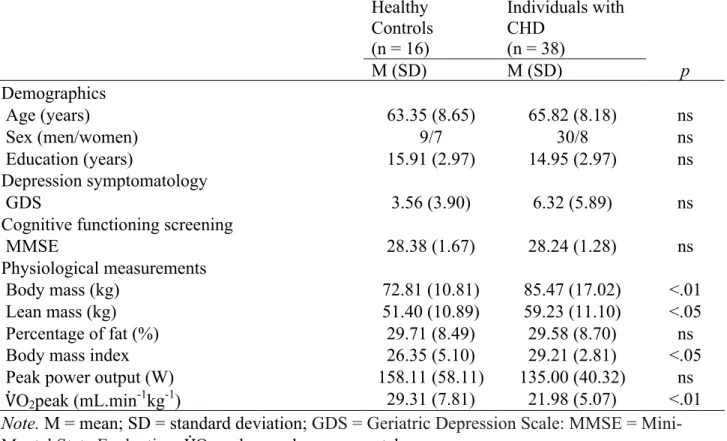
Table 1. Means and standard deviations for demographics, neuropsychological,
psychological and physiological data for individuals with CHD and healthy control groups
Healthy Controls (n = 16) Individuals with CHD (n = 38) M (SD) M (SD) p Demographics Age (years) 63.35 (8.65) 65.82 (8.18) ns Sex (men/women) 9/7 30/8 ns Education (years) 15.91 (2.97) 14.95 (2.97) ns Depression symptomatology GDS 3.56 (3.90) 6.32 (5.89) ns
Cognitive functioning screening
MMSE 28.38 (1.67) 28.24 (1.28) ns
Physiological measurements
Body mass (kg) 72.81 (10.81) 85.47 (17.02) <.01
Lean mass (kg) 51.40 (10.89) 59.23 (11.10) <.05
Percentage of fat (%) 29.71 (8.49) 29.58 (8.70) ns
Body mass index 26.35 (5.10) 29.21 (2.81) <.05
Peak power output (W) 158.11 (58.11) 135.00 (40.32) ns
VO2peak (mL.min-1kg-1) 29.31 (7.81) 21.98 (5.07) <.01 Note. M = mean; SD = standard deviation; GDS = Geriatric Depression Scale: MMSE = Mini-Mental State Evaluation; VO2peak = peak oxygen uptake.
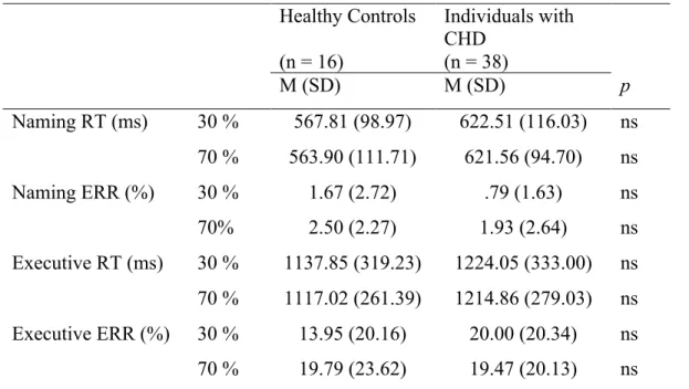
Table 2. Means and standard deviations for Stroop task performance for individuals with CHD and healthy control group
Healthy Controls (n = 16) Individuals with CHD (n = 38) M (SD) M (SD) p Naming RT (ms) 30 % 567.81 (98.97) 622.51 (116.03) ns 70 % 563.90 (111.71) 621.56 (94.70) ns Naming ERR (%) 30 % 1.67 (2.72) .79 (1.63) ns 70% 2.50 (2.27) 1.93 (2.64) ns Executive RT (ms) 30 % 1137.85 (319.23) 1224.05 (333.00) ns 70 % 1117.02 (261.39) 1214.86 (279.03) ns Executive ERR (%) 30 % 13.95 (20.16) 20.00 (20.34) ns 70 % 19.79 (23.62) 19.47 (20.13) ns Note. RT: Reaction Time; ERR: Error Rate; M: mean; SD: Standard Deviation.
Figure 1. Means and Standard Errors of [HbO] Variations as a Function of Group, Intensity and Stroop Task Condition
* Main condition effect at 30 % of PPO, p < .05
Figure 2. Means and Standard Errors of [HbR] Variations as a Function of Group, Intensity and Stroop Task Condition
* Simple group effect at 70 % of PPO, p < .05
Figure 3. Means and Standard Errors of [HbT] Variations as a Function of Group, Intensity and Stroop Task Condition
* Simple condition effect at 30 % of PPO, p < .05 α Main group effect at 70 % of PPO, p < .00
References
1. Mathers, C.D. and D. Loncar, Projections of global mortality and burden of disease from 2002 to 2030. PLoS Med, 2006. 3(11): p. e442.
2. Canada, P.H.A.o., Heart Diseases and Conditions. 2017: Canada.
3. Burkauskas, J., et al., Cognitive function in patients with coronary artery disease: A literature review. J Int Med Res, 2018. 46(10): p. 4019-4031.
4. Claassen, J.A.H.R., New cardiovascular targets to prevent late onset Alzheimer disease. European Journal of Pharmacology, 2015. 763: p. 131-134.
5. Labelle, V., et al., Decline in executive control during acute bouts of exercise as a function of exercise intensity and fitness level. Brain Cogn, 2013. 81(1): p. 10-7.
6. Chang, Y.K., et al., The effects of acute exercise on cognitive performance: a meta-analysis. Brain Res, 2012. 1453: p. 87-101.
7. Ando, S., Chapter 6 - Acute Exercise and Cognition: Effects of Cerebral Oxygenation and Blood Flow A2 - McMorris, Terry, in Exercise-Cognition Interaction. 2016, Academic Press: San Diego. p. 131-145.
8. Mekari, S., et al., The relationship between exercise intensity, cerebral oxygenation and cognitive performance in young adults. Eur J Appl Physiol, 2015. 115(10): p. 2189-97. 9. Ekkekakis, P., Illuminating the black box: investigating prefrontal cortical hemodynamics
during exercise with near-infrared spectroscopy. J Sport Exerc Psychol, 2009. 31(4): p. 505-53.
10. Braz, I.D. and J.P. Fisher, The impact of age on cerebral perfusion, oxygenation and metabolism during exercise in humans. J Physiol, 2016. 594(16): p. 4471-83.
11. Ando, S., et al., Reaction time to peripheral visual stimuli during exercise under normoxia and hyperoxia. Eur J Appl Physiol, 2009. 106(1): p. 61-9.
![Figure 1. Means and Standard Errors of [HbO] Variations as a Function of Group, Intensity and Stroop Task Condition](https://thumb-eu.123doks.com/thumbv2/123doknet/7719637.248235/50.918.120.627.150.478/figure-standard-errors-variations-function-intensity-stroop-condition.webp)
![Figure 2. Means and Standard Errors of [HbR] Variations as a Function of Group, Intensity and Stroop Task Condition](https://thumb-eu.123doks.com/thumbv2/123doknet/7719637.248235/51.918.110.802.112.429/figure-standard-errors-variations-function-intensity-stroop-condition.webp)
![Figure 3. Means and Standard Errors of [HbT] Variations as a Function of Group, Intensity and Stroop Task Condition](https://thumb-eu.123doks.com/thumbv2/123doknet/7719637.248235/52.918.110.808.111.445/figure-standard-errors-variations-function-intensity-stroop-condition.webp)
