Université de Montréal
Effect of Osteogenesis Imperfecta on Orthodontic Tooth
Movement in a Mouse Model
par Jean Rizkallah
Université de Montréal Faculté de médecine dentaire
Mémoire présenté à la Faculté de médecine dentaire en vue de l’obtention du grade de maîtrise scientifique
en médecine dentaire option orthodontie
Mai, 2014
Résumé
IntroductionL'ostéogenèse imparfaite (OI) est une maladie osseuse héréditaire qui affecte la production du collagène de type I et le remodelage osseux. Les biphosphonates sont administrés aux enfants atteints d'OI dans le but d’augmenter la masse osseuse et de réduire les fractures osseuses. Les patients atteints d’OI ont des malocclusions sévères qui affectent leur qualité de vie. Plusieurs processus biologiques de remodelage osseux qui sont nécessaires pour un mouvement dentaire orthodontique sont affectés chez les gens atteints d’OI. L'objectif de cette étude est d'évaluer le mouvement dentaire orthodontique dans un modèle de souris avec OI et traitées aux biphosphonates.
Matériels et méthodes
Vingt-quatre souris femelles âgées de 10 semaines ont été divisés en 4 groupes :
1 - OI traitées par zolédronate (n=6); 2 - OI non traitées (n=6); 3 - Type sauvage traitées par zolédronate (n=6); 4 – Type sauvage non traitées (n=6)
Un ressort de nickel-titane activé à 10 g de force a été cimenté entre les incisives et la 1ère molaire maxillaire droite. Le côté contralatéral a été utilisé comme témoin. Une dose de 0,05 mg de zolédronate a été administrée par voie sous-cutanée un jour avant la chirurgie. Sept jours après l'intervention, les souris ont été euthanasiées et la distance entre la 1ère et la 2e molaire a été mesurée par analyse microtomographique.
Résultats
Le mouvement dentaire orthodontique était significativement plus important chez les souris OI que celles de types sauvages dans les groupes non traités (p < 0,05). Le traitement par zolédronate n'a eu aucun effet significatif sur le mouvement dentaire orthodontique au sein des groupes OI et type sauvages.
Conclusions
Ces résultats suggèrent une augmentation du mouvement dentaire orthodontique chez les souris avec l’ostéogenèse imparfaite.
Mots-clés : ostéogenèse imparfaite, mouvement dentaire orthodontique, souris,
Abstract
INTRODUCTIONOsteogenesis imperfecta (OI) is a heritable bone disorder that affects collagen type I production and bone remodeling. Orthodontic tooth movement (OTM) involves the underlying process of alveolar bone remodeling. The objective of this study is to evaluate OTM in a mouse model of OI.
METHODS
Twenty four, 10 week-old female mice were divided into 4 groups: 1- OI treated with zoledronate, 2- OI untreated, 3- Wild-type (WT) treated with zoledronate and 4- WT untreated. A nickel-titanium closed coil spring (10 g) was attached between the incisors and the right maxillary 1st molar. The contralateral side was used as control. Zoledronate (0.05mg/kg) was administered sub-cutaneously 1 day prior to surgery. Seven days after the procedure, the distance between 1st – 2nd molars was measured by micro-CT.
RESULTS
OI mice presented significantly more OTM than WT mice when comparing within untreated groups (p<0.05). Zoledronate treatment had no significant effect on OTM within OI and WT groups.
CONCLUSIONS
These results suggest increased OTM in mice with OI. The dose of zoledronate administrated 1 day prior to surgery had no significant effect on OTM.
Keywords: osteogenesis imperfecta, orthodontic tooth movement, mice, bisphosphonates,
Table of contents
Résumé ... ii
Abstract... iii
Table of contents... iv
List of tables ... vi
List of figures... vii
List of abbreviations ... viii
Aknowledgements ...x Introduction...1 Osteogenesis Imperfecta...1 Overview...1 Classification ...2 Oral manifestations...3 Pathogenesis ...4 Treatments ...4 Bisphosphonates ...5 Overview...5
Bisphosphonate-related osteonecrosis of the jaw ...9
Orthodontic considerations...9
Orthodontic tooth movement...10
Theories of tooth movement...11
Factors influence tooth movement...13
Biology of tooth movement ...14
Hypothesis and study aims ...21
Materials and methods...23
Results...32
Discussion...37
Study limitations...39
Future studies...40 Conclusion ...41 Bibliography ... i
List of tables
Table I Expanded Sillence classification of osteogenesis imperfecta ...2
Table II Physiologic response to sustained pressure against a tooth ...13
Table III Group description ...24
Table IV Individual weight fluctuations for all 24 mice ...32
List of figures
Figure 1 Bowing of the radius (a) and tibia (b) in a baby with osteogenesis imperfecta type III
...1
Figure 2 Chemical structure of bisphosphonates...6
Figure 3 Mechanisms by which biphosphonates inhibit bone resorption...7
Figure 4 Molecular effect of bisphosphonates...8
Figure 5 Diagrammatic representation of the increasing compression of blood vessels as pressure increases in the PDL...14
Figure 6 Sequence of bone remodeling stages during orthodontic treatment ...16
Figure 7 The responses of periodontal ligament fibroblasts to orthodontic forces ...17
Figure 8 The sequence followed by cells and tissues in mechanosensing, transduction, and response ...19
Figure 9 The sequence of orthodontic tooth movement, illustrating the roles played by mineralized and non-mineralized tissues along with the associated blood vessels and neural elements ...20
Figure 10 NiTi coil cemented from right maxillary 1st molar to central incisors...25
Figure 11 Etching of right maxillary 1st molar ...26
Figure 12 NiTi coil cemented on right maxillary 1st molar ...27
Figure 13 Etching of central incisors...28
Figure 14 Trimming of lower incisors...29
Figure 15 Study timeline ...30
Figure 16 Illustration of measurement of tooth displacement by micro-CT scan ...31
Figure 17 First and second molar distances during orthodontic tooth movement on right maxilla. ...35
Figure 18 3D micro-CT reconstructions of the right maxillas for the OI mouse (i) and wild type mouse (ii) after 7 days of tooth movement ...36
List of abbreviations
CO2: carbon dioxideFVB: friend Virus B-Type
Micro-CT: micro computed tomography MMA: methylmetacrylate
NiTi: nickel titanium OI: osteogenesis imperfecta
OTM: orthodontic tooth movement PB: phosphate buffer
WT: wild type i.p.: intraperitoneally
I would like to dedicate this memoire to my family for the many hours this study has kept me from them.
And to the patients affected with osteogenesis imperfecta. I hope this study will bring us closer to treating their malocclusions; may it be one less issue to deal with…
Aknowledgements
First and foremost, I would like to thank my director, Dr. Nelly Huynh, for her guidance, support and making this study a very enjoyable experience.
I would also like to thank my codirector, Dr. Frank Rauch, for his invaluable contribution as well as supplying the mice for the study.
Dr. Clarice Nishio, my friend and colleague, thank you for the countless hours you spent helping us.
Thank you Dr. Antonio Nanci for allowing us access to your laboratory and for your advice.
Thank you Dr. Retrouvey, Dr. Vu and Dr. Schwartz for introducing me to research and specifically on osteogenesis imperfecta. They originally had the idea of doing a study on mice affected with osteogenesis imperfecta
Thank you Mr. Rompré for clarifying the very confusing statistical analyses that were used in this study.
A special thank you to:
- Sami Abdullah, Mia Esser, Pierre Moffat and the entire team at the Shriners Hospital in Montreal for their support
- Ultimate Wire Technologies for gracefully offering all of the NiTi coils. This study would not of been possible without this great team’s effort.
Introduction
Osteogenesis Imperfecta
Overview
Osteogenesis imperfecta (OI), also known as ‘brittle bone disease’, is a clinically heterogeneous heritable disorder affecting the connective tissue. It has a cumulative incidence of approximately 1 in 15,000-20,000 with the majority of patients having dominant mutations in the genes COL1A1 and COL1A2.1 Mutations in other genes, such as CRTAP and LEPRE1, lead to severe recessive forms of osteogenesis imperfecta. The severity is variable, ranging from a severe lethal form to milder forms without fractures. In general, the clinical manifestations are multiple fractures, muscle weakness, joint laxity, curved bones, blue sclera, hearing loss, presence of wormian bones on skull radiographs and dentinogenesis imperfecta (figure 1).
Classification
The original classification by Sillence et al in 1979 divided osteogenesis imperfecta into 4 types and is still the most widely used classification to date. The classification has since been expanded to include the more recent types of OI. Bone fragility in the most important characteristic of all types of osteogenesis imperfecta, its severity increases in the following order: type I < types IV, V, VI, VII < type III < type II.3
Table I Expanded Sillence classification of osteogenesis imperfecta3
Type Clinical severity Typical features Typically associated mutations*
I Mild non-deforming osteogenesis
imperfecta
Normal height or mild short stature; blue sclera; no dentinogenesis imperfecta
Premature stop codon in COL1A1
II Perinatal lethal Multiple rib and long-bone fractures at birth; pronounced deformities; broad long bones; low density of skull bones on radiographs; dark sclera
Glycine
substitutions in COL1A1 or COL1A2
III Severely deforming Very short; triangular face; severe scoliosis; greyish sclera; dentinogenesis imperfecta Glycine substitutions in COL1A1 or COL1A2 IV Moderately deforming
Moderately short; mild to moderate scoliosis; grayish or white sclera;
Glycine
dentinogenesis imperfecta COL1A1 or COL1A2 V Moderately
deforming
Mild to moderate short stature;
dislocation of radial head; mineralised interosseous membrane; hyperplastic callus; white sclera; no dentinogenesis imperfecta
Unknown
VI Moderately to severely deforming
Moderately short; scoliosis; accumulation of osteoid in bone tissue, fish-scale pattern of bone lamellation; white sclera; no dentinogenesis imperfecta Homozygous SERPINF1 mutations VII Moderately deforming
Mild short stature; short humeri and femora; white sclera; no dentinogenesis imperfecta
Homozygous CRTAP mutations
*May or may not be detectable in a given patient
Oral manifestations
Many oral manifestations are associated to osteogenesis imperfecta. Dentinogenesis imperfecta has been well documented in the literature. Many authors have reported an overrepresentation of Class III malocclusions, posterior and anterior openbites, crossbites and impacted teeth. Recently, our group showed that the patients with severe types of osteogenesis imperfecta have more severe malocclusions.4 One of the most challenging types of malocclusions that require early orthodontic treatment is the Class III malocclusion. This malocclusion is manifested by a more anterior position of the mandibular teeth relative to the maxillary teeth. In this type of bite, it is common for the lower anterior teeth to bite ahead of
the upper anterior teeth; this condition is also called anterior cross-bite. There are three causes of Class III malocclusions: an overdeveloped mandible (prognatism), an underdeveloped maxilla or a combination of both. Many environmental factors have been suggested as contributory to the development of mandibular prognatism. They include congenital anatomic defects, a habit of protruding the mandible, posture, hormonal disturbances and irregular eruption of the permanent incisors or loss of deciduous incisors. Substantial evidence has been advanced to support the theory of familial influence in mandibular prognatism and Class III malocclusion.
Pathogenesis
The pathogenesis of the forms of osteogenesis imperfecta with type 1 collagen mutations is well known. A collagen type I molecule comprises of 3 polypeptides (two α 1 and one α 2 chain) that form a triple-helical structure.5 In order for the three chains to perfectly intertwine, there must be a glycine residue at every third position. The most typical sequence abnormality associated with osteogenesis imperfecta is a point mutation that affects a glycine residue in either COL1A1 or COL1A2.3 Cells enclosing this kind of mutation
produce a mixture of normal and abnormal type I collagen. The resulting phenotype will be altered by which α chain is affected, the position in the triple helix at which the substitution arises, and which aminoacid is substituted for glycine. In most cases, mutations that form a premature stop codon within COL1A1 result in an osteogenesis imperfecta type I phenotype.
Treatments
Medical management of patients affected by osteogenesis imperfecta is directed by a multidisciplinary approach consisting of physical therapy, rehabilitation and orthopaedic
surgery. Till now, there is no cure for this disease. Physical activity is encouraged, with an increased risk of fracture, to prevent bone loss due to inactivity.6 Some patients require intramedullary rods surgically placed in their tibiae and femora in order to stand and walk.6
Historically, pharmacological treatment of osteogenesis imperfecta has been unsuccessful. Three decades ago, Albright7 evaluated 96 reports of 20 different treatments including hormones (calcitonin, cortisone, oestrogens, androgens, and thyroxine), vitamins (A, C, and D), minerals (aluminium, calcium, fluoride, magnesium, phosphate, and strontium), and some more exotic approaches (such as arsenic, radiation, dilute hydrochloric acid, and calf-bone extract). Although researchers claimed some clinical effectiveness, none stood the test of time.3
In 1987, Devogelear and colleagues reported pronounced clinical and radiological improvement in a case of a 12-year-old girl suffering from osteogenesis imperfecta and treated with oral pamidronate for 1 year. Pamidronate is a member of the bisphosphonate family of drugs, potent antiresorptive compounds that inhibit osteoclastic activity. Bisphosphonates are administered to children with osteogenesis imperfecta with the rationale that an increased bone mass will reduce bone fractures. Traditionally intravenous pamidronate has been used in children, however newer bisphosphonates with greater potency such as zoledronic acid have started to be used in clinical practice.2
Bisphosphonates
Overview
diseases such as osteoporosis or in malignomas and bone metastases. Bisphosphonates have an inhibitory effect on osteoclasts and thus decrease bone resorption. All bisphosphonates contain two phosphate groups attached to a single carbon atom to form a P-C-P core structure (figure 2).8 They are stable analogues of pyrophosphate compounds, which are commonly found in nature. Inorganic pyrophosphates are the simplest, naturally occurring pyrophosphates.9 Bisphosphonates differ from inorganic pyrophosphates (P–O–P) in that the oxygen has been replaced by a non-hydrolysable carbon. The phosphate and hydroxyl group create a tertiary interaction between the BP and the bone matrix, giving BPs their specificity for bone. Bisphosphonates also have a high affinity for bone mineral, allowing them to achieve a high local concentration throughout the entire skeleton.10 The half-life can exceed 10 years.11
Figure 2 Chemical structure of bisphosphonates.8 The two phosphonate groups form covalent bonds to the carbon atom. The carbon atom forms two other covalent bonds, the resulting R1
and R2 chains. The P-C-P core is responsible for the high affinity of BPs to calcium ions.
Bisphosphonates reduce the risk of fracture by inhibiting osteoclastic activity, thus reducing bone remodeling and increasing bone mineral density.
Bisphosphonates are readily adsorbed to the mineral surface of the bone and cleared from the circulation. Their preference to bone rather than soft tissue brings them into close
contact with osteoclasts. This results in biphosphonates being stored in the bone and only released during bone resorption. There are many mechanisms by which bisphosphonates can affect osteoclast-mediated bone resorption (figure 3). The most likely route through which bisphosphonates inhibit bone resorption is by their direct effects on mature osteoclasts.8 In addition to affecting mature osteoclasts, bisphosphonates can inhibit bone resorption by preventing osteoclast formation. Previous studies have demonstrated that some bisphosphonates can inhibit the formation of osteoclasts, probably by preventing the fusion of osteoclast precursors.9
Figure 3 Mechanisms by which biphosphonates inhibit bone resorption8
Nitrogen containing bisphosphonates, such as pamidronate and zoledronate, are taken up by the osteoclasts and inhibit farnesyl pyrophosphotase synthase, an enzyme of the
mevalonate pathway. This leads to the inhibition of isoprenoid geranyl pyrophosphate and thereby of the prenylation of small GTP-binding proteins (i.e.,Ras and Rho) that are responsible for cytoskeletal integrity and intracellular signaling (figure 4).8 The consequence
of these events initiates a series of results including the suppression of osteoclastic activity, loss of osteoclast cytoskeletal integrity and ruffled border, and ultimately apoptosis.8
Figure 4 Molecular effect of bisphosphonates.8 Nitrogen-containing bisphosphonates mediate their action by inhibiting the mevalonate pathway involved in cholesterol synthesis. Bisphosphonates inhibit farsnesylpyrophosphate (Farnesyl-PP) synthase, an enzyme that catalyzes conversion of geranylpyrophosphate to farnesylpyrophosphate.
Bisphosphonate-related osteonecrosis of the jaw
A significant complication related to bisphosphonate use is osteonecrosis of the jaw. The incidence of bisphosphonate-related osteonecrosis of the jaw (BRONJ) in osteoporotic patients treated with oral bisphosphonates is very low, with an estimated 0-0.04%.12 In a study by Marx et al.13 one hundred and nineteen cases of BRONJ were thoroughly reviewed. The vast majority of patients were treated for cancer. Aggravating factors included smoking, alcohol use, and ongoing chemotherapy. Migliorati et al.14 reported BRONJ in 17 cases of cancer patients receiving intravenous zoledronate or pamidronate. There were 2 spontaneous cases but the rest appeared following an oral surgical procedure, primarly dental extractions. There has yet to be a case of BRONJ reported in children. Also, no cases of osteonecrosis of the jaw have been reported in patients with osteogenesis imperfecta.15
Orthodontic considerations
Successful orthodontic therapy is dependant on bone remodeling. In order to move teeth, adequately functioning osteoblasts and osteoclasts are required in the area surrounding the periodontal ligament. It would be logical to assume that an inhibition of osteoclastic activity would result in reduced tooth movement. In a study on 9-10 weeks old rats, a 3-week systemic bisphosphonate treatment reduced tooth movement by 40%.16 In a study on mice, a daily local injection of bisphosphonates for 12 days reduced the amount of tooth movement and the number of osteoclasts. In addition, it also reduced root resorption.17 In 2005, Schwartz18 reported a case of a female orthodontic patient treated with bisphosphonates to
control bone metastases related to breast cancer. When the patient initiated orthodontic therapy, the premolar spaces were one-third closed, however, no significant space closure was observed after bisphosphonate therapy was initiated. Rinchuse et al.19 described the
orthodontic treatment and outcome of two patients who had received bisphosphonates. Both patients experienced impeded tooth movement and one patient also had osteonecrosis of the mandible. Recently, a pilot study from the University of Alberta has demonstrated that the bone burden from previous bisphosphonate use will significantly inhibit orthodontic tooth movement (OTM).20 It is reasonable to conclude that patients receiving bisphosphonate treatment pose a greater challenge to the orthodontist because of the possible inhibition of tooth movement and the potential of developing osteonecrosis of the jaw.
Orthodontic tooth movement
The general principle of orthodontic tooth movement is that if a prolonged force is applied to a tooth, tooth movement will occur as the surrounding bone remodels. Bone is selectively resorbed in some areas and added in other areas. Essentially, as the tooth migrates through the bone, it drags its attachment apparatus along with it. The periodontal ligament, the intermediate between the teeth and bone, mediates the bony response. According to Proffit, tooth movement is believed to be primarily a periodontal ligament phenomenon.
Each tooth is attached to its supporting alveolar bone by a network of predominantly collagenous structure, the periodontal ligament. Although most of the space is taken up by collagen there are also two other major components of the ligament. There are the cellular elements, including various types of mesenchymal cells along with vascular and neural
elements; and tissue fluids. Both play an important role in normal function as well as allowing orthodontic tooth movement.
The principal cells found in the periodontal ligament are fibroblasts, osteoblasts and their precursors, mesenchymal cells. During normal function, fibroblasts are constantly renewing the collagenous matrix. At the same time, osteoblasts are adding new bone to the dental socket. Bone and cementum are removed by specialized osteoclasts and cementoclasts, respectively. The periodontal ligament, though not highly vascular, does receive its nutrition through a vascular system. It is also important to recognize that the periodontal ligament is filled with fluid. Many describe the periodontal ligament as a gel-like shock absorber.
Theories of tooth movement
Proffit describes two main theories that attempt to explain orthodontic tooth movement. The bioelectric theory relates tooth movement to changes in bone metabolism that are dictated by the electric signals produced when alveolar bone is bended. The pressure-tension theory relates tooth movement to cellular, chemical and blood flow changes in the periodontal ligament. The theories are not mutually exclusive, it would seem that both mechanisms might play a role in the biologic control of tooth movement.
Electrical signals that could initiate tooth movement were thought to be piezoelectric. Piezoelectricity is a phenomenon that can be observed in any crystalline structure. Essentially piezoelectricity means a charge produced from pressure. Piezoelectricity has two unusual characteristics: 1- A quick decay rate and 2- The production of an equal and opposite charge when the force is released. Due to these characteristics, only rhythmic activity could generate an important exchange of electric signals, whereas constant forces would only produce a charge at the moment of application and release of the force. It would seem that this type of
force is more important in the general maintenance of the skeleton and less so in orthodontic tooth movement. Another type of endogenous electric signal called the ‘‘bioelectric potential’’ can be observed in bone that is not being stressed. Although this process is not well known, cellular activity can be modified by the application of exogenous electric signals. In animal and human studies, it has been demonstrated that tooth movement can be accelerated when a low voltage current is applied.21
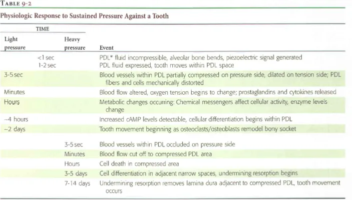
The classical theory of orthodontic tooth movement, the pressure-tension theory, relies on chemical rather than electrical signals as the trigger to the cascade of events that ultimately lead to tooth movement. In this theory, an alteration in blood flow is caused by the movement of the tooth in the periodontal ligament space, creating areas of compression and areas of tension. Generally, blood flow is decreased in areas of compression while remaining constant or increased in the areas of tension. The alterations in blood flow quickly alter the chemical composition of the local environment. For example, oxygen levels are decreased in areas of low blood flow but might increase in areas of higher blood flow. These chemical changes, acting either directly or by stimulating the release of secondary messengers, then would stimulate cellular differentiation and activity. Essentially, this theory shows three stages; 1- alterations in blood flow, 2- the formation and/o release of chemical messengers, and 3- activation of cells (Table II).22
Table II Physiologic response to sustained pressure against a tooth22
Factors influence tooth movement
Orthodontic tooth movement is also modulated by the magnitude, the duration, the origin of application and the direction of the force. There are two different types of applied forces: continuous and intermittent. Contemporary orthodontic appliances are based on light continuous forces. The magnitude and the duration of forces have an important effect on the tissue response. Up to a certain stress level, the reactions occur mainly in the periodontal ligament with increasing vascularization, cell proliferation, fiber and bone formation but beyond that level, decreased vascularization and cell destruction occurs. At a certain magnitude of continuous force, blood vessels appear completely occluded and a sterile necrosis ensues (figure 5). Different directions and application of the force will result in different types of movements. Movements are often presented in terms of tipping, translation
or bodily movement, intrusion, extrusion, rotation and torquing.
Figure 5 Diagrammatic representation of the increasing compression of blood vessels as pressure increases in the PDL.22 At a certain magnitude of continuous pressure, blood vessels are totally occluded and a sterile necrosis of PDL tissue ensues.
Biology of tooth movement
At the cellular level, the application of forces on the crown of a tooth, may alter the internal forces acting on resident cells, leading to changes in gene expression, and production of proteins that ultimately alter the structure and function of the extracellular matrix, as well as the jaw bones.23 As previously mentioned, the most important macromolecules of the extracellular matrix are collagen (the most abundant), proteoglycans, laminin, fibronectin,
elastin, and hyaluronic acid. These molecules bind to cell adhesion foci to transfer signals intracellularly.24 Thus, we can consider the extracellular matrix as a tissue that relays extracellular and intracellular strains to effect changes in organ structure and function, through mechanotransduction.
Bone cells, especially osteocytes, are sensitive to their environment. They play the important role of mechanosensing and transducing in response to mechanical stress.25 Osteocytes are connected to one another and to cells of the bone surface by a network of cytoplasmic projections or dendrites. Soon after bone loading, osteocytic metabolism is altered. Many anabolic signals, such asnitric oxide, prostaglandins, and ATP, are released within seconds of osteocyte loading.26 This triggers a cascade of events capable of stimulating osteoblastogenesis.
The fluid flow theory states that osteocytes are sensitive to strains evoked from locally displaced fluid in the canaliculi (figure 6).23 When loading occurs, interstitial fluid is squeezed through the thin layer of non-mineralized matrix surrounding the cell bodies and cell processes, resulting in local strain at the cell membrane and activation of the affected osteocytes.23 Osteocytes may also send signals to activate the bone resorption cascade through expression of NF-κB ligand, secretion of macrophage colony-stimulating factors and through their own apoptosis.23 It is now thought that osteocytes act as the chief mechanosensors in bone.
Figure 6 Sequence of bone remodeling stages during orthodontic treatment.23 The roles played by osteocytes, osteoblasts, and osteoclasts are illustrated.
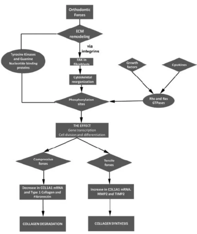
Similarly to osteocytes in bone, fibroblasts mediate the changes in connective tissues in response to mechanical loading. Fibroblasts are thought to be mechanoresponsive, meaning that the mechanical stress that is transmitted through the extracellular matrix to the fibroblasts via integrins influence their morphology, cytoskeletal organization, proliferation, differentiation, and gene expression.27 They contain proteins at their surface, called focal
adhesion kinases, which act as stress gauges. Once loaded, fibroblasts generate a cascade of events that ultimately lead to collagen synthesis or collagen degradation (figure 7).23
In summary, orthodontic forces strain the extracellular matrix and cells of the alveolar bone and periodontal ligament. Through mechanotransduction, changes in the form and function of the extracellular matrix, the cell membrane, the cytoskeleton, the nucleus and other cytoplasmic organelles trigger a cascade of events.28 Cell adhesion molecules, such as integrins, transmit the mechanical stress from the extracellular matrix into the cell and vice versa. This is the general process by which a mechanical stimulus is converted into a biological signal. These biochemical or electric signals are rapidly relayed to the nucleus. The outcome of these events is either stimulation or suppression of gene expression and protein synthesis (figures 8 & 9).
Figure 8 The sequence followed by cells and tissues in mechanosensing, transduction, and response23
Figure 9 The sequence of orthodontic tooth movement, illustrating the roles played by mineralized and non-mineralized tissues along with the associated blood vessels and neural elements23
Hypothesis and study aims
The objective of the current study is first to evaluate orthodontic tooth movement in a mouse model with osteogenesis imperfecta. The research hypothesis was that orthodontic tooth movement would be inhibited in the OI mouse model. The reasoning was that orthodontic forces trigger a series of events that involve collagen metabolism, among others. The mutant collagen would create a ‘‘kink’’ in the chain, translating to an inhibition of tooth movement. We would not expect to see the normal patterns of apposed immature bone on the areas of tension and bone resorption on the areas of compression.
A second theory involves the periodontal ligament. Orthodontic forces strain the extracellular matrix and cells of the alveolar bone and periodontal ligament. Through mechanotransduction, changes in the form and function of the extracellular matrix are relayed to the cells of the alveolar bone and periodontal ligament. The most abundant macromolecules of the extracellular matrix are collagen. We thought that the mutant collagen would make the ECM insensitive to orthodontic forces and thus impede mechanotransduction. An analogy could be made with driving a car with a stiff suspension compared to a car with a loose suspension. The threshold of orthodontic force to induce tooth movement would be much higher in the OI mice just like we do not feel small bumps while driving a car with a loose suspension.
Another objective that was studied is the effect of bisphosphonates on orthodontic tooth movement. Specifically, we wanted to study the effect of one systemic dose of zoledronate, equivalent to one treatment given in humans affected by OI, administered one day pre-op. The general consensus in the orthodontic literature is that bisphosphonates have an
inhibitory effect on orthodontic tooth movement, at least in adults. Recent studies have also established a dose dependant association. In our study, we expected a limited inhibitory effect of the orthodontic tooth movement. The OI patients followed at the Montreal Shriners Hospital receive 2 doses per year and typically for many years. No equivalencies have yet been established between mice drug metabolism and humans. The researchers are aware that the bisphosphonate protocol used on mice in this study cannot be compared to the one given to humans.
Materials and methods
A novel mouse model of osteogenesis imperfecta was recently developed in a laboratory at the University of Toronto (Laboratory Dr. J. Aubin). A mutation in the COL1A1 gene was obtained by N-nitroso-N-ethylurea exposure. The OI mouse model used in our study mimics type IV OI in humans. Genotyping was performed on all mice to confirm for the mutation.
A total of 24 ten-week-old female mice were used. These mice were divided into 2 groups: 12 FVB mice (wild type) and 12 Col1a1Jrt/+ (OI). They were further randomly divided
into 2 groups: 6 mice of each group received a subcutaneous injection of 0.05 mg/kg zoledronate and 6 were injected with saline one day prior to the experiment (table III). A 5 mm NiTi closed coil spring was expanded to produce 10 g of force and attached between the right first molar and the central incisors (figure 10).
The mice were put under general anesthesia with a Ketamine/Xylazine/Acepromazine cocktail administered intraperitoneally (i.p.). The mouth was maintained open with Andy orthodontic elastics (3M, Unitek) extending from the operating table to the incisors. The teeth were then isolated by holding the tongue and cheeks with college pliers. The entire procedure was a 4-hand procedure. The first part of the procedure consisted of cementing the intra-oral coil spring to the 1st molar. Prior to cementation, the coil spring was adjusted to exert 10 g of force by a tension gauge. The 1st molar was etched with 37% phosphoric acid (Scotchbond,
3M ESPE) for 20 seconds (figure 11). We then rinsed off the acid with a wet cotton pellet and dried the tooth surface with an air compressor. A bonding agent (Transbond plus self etching primer, 3M ESPE) was then applied to the tooth. Finally, the coil spring was cemented with a
flowable composite cement (Filtek Supreme Ultra, 3M ESPE) (figure 12). The second part of the procedure consisted of attaching the coil to the central incisors. To cement the coil on the incisors, the same manipulations as described above were used but on the incisors (figures 10 and 13)
Table III Group description
WT: wild type, OI: osteogenesis imperfecta 24 Total mice 12 FVB (WT) 6 Treated with zoledronate 6 Not treated 12 Col1a1Jrt/+ (OI) 6 Treated with zoledronate 6 Not treated
Figure 10 NiTi coil cemented from right maxillary 1st molar to central incisors © Jean Rizkallah
Figure 14 Trimming of lower incisors © Jean Rizkallah
In order to minimize complications, the mice were given a soft food diet 3 days prior to cementing the orthodontic appliance and continued during the duration of the experiment. The lower incisors were also trimmed to minimize appliance breakage (figure 14). The 24 mice received daily injections of carprofen (5 mg/kg) and saline for the first 4 days. After 7 days, the mice were euthanized by CO2 in a gas chamber and the maxillas and mandibles were
dissected and fixed in 4% paraformaldehyde and 0.1% glutaraldehyde for 24 hours and then washed in PB 0.1 M. The different timepoints are illustrated in figure 15. The calcified dissections were maintained in PB 0.1 M during micro-CT scanning to finally be embedded in methylmetacrylate (MMA).
Figure 15 Study timeline
The exclusion criteria were: broken or debonded coil and spacing between left 1st and
2nd maxillary molars.
The distance between the maxillary 1st molars and the 2nd molars were measured by micro-CT scans (figure 16). The maxillas were scanned with the skyscan 1172 (Bruker microCT) at 5 microns of resolution. We assumed that the teeth were contacting prior to the cementation of the appliance. Total movement was obtained by measuring the shortest distance between the molar surfaces in both sagittal and occlusal views. Since the results were practically the same in both views, only the sagittal measurements were kept for statistical purposes. The maxillary left side was also measured and used as control.
Figure 16 Illustration of measurement of tooth displacement by micro-CT scan. The red line represents the shortest distance between the 1st and 2nd molar; and was used as the measure of tooth displacement. © Jean Rizkallah
Statistical analysis
Statistical differences of the right and left sides were determined by using a related-samples Wilcoxon test (p<0.05). Statistical analysis of the data was obtained by using independent-samples Kruskal-Wallis test (p<0.05). An intraclass correlation coefficient of 0.99 was calculated to test intrarater reliability, based on the measure for a set of 15 mice. This shows excellent reliability in the measurement method.
Results
Table IV describes the weight fluctuations for all 24 mice. A decrease in body weight was observed for the first 3 days but was quickly followed by an increase for the remainder of the experiment. Table V describes the average tooth movement measured for each group. Differences between left and right sides were statistically significant in all 4 groups (Wilcoxon, p≤0.028). On the experimental side, tooth movement was significantly higher in the OI group compared to the control (figures 17 and 18). No statistical difference was measured between the groups treated with zoledronate and the untreated groups.
Table IV Individual weight fluctuations for all 24 mice
Animal # (Group) Day 1 body weight (g) Day 2 body weight (g) Day 3 body weight (g) Day 4 body weight (g) Day 5 body weight (g) Day 6 body weight (g) Day 7 body weight (g) Group 1 (OI+Z) 588 16.5 15.4 15.4 15.5 16.2 16.1 17.1 589 17.3 15.1 13.7 15.3 16.6 17.5 17.3 738 16.8 15.7 16.9 17.2 17.5 17.5 17.7 746 18.3 16.7 17.3 18.0 18.4 18.9 18.7 748 18.1 15.9 15.7 16.2 16.7 17.3 17.5 751 18.0 17.7 17.3 17.4 17.8 17.9 17.7 Group 2 (OI)
599 16.2 14.2 12.8 13.3 14.4 15.3 16.0 702 14.9 13.4 13.5 13.8 14.0 14.4 14.4 708 18.8 18.1 17.8 17.8 17.6 18.0 18.0 715 18.7 17.7 18.2 18.4 18.5 19.2 19.5 832 18.6 16.7 17.0 16.9 17.8 19.1 18.2 834 19.6 17.0 17.9 18.7 19.5 19.6 19.3 Group 3 (WT+Z) 470 20.8 17.5 15.9 16.6 16.2 17.8 17.9 484 21.0 17.7 17.1 17.6 18.4 19.1 18.6 564 20.5 19.0 18.5 18.6 19.4 19.6 19.8 565 22.8 20.9 19.4 18.9 19.1 20.2 20.9 566 23.4 21.0 20.3 20.2 21.0 21.2 21.4 568 24.9 22.2 20.7 21.7 22.4 22.8 23.3 Group 4 (WT) 452 21.7 18.6 18.1 19.2 19.1 19.2 18.9 453 21.1 17.8 17.2 17.6 17.8 18.4 18.2 553 19.8 18.2 18.2 17.5 17.8 18.2 18.5 555 21.6 19.3 18.7 19.1 20.1 20.2 19.7 559 21.0 19.1 19.3 18.8 20.2 20.9 20.9 560 21.8 19.8 19.2 20.0 20.5 21.2 21.5
Table V Average measurements for distance between 1st and 2nd maxillary molars
Groups (n=6)
Right (Experimental) Mean maxillary molar
distance (SD) (µm)
Left (control) Mean maxillary molar
distance (SD) (µm) p-value
1 91.45 (60.88) 10.04 (14.20) 0.028*
2 159.60 (84.98) 9.43 (20.46) 0.027*
3 46.66 (27.13) 0.94 (2.30) 0.028*
4 46.22 (21.64) 0.94 (2.30) 0.027*
* p< 0.05 considered statistically significant
1: osteogenesis imperfecta treated with zoledronate; 2: osteogenesis imperfecta not treated; 3: wild type treated with zoledronate; 4: wild type not treated.
Figure 17 First and second molar distances during orthodontic tooth movement on right maxilla. Increase tooth movement in osteogenesis imperfecta (n=6) compared to wild type (n=6). 1: osteogenesis imperfecta treated with zoledronate; 2: osteogenesis imperfecta not treated; 3: wild type treated with zoledronate; 4: wild type not treated.
0.00 50.00 100.00 150.00 200.00 250.00 300.00 1 2 3 4 Orthodontic tooth movement (μm) Groups
Orthodontic Tooth Movement in OI vs WT
Pairwise comparison of groups 2 & 4 p = 0.048Figure 18 3D micro-CT reconstructions of the right maxillas for the OI mouse (i) and wild type mouse (ii) after 7 days of tooth movement. A: Anterior; P: Posterior; F: Force; M: Molar © Jean Rizkallah
Discussion
This is the first study of orthodontic tooth movement on an osteogenesis imperfecta animal model. Bone cell function and the remodeling mechanisms which normally maintain bone homeostasis are altered in OI.29 Orthodontic tooth movement depends on the underlying processes of alveolar bone remodeling.30, 31 In light of this, we hypothesized that OI might affect OTM. Our results demonstrated that OI mice presented a greater amount of tooth movement than control mice. Interestingly, the results were opposite to the original hypothesis that OTM would be inhibited in mice with OI. The original reasoning was that the mutant collagen would disrupt the mechanotransduction processes during force application and thus, reduce tooth movement. However, our results suggest that tooth movement is not only possible in OI but also accelerated. Increased tooth movement in mice with OI could be due to the lower density of cortical bone and higher bone turnover.
Furthermore, the standard deviations for the osteogenesis imperfecta groups were larger than the controls. This observation could be explained by the variable penetrance of the collagen mutation. Assuming the fact that the disease affected the OI mice differently, we could also expect a difference in tooth movement in these mice. Since the standard deviations for the wild type groups were reduced, we assumed that the procedure was well controlled. The large standard deviations found in the OI groups could not be explained by a faulty procedure. Therefore, we hypothesized that the standard deviations observed in the OI groups are a reflection of the variance in the disease penetrance.
We also observed more dental tipping in the OI mice compared to the control mice. One of the factors that affect tooth movement is the supporting bone. A softer bone will move
the center of resistance of the tooth more apically. Since the point of application of the force is the same in all mice, a larger distance between the force and the center of resistance would result in more dental tipping.
The effect of bisphosphonates on OTM has been studied extensively. It has previously been reported that bisphosphonates inhibit tooth movement.8, 32 This is the first study to investigate the effect of zoledronate on OTM in OI. Conversely to what is well described in the orthodontic literature, our study revealed that zoledronate had no significant inhibiting effect on OTM. Previous studies have demonstrated the dose dependent effect of bisphosphonates.17, 20 The drug regimen used in this study could be described as a relatively low systemic dose. A probable explanation for our results is that the dosage of zoledronate was too low to significantly reduce tooth movement. However, a non-significant tooth movement reduction was observed in the OI mice treated with zoledronate.
Bisphosphonates are commonly used to treat patients affected with OI. The typical mode of administration is chronic intra-venous injections. This study did not investigate the effect of multiple doses of zoledronate on OTM in OI. Instead, the purpose of this study was to evaluate the effect on OTM of a minimally effective dose to prevent bone loss.33 Theoretically, bone loss can be limited in mice with OI without significantly inhibiting tooth movement. Bisphosphonates are highly potent drugs with possible side effects like osteonecrosis of the jaw. A noble goal would be to evaluate the minimal dose to maintain the positive effects while minimizing the side effects. The effect of chronic administration of bisphosphonates on OTM in mice with OI was not evaluated in this study.
These results clearly demonstrate that OTM is accelerated in mice with osteogenesis imperfecta in the absence of other factors. It is difficult to draw any conclusions from this
study on humans. However, one could hypothesize that orthodontic tooth movement would also be accelerated in humans affected by OI in the absence of bisphosphonates. If the latter was proven to be true, then orthodontic treatment could be offered to patients affected by OI. What remains unclear is the effect of bisphosphonates in OI during orthodontic treatment. This question requires more extensive research.
Study limitations
There are several limitations to acknowledge. The low dosage of bisphosphonates in our protocol did not permit to evaluate a significant inhibiting effect on tooth movement; future studies may consider looking at different dosages of bisphosphonates. No biochemical tests were performed on the mice to analyze the blood level of zoledronate. Furthermore, our groups were relatively small; and although an inhibiting effect of OTM was noticed in OI treated with zoledronate, it was not significant. Due to a lack of time and resources we were only able to study one time point, after 7 days; future studies could consider looking at different timepoints.
Difficulties encountered
This study also had its share of difficulties. Breeding of the OI mice was a long process and the OI pups had a lower survival rate. Another issue was maintaining mouse body weight. Different strategies were combined to minimize weight loss. First, the mice were given a soft food diet 3 days prior to the procedure and during the entire length of the study. And second, daily injections of saline and analgesics were administered for the first 4 days following the surgery. Furthermore, the intra-oral appliance survival was also an issue due to coil breakage.
In order to palliate the latter issue, we added more cement on the anterior portion of the coil and trimmed the lower incisors.
Future studies
Future studies could look at the effect on OTM of multiple systemic doses of zoledronate administered in an OI mouse model, for example, 3 weeks, 2 weeks, 1 week and 1 day prior to starting OTM. It would also be interesting to compare with WT groups and evaluate if the inhibiting effect of zoledronate on OTM is similar in both OI and WT mice. With the current micro-CT data, we could study bone height, bone width and compare between groups.
It would also be interesting to study bone density, however, that would require redoing the CT scans of the entire sample. This data would allow us to evaluate the center of resistance of the molars and could explain the observation of increased tooth tipping that was noticed in OI.
Furthermore, an immunohistochemical analyses would answer many questions that pertain to quantity of osteoclasts and osteoblasts. Markers such as TRAP can be used to identify osteoclasts and Runx2 to identify osteoblasts. Bril could also be used to identify new bone formation.
Conclusion
Orthodontic tooth movement is increased in mice with osteogenesis imperfecta. A unique low systemic dose administered one day prior to force application had no significant effect on OTM. These results give further evidence to the dose dependent effect of bisphosphonates. Future studies using immunohistochemistry might help elucidate the underlying cellular mechanisms of orthodontic tooth movement in mice with OI.
Bibliography
1. Forlino A, Cabral WA, Barnes AM, Marini JC. New perspectives on osteogenesis imperfecta. Nat Rev Endocrinol 2011;7(9):540-57.
2. Cheung MS, Glorieux FH. Osteogenesis Imperfecta: update on presentation and management. Rev Endocr Metab Disord 2008;9(2):153-60.
3. Rauch F, Glorieux FH. Osteogenesis imperfecta. Lancet 2004;363(9418):1377-85. 4. Rizkallah J, Schwartz S, Rauch F, Glorieux F, Vu DD, Muller K, et al. Evaluation of
the severity of malocclusions in children affected by osteogenesis imperfecta with the peer assessment rating and discrepancy indexes. Am J Orthod Dentofacial Orthop 2013;143(3):336-41.
5. Kadler KE, Holmes DF, Trotter JA, Chapman JA. Collagen fibril formation. Biochem J 1996;316 ( Pt 1):1-11.
6. Zeitlin L, Fassier F, Glorieux FH. Modern approach to children with osteogenesis imperfecta. J Pediatr Orthop B 2003;12(2):77-87.
7. Albright JA. Systemic treatment of osteogenesis imperfecta. Clin Orthop Relat Res 1981(159):88-96.
8. Ghoneima AA, Allam ES, Zunt SL, Windsor LJ. Bisphosphonates treatment and orthodontic considerations. Orthodontics & craniofacial research 2010;13(1):1-10. 9. Russell RG. Bisphosphonates: the first 40 years. Bone 2011;49(1):2-19.
10. Abela S, Chotai M, Bister D. What you need to know about bisphosphonates: an overview and general recommendations for orthodontic treatment. Journal of orthodontics 2012;39(3):186-92.
11. Zahrowski JJ. Bisphosphonate treatment: an orthodontic concern calling for a proactive approach. Am J Orthod Dentofacial Orthop 2007;131(3):311-20.
12. Rizzolo D, Sedrak M. Managing the adverse effects of bisphosphonate therapy on the jaw. JAAPA 2009;22(11):48-52.
13. Marx RE, Sawatari Y, Fortin M, Broumand V. Bisphosphonate-induced exposed bone (osteonecrosis/osteopetrosis) of the jaws: risk factors, recognition, prevention, and treatment. Journal of oral and maxillofacial surgery : official journal of the American Association of Oral and Maxillofacial Surgeons 2005;63(11):1567-75.
14. Migliorati CA, Schubert MM, Peterson DE, Seneda LM. Bisphosphonate-associated osteonecrosis of mandibular and maxillary bone: an emerging oral complication of supportive cancer therapy. Cancer 2005;104(1):83-93.
15. Malmgren B, Astrom E, Soderhall S. No osteonecrosis in jaws of young patients with osteogenesis imperfecta treated with bisphosphonates. J Oral Pathol Med 2008;37(4):196-200.
16. Igarashi K, Mitani H, Adachi H, Shinoda H. Anchorage and retentive effects of a bisphosphonate (AHBuBP) on tooth movements in rats. Am J Orthod Dentofacial Orthop 1994;106(3):279-89.
17. Fujimura Y, Kitaura H, Yoshimatsu M, Eguchi T, Kohara H, Morita Y, et al. Influence of bisphosphonates on orthodontic tooth movement in mice. European journal of orthodontics 2009;31(6):572-7.
18. Schwartz JE. Ask us: Some drugs affect tooth movement. Am J Orthod Dentofacial Orthop 2005;127(6):644.
19. Rinchuse DJ, Sosovicka MF, Robison JM, Pendleton R. Orthodontic treatment of patients using bisphosphonates: a report of 2 cases. Am J Orthod Dentofacial Orthop 2007;131(3):321-6.
20. Kaipatur NR, Wu Y, Adeeb S, Stevenson TR, Major PW, Doschak MR. Impact of bisphosphonate drug burden in alveolar bone during orthodontic tooth movement in a rat model: a pilot study. Am J Orthod Dentofacial Orthop 2013;144(4):557-67.
21. Giovanelli S, Festa F. Effect of electric stimulation on tooth movement in clinical application. In: Davidovitch Z, Norton LA, eds. Biological Mechanisms of Tooth Movement and Craniofacial Adaptation. Boston: Harvard Society for Advancement of Orthodontics; 1996.
22. Proffit W, Fields H, Sarver D. Contemporary Orthodontics. Philadelphia, PA: Elsevier; 2007.
23. Krishnan V, Davidovitch Z. On a path to unfolding the biological mechanisms of orthodontic tooth movement. Journal of dental research 2009;88(7):597-608.
24. Li J, Zhao Z, Wang J, Chen G, Yang J, Luo S. The role of extracellular matrix, integrins, and cytoskeleton in mechanotransduction of centrifugal loading. Mol Cell Biochem 2008;309(1-2):41-8.
25. Bonewald LF, Johnson ML. Osteocytes, mechanosensing and Wnt signaling. Bone 2008;42(4):606-15.
26. Bakker AD, Soejima K, Klein-Nulend J, Burger EH. The production of nitric oxide and prostaglandin E(2) by primary bone cells is shear stress dependent. J Biomech 2001;34(5):671-7.
27. Wang JH, Thampatty BP, Lin JS, Im HJ. Mechanoregulation of gene expression in fibroblasts. Gene 2007;391(1-2):1-15.
28. Masella RS, Meister M. Current concepts in the biology of orthodontic tooth movement. Am J Orthod Dentofacial Orthop 2006;129(4):458-68.
29. Rauch F, Lalic L, Roughley P, Glorieux FH. Relationship between genotype and skeletal phenotype in children and adolescents with osteogenesis imperfecta. Journal of bone and mineral research : the official journal of the American Society for Bone and Mineral Research 2010;25(6):1367-74.
30. Krishnan V, Davidovitch Z. Cellular, molecular, and tissue-level reactions to orthodontic force. Am J Orthod Dentofacial Orthop 2006;129(4):469 e1-32.
31. Wise G, King G. Mechanisms of tooth eruption and orthodontic tooth movement. Journal of dental research 2008;87(5):414-34.
32. Iglesias-Linares A, Yanez-Vico RM, Solano-Reina E, Torres-Lagares D, Gonzalez Moles MA. Influence of bisphosphonates in orthodontic therapy: Systematic review. J Dent 2010;38(8):603-11.
33. Lloyd SA, Travis ND, Lu T, Bateman TA. Development of a low-dose anti-resorptive drug regimen reveals synergistic suppression of bone formation when coupled with disuse. J Appl Physiol 2008;104(3):729-38.