HAL Id: inserm-02156686
https://www.hal.inserm.fr/inserm-02156686
Submitted on 14 Jun 2019
HAL is a multi-disciplinary open access archive for the deposit and dissemination of sci-entific research documents, whether they are pub-lished or not. The documents may come from teaching and research institutions in France or abroad, or from public or private research centers.
L’archive ouverte pluridisciplinaire HAL, est destinée au dépôt et à la diffusion de documents scientifiques de niveau recherche, publiés ou non, émanant des établissements d’enseignement et de recherche français ou étrangers, des laboratoires publics ou privés.
tissue homeostasis and promoting tolerance in
autoimmunity, inflammatory disease and transplantation
Giada Amodio, Joanna Cichy, Patricia Conde, Gianluca Matteoli, Aurélie
Moreau, Jordi Ochando, Barbaros Oral, Michaela Pekarova, Elizabeth Ryan,
Johannes Roth, et al.
To cite this version:
Giada Amodio, Joanna Cichy, Patricia Conde, Gianluca Matteoli, Aurélie Moreau, et al.. Role of myeloid regulatory cells (MRCs) in maintaining tissue homeostasis and promoting tolerance in autoim-munity, inflammatory disease and transplantation. Cancer Immunology, Immunotherapy, Springer Verlag, 2019, 68 (4), pp.661-672. �10.1007/s00262-018-2264-3�. �inserm-02156686�
https://doi.org/10.1007/s00262-018-2264-3
SYMPOSIUM-IN-WRITING PAPER
Role of myeloid regulatory cells (MRCs) in maintaining tissue
homeostasis and promoting tolerance in autoimmunity, inflammatory
disease and transplantation
Giada Amodio1 · Joanna Cichy2 · Patricia Conde3 · Gianluca Matteoli4 · Aurélie Moreau5,6 ·
Jordi Ochando3 · Barbaros H. Oral7 · Michaela Pekarova8 · Elizabeth J. Ryan9 · Johannes Roth10 ·
Yahya Sohrabi11 · Maria‑Cristina Cuturi5,6 · Silvia Gregori1
Received: 20 February 2018 / Accepted: 16 October 2018 / Published online: 24 October 2018 © The Author(s) 2018
Abstract
Myeloid cells play a pivotal role in regulating innate and adaptive immune responses. In inflammation, autoimmunity, and after transplantation, myeloid cells have contrasting roles: on the one hand they initiate the immune response, promoting acti-vation and expansion of effector T-cells, and on the other, they counter-regulate inflammation, maintain tissue homeostasis, and promote tolerance. The latter activities are mediated by several myeloid cells including polymorphonuclear neutrophils, macrophages, myeloid-derived suppressor cells, and dendritic cells. Since these cells have been associated with immune suppression and tolerance, they will be further referred to as myeloid regulatory cells (MRCs). In recent years, MRCs have emerged as a therapeutic target or have been regarded as a potential cellular therapeutic product for tolerance induction. However, several open questions must be addressed to enable the therapeutic application of MRCs including: how do they function at the site of inflammation, how to best target these cells to modulate their activities, and how to isolate or to gener-ate pure populations for adoptive cell therapies. In this review, we will give an overview of the current knowledge on MRCs in inflammation, autoimmunity, and transplantation. We will discuss current strategies to target MRCs and to exploit their tolerogenic potential as a cell-based therapy.
Keywords Myeloid regulatory cells (MRCs) · Polymorphonuclear neutrophils · Monocytes/macrophages · Dendritic cells · Tolerance · Mye-EUNITER
Abbreviations
CNS Central nervous system Dexa Dexamethasone
EAE Experimental autoimmune encephalomyelitis
EBI3 Epstein-Barr virus-induced gene 3 HO-1 Heme-oxygenase 1
IBD Inflammatory bowel disease
M-MDSCs Monocytic myeloid-derived suppressor cells
M1 Pro-inflammatory macrophages M2 Anti-inflammatory macrophages MRCs Myeloid regulatory cells Mregs Regulatory macrophages MS Multiple sclerosis
NETs Neutrophil extracellular traps pDCs Plasmacytoid dendritic cells PMN Polymorphonuclear neutrophils
PMN-MDSCs Granulocytic myeloid-derived suppressor cells
RA Rheumatoid arthritis
SLE Systemic lupus erythematosus T1D Type 1 Diabetes
tolDCs Tolerogenic dendritic cells VitD3 Vitamin D3
This paper is part of a Symposium-in-Writing in Cancer
Immunology, Immunotherapy by members of the European Network of Investigators Triggering Exploratory Research on Myeloid Regulatory Cells (Mye-EUNITER network), funded by
the COST programme of the European Union ( http://www.mye-eunit er.eu).
* Silvia Gregori gregori.silvia@hsr.it
Introduction
Dysregulation of the immune system and uncontrolled inflammation contribute to disease pathology. Myeloid cells play a key role in this process: they initiate effective and controlled immune responses that protect the host. However, under certain circumstances, they contribute to the inflammatory process, exacerbating disease pathology. Alternatively, myeloid cells with regulatory properties can protect the host from uncontrolled inflammation that might be triggered by pathogens or self-antigens (Ags). These cells, referred to as myeloid regulatory cells (MRCs), have been described within all the major myeloid cell lineages: polymorphonuclear neutrophils (PMN), macrophages, and dendritic cells (DCs). Moreover, a particular sub-set of MRCs, termed myeloid-derived suppressor cells (MDSCs) according to their regulatory activity, has been described. MRCs promote a tolerogenic microenvironment that sustains the generation of T-regulatory cells (Tregs), thereby, the induction of tolerance. The ability of MRCs to control immune responses and to promote tolerance has prompted an interest in exploiting them therapeutically to treat inflammation, autoimmunity, or to improve out-comes in transplantation. Here, we present an overview of the role of different MRCs in inflammation, autoim-munity (see “Myeloid regulatory cells in inflammation and autoimmunity”), and in organ transplantation (see “ Mye-loid regulatory cells in allo-reactive T-cell responses”). We include data from experimental disease models and patients, if available.
Myeloid regulatory cells in inflammation
and autoimmunity
The inflammatory response is a self-limiting process, culminating in the complete resolution of inflammation and a rapid return to tissue homeostasis. Disruption of the tightly regulated mechanisms that control the resolu-tion of inflammaresolu-tion can result in excessive and persistent immune activation, which may cause tissue damage and promote the onset of autoimmune disease. Originally, the resolution of inflammation was considered a passive pro-cess. However, strong evidence is emerging that the reso-lution of inflammation is an active process crucial for pre-venting uncontrolled inflammation and collateral damage. Myeloid cells, including MRCs, are a key component of the regulatory response and it is imperative to understand the mechanisms underpinning their recruitment and acti-vation. While the suppression inhibition of inflammation in a myeloid cell-dependent manner can be detrimental in
cancer and chronic infections (covered/reviewed by Uman-sky et al. and Dorhoi et al. in companion reviews in this “symposium-in-writing”), it plays a key role in modulat-ing T-cell responses and promotmodulat-ing/maintainmodulat-ing tolerance. In the following sections, we will discuss the role that different MRCs play in modulating inflammation and auto-immunity both in experimental models and patients. We also review therapeutic approaches targeting MRCs or exploiting MRC-based cell therapy to restore tolerance.
Contribution of polymorphonuclear neutrophils to inflammation in autoimmunity
Neutrophils are the most abundant circulating leukocytes in humans and the first line of defense against pathogens. They are present in large numbers at sites of autoimmune damage, such as the Rheumatoid Arthritis (RA) synovium, psoriatic skin, or Systemic Lupus Erythematosus (SLE) affected sites, where they contribute to pathology [1]. A reduced frequency of neutrophils in experimental models can lead to different outcomes: in Type 1 diabetes (T1D), this attenuates disease development [2], whereas in Genista mice, it is associated with spontaneous lupus-like autoimmunity [3]. Lower levels of neutrophils are typically associated with reduced disease severity, suggesting that neutrophils participate in promoting inflammation and autoimmunity.
Autoimmune disorders often involve organs that are densely colonized by microbes and frequently exposed to pathogens, e.g., the gastrointestinal tract or skin. Neutro-phils are recruited to these sites to fight infection, frequently being the first cells recruited, where they act as effector cells via phagocytosis of the pathogens, release of lytic enzymes, and production of reactive oxygen species and inflammatory mediators [1]. Neutrophils mediate tissue damage by expos-ing autoAgs (e.g., in autoimmune vasculitis where neutro-phils become the target of myeloperoxidase or proteinase three specific autoantibodies), or releasing autoAgs, primar-ily when dying by apoptosis or through the formation of neu-trophil extracellular traps (NETs) [4]. During inflammatory responses, neutrophils interact with natural killer (NK) cells, macrophages, plasmacytoid (p)DCs, T- and B-lymphocytes, or can home to secondary lymphoid organs, where they serve as antigen-presenting cells (APCs) [5], activate auto-reactive T-cells [5], and promote B-cell differentiation [6]. In autoimmunity, the best characterized neutrophil cellular partners are pDCs, the main producers of IFN-α and induc-ers of Th17-mediated inflammation [7]. IFN-α production by pDCs requires the formation of nucleic acid complexes with specific peptides/proteins (e.g., anti-microbial peptide LL37, or neutrophil elastase together with secretory leukocyte pro-tease inhibitor), which activate intracellular Toll-like recep-tors (TLRs). NETs and NET-like structures containing neu-trophil DNA, peptides, and proteins, directly activate pDCs
to produce IFN-α [8]. Several lines of evidence indicate that the neutrophil/pDC axis is active in autoimmunity: in psori-atic patients, pDCs are in close proximity to neutrophils and NETs [9]; in SLE patients, neutrophils by extruding oxidized DNA within NETs stimulate pDCs to produce IFN-α [10]; in experimental models of T1D, neutrophils and pDCs accu-mulate within the pancreas, where they contribute to tissue inflammation and autoantibody production [2].
The abnormalities in neutrophil phenotype and function reported in autoimmune diseases indicate that these cells play an important role in promoting/maintaining aberrant immune responses and tissue damage. However, recent evi-dence indicates that neutrophils with regulatory activity also exist and can act to suppress T-cell responses [11], opening up the possibility that regulatory neutrophils are involved in dampening/controlling inflammatory responses in auto-immunity. Neutrophils display phenotypic and functional heterogeneity, exemplified in humans by their sub-classi-fication into low-density and “conventional” polymorpho-nuclear neutrophils (PMNs) [11]. In autoimmune disease, low-density neutrophils promote inflammation. However, under certain conditions, e.g., in cancer, low-density granu-locytes are a major constituent of the immunosuppressive cell subset, termed MDSCs. However, some PMN-MDSCs can pass through the gradient and contaminate the high-density fraction of cells that is generally enriched in conventional PMNs [12]. Thereby, to clarify the role neutro-phils play in autoimmune inflammation or tissue homeosta-sis, a more complete characterization of neutrophil diversity and plasticity is needed. New tools currently under develop-ment to dissect neutrophil phenotype and function in vivo will address these questions in the near future (covered/ reviewed by Cassetta et al. and [13] in companion reviews in this “symposium-in-writing”).
Role of monocytes/macrophages to promote/ control inflammation in autoimmunity
Circulating monocytes and tissue-resident macrophages are key cells of the innate immune system involved in the patho-genesis of inflammatory and autoimmune diseases. Mono-cytes and macrophages display a variety of effector functions depending on the activation of specific signaling pathways and on their metabolic adaptation.
Monocytes are highly plastic and heterogeneous, and can be classified into distinct subsets, based on pheno-type and function. Human monocytes are classified as follows: CD14highCD16− ‘classical’ inflammatory
cytes, the prevalent predominant subset of blood mono-cytes, CD14+CD16+ ‘intermediate’ monocytes, and
CD14lowCD16+ ‘non-classical’ monocytes [14]. While
all monocyte subsets have phagocytic potential and secrete pro-inflammatory cytokines, the ‘intermediate’
CD14+CD16+ cells and ‘non-classical’ CD14lowCD16+ cells
display distinct gene expression profiles from ‘classical’ CD14highCD16− monocytes. The majority of
IL-10-produc-ing cells are CD14+CD16+ ‘intermediate’ monocytes and
these cells are selectively expanded in different patholo-gies. In contrast, CD14lowCD16+ ‘non-classical’ monocytes
have a reduced phagocytic capacity, produce low amounts of reactive oxygen species, and have the unique ability to patrol the endothelium for signs of damage and infection [14]. The classification of human monocytes resembles that proposed in mice, with ‘classical’ monocytes being Ly6ChighCX
3CR1lowCCR2highCD43low, ‘intermediate’
mono-cytes being Ly6ChighCD43high, and ‘non-classical’
mono-cytes defined as Ly6ClowCX
3CR1highCCR2lowCD43high [14].
In recent years, the belief that adult tissue-resident mac-rophages are replenished by monocytes from the bone mar-row has been revised. New evidence has emerged indicat-ing that these immune cells have an embryonic origin and are self-maintaining regardless of bone marrow contribu-tion. This new paradigm increases the complexity of tissue macrophages, indicating that in addition to the phenotypic and functional heterogeneity, populations of macrophages with different ontology co-exist at steady state and during inflammation within tissue [15]. Despite their origin, tissue-resident macrophages have been categorized into classically activated, or pro-inflammatory (M1 and murine Ly6Chigh)
and alternatively activated, or anti-inflammatory (M2 and murine Ly6Clow) macrophages. M1 and murine Ly6Chigh
macrophages are linked to inflammation and autoimmune development, whereas M2 and murine Ly6Clow macrophages
are associated with fibrosis, allergies, and tumor progression (covered/reviewed by Umansky et al. in companion reviews in this “symposium-in-writing”).
The selective expansion of peripheral blood CD14+CD16+ monocytes correlates with disease severity in
RA, Inflammatory Bowel Disease (IBD), and psoriasis [16]. A specific reduction of circulating CD14+CD16− monocytes
in favor of CD14lowCD16+ monocytes has been observed
in RA patients responding to therapy [17]. Conversely, activated monocytes are expanded in the synovial fluid of RA patients [18], in the inflamed mucosa of IBD patients [19], and in the central nervous system (CNS) of relapsing remitting Multiple Sclerosis (MS) patients [20]. The mas-sive infiltration of activated CD14highCD16− monocytes is
a major source of cytokines that disrupts tissue homeosta-sis by promoting conversion of resident M2 into M1 mac-rophages (Fig. 1). The role of resident M2 macrophages in maintaining tissue integrity and limiting/resolving inflam-mation is supported by several lines of evidence both in non-inflamed tissues and experimental models, in which M2 and murine Ly6Clow macrophage depletion results in
worsened disease [21]. Conversely, experimental models of intestinal inflammation and autoimmune encephalomyelitis
(EAE) showed that increased frequency of inflammatory Ly6Chigh macrophages promotes and sustains tissue damage
and aggravates disease symptoms [19, 22]. These examples underline that the balance between M1/M2 macrophages is important for controlling/resolving inflammation. In IBD patients responding to anti-TNFα therapy accumulation of anti-inflammatory M2 macrophages has indeed been associ-ated with mucosal healing [23].
Overall, accumulation and/or persistence of inflamma-tory monocytes/macrophages within the target organ leads to excessive inflammation and induction of pathogenic cells in autoimmunity. Moreover, the concomitant reduc-tion/impairment of macrophages with immunomodulatory activity sustains inflammation and contributes to disease progression. From a therapeutic point of view, this observa-tion implies that to suppress inflammaobserva-tion and restore tissue
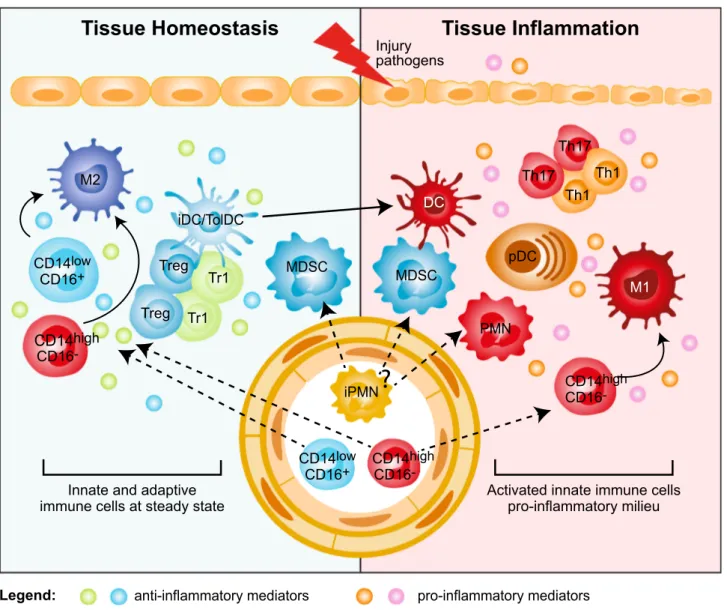
Tissue Homeostasis
Tissue Inflammation
Injury pathogens
M2
DC
Innate and adaptive immune cells at steady state
Legend: M1 Th1 Th1 Th17 Th17 Treg Tr1 Tr1 Treg
anti-inflammatory mediators pro-inflammatory mediators MDSC MDSC PMN pDC iDC/TolDC iPMN CD14low CD16+ CD14high CD16 -CD14low CD16+ CD14CD16high
-Activated innate immune cells pro-inflammatory milieu
CD14high CD16
-?
Fig. 1 Myeloid regulatory cell contribution in tissue homeostasis and
inflammation. Several subsets of myeloid regulatory cells (MRCs) are involved in preventing uncontrolled responses, in maintaining tissue homeostasis, and in promoting resolution of inflammation. Tissue homeostasis. Tissue-resident non-inflammatory M2 mac-rophages, immature and specialized DC subsets (iDC/TolDC), and MDSCs promote tissue homeostasis via different mechanisms: (1) secretion of anti-inflammatory mediators, such as IL-10 and TGF-β, and expression of IDO; (2) induction of T-regulatory cells, both FOXP3+ Tregs and Tr1 cells; (3) generation of a non-inflammatory milieu that leads to the differentiation of migrating classical inflam-matory CD14highCD16− and not classical CD14lowCD16+
mono-cytes into anti-inflammatory M2 macrophages, which contribute to T-regulatory cell induction. Tissue inflammation. Upon tissue injury or pathogen entry, PMNs are recruited at the site of inflammation and, by secreting pro-inflammatory mediators, lead to the activation of plasmacytoid DC (pDCs), which consequently release IFN-α. The inflammatory milieu promotes the recruitment of classical inflam-matory CD14highCD16− monocytes to the site of inflammation and their differentiation into pro-inflammatory M1 macrophages, and the activation and maturation of DCs. These cells in turn promote Th1 and Th17 cell responses via secretion of pro-inflammatory cytokines, such as IL-12 and IL-23. It still remains to be clarified whether MDSCs contribute to tissue inflammation
homeostasis, the accumulation of anti-inflammatory mono-cytes/macrophages in the target organ is critical. Moreover, strategies aimed at targeting factors driving the selective dif-ferentiation of migrating monocytes into M2 macrophages or preventing the conversion of tissue-resident M2 into M1 macrophages, as already reported in transplantation settings (see below), may be more effective than blocking the devel-opment of inflammatory cells.
Impact of myeloid‑derived suppressor cells in autoimmunity
MDSCs are a heterogeneous population of myeloid cells with different maturation stages, and the capacity to sup-press immune responses [24]. MDSCs accumulate in the blood, bone marrow, and secondary lymphoid organs of tumor-bearing mice and cancer patients, in whom circulat-ing levels of MDSCs correlate with clinical stage and meta-static burden [24]. In mice, MDSCs are broadly defined as CD11b+Gr-1+ cells, although they comprise subsets known
as granulocytic (PMN)-MDSC (CD11b+Ly6G+Ly6Clow) and
monocytic (M)-MDSC (CD11b+Ly6G−Ly6Chigh) cells.
Sim-ilar to their murine counterpart, human MDSCs comprise two cell subtypes with either granulocyte or monocyte mor-phology. Human PMN-MDSCs (CD11b+CD14−CD15+ or
CD66b+) and M-MDSCs (CD11b+CD14+HLA-DR−CD15−)
are phenotypically overlapping with neutrophils and mono-cytes, respectively. However, these cells are defined as MDSCs as they display immunosuppressive functions [25]. The lack of consensus on specific markers, which would allow precise MDSC identification, and their phenotypic het-erogeneity have generated controversial results regarding the role of MDSC in autoimmune diseases.
In experimental models of autoimmunity, accumulation of PMN-MDSCs in lymphoid and target organs is associ-ated with inhibition of T-cell proliferation and reduction of pro-inflammatory cytokine release [26, 27]. Expansion of PMN-MDSCs has been described in the synovial fluid of RA patients, where they contribute to limiting the expan-sion of autoreactive T-cells [28], and in the peripheral blood of MS patients with active disease. Furthermore, they sup-press the activation and ex vivo proliferation of autologous CD4+ T-cells [27]. Moreover, the proportion of MDSCs,
comprising both PMN-MDSC and M-MDSC, correlated with disease course in EAE: both MDSC subsets decrease significantly during the remitting phase, and MDSCs com-pletely disappear during the chronic phase [29]. Overall, these examples indicate that MDSCs play an important role in limiting inflammation. However, MDSCs have also been associated with increased inflammatory responses in autoimmunity. Indeed, an accumulation of MDSCs with the ability to promote an inflammatory microenvironment and pathogenic Th17 cells has been described in target tissues
in experimental models of RA, EAE, and SLE [24]. Moreo-ver, an increased frequency of circulating M-MDSCs in RA patients and of circulating PMN-MDSCs and M-MDSCs in SLE patients has been correlated with Th17 responses and disease severity [24].
These discrepancies may be explained by the differ-ent strategies and markers used to iddiffer-entify MDSCs. Only recently, suggestions on the standardization of gating strate-gies and markers to be used to distinguish PMN-MDSCs and M-MDSCs have been proposed [25]. Importantly, one of the key characteristics allowing the classification of both PMN-MDSCs and M-PMN-MDSCs is their suppressive activity [24, 25]. However, the lack of consensus on the suppressive assays to be used to assess MDSC regulatory activity, as discussed in [13], has limited their definitive classification to date. Thus, to draw conclusions regarding MDSC contribution in the suppression or induction of autoreactive immune responses, consensus on biomarkers to distinguish MDSCs from other myeloid cell types and to discriminate the different MDSC subsets, and standardized methods to define their suppres-sive properties are warranted.
Role of dendritic cells in promoting/regulating autoreactive T‑cell responses
DCs are professional APCs specialized in the uptake, pro-cessing, and presentation of Ags to T-cells. Conventional, e.g. immunogenic DCs, are involved in the initiation of adaptive immune responses. However, in steady state, these conventional DCs or specialized subsets of DCs, termed tolerogenic (tol)DCs, control tissue homeostasis and induce/ maintain tolerance [30]. Aberrant activation of immunogenic DCs or defects in the function of tolDCs are involved in breaking self-tolerance in autoimmune disease [30].
Accumulation and activation of conventional DCs in target organs promote autoreactive T-cell activation, and contribute to local inflammation in autoimmunity [30]. Increased numbers of activated conventional DCs with the ability to stimulate autoreactive T-cells and to secrete pro-inflammatory cytokines are evident in synovial fluid of RA patients, in the demyelinating regions of the CNS, in psori-atic skin lesions, and in intestinal mucosa in IBD patients. These cells contribute to effector Th1 and Th17 cell activa-tion and disease progression [30].
Tissue-resident conventional DCs, characterized by the expression of specific markers, such as langerin (CD207) in the skin (Langerhans cells) or CD103+ DCs in the
intes-tinal mucosa, perform tolerogenic functions and maintain tissue homeostasis [30]. An additional subset of tolerogenic DCs, are DC-10, characterized by the expression of HLA-G and Ig-like transcript-4 (ILT4) and the ability to promote IL-10-mediated tolerance [31]. These cells are present in secondary lymphoid organs [32] and in human decidua
during pregnancy, where they participate in maintaining fetal–maternal tolerance [33].
The regulatory activity of DCs depends both on their immature state and expression of immune-modulatory factors [e.g., IL-10, TGF-β, indoleamine 2,3-dioxygenase (IDO), aryl-hydrocarbon receptor]. These features are con-trolled and induced by several environmental signals and crosstalk with local immune cells that, as described above, are dysregulated in inflamed tissues (Fig. 1). Therefore, we can speculate that in autoimmunity a pro-inflammatory envi-ronment in the target organ, enriched in immune effector cells, leads to an increased number of inflammatory DCs and a reduced frequency of tolDCs or to a breakdown in tolDC regulatory activities. TolDCs and immunogenic DCs express many overlapping cell-surface markers. Therefore, only functional analysis, e.g., cytokine profile, stimulatory or suppressive activity, can be used to fully define them. The identification of specific biomarkers and consensus on the assays to determine tolDC suppressive activity are critical to better define their role in different autoimmune diseases. This knowledge is required to enable the development of targeted interventions to promote tolDC differentiation, recruitment to sites of inflammation, and maintenance of their regulatory function.
Therapeutic intervention to restore tolerance in autoimmunity
Current therapies to treat autoimmunity are based on sys-temic administration of immunosuppressive drugs. While often leading to the amelioration of symptoms, these drugs can have widespread side effects and, in many cases, do not promote durable disease remission. Alternatively, biological therapies consisting of antibodies targeting pro-inflamma-tory cytokines and their receptors can dampen inflammation and may prevent myeloid cell hyperactivation. While effica-cious, these treatments are expensive and long-term admin-istration can result in loss of response and cumulative side effects. An innovative and challenging approach to control auto-reactive T-cells and restore tolerance in autoimmun-ity is to boost the regulatory arm of the immune system by suppling ex vivo generated MRCs (i.e., MDSCs or tolDCs).
Adoptive transfer of ex vivo isolated or in vitro induced M-MDSCs or PMN-MDSCs in experimental models of RA and EAE ameliorated disease severity by reducing Th1 and Th17 immune responses [24]. Conversely, the therapeu-tic potential of MDSCs in T1D remains an open question: while adoptive transfer of MDSCs cells improved glucose tolerance and insulin resistance [34], in vitro bone marrow (BM)-derived MDSCs cells failed to prevent autoimmunity in vivo [35]. Ag specificity is likely one of the factors con-tributing to these discrepancies, since infusion of MDSCs conferred protection only in the presence of cognate Ag
[36]. The translation of effective MDSC-based therapies into clinical application faces several hurdles: how in vitro induced MDSCs respond to different inflammatory media-tors; whether inflammatory mediators may inhibit MDSCs activity in vivo; and, importantly, whether in vitro induced MDSCs can mature and differentiate into conventional DCs and M1 macrophages, thus acquiring the ability to present autoAgs and exacerbate disease.
Human tolDCs potentially suitable for cell-based therapies can be differentiated in vitro using a plethora of agents [37]. The first clinical trial, performed in T1D patients, demonstrated the safety of this tolDC-based cell therapy approach but with limited effects on the patients’ insulin requirements [38]. Monocyte-derived DCs cultured with a nuclear factor-kB (NF-kB) inhibitor, pulsed with citrullinated peptide Ags, and injected in RA patients sig-nificantly reduced levels of activated effector T-cells and pro-inflammatory cytokines [39]. DCs differentiated in the presence of vitamin D3 and dexamethasone (VitD3/Dexa) injected into the knee joints of RA patients can stabilize dis-ease symptoms [40]. VitD3/Dexa DCs were also safely intra-peritoneally administered to Crohn’s disease patients with active disease, but no clear clinical benefit was observed [41]. A similar approach is currently being used to treat MS patients with active disease: VitD3 DCs will be administered in MS patients via intradermal injection close to cervical lymph nodes (ClinicalTrials.gov identifier: NCT02618902) or directly injected in cervical lymph nodes (TOLERVIT-MS, ClinicalTrials.gov identifier: NCT02903537).
These completed and ongoing studies have demonstrated the safety of the cell-based approach and some clinical ben-efit. However, several questions remain before tolDC-based cell therapies can be routinely used to treat or cure auto-immune disease, including the route of administration, the maintenance of the tolerogenic cell properties in vivo, and the ability to stably present autoAgs to inhibit auto-reactive T-cells while promoting autoAg-specific Tregs, thus re-establishing long-standing tolerance.
Myeloid regulatory cells in allo‑reactive
T‑cell responses
Organ transplantation is the most efficient treatment to replace the loss of organ function in patients suffering from end-stage diseases. Graft rejection remains a major limita-tion of organ transplantalimita-tion. Myeloid cells are involved both in innate non-specific reactions and donor-specific adaptive responses during allograft rejection. Three pathways pro-mote allo-specific T-cell activation after organ transplanta-tion [42]. In the direct pathway, after transplantation, donor APCs, mainly DCs, migrate from the graft into recipient secondary lymphoid organs, where they present alloAgs to
host naive T-cells. These activated T-cells differentiate into effector T-cells, that migrate back to the graft, where they can mediate rejection (Fig. 2). In the indirect pathway, host T-cells are primed in the secondary lymphoid organs by host APCs that uptake alloAg derived from dying migrated donor DCs (Fig. 2) [43]. Finally, in the semi-direct pathway host APCs acquire intact allogeneic MHC-peptide complex from donor APCs by direct cell-to-cell contact or via exosomes, leading to host T-cell stimulation [44]. The direct and the indirect pathway are mainly involved in early acute and chronic graft rejection, respectively.
Current pharmacological approaches to prevent graft rejection rely on long-term and non-specific therapies that result in metabolic toxicity and other undesirable side effects, such as infection and cancer. Consequently, graft survival outcomes are suboptimal. Novel therapeu-tic approaches that target adaptive immune responses are
currently under clinical testing. These approaches include the use of costimulatory blockade, lymphodepletion, or in vivo induction of Tregs. While promising results have been obtained, transplantation tolerance, defined as a state of donor-specific unresponsiveness, remains elusive [45]. This underlines the need of developing alternative tolerance-inducing protocols. In the present section, we discuss the role of different subtypes of MRCs in modulating allograft rejection in experimental models and in patients. Moreover, we discuss MRC-based cell therapy to prevent graft rejection and promote tolerance.
Role of regulatory macrophages in organ transplantation
Historically, transplant immunologists have attempted to develop tolerogenic protocols by targeting the adaptive
CD8+ CD8+ CD8+ CD8+ CD8+ CD8+ CD8+ Treg Treg Treg Treg Treg Tr1 Th1 Th1 Th1 dDC CD8+CD8+ Tr1 Tr1 Tr1 Tr1 M1 hDC Th1 dDC M2 iDC/ToIDC GRAFT
IMMUNITY/ALLOGRAFT REJECTION TOLERANCE
GRAFT
CD14lowCD16
-CD14highCD16
-CD14highCD16- CD14highCD16
-Fig. 2 Myeloid cells in allograft rejection and tolerance. Myeloid cells play a central role in allograft rejection and tolerance induction after transplantation. Immunity/allograft rejection. Donor myeloid DCs (dDCs) migrate to the secondary lymphoid organs and activate recipient allo-reactive effector CD8+ and CD4+ (Th1) cells, which migrate back into the graft where they mediate rejection. Moreover, dying dDCs in the draining lymph nodes release alloAgs, host DCs (hDCs) uptake donor-derived alloAgs and contribute to the activa-tion of alloAgs-specific effector CD8+ and CD4+ (Th1) cells. Within the graft, classical inflammatory CD14highCD16− monocytes are recruited from the circulation and differentiate into M1 macrophages that, by secreting pro-inflammatory mediators, contribute to the
expansion of effector alloAg-specific T-cells. The limited number of T-regulatory cells (Tregs and Tr1 cells) present within the graft is not sufficient to control the massive infiltration of effector alloAg-specific T-cells. Tolerance. The graft microenvironment enriched of anti-inflammatory mediators, including IL-10, TGF-β, and CSF-1, leads to the differentiation of migrating classical inflammatory CD14highCD16− monocytes into anti-inflammatory M2 macrophages, which promote alloAgs-specific T-regulatory (Treg and Tr1) cells. In addition, the recruitment and/or induction of immature and tolero-genic DC subsets (iDC/TolDC) within the graft sustains the expan-sion/induction of alloAgs-specific T-regulatory (Treg and Tr1) cells, leading to long-term transplantation tolerance
immune response. However, macrophage accumulation in the transplanted organ has long been recognized as a feature of allograft rejection. Inflammatory macrophages indeed initiate the allo-response against the transplanted organ [46] and represent a major cell subset during organ rejection (Fig. 2). Recent evidence suggests that mac-rophages are also important during the induction of trans-plantation tolerance [36]. The presence of graft-infiltrating macrophages with immunosuppressive activity has been described in long term surviving transplant recipients and is associated with unresponsiveness to the transplanted organ [47] (Fig. 2). In mice, inflammatory graft-infiltrating macrophages are Ly6Chigh [48], while suppressive
mac-rophages are Ly6Clow [49]. Inflammatory monocytes are
rapidly recruited within the graft, where they can differ-entiate into either immunogenic Ly6Chigh or tolerogenic
Ly6Clow macrophages, depending on the local environment
(Fig. 2). The behavior of migrating inflammatory mono-cytes observed in organ transplantation is similar to that reported in autoimmunity. In both conditions, the local microenvironment dictates the differentiation program of infiltrating monocytes: in inflamed tissues (organs early after transplantation or injured tissues) migrating mono-cytes acquire an inflammatory phenotype and differentiate into M1 or immunogenic DCs, that in turn promote T-cell activation and tissue damage. In steady-state conditions (engrafted transplanted organs or healthy tissues) migrat-ing monocytes differentiate into anti-inflammatory/tolero-genic cells or M2 macrophages and contribute to tissue homeostasis and tolerance induction.
Therapeutic approaches preventing the accumulation of inflammatory monocytes/macrophages in the transplanted organs or improving their differentiation into suppressive cells have given promising results: selective depletion of inflammatory Ly6ChighCCR2high monocytes prolonged
normoglycemia after allogeneic islet transplantation in streptozotocin-induced diabetic mice [50]. Alternatively, tolerogenic treatment with costimulatory blockade allowed inflammatory Ly6Chigh monocytes infiltrating the allograft,
early after transplantation, to differentiate into suppressive Ly6Clow macrophages through a CSF1-dependent
mecha-nism [49]. These pre-clinical experiments show that target-ing either donor- or recipient-derived monocytes represents a promising therapeutic approach to promote long-term graft acceptance in organ transplantation. However, there are no pharmacological agents that target monocyte/macrophages in clinical use. We believe that future experiments should consider the clinical development of immunotherapies that target myeloid cells within transplanted organs. One exam-ple is the potential use of drug-loaded nanoparticles [51]. The identification of new markers that allow specific tar-geting of myeloid cell subsets are required to facilitate the development of such innovative approaches.
Contribution of dendritic cells in promoting/ maintaining tolerance towards allo‑antigens
As discussed above, tolDCs are essential for tolerance in autoimmunity, and recent evidence indicates that they also promote tolerance in the setting of transplantation (Fig. 2). TolDCs prevent pathogenic responses using a large arsenal of mechanisms: they promote T-cell anergy, clonal deletion, and apoptosis; they express and secrete immunomodulatory mediators that generate a pro-tolerogenic microenvironment that supports T-cell unresponsiveness and induction of Tregs (as reviewed in [52]) (Fig. 2). TolDCs generated with low doses of GM-CSF express heme-oxygenase 1 (HO-1), whose engagement prevents allogenic T-cell proliferation [53] and expression of Epstein-Barr virus-induced gene 3 (EBI3), a member of the IL-12 family [54]. Cardiac allograft survival induced by tolDC immunotherapy is abrogated by specific inhibition of HO-1 [55] or anti-EBI3 treatment [54]. These examples demonstrate that tolDCs generated with low doses of GM-CSF modulate T-cell responses and promote allo-graft tolerance via several mechanisms.
The transplantation procedure promotes a pro-inflam-matory environment similar to that observed in the target organs of autoimmunity. In both instances, a hostile environ-ment contributes to a reduction of the frequency of tolDCs and impairment of their immunosuppressive function. How-ever, in contrast to most autoimmune settings, inflamma-tion after organ transplantainflamma-tion is kept under control by the administration of immunosuppressive drugs that prevent immunogenic DC activation and consequently allo-reactive T-cell induction and function. Of note, these medications may also impair the induction and activity of tolDCs. Thus, the development of immunomodulatory agents that prevent inflammatory cell activation while favoring tolDC induction and function is a research priority in the setting of transplan-tation and autoimmune disease.
Cell‑based approaches to promote tolerance after allogeneic transplantation
The results obtained in experimental models of allo-transplantation and in proof-of-principle clinical trials in autoimmune diseases have prompted investigators to apply MRC-based cell approaches in the prevention of organ rejection. Hutchinson et al. developed a CSF1-dependent human suppressive myeloid cell-based medicinal product, called regulatory macrophages (Mregs) [56] and dem-onstrated the feasibility and safety of Mreg administra-tion in a proof-of-principle clinical trial in living-donor renal transplant recipients [56]. A clinical-grade proto-col for the manufacturing of Mregs has been optimized and donor-derived Mregs are currently pre-operatively administered to living-donor kidney transplant recipients
under the umbrella of the ONE study (Clinicaltrials. gov: NCT02085629). The ONE study consortium (http:// www.onest udy.org) is a European initiative of the FP7 7th Framework Programme ofthe European Union that aims at developing and comparing the safety and efficacy of various immunoregulatory cell products, including Mregs and tolDCs, as cell-based therapy in organ transplantation.
TolDCs suitable for cell-based therapy in transplanta-tion can be generated by culturing precursors with several molecules including IL-10, TGF-β, VitD3, low dose of GM-CSF, rapamycin, tacrolimus or Dexa, or by down-regulating costimulatory molecules (“Therapeutic inter-vention to restore tolerance in autoimmunity”). Regardless of the treatment, differentiated tolDCs express low levels of MHC-II and costimulatory molecules, are refractory to maturation, induce allogenic T-cell hypo-responsiveness in a mixed lymphocyte reaction, produce immunomodulatory mediators, and support Treg differentiation and prolifera-tion [57].
The seminal study that led to the use of tolDCs as cell therapy in the field of transplantation stemmed from data demonstrating that adoptive transfer of donor-derived tolDCs prolonged heart graft survival in mice [58]. Since then, several studies in pre-clinical models of transplanta-tion confirmed the immunosuppressive capacity of tolDCs, and, more recently, a meta-analysis showed the effective-ness of tolDCs in prolonging allograft survival [59]. Inter-estingly, donor-derived tolDCs, alone or in combination with immunosuppressive drugs, prolong cardiac allograft survival [52], but it was shown that recipient-derived tolDCs have a superior activity [60]. Adoptive transfer of tolDCs generated with VitD3 and IL-10 prolonged kidney allograft survival in a clinically relevant rhesus macaque model [61]. More recently, Cuturi et al. performed a proof-of-principle clinical trial with tolDCs generated in the presence of a low dose of GM-CSF as immunotherapy in kidney transplantation under the umbrella of the ONE study (Clinicaltrials.gov: NCT02252055).
The feasibility of generating ex vivo tolDCs for cell-based approaches has now been proven. However, the presence of inflammation generated by the transplant procedure and the use of immunosuppressive drugs may hinder the tolerogenic effects of tolDCs [52]. One way to counter this possibility is to inject semi-mature tolDCs, that are more resistant to this inflammatory environment and have been demonstrated to be effective in prolonging organ graft survival [52]. Furthermore, pre-clinical studies demonstrated that co-administration of immunosuppres-sive drugs in combination with tolDCs did not impair their activity (reviewed in [52]). The selection of the optimum immunosuppressive regimen that can sustain tolerance is an important consideration for the clinical application of tolDC-based therapy to prevent graft rejection.
Conclusions and perspectives
Myeloid cells play a pivotal role in regulating innate and adaptive immune responses. They have a dual purpose, they can initiate effective controlled inflammation leading to activation of appropriate protective immune responses and they are involved in the resolution of inflammation and the promotion of tissue homeostasis and tolerance. Failure in either capacity has important consequences potentially leading to pathology. The discovery that several subtypes of myeloid cells with regulatory activity (MRCs) exist in vivo and can be induced in vitro opens the prospect of clinical interventions designed to induce/modulate these cells in vivo and use them as tolerogenic tool to re-estab-lish/promote tolerance in autoimmune diseases and after transplantation. Recently, tolDC- and Mreg-based cell therapies have entered the clinical arena demonstrating the feasibility and safety of the approach. These encourag-ing results support the potential of usencourag-ing other subtypes of MRCs as a tolerogenic cell therapy in clinical practice.
A number of open questions remain regarding MRCs and their contribution to autoimmunity and transplanta-tion: the most important is the identification of the relative contribution of each MRC subset in different pathological settings. The lack of consensus on markers to identify each MRC subset not only makes their identification in patients difficult, but also hampers the ability to specifically target the optimum cell type. In 2014, a European network (Mye-EUNITER, http://www.mye-eunit er.eu/) was initiated under the umbrella of COST (European cooperation in science and technology) with the aim of joining forces to standardize the phenotypical and functional characteriza-tion of MRCs with the overall objective of expediting the application of this knowledge in diagnosis and treatment of a broad spectrum of disease. This is the first European initiative bringing together MRC experts from different research domains: basic, translational and clinical. By creating a forum for knowledge and expertise exchange, Mye-EUNITER will standardize tools to improve MRCs identification and characterization of their role in healthy and pathological conditions. This effort will contribute to increasing our knowledge about these particular subsets of myeloid cells and to identify strategies to target them at best or to use them as a cell-based product.
Acknowledgements All Mye-EUNITER members are acknowledged
for their inspiring discussions.
Author contributions GA, JC, PC, GM, AM, JO, HBO, MP, EJR, JR, YS wrote parts of the manuscript. MCC and SG designed, supervised and coordinated the contributions of the authors and wrote the manu-script. EJR performed English editing. SG and GA revised the final version of the manuscript. All authors checked and approved the final version of this the manuscript.
Funding This work was supported by COST (European Cooperation in Science and Technology) and the COST Action BM1404 Mye-EUNITER (https ://www.mye-eunit er.eu). COST is part of the EU Framework Programme Horizon 2020. H. Barbaros Oral is supported by The Scientific and Technical Research Council of Turkey (TUBI-TAK) (Project no: 114S354). Elizabeth J. Ryan is supported by the Irish Health Research Board (#POR-2013-281). Joanna Cichy was sup-ported by Polish National Science Center grant 2011/02/A/NZ5/00337. Michaela Pekarova was supported by the Ministry of Education, Youth and Sports (no. LTAUSA17160).
Compliance with ethical standards
Conflict of interest The authors declare that they have no conflict of interests.
Open Access This article is distributed under the terms of the Crea-tive Commons Attribution 4.0 International License (http://creat iveco mmons .org/licen ses/by/4.0/), which permits unrestricted use, distribu-tion, and reproduction in any medium, provided you give appropriate credit to the original author(s) and the source, provide a link to the Creative Commons license, and indicate if changes were made.
References
1. Majewski P, Majchrzak-Gorecka M, Grygier B, Skrzeczynska-Moncznik J, Osiecka O, Cichy J (2016) Inhibitors of serine pro-teases in regulating the production and function of neutrophil extracellular traps. Front Immunol 7:261. https ://doi.org/10.3389/ fimmu .2016.00261
2. Diana J, Simoni Y, Furio L, Beaudoin L, Agerberth B, Barrat F, Lehuen A (2013) Crosstalk between neutrophils, B-1a cells and plasmacytoid dendritic cells initiates autoimmune diabetes. Nat Med 19:65–73. https ://doi.org/10.1038/nm.3042
3. Desnues B, Macedo AB, Ordonez-Rueda D, Roussel-Queval A, Malissen B, Bruhns P, Malissen M, Alexopoulou L (2016) The transcriptional repressor Gfi1 prevents lupus autoimmunity by restraining TLR7 signaling. Eur J Immunol 46:2801–2811. https ://doi.org/10.1002/eji.20164 6573
4. Kessenbrock K, Krumbholz M, Schonermarck U, Back W, Gross WL, Werb Z, Grone HJ, Brinkmann V, Jenne DE (2009) Net-ting neutrophils in autoimmune small-vessel vasculitis. Nat Med 15:623–625. https ://doi.org/10.1038/nm.1959
5. Vono M, Lin A, Norrby-Teglund A, Koup RA, Liang F, Lore K (2017) Neutrophils acquire the capacity for antigen presentation to memory CD4(+) T cells in vitro and ex vivo. Blood 129:1991– 2001. https ://doi.org/10.1182/blood -2016-10-74444 1
6. Puga I, Cols M, Barra CM et al (2011) B cell-helper neutrophils stimulate the diversification and production of immunoglobulin in the marginal zone of the spleen. Nat Immunol 13:170–180. https ://doi.org/10.1038/ni.2194
7. Lowes MA, Suarez-Farinas M, Krueger JG (2014) Immunol-ogy of psoriasis. Annu Rev Immunol 32:227–255. https ://doi. org/10.1146/annur ev-immun ol-03271 3-12022 5
8. Majchrzak-Gorecka M, Majewski P, Grygier B, Murzyn K, Cichy J (2016) Secretory leukocyte protease inhibitor (SLPI), a multifunctional protein in the host defense response. Cytokine Growth Factor Rev 28:79–93. https ://doi.org/10.1016/j.cytog fr.2015.12.001
9. Skrzeczynska-Moncznik J, Wlodarczyk A, Zabieglo K, Kapinska-Mrowiecka M, Marewicz E, Dubin A, Potempa J, Cichy J (2012)
Secretory leukocyte proteinase inhibitor-competent DNA deposits are potent stimulators of plasmacytoid dendritic cells: implication for psoriasis. J Immunol 189:1611–1617. https ://doi.org/10.4049/ jimmu nol.11032 93
10. Caielli S, Athale S, Domic B et al (2016) Oxidized mitochon-drial nucleoids released by neutrophils drive type I interferon production in human lupus. J Exp Med 213:697–713. https ://doi. org/10.1084/jem.20151 876
11. Carmona-Rivera C, Kaplan MJ (2013) Low-density granulo-cytes: a distinct class of neutrophils in systemic autoimmun-ity. Semin Immunopathol 35:455–463. https ://doi.org/10.1007/ s0028 1-013-0375-7
12. Rosales C (2018) Neutrophil: a cell with many roles in inflam-mation or several cell types? Front Physiol 9:113. https ://doi. org/10.3389/fphys .2018.00113
13. Bruger AM, Dorhoi A, Esendagli G et al (2018) How to measure the immunosuppressive activity of MDSC: assays, problems and potential solutions. Cancer Immunol Immunother. https ://doi. org/10.1007/s0026 2-018-2170-8
14. Ziegler-Heitbrock L, Ancuta P, Crowe S et al (2010) Nomencla-ture of monocytes and dendritic cells in blood. Blood 116:e74– e80. https ://doi.org/10.1182/blood -2010-02-25855 8
15. Ginhoux F, Guilliams M (2016) Tissue-resident macrophage ontogeny and homeostasis. Immunity 44: 439–449. https ://doi. org/10.1016/j.immun i.2016.02.024
16. Morell M, Varela N, Maranon C (2017) Myeloid populations in systemic autoimmune diseases. Clin Rev Allergy Immunol 53:198–218. https ://doi.org/10.1007/s1201 6-017-8606-7 17. Amoruso A, Sola D, Rossi L, Obeng JA, Fresu LG, Sainaghi
PP, Pirisi M, Brunelleschi S (2016) Relation among anti-rheu-matic drug therapy, CD14(+)CD16(+) blood monocytes and disease activity markers (DAS28 and US7 scores) in rheumatoid arthritis: a pilot study. Pharmacol Res 107:308–314. https ://doi. org/10.1016/j.phrs.2016.03.034
18. Yoon BR, Yoo SJ, Choi Y, Chung YH, Kim J, Yoo IS, Kang SW, Lee WW (2014) Functional phenotype of synovial mono-cytes modulating inflammatory T-cell responses in rheumatoid arthritis (RA). PloS One 9:e109775. https ://doi.org/10.1371/ journ al.pone.01097 75
19. Bain CC, Scott CL, Uronen-Hansson H et al (2013) Resident and pro-inflammatory macrophages in the colon represent alternative context-dependent fates of the same Ly6Chi mono-cyte precursors. Mucosal Immunol 6:498–510. https ://doi. org/10.1038/mi.2012.89
20. Waschbisch A, Schroder S, Schraudner D et al (2016) Pivotal role for CD16+ monocytes in immune surveillance of the central nerv-ous system. J Immunol 196:1558–1567. https ://doi.org/10.4049/ jimmu nol.15019 60
21. McInnes IB, Schett G (2011) The pathogenesis of rheumatoid arthritis. N Engl J Med 365:2205–2219. https ://doi.org/10.1056/ NEJMr a1004 965
22. Vogel DY, Vereyken EJ, Glim JE et al (2013) Macrophages in inflammatory multiple sclerosis lesions have an intermedi-ate activation status. J Neuroinflammation 10:35. https ://doi. org/10.1186/1742-2094-10-35
23. Vos AC, Wildenberg ME, Arijs I et al (2012) Regulatory mac-rophages induced by infliximab are involved in healing in vivo and in vitro. Inflamm Bowel Dis 18:401–408. https ://doi.org/10.1002/ ibd.21818
24. Veglia F, Perego M, Gabrilovich D (2018) Myeloid-derived sup-pressor cells coming of age. Nat Immunol 19:108–119. https :// doi.org/10.1038/s4159 0-017-0022-x
25. Bronte V, Brandau S, Chen SH et al (2016) Recommendations for myeloid-derived suppressor cell nomenclature and characteriza-tion standards. Nat Commun 7:12150. https ://doi.org/10.1038/ ncomm s1215 0
26. Fujii W, Ashihara E, Hirai H et al (2013) Myeloid-derived sup-pressor cells play crucial roles in the regulation of mouse col-lagen-induced arthritis. J Immunol 191:1073–1081. https ://doi. org/10.4049/jimmu nol.12035 35
27. Ioannou M, Alissafi T, Lazaridis I et al (2012) Crucial role of granulocytic myeloid-derived suppressor cells in the regula-tion of central nervous system autoimmune disease. J Immunol 188:1136–1146. https ://doi.org/10.4049/jimmu nol.11018 16 28. Kurko J, Vida A, Glant TT, Scanzello CR, Katz RS, Nair A,
Szekanecz Z, Mikecz K (2014) Identification of myeloid-derived suppressor cells in the synovial fluid of patients with rheumatoid arthritis: a pilot study. BMC Musculoskelet Disord 15:281. https ://doi.org/10.1186/1471-2474-15-281
29. Moline-Velazquez V, Cuervo H, Vila-Del Sol V, Ortega MC, Clemente D, de Castro F (2011) Myeloid-derived suppres-sor cells limit the inflammation by promoting T lymphocyte apoptosis in the spinal cord of a murine model of multiple sclerosis. Brain Pathol 21:678–691. https ://doi.org/10.111 1/j.1750-3639.2011.00495 .x
30. Amodio G, Gregori S (2012) Dendritic cells: a doubleedge sword in autoimmune responses. Front Immunol. 3. https ://doi. org/10.3389/fimmu .2012.00233
31. Amodio G, Comi M, Tomasoni D, Gianolini ME, Rizzo R, Lemaoult J, Roncarolo MG, Gregori S (2015) Hla-g expres-sion levels influence the tolerogenic activity of human DC-10. Haematologica 100:548–557. https ://doi.org/10.3324/haema tol.2014.11380 3
32. Gregori S, Tomasoni D, Pacciani V, Scirpoli M, Battaglia M, Magnani CF, Hauben E, Roncarolo MG (2010) Differentiation of type 1 T regulatory cells (Tr1) by tolerogenic DC-10 requires the IL-10-dependent ILT4/HLA-G pathway. Blood 116:935– 944. https ://doi.org/10.1182/blood -2009-07-23487 2
33. Amodio G, Mugione A, Sanchez AM, Vigano P, Candiani M, Somigliana E, Roncarolo MG, Panina-Bordignon P, Gregori S (2013) HLA-G expressing DC-10 and CD4(+) T cells accu-mulate in human decidua during pregnancy. Hum Immunol 74:406–411. https ://doi.org/10.1016/j.humim m.2012.11.031 34. Xia S, Sha H, Yang L, Ji Y, Ostrand-Rosenberg S, Qi L (2011)
Gr-1+ CD11b+ myeloid-derived suppressor cells suppress inflammation and promote insulin sensitivity in obesity. J Biol Chem 286:23591–23599. https ://doi.org/10.1074/jbc. M111.23712 3
35. Drujont L, Carretero-Iglesia L, Bouchet-Delbos L, Beriou G, Merieau E, Hill M, Delneste Y, Cuturi MC, Louvet C (2014) Evaluation of the therapeutic potential of bone marrow-derived myeloid suppressor cell (MDSC) adoptive transfer in mouse mod-els of autoimmunity and allograft rejection. PloS One 9:e100013. https ://doi.org/10.1371/journ al.pone.01000 13
36. Garcia MR, Ledgerwood L, Yang Y et al (2010) Monocytic sup-pressive cells mediate cardiovascular transplantation tolerance in mice. J Clin Investig 120:2486–2496. https ://doi.org/10.1172/ JCI41 628
37. Boks MA, Kager-Groenland JR, Haasjes MS, Zwaginga JJ, van Ham SM, ten Brinke A (2012) IL-10-generated tolerogenic den-dritic cells are optimal for functional regulatory T cell induction— a comparative study of human clinical-applicable DC. Clin Immu-nol 142:332–342. https ://doi.org/10.1016/j.clim.2011.11.011 38. Giannoukakis N, Phillips B, Finegold D, Harnaha J, Trucco M
(2011) Phase I (safety) study of autologous tolerogenic dendritic cells in type 1 diabetic patients. Diabetes Care 34:2026–2032. https ://doi.org/10.2337/dc11-0472
39. Benham H, Nel HJ, Law SC et al (2015) Citrullinated peptide dendritic cell immunotherapy in HLA risk genotype-positive rheumatoid arthritis patients. Sci Transl Med 7:290ra87. https :// doi.org/10.1126/scitr anslm ed.aaa93 01
40. Hilkens CM, Isaacs JD (2013) Tolerogenic dendritic cell therapy for rheumatoid arthritis: where are we now? Clin Exp Immunol 172:148–157. https ://doi.org/10.1111/cei.12038
41. Jauregui-Amezaga A, Cabezon R, Ramirez-Morros A et al (2015) Intraperitoneal administration of autologous tolerogenic dendritic cells for refractory Crohn’s disease: a phase I study. J Crohns Colitis 9:1071–1078. https ://doi.org/10.1093/ecco-jcc/jjv14 4 42. Ochando JC, Krieger NR, Bromberg JS (2006) Direct versus
indi-rect allorecognition: visualization of dendritic cell distribution and interactions during rejection and tolerization. Am J Transplant 6:2488–2496. https ://doi.org/10.1111/j.1600-6143.2006.01494 .x 43. Celli S, Albert ML, Bousso P (2011) Visualizing the innate and
adaptive immune responses underlying allograft rejection by two-photon microscopy. Nat Med 17:744–749. https ://doi.org/10.1038/ nm.2376
44. Liu H, Gao W, Yuan J et al (2016) Exosomes derived from den-dritic cells improve cardiac function via activation of CD4(+) T lymphocytes after myocardial infarction. J Mol Cell Cardiol 91:123–133. https ://doi.org/10.1016/j.yjmcc .2015.12.028 45. Espinosa JR, Samy KP, Kirk AD (2016) Memory T cells in
organ transplantation: progress and challenges. Nat Rev Nephrol 12:339–347. https ://doi.org/10.1038/nrnep h.2016.9
46. Oberbarnscheidt MH, Zeng Q, Li Q, Dai H, Williams AL, Shlom-chik WD, Rothstein DM, Lakkis FG (2014) Non-self recogni-tion by monocytes initiates allograft rejecrecogni-tion. J Clin Investig 124:3579–3589. https ://doi.org/10.1172/JCI74 370
47. Ochando J, Conde P, Bronte V (2015) Monocyte-derived suppres-sor cells in transplantation. Curr Transplant Rep 2:176–183. https ://doi.org/10.1007/s4047 2-015-0054-9
48. Swirski FK, Wildgruber M, Ueno T et al (2010) Myeloperoxidase-rich Ly-6C+ myeloid cells infiltrate allografts and contribute to an imaging signature of organ rejection in mice. J Clin Investig 120:2627–2634. https ://doi.org/10.1172/JCI42 304
49. Conde P, Rodriguez M, van der Touw W et al (2015) DC-SIGN(+) macrophages control the induction of transplantation tolerance. Immunity 42:1143–1158. https ://doi.org/10.1016/j. immun i.2015.05.009
50. Ochando J, Braza MS (2017) Nanoparticle-based modulation and monitoring of antigen-presenting cells in organ transplan-tation. Front Immunol 8:1888. https ://doi.org/10.3389/fimmu .2017.01888
51. Fisher JD, Acharya AP, Little SR (2015) Micro and nanoparticle drug delivery systems for preventing allotransplant rejection. Clin Immunol 160:24–35. https ://doi.org/10.1016/j.clim.2015.04.013 52. Moreau A, Alliot-Licht B, Cuturi MC, Blancho G (2017) Tolero-genic dendritic cell therapy in organ transplantation. Transpl Int 30:754–764. https ://doi.org/10.1111/tri.12889
53. Moreau A, Chiffoleau E, Beriou G et al (2008) Superiority of bone marrow-derived dendritic cells over monocyte-derived ones for the expansion of regulatory T cells in the macaque. Transplanta-tion 85:1351–1356. https ://doi.org/10.1097/TP.0b013 e3181 6f22d 6
54. Hill M, Thebault P, Segovia M et al (2011) Cell therapy with autologous tolerogenic dendritic cells induces allograft toler-ance through interferon-gamma and epstein-barr virus-induced gene 3. Am J Transplant 11:2036–2045. https ://doi.org/10.111 1/j.1600-6143.2011.03651 .x
55. Moreau A, Hill M, Thebault P et al (2009) Tolerogenic dendritic cells actively inhibit T cells through heme oxygenase-1 in rodents and in nonhuman primates. FASEB J 23:3070–3077. https ://doi. org/10.1096/fj.08-12817 3
56. Hutchinson JA, Riquelme P, Sawitzki B et al (2011) Cutting edge: immunological consequences and trafficking of human regulatory macrophages administered to renal transplant recipients. J Immu-nol 187:2072–2078. https ://doi.org/10.4049/jimmu nol.11007 62
57. Moreau A, Varey E, Beriou G, Hill M, Bouchet-Delbos L, Segovia M, Cuturi MC (2012) Tolerogenic dendritic cells and negative vaccination in transplantation: from rodents to clinical trials. Front Immunol 3:218. https ://doi.org/10.3389/fimmu .2012.00218 58. Fu F, Thai NL, Li Y, Lu L, Thomson AW, Fung JJ, Qian S (1996)
Second-set rejection of mouse liver allografts is dependent on radiation-sensitive nonparenchymal cells of graft bone marrow origin. Transplantation 61:1228–1233
59. Zhou Y, Shan J, Guo Y, Li S, Long D, Li Y, Feng L (2016) Effects of adoptive transfer of tolerogenic dendritic cells on allo-graft survival in organ transplantation models: an overview of systematic reviews. J Immunol Res 2016:5730674. https ://doi. org/10.1155/2016/57306 74
60. Peche H, Trinite B, Martinet B, Cuturi MC (2005) Prolongation of heart allograft survival by immature dendritic cells generated from recipient type bone marrow progenitors. Am J Transplant 5:255–267. https ://doi.org/10.1111/j.1600-6143.2004.00683 .x 61. Ezzelarab MB, Lu L, Guo H, Zahorchak AF, Shufesky WF,
Cooper DK, Morelli AE, Thomson AW (2016) Eomesodermin(lo) CTLA4(hi) alloreactive CD8+ memory T cells are associated with prolonged renal transplant survival induced by regulatory den-dritic cell infusion in CTLA4 immunoglobulin-treated nonhuman primates. Transplantation 100:91–102
Affiliations
Giada Amodio1 · Joanna Cichy2 · Patricia Conde3 · Gianluca Matteoli4 · Aurélie Moreau5,6 ·
Jordi Ochando3 · Barbaros H. Oral7 · Michaela Pekarova8 · Elizabeth J. Ryan9 · Johannes Roth10 ·
Yahya Sohrabi11 · Maria‑Cristina Cuturi5,6 · Silvia Gregori1
1 San Raffaele Telethon Institute for Gene Therapy (SR-Tiget), San Raffaele Scientific Institute IRCCS, Via Olgettina, 58, 20132 Milan, Italy
2 Faculty of Biochemistry, Biophysics and Biotechnology, Jagiellonian University, Krakow, Poland
3 Centro Nacional de Microbiologia, Instituto de Salud Carlos III, Majadahonda, 28220 , Madrid, Spain
4 Translational Research in Gastrointestinal Disorders (TARGID), Department of Chronic Diseases, Metabolism and Ageing, KU Leuven, Leuven, Belgium
5 Centre de Recherche en Transplantation et Immunologie UMR1064, INSERM, Université de Nantes, Nantes, France 6 Institut de Transplantation Urologie Nephrologie (ITUN),
CHU Nantes, Nantes, France
7 Department of Immunology, Faculty of Medicine, Uludag University, Bursa, Turkey
8 Institute of Biophysics, The Czech Academy of Sciences, Brno, Czech Republic
9 Department of Biological Sciences, Faculty of Science and Engineering, University of Limerick, Limerick, Ireland 10 Institute of Immunology, University of Münster, Münster,
Germany
11 Molecular and Translational Cardiology, Department of Cardiovascular Medicine, University Hospital Münster, Münster, Germany