In vivo effect of deoxynivalenol (DON) naturally contaminated feed on
1porcine reproductive and respiratory syndrome virus (PRRSV)
2infection.
34
Christian Savard1, Vicente Pinilla2,Chantale Provost1, Carl A. Gagnon1, Younes Chorfi2.
5
1 Groupe de recherche sur les maladies infectieuses du porc (GREMIP);
6
Swine and Poultry Infectious Diseases Research Center (CRIPA), Saint-Hyacinthe,
7
Québec, Canada
8
2 Département de biomédecine vétérinaire, Saint-Hyacinthe, Québec, Canada.
9 10 Corresponding authors 11 Younes.chorfi@umontreal.ca 12
Faculté de médecine vétérinaire, Université de Montréal, 3200 rue Sicotte,
Saint-13
Hyacinthe, Québec, Canada, J2S 7C6
14
*Revised manuscript without changes marked (clean version) Click here to view linked References
Abstract
1
Deoxynivalenol (DON), also known as vomitoxin, is the most prevalent type B
2
trichothecene mycotoxin worldwide. Pigs show a great sensitivity to DON, and because
3
of the high proportion of grains in their diets, they are frequently exposed to this
4
mycotoxin. The objective of this study was to determine the impact of DON naturally
5
contaminated feed on porcine reproductive and respiratory syndrome virus (PRRSV)
6
infection, the most important porcine viral pathogens in swine. Experimental infections
7
were performed with 30 animals. Piglets were randomly divided into three groups of 10
8
animals based on DON content of diets (0, 2.5 and 3.5 mg/Kg DON). All experimental
9
groups were further divided into subgroups of 6 pigs and were inoculated with PRRSV.
10
The remaining pigs (control) were sham-inoculated with PBS. Pigs were daily monitored
11
for temperature, weight and clinical signs for 21 days. Blood samples were collected and
12
tested for PRRSV RNA and for virus specific antibodies. Results of PRRSV infection
13
showed that ingestion of diet highly contaminated with DON greatly increases the effect
14
of PRRSV infection on weight gain, lung lesions and mortality, without increasing
15
significantly viral replication, for which the tendency is rather directed towards a
16
decrease of replication. These results suggest that PRRSV infection could exacerbate
17
anorectic effect of DON, when ingested in large doses. Results also demonstrate a DON
18
negative effect on PRRSV-specific humoral responses. This study demonstrate that high
19
concentrations of DON naturally contaminated feed decreased the immune response
20
against PRRSV and influenced the course of PRRSV infection in pigs.
21
Keywords: Pig; DON mycotoxin; PRRSV; predisposition to infection
1. Introduction
1
Various commodities for animal feeding are frequently contaminated with mycotoxins
2
produced by the secondary metabolism of diverse strains of filamentous fungi. Among
3
them, Fusarium spp. are the most prevalent mycotoxin producing fungi in temperate
4
regions (Binder et al., 2007). Several toxins are produced by Fusarium spp. including
5
trichothecenes deoxynivalenol (DON), nivalenol, and T-2 toxin. Fusarium spp. also
6
produces other toxins such as zearalenone (ZEA), fumonisin B1 (FB1), beauvericin and
7
enniatins (Glenn, 2007). Consequently, Fusarium spp. naturally infected cereals are
8
frequently contaminated with low levels of several different mycotoxins (Binder et al.,
9
2007).
10
DON, also known as vomitoxin, is the most encountered mycotoxin contaminating cereal
11
worldwide including Canada (Tran et al., 2012). Among farm animals, pigs are the most
12
sensitive animals to DON; dietary concentrations between 2 to 5 mg DON/kg are
13
frequently associated with feed refusal and concentrations over 20 mg DON/kg induce
14
vomiting (Bryden, 2012). DON has a unique effect on immune system as it has the
15
capacity to up and down regulate immune function depending on dose, exposure
16
frequency, timing and the functional immune assay being employed (Pestka, 2008). The
17
molecular mechanism of action of DON imply phosphorylation of mitogen-activated
18
protein kinases (MAPKs) which in turn modulates expression of genes associated with
19
immune response, inflammation and apoptosis. Leukocytes are among the most sensitive
20
cells to DON effect as low concentration of this toxin upregulates immune and
21
inflammatory genes and high concentration triggers cell death, typically by apoptosis,
22
which leads to immunosuppression (Pestka et al., 2004). Immunosuppression engendered
by DON has the potential to decrease resistance to infectious diseases (Oswald et al.,
1
2005). Previous reports in mice have shown that DON could increase reovirus
2
replication, a double stranded RNA virus, in enteric (Li et al., 2005) and respiratory (Li et
3
al., 2007) infection models. In this latest model, DON exacerbated viral-induced
4
inflammation and pulmonary damage by suppressing type-1 interferon (IFN) response
5
and elevating expression of proinflammatory cytokines (Li et al., 2007). DON has also
6
been shown to modulate the virulence-dependent pathogenesis of infectious bursal
7
disease virus (IBDV) in infected broiler (Danicke et al., 2011). In that study, Fusarium
8
contaminated diet, containing predominantly DON at 10.7 mg/kg of diet, significantly
9
increased histopathological lesions of IBDV infected birds. Previous report showed that
10
oral administration of purified FB1 significantly increased the severity of pulmonary
11
lesions following porcine reproductive and respiratory syndrome virus (PRRSV)
12
infection (Ramos et al., 2010). Up to date, no study has reported an interaction of DON
13
with swine viral infections. However, our recent in vitro results showed that doses over
14
140 ng/mL of DON could inhibit PRRSV replication (Savard et al., 2014).
15
Swine industry faces many diseases that threaten animal health and the economy of the
16
industry. Among these diseases, PRRS represents the most economically important viral
17
disease of swine industry in North America (Holtkamp et al., 2013). PRRSV causes
18
common clinical signs such as anorexia, fever, and lethargy. In sows, PRRSV is
19
responsible of reproductive failure, characterized by late-term abortions, increased
20
numbers of stillborn fetuses, and/or premature, weak pigs. Furthermore, PRRSV is
21
responsible of respiratory problems in growing and finishing pigs. Respiratory problems
22
induce by PRRSV are usually more severe in young piglets and often aggravated by
infections with bacterial and viral pathogens (Chand et al., 2012; Dorr et al., 2007).
1
PRRSV is an enveloped, single stranded, positive sense RNA virus belonging to the
2
Arteriviridae viral family, which includes lactate dehydrogenase-elevating virus (LDV)
3
of mice, simian hemorrhagic fever virus (SHFV) and equine arteritis virus (EAV)
4
(Meulenberg et al., 1994).
5
Pigs are frequently exposed to DON because of their cereal-rich diet that is frequently
6
contaminated by Fusarium spp. mycotoxins. Chronic exposure of pigs to DON could
7
impair immunity and decrease resistance to infectious diseases, on the other hand in vitro
8
exposure to DON could inhibit PRRSV replication. Therefore the objective of this study
9
was to evaluate the impact of DON naturally contaminated feed on PRRSV in vivo
10
infection.
11
2. Materials and Methods.
1
2.1. Animals
2
The experiment was conducted at the Faculté de médecine vétérinaire, Université de
3
Montréal. Animal care procedures followed the guidelines of the Canadian Council on
4
Animal Care and the protocol was approved by the Institutional Animal Care Committee
5
(Protocol #11-Rech-1609). Thirty commercial crossbred piglets, negative for PRRSV
6
were purchased locally at 4 weeks of age. After one week of acclimation on a commercial
7
ration, piglets were randomly divided into 3 experimental groups of 10 animals, housed
8
separately and fed naturally contaminated diets containing 0, 2.5 or 3.5 mg/kg of DON
9
for all the duration of the experiment.
10
2.2. Experimental diets
11
Experimental diets (Table 1) were formulated according to the energy and amino acid
12
requirements for piglets as previously described in the National Swine Nutrition Guide
13
(2010). Wheat used in experimental diets was naturally contaminated with DON. Dietary
14
contents of mycotoxins (Table 2) were analysed in the final diet through
ultra-15
performance liquid chromatography/electrospray ionization tandem mass spectrometry,
16
based on method of (Jackson et al., 2012).
17
2.3. PRRSV challenge strain and experimental infection
18
PRRSV isolate used in this study was FMV12-1425619 (Genbank accession number
19
KJ888950), obtained from one serum sample originating from a PRRS clinical case.
20
Based on ORF5 phylogenic analyses, this strain, often associated with clinical signs
21
reported from the field, was classified within a lineage 1 of type II genotype cluster that is
22
frequently found in Quebec over the past 2 years (data not shown). Since several different
attempts to isolate the virus have failed, the viral inoculum was obtained from a lung
1
tissue homogenate after infection of piglet with 3 mL of PRRSV positive serum, 2 mL
2
intranasally (i.n) and 1 mL intramuscularly (i.m), containing 1.7 X 10³ TCID50/mL
3
PRRSV. PRRSV concentration in filtrated lung tissue homogenate was determined by
4
RT-qPCR to be 1.5 X 10⁴ TCID50/mL. It was also determined by PCR that the tissue
5
homogenate was negative for bacteria (with a 16S gene amplification by PCR) (Cai et al.,
6
2003), swine influenza virus (Tremblay et al., 2011), porcine parvovirus (Gagnon et al.,
7
2007) and porcine circovirus (Gagnon et al., 2008). A pilot study using four animals
8
confirmed the capacity of the virus containing inoculum to induce PRRSV-specific
9
clinical signs, viremia, and lung lesions (data not shown).
10
After 2 weeks on the experimental diets, all experimental groups were further subdivided
11
in groups of 6 pigs, kept in separated rooms and inoculated i.m with 1mL containing
12
1.5x10⁴ TCID50 PRRSV and i.n with 1mL of the same inoculums in each nostril.
13
Remaining pigs of each experimental groups (4) were housed separately and were
sham-14
inoculated with PBS buffer.
15
2.4. Body weight, rectal temperature and blood collection.
16
Pigs were daily monitored for rectal temperature, body weight and clinical signs for 21
17
days post-infection (p.i). The average daily gain (ADG) was calculated by subtracting the
18
initial body weight of the final body weight and divided by the number of experimental
19
days. Fever was defined as body temperature higher than 40˚C for two consecutive days.
20
Blood samples were collected at days -1, 3, 6, 9, 14 and 21 p.i and PRRSV viremia was
21
evaluated by RT-qPCR and serological response by ELISA. Serum samples were stored
22
frozen for further analysis.
2.5. Macroscopic and microscopic lung lesions evaluation.
1
Pigs were euthanized on day 21 p.i, and macroscopic lung lesion scores were recorded to
2
estimate the percentage of lung affected by pneumonia (scores vary from 0 to 100%).
3
Each lung lobe was assigned a percentage to reflect the approximate volume of the lobe
4
on the entire lung, based on lung schematisation of (Sorensen et al., 2006). Lung samples
5
and tracheobronchial lymph nodes were fixed in 10% neutral buffered formalin to
6
evaluate specific microscopic lesions. Lung samples were collected to evaluate viral load
7
and stored frozen until tested. Lung sections were scored for severity of interstitial
8
pneumonia as follows 0= normal, 1= mild, 2=moderate, 3= severe, 4= severe with
9
alveolar disappearance. Presence of leucocytes, serum, or necrotic debris in alveolar
10
exsudate were also scored as follows 0=normal, 0.5 rare, 1= mild, 2= moderate, 3=
11
important, and 4= severe. Finally lymphoid follicular hyperplasia were scored as follows
12
0=normal, 1=mild, 2=moderate, 3= severe.
13
2.6. PRRSV quantification
14
Sera and lung homogenates were analyzed for the presence of PRRSV RNA using
RT-15
qPCR assay as described by (Gagnon et al., 2008). Lung tissues were weighed, an equal
16
volume of PBS added to sample and tissues were homogenized before viral RNA
17
isolation. QIAamp Viral RNA kit (Qiagen) was used to isolate viral RNA from serum
18
samples and lung homogenates according to the manufacturer’s instructions. A
19
commercial PRRSV RT-qPCR diagnostic kit (NextGen, Tetracore Inc., Gaithersburg,
20
MD, USA) was used for PRRSV quantification as recommended by the manufacturer.
21
The quantification of PRRSV was determined by comparing the sample results with a
22
standard curve based on the amount of serially diluted PRRSV IAF-Klop reference strain
produced in MARC-145 cells and titrated as TCID50/mL in the MARC-145-infected cell
1
(Gagnon et al., 2008). The PRRSV RT-qPCR results were expressed in TCID50/mL of
2
serum or g of lung tissues.
3
2.7. PRRSV specific antibodies
4
Sera were assayed for virus-specific antibody by ELISA with the Herdchek PRRS X3
5
diagnostic kits (IDEXX Laboratories, Portland, Maine, USA). Serum were diluted 1/40 in
6
diluents supplied by the manufacturer and the assay was performed following the
7
manufacturers' instructions. A sample-to-positive (S:P) ratio equal or greater than 0.4 was
8
considered positive.
9
2.8. Statistical analysis
10
Results are expressed as the mean ± SEM. All statistical analyses were performed using
11
GraphPad Prism software (version 5.03, GraphPad Prism software Inc., San Diego, CA).
12
Data were statistically analysed using a one-way ANOVA with Dunnett’s multiple
13
comparison test, using animal receiving control diet as control group. For the ADG data,
14
noninfected versus infected animals were compared by applying Student′s unpaired ′t′
15
test, for each DON concentration. For viremia, lung viral load and microscopic lesions,
16
pair-wise mean comparisons between control and DON treated animals were made using
17
Student′s unpaired ′t′ test. P<0.05 was considered to reflect statistically significant
18
differences.
19 20
3. Results
1
3.1. Growth performance
2
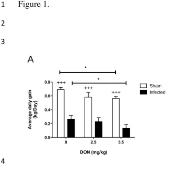
Results of growth performance showed that noninfected pigs had significantly higher
3
ADG than PRRSV infected pigs (Fig. 1A) regardless of DON contamination of the diets.
4
Severe growth retardation was observed following PRRSV infection suggesting a high
5
degree of virulence associated to FMV12-1425619 strain used for the experimental
6
infection. Contaminated diet with DON at 3.5 mg/kg decreased significantly (P<0.05) the
7
ADG of noninfected group with a loss of approximately 19% of kg/day compared to
8
uncontaminated noninfected group (Fig. 1A). Same level of contamination in infected
9
animals had severe impact since they had a 40% of ADG less than PRRSV infected
10
animals fed control diet (P<0.05) (Fig. 1A). These data suggest that pigs fed high
11
mycotoxin diet are more affected by PRRSV than those fed uncontaminated diet, and the
12
anorectic effect of DON could be additive to that of PRRSV infection.
13
One PRRSV infected pig in control diet group and one PRRSV infected pig in 2.5-mg/kg
14
group were humanely sacrificed at day 16 and 17 p.i, respectively. At necropsy, lungs of
15
both animals showed PRRSV associated lesions covering over 30% of the organ. At
16
microscopic level, lungs section of both animals showed interstitial pneumonia with
17
presence of leucocytes and necrotic debris in alveolar exudate. Two PRRSV infected pigs
18
in 3.5-mg/kg group were found dead at day 12 and 20 p.i. Due to damage caused by post
19
mortem freezing, the accurate assessment of PRRSV specific lesions has been made
20
difficult. All data from these pigs were excluded from further analysis. Mortality rate due
21
to infection was relatively high, being above 15% for all infected groups (Fig. 1B).
22
Mortality in the PRRSV infected group ingesting 3.5 mg/kg of DON contaminated diet
was at 33% but not statically significant, due to the small number of pigs included in the
1
study. All noninfected pigs survived the experiment (data not shown).
2
3.2. Body temperature, viremia and viral loads in the lungs.
3
Five out of 6 PRRSV infected pigs fed uncontaminated diet and 4 out of 6 fed 2.5 mg/kg
4
diet had fever over 6 days (Fig. 2A). In contrast, no pig from PRRSV infected group
5
receiving diet contaminated at 3.5 mg/kg of DON experienced fever episode.
6
Temperature of all noninfected animals remains normal for all duration of the study (Fig.
7
2B).
8
All experimentally infected pigs, regardless of DON contamination levels of the diet,
9
developed viremia from day 3 p.i to the end of the experiment (Fig. 3A). Viremia was
10
significantly higher in DON contaminated groups early after experimental infection at
11
day 3 p.i for 2.5 mg/kg DON treated group and at day 6 p.i for 3.5 mg/kg DON treated
12
group suggesting an acceleration of viral replication kinetics in presence of DON (Fig.
13
3A). However, DON had a limited impact on viremia after day 9 p.i since viremia was
14
similar between all experimental groups (Fig. 3A). Evaluated at day 21 p.i, viral load in
15
the lungs was also increased in all experimentally infected animals (Fig. 3B) compared to
16
noninfected animals that were negative (data not shown). DON contamination appeared
17
to be associated to PRRSV elimination in the lungs, since viral loads of this organ were
18
respectively 5 and 6 times lower in pigs fed diet 2.5 mg/kg (P=0.0965) and 3.5 mg/kg
19
(P=0.0195) of DON than pigs receiving uncontaminated diet (Fig. 3B).
20
3.3. Macroscopic and microscopic lesions
21
Following necropsy, observations were made of the macroscopic lung lesions of each
22
piglet. Extents of lesions were recorded as percentage of lungs affected by pneumonia.
Macroscopic lesions were observed on most of the lungs of animals that survived the
1
infection. The extent of lung lesions was significantly more important in animals fed 3.5
2
mg/kg DON diets (Fig. 4A) than the other groups. No significant PRRSV-associated lung
3
lesions were observed in noninfected animals (data not shown). Microscopically, lungs of
4
all infected pigs showed a severe interstitial pneumonia. However, infected group fed 3.5
5
mg/kg DON had significantly higher presence of leucocytes, serum, and or necrotic
6
debris in alveolar exsudate (Fig. 4B). Analysis of lymph nodes did not reveal significant
7
changes (data not shown).
8
3.4. Antibody response
9
As demonstrated by measured specific antibodies against PRRSV in the serum, all
10
experimentally infected animals, regardless of DON contamination, had seroconverted
11
(Fig. 5). In animals fed uncontaminated diet, humoral response was significantly lower
12
(P<0.05) in animals ingesting 2.5 mg/kg of DON but was similar to that of animals fed
13
3.5 mg/kg (Fig. 5). Noninfected animals did not PRRSV seroconverted.
4. Discussion.
1
Mycotoxins are frequently detected in different sources of grains designated to animal
2
feeding. Among these mycotoxins, DON is the one that draws the most attention because
3
of its frequent occurrence at levels high enough to cause adverse effects, particularly in
4
pigs. The main clinical effect of DON at lower dietary concentrations is anorexia and
5
decreased weight gain (Danicke et al., 2004; Goyarts et al., 2005; Rotter et al., 1994). The
6
reduced weight gain observed following ingestion of our experimental diets is consistent
7
with what has been previously observed.
8
In this study, growing young piglets have been used as a model for experimental infection
9
with PRRSV that can cause many different clinical manifestations including fever,
10
lethargy and severe pneumonia often complicated by concurrent bacterial infections
11
(Done et al., 1996; Rossow, 1998). But this clinical picture is highly variable, ranging
12
from mild subclinical infection to acute death of infected animals (Mengeling and Lager,
13
2000). Several factors can influence the severity of PRRSV infection including host
14
genetics, management practices, environmental factors, concurrent bacterial infections
15
and virus strain heterogeneity (Goldberg et al., 2000; Halbur et al., 1995; Halbur et al.,
16
1996). Environmental factor such as the presence of DON in animals feed could impact
17
on the severity of PRRS disease following an experimental infection. Like DON, PRRSV
18
has been shown to cause anorexia and reduction of weight gain (Done et al., 1996;
19
Rossow, 1998). Results of experimental infections showed that ingestion of diets highly
20
contaminated with DON greatly increases the effect of PRRSV infection on reduction of
21
weight gain. Both effect appears to be additive.
Nonetheless, animals in this study were fed naturally contaminated diets. It is well known
1
that Fusarium naturally infected cereals are commonly contaminated with low levels of
2
several different mycotoxins (Binder et al., 2007). In this study, FB1 was the second most
3
abundant mycotoxin found after DON. In swine, FB1 can have negative impact on
4
production by causing pulmonary edema, liver failure or cardiovascular toxicity (Haschek
5
et al., 2001). FB1 has also been shown to increase the severity of pulmonary lesions
6
following porcine reproductive and respiratory syndrome virus (PRRSV) infection
7
(Ramos et al., 2010). However, the FB1 concentrations found in our experimental diets
8
are well below levels that may cause clinical signs according to Ramos et al. (2010).
9
However, we cannot exclude that they could, in part, be responsible for the observed
10
effect. Co-occurrence of FB1 and DON in animal feedstuffs is common and it has been
11
shown that subclinical co-exposure of pigs to these toxins resulted in greater immune
12
suppression than exposure of a single toxin (Grenier and Oswald, 2011). A significant
13
amount of DON was also found in a conjugated form of DON, the DON-3-glucoside. It is
14
not surprising to found this form of conjugate because it was estimated that this conjugate
15
could constitute up to 20% of the total content of its mycotoxin precursor (Berthiller et
16
al., 2006). Hydrolysis of this conjugate, following ingestion, may thus increase exposure
17
to the precursor toxin.
18
In addition to anorexia, DON has also demonstrated a unique ability to up- and
down-19
regulate immune functions (Pestka et al., 2004). As consequence, DON has been shown
20
to exacerbate some viral infection in mice (Li et al., 2007; Li et al., 2005) and in broiler
21
chicken (Danicke et al., 2011). Here, results of the study showed an increase of viremia
22
during the first days’ post-infection following chronic ingestion of DON. This suggests
that DON could accelerate the kinetics of viral replication by an unknown mechanism.
1
This result is in contradiction with our previous in vitro study showing that DON, at
2
concentration over 140 ng/mL could inhibit PRRSV replication (Savard et al., 2014).
3
This could be explained by the fact that in vivo exposure to DON in this study was not the
4
same under in vitro conditions. Indeed, it was previously shown that the maximum blood
5
level of DON was approximately 20 ng/mL following ingestion of contaminated diet at
6
4.5 mg / kg of DON (Goyarts and Danicke, 2006), this level similar to the highest
7
contamination doses used in the present study, which is probably insufficient to have the
8
DON inhibitory effect. However, the viremia was not modulated by DON, at the end of
9
the viremic period, between days 9 and 21 p.i. Viral load in the lungs, observed at 21
10
days p.i was significantly decreased by chronic consumption of DON. This suggests that
11
virus elimination could be accelerated in the presence of DON. This is in contradiction
12
with previous reports which reported an increased lung and intestinal reovirus burden and
13
suppression of viral clearance in mice (Li et al., 2007). Unlike its effect on blood viremia,
14
DON is more inhibitor on viral load in the lungs, which is in agreement with our previous
15
study. PRRSV is responsible of specific lung lesions that vary from no apparent to severe
16
tan consolidation lesions that are frequently aggravated by lesions resulting from
17
concurrent bacterial infection (Rossow, 1998). Here, PRRSV specific macroscopic lung
18
lesions were significantly increased by the chronic absorption of DON, when present in
19
larger quantity. Since lung viral load appears to be lower in DON exposed animals, the
20
increased clinical effects in these groups could not be explain by increased viral
21
replication. To some extent, the increased clinical and pathological effects of PRRSV can
22
be caused by enhanced inflammatory immune response rather than the higher levels of
virus replication (Morgan et al., 2013; van Reeth and Nauwynck, 2000). Since DON has
1
been previously shown to stimulate the expression of pro-inflammatory cytokines in
2
lungs of mice (Amuzie et al., 2008), excess production of pro-inflammatory cytokines
3
following DON exposure can possibly explain the higher clinical and pathological effects
4
seen in PRRSV infected animal fed high DON diet. Further investigations will be needed
5
to confirm this hypothesis. Microscopically, PRRSV-specific lung lesions were
6
characterized by septal tickening and presence of alveolar necrotic debris, macrophages
7
and other mononuclear cells. Higher level of DON contamination significantly increases
8
the presence of alveolar necrotic debris, macrophages and other mononuclear cells. These
9
results are consistent with the previous hypothesis.
10
PRRSV infections normally induce an abundant virus-specific antibody response with
11
minimal virus neutralization activity (Kimman et al., 2009). Our results showed that
12
dietary DON at 2.5 mg/kg significantly decreases PRRSV specific humoral responses.
13
PRRSV vaccines are important tools in the effort to control the disease. Up to date many
14
PRRSV vaccines have been developed, including products that contain modified live
15
virus (MLV) derived from cell culture attenuation of virulent field isolates (e.g.
16
Ingelvac® PRRS MLV and Porcilis® PRRS) (Murtaugh and Genzow, 2011). Presently,
17
only MLV-PRRSV vaccines offer good protection towards homologous strains and
18
mitigated efficiency against anti-genically distant heterologous isolates (Murtaugh and
19
Genzow, 2011; Zuckermann et al., 2007). Here we showed some indication that immune
20
response against virulent strain could be affected in presence of mycotoxins and that
21
could potentially affect the efficiency of MLV vaccines.
In conclusion, results of this study reveals that anorectic effect of DON is additive to
1
anorectic effect of PRRSV, aggravating clinical signs of the infection, when ingested in
2
higher concentration. At level frequently encounter in the field, anorectic effect of DON
3
had more important impact than its inhibitor effect on PRRSV replication.
4
5
Acknowledgements
1
The authors gratefully thank the Canadian Swine Research and Development Cluster for
2
funding this project. C Savard and C Provost received postdoctoral fellowships from the
3
Canadian Swine Health Board (CSHB) and C Savard from the Fonds de recherche du
4
Québec - Nature et technologies (FRQNT). V Pinilla received a scholarship from
5
Innovagrains Network. The authors would also like to thank Dr Sonia Chénier for her
6
assistance in histopathology analysis. PRRSV stain production was supported by Zoetis
7
Canada.
8 9
References
1
Amuzie, C.J., Harkema, J.R., Pestka, J.J., 2008, Tissue distribution and proinflammatory cytokine 2
induction by the trichothecene deoxynivalenol in the mouse: comparison of nasal vs. 3
oral exposure. Toxicology 248, 39-44. 4
Berthiller, F., Schuhmacher, R., Poppenberger, B., Lucyshyn, D., Lemmens, M., Adam, G., Krska, 5
R., 2006, Determination of masked mycotoxins using HPLC-tandem mass spectrometry. 6
Mycotoxins and Phycotoxins, Proceedings, 125-132. 7
Binder, E.M., Tan, L.M., Chin, L.J., Handl, J., Richard, J., 2007, Worldwide occurrence of 8
mycotoxins in commodities, feeds and feed ingredients. Animal Feed Science and 9
Technology 137, 265-282. 10
Bryden, W.L., 2012, Mycotoxin contamination of the feed supply chain: Implications for animal 11
productivity and feed security. Animal Feed Science and Technology 173, 134-158. 12
Cai, H., Archambault, M., Prescott, J.F., 2003, 16S ribosomal RNA sequence-based identification 13
of veterinary clinical bacteria. J Vet Diagn Invest 15, 465-469. 14
Chand, R.J., Trible, B.R., Rowland, R.R.R., 2012, Pathogenesis of porcine reproductive and 15
respiratory syndrome virus. Current Opinion in Virology 2, 256-263. 16
Danicke, S., Pappritz, J., Goyarts, T., Xu, B., Rautenschlein, S., 2011, Effects of feeding a Fusarium 17
toxin-contaminated diet to infectious bursal disease virus-infected broilers on the 18
protein turnover of the bursa of Fabricius and spleen. Arch Anim Nutr 65, 1-20. 19
Danicke, S., Valenta, H., Klobasa, F., Doll, S., Ganter, M., Flachowsky, G., 2004, Effects of graded 20
levels of Fusarium toxin contaminated wheat in diets for fattening pigs on growth 21
performance, nutrient digestibility, deoxynivalenol balance and clinical serum 22
characteristics. Archives of Animal Nutrition-Archiv Fur Tierernahrung 58, 1-17. 23
Done, S.H., Paton, D.J., White, M.E.C., 1996, Porcine reproductive and respiratory syndrome 24
(PRRS): A review, with emphasis on pathological, virological and diagnostic aspects. 25
British Veterinary Journal 152, 153-174. 26
Dorr, P.M., Gebreyes, W.A., Almond, G.W., 2007, Porcine reproductive and respiratory 27
syndrome virus: Age and management system disease modeling for pathogenic co-28
infection. Journal of Swine Health and Production 15, 258-263. 29
Gagnon, C.A., del Castillo, J.R., Music, N., Fontaine, G., Harel, J., Tremblay, D., 2008, 30
Development and use of a multiplex real-time quantitative polymerase chain reaction 31
assay for detection and differentiation of Porcine circovirus-2 genotypes 2a and 2b in an 32
epidemiological survey. J Vet Diagn Invest 20, 545-558. 33
Gagnon, C.A., Tremblay, D., Tijssen, P., Venne, M.H., Houde, A., Elahi, S.M., 2007, The 34
emergence of porcine circovirus 2b genotype (PCV-2b) in swine in Canada. Can Vet J 48, 35
811-819. 36
Glenn, A.E., 2007, Mycotoxigenic Fusarium species in animal feed. Animal Feed Science and 37
Technology 137, 213-240. 38
Goldberg, T.L., Weigel, R.M., Hahn, E.C., Scherba, G., 2000, Associations between genetics, farm 39
characteristics and clinical disease in field outbreaks of porcine reproductive and 40
respiratory syndrome virus. Preventive Veterinary Medicine 43, 293-302. 41
Goyarts, T., Danicke, S., 2006, Bioavailability of the Fusarium toxin deoxynivalenol (DON) from 42
naturally contaminated wheat for the pig. Toxicol Lett 163, 171-182. 43
Goyarts, T., Danicke, S., Rothkotter, H.J., Spilke, J., Tiemann, U., Schollenberger, M., 2005, On 44
the effects of a chronic deoxynivalenol intoxication on performance, haematological and 45
serum parameters of pigs when diets are offered either for ad libitum consumption or 46
fed restrictively. Journal of Veterinary Medicine Series a-Physiology Pathology Clinical 1
Medicine 52, 305-314. 2
Grenier, B., Oswald, I.P., 2011, Mycotoxin co-contamination of food and feed: meta-analysis of 3
publications describing toxicological interactions. World Mycotoxin Journal 4, 285-313. 4
Halbur, P.G., Paul, P.S., Frey, M.L., Landgraf, J., Eernisse, K., Meng, X.J., Lum, M.A., Andrews, J.J., 5
Rathje, J.A., 1995, Comparison of the Pathogenicity of 2 Us Porcine Reproductive and 6
Respiratory Syndrome Virus Isolates with That of the Lelystad Virus. Veterinary 7
Pathology 32, 648-660. 8
Halbur, P.G., Paul, P.S., Meng, X.J., Lum, M.A., Andrews, J.J., Rathje, J.A., 1996, Comparative 9
pathogenicity of nine US porcine reproductive and respiratory syndrome virus (PRRSV) 10
isolates in a five-week-old cesarean-derived, colostrum-deprived pig model. Journal of 11
Veterinary Diagnostic Investigation 8, 11-20. 12
Haschek, W.M., Gumprecht, L.A., Smith, G., Tumbleson, M.E., Constable, P.D., 2001, Fumonisin 13
toxicosis in swine: An overview of porcine pulmonary edema and current perspectives. 14
Environmental Health Perspectives 109, 251-257. 15
Holtkamp, D.J., Kliebenstein, J.B., Neumann, E.J., Zimmerman, J.J., Rotto, H.F., Yoder, T.K., Wang, 16
C., Yeske, P.E., Mowrer, C.L., Haley, C.A., 2013, Assessment of the economic impact of 17
porcine reproductive and respiratory syndrome virus on United States pork producers. 18
Journal of Swine Health and Production 21, 72-84. 19
Jackson, L.C., Kudupoje, M.B., Yiannikouris, A., 2012, Simultaneous multiple mycotoxin 20
quantification in feed samples using three isotopically labeled internal standards applied 21
for isotopic dilution and data normalization through ultra-performance liquid 22
chromatography/electrospray ionization tandem mass spectrometry. Rapid 23
Communications in Mass Spectrometry 26, 2697-2713. 24
Kimman, T.G., Cornelissen, L.A., Moormann, R.J., Rebel, J.M.J., Stochofe-Zurwieden, N., 2009, 25
Challenges for porcine reproductive and respiratory syndrome virus (PRRSV) 26
vaccinology. Vaccine 27, 3704-3718. 27
Li, M., Harkema, J.R., Cuff, C.F., Pestka, J.J., 2007, Deoxynivalenol exacerbates viral 28
bronchopneumonia induced by respiratory reovirus infection. Toxicol Sci 95, 412-426. 29
Li, M.X., Cuff, C.F., Pestka, J., 2005, Modulation of murine host response to enteric reovirus 30
infection by the trichothecene deoxynivalenol. Toxicological Sciences 87, 134-145. 31
Mengeling, W.L., Lager, K.M., 2000, A brief review of procedures and potential problems 32
associated with the diagnosis of porcine reproductive and respiratory syndrome. 33
Veterinary Research 31, 61-69. 34
Meulenberg, J.J.M., Hulst, M.M., Demeijer, E.J., Moonen, P.L.J.M., Denbesten, A., Dekluyver, 35
E.P., Wensvoort, G., Moormann, R.J.M., 1994, Lelystad Virus Belongs to a New Virus 36
Family, Comprising Lactate Dehydrogenase-Elevating Virus, Equine Arteritis Virus, and 37
Simian Hemorrhagic-Fever Virus. Archives of Virology, 441-448. 38
Morgan, S.B., Graham, S.P., Salguero, F.J., Sanchez Cordon, P.J., Mokhtar, H., Rebel, J.M., 39
Weesendorp, E., Bodman-Smith, K.B., Steinbach, F., Frossard, J.P., 2013, Increased 40
pathogenicity of European porcine reproductive and respiratory syndrome virus is 41
associated with enhanced adaptive responses and viral clearance. Vet Microbiol 163, 13-42
22. 43
Murtaugh, M.P., Genzow, M., 2011, Immunological solutions for treatment and prevention of 44
porcine reproductive and respiratory syndrome (PRRS). Vaccine 29, 8192-8204. 45
Oswald, I.P., Marin, D.E., Bouhet, S., Pinton, P., Taranu, I., Accensi, F., 2005, Immunotoxicological 46
risk of mycotoxins for domestic animals. Food Addit Contam 22, 354-360. 47
Pestka, J.J., 2008, Mechanisms of deoxynivalenol-induced gene expression and apoptosis. Food 1
Addit Contam Part A Chem Anal Control Expo Risk Assess 25, 1128-1140. 2
Pestka, J.J., Zhou, H.R., Moon, Y., Chung, Y.J., 2004, Cellular and molecular mechanisms for 3
immune modulation by deoxynivalenol and other trichothecenes: unraveling a paradox. 4
Toxicology Letters 153, 61-73. 5
Ramos, C.M., Martinez, E.M., Carrasco, A.C., Puente, J.H.L., Quezada, F., Perez, J.T., Oswald, I.P., 6
Elvira, S.M., 2010, Experimental Trial of the Effect of Fumonisin B-1 and the PRRS Virus 7
in Swine. Journal of Animal and Veterinary Advances 9, 1301-1310. 8
Rossow, K.D., 1998, Porcine reproductive and respiratory syndrome. Veterinary Pathology 35, 1-9
20. 10
Rotter, B.A., Thompson, B.K., Lessard, M., Trenholm, H.L., Tryphonas, H., 1994, Influence of Low-11
Level Exposure to Fusarium Mycotoxins on Selected Immunological and Hematological 12
Parameters in Young Swine. Fundamental and Applied Toxicology 23, 117-124. 13
Savard, C., Pinilla, V., Provost, C., Segura, M., Gagnon, C.A., Chorfi, Y., 2014, In vitro effect of 14
deoxynivalenol (DON) mycotoxin on porcine reproductive and respiratory syndrome 15
virus replication. Food Chem Toxicol 65C, 219-226. 16
Sorensen, V., Jorsal, S.E., Mousing, J., 2006, Diseases of the respiratory system, In: Straw, B.E., 17
Zimmerman, J.J., D'Allaire, S., Taylor, D.J. (Eds.) Diseases of swine. Blackwell publishing, 18
Ames, pp. 149-177. 19
Tran, S.T., Smith, T.K., Girgis, G.N., 2012, A survey of free and conjugated deoxynivalenol in the 20
2008 corn crop in Ontario, Canada. J Sci Food Agric 92, 37-41. 21
Tremblay, D., Allard, V., Doyon, J.F., Bellehumeur, C., Spearman, J.G., Harel, J., Gagnon, C.A., 22
2011, Emergence of a new swine H3N2 and pandemic (H1N1) 2009 influenza A virus 23
reassortant in two Canadian animal populations, mink and swine. J Clin Microbiol 49, 24
4386-4390. 25
van Reeth, K., Nauwynck, H., 2000, Proinflammatory cytokines and viral respiratory disease in 26
pigs. Vet Res 31, 187-213. 27
Zuckermann, F.A., Garcia, E.A., Luque, I.D., Christopher-Hennings, J., Doster, A., Brito, M., 28
Osorio, F., 2007, Assessment of the efficacy of commercial porcine reproductive and 29
respiratory syndrome virus (PRRSV) vaccines based on measurement of serologic 30
response, frequency of gamma-IFN-producing cells and virological parameters of 31
protection upon challenge. Veterinary Microbiology 123, 69-85. 32
33 34
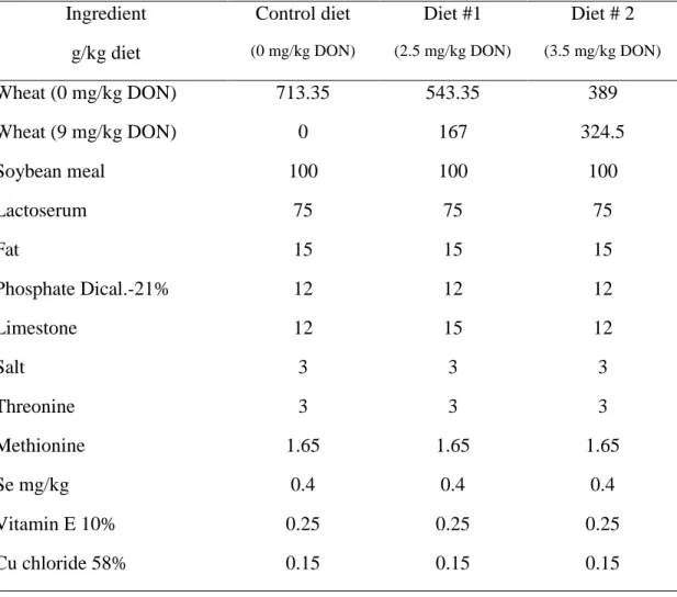
Table 1. Diet compositions. 1 Ingredient g/kg diet Control diet (0 mg/kg DON) Diet #1 (2.5 mg/kg DON) Diet # 2 (3.5 mg/kg DON) Wheat (0 mg/kg DON) 713.35 543.35 389 Wheat (9 mg/kg DON) 0 167 324.5 Soybean meal 100 100 100 Lactoserum 75 75 75 Fat 15 15 15 Phosphate Dical.-21% 12 12 12 Limestone 12 15 12 Salt 3 3 3 Threonine 3 3 3 Methionine 1.65 1.65 1.65 Se mg/kg 0.4 0.4 0.4 Vitamin E 10% 0.25 0.25 0.25 Cu chloride 58% 0.15 0.15 0.15 2
All diets were formulated to reach the following requirement: metabolisable energy 3200 kcal/kg,
3
protein 19%, fat 3%, fiber 2.5%, moisture 10%, Ca 0.8%, Mg 509 mg/Kg, total P 0.7%, K 0.7%,
4
Na 0.2%, Se 0.6 mg/Kg, Cu 120.4 mg/kg, Zn 250 mg/Kg, vitamin A 18.4 KIU/Kg, vitamin D 2.5
5
KIU/Kg, vitamin E 106.7 mg/Kg, biotin, 0.3 mg/Kg.
6
7
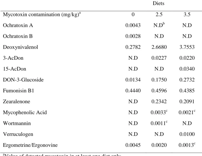
Table 2. Mycotoxins’ content of the diets. 1 Diets Mycotoxin contamination (mg/kg)a 0 2.5 3.5 Ochratoxin A 0.0043 N.Db N.D Ochratoxin B 0.0028 N.D N.D Deoxynivalenol 0.2782 2.6680 3.7553 3-AcDon N.D 0.0227 0.0220 15-AcDon N.D N.D 0.0340 DON-3-Glucoside 0.0134 0.1750 0.2732 Fumonisin B1 0.4440 0.4596 0.4385 Zearalenone N.D 0.2342 0.2091 Mycophenolic Acid N.D 0.0033c 0.0021c Wortmannin N.D 0.0011c N.D Verruculogen N.D N.D 0.0100 Ergometrine/Ergonovine 0.0045 0.0020 0.0013c a
Value of detected mycotoxin in at least one diet only.
2
b
N.D: Not detected, values below limit of detection.
3
c
Values under the limit of quantitation but above limit of detection.
Figure1. Effect of DON naturally contaminated diets on piglet’s growth following PRRSV
1
infection.
2
Groups of piglets (10) were fed with DON naturally contaminated diets (2.5 and 3.5 mg/kg of
3
feed) for 2 weeks. A control group of piglets received uncontaminated diet for the same period of
4
time. A subgroup of piglets (n=6) was PRRSV infected and the remaining piglets (4) were sham
5
infected with PBS. Average daily gain was calculated by dividing the total weight gain on the
6
number of days of the study (A). Kaplan-Meier survival curve in PRRSV infected animals with
7
end points piglet death (B). +++significant when compared to respective PRRSV infected group
8
(P<0.001).
9
*
significant when compared to control group (0 mg DON/kg) (P<0.05).
10
Figure2. Time course of body temperature (°C) during experimental infection.
11
PRRSV infected (A) and noninfected (B) pigs were monitored daily for rectal body temperature
12
during 21 days post-infection. Fever was defined as rectal temperature higher than 40˚C for two
13
consecutive days.
14
Figure3. Effect of DON naturally contaminated diets on PRRSV viremia and lungs viral load.
15
Blood was collected at day -1,3, 6, 9, 13, and 21 pi and serum tested for the presence of PRRSV
16
RNA by RT-qPCR (A). At necropsy, sections of lung were collected to determine the pulmonary
17
viral load by RT-qPCR (B). Data are expressed in TCID50/mL. *significant when compared to
18
control group (0 mg DON/kg) at the same day (P<0.05). Note: animals sacrificed during the
19
experiment were excluded from the analysis.
20
Figure 4. Effect of DON naturally contaminated diets on macroscopic and microscopic lung
21
At necropsy, on day 21 p.i, macroscopic lung lesions were recorded as percentage of lungs
1
affected by pneumonia (A) and microscopic presence of leucocytes, serum, or necrotic debris in
2
alveolar exsudate were scored (B). *significant when compared to control group (0 mg DON/kg)
3
at the same day (P<0.05). Note: animals sacrificed during the experiment were excluded from the
4
analysis.
5
Figure 5. Effect of DON naturally contaminated diets on PRRSV specific antibody response.
6
Blood was collected at day -1, 9, 13, and 21 p.i and sera were tested for the presence of specific
7
PRRSV antibodies using a commercial ELISA kit (HerdChek-PRRS®,IDEXX). Data are
8
expressed in sample to positive ratio. *significant when compared to control group (0 mg
9
DON/kg) at the same day (P<0.05). #tendency when compared to control group (0 mg DON/kg)
10
at day 21 (P=0.09)
11
Figure 1. 1 2 3 4 5 0 2.5 3.5 0.0 0.2 0.4 0.6 0.8 Sham Infected * +++ +++ +++ * DON (mg/kg) A v e ra g e d a ily g a in (k g /D a y ) 0 5 10 15 20 60 70 80 90 100 0 2.5 3.5 Days P e rc e n t s u rv iv a l A B
Figure 2. 1 2 3 4 5 0 5 10 15 20 38 39 40 41 0 2.5 3.5 Days B o d y te m p e ra tu re ( ° C ) 0 5 10 15 20 38 39 40 41 0 2.5 3.5 Days B o d y te m p e ra tu re ( ° C ) A B
Figure 3. 1 2 3 4 5 -1 3 6 9 14 21 0 1 2 3 4 5 0 2.5 3.5 * * Days P R R S V ti te r (L o g10 T C ID50 /m l) 0 2.5 3.5 0 1 2 3 4 5 * P=0.1 DON (mg/kg) P R R S V ti te r (L o g10 T C ID50 /m l) A B
Figure 4 1 2 3 4 5 6 0 2.5 3.5 0 10 20 30 40 50 * DON (mg/kg) M a c ro s c o p ic lu n g le s io n s (% ± S E M ) 0 2.5 3.5 0 1 2 3 4 * DON (mg/kg) M ic ro s c o p ic le s io n s c o re s A B
Figure 5 1 2 3 -1 9 14 21 0.0 0.5 1.0 1.5 2.0 2.5