Université de Montréal
%4i,’,/,
3c ? z
Study of Thermosensitive Microspheres for Potential
Applications in Biosensing
Par
Yilong Chen
Département de chimie
Faculté des arts et des sciences
Mémoire présenté à la faculté des études supérieures en vue de l’obtention
du grade de maîtrise en chimie
Août 2006
©Yilong Chen, 2006
3
o
‘V
o) o
Université
de Montréal
Direction des bib1othèques
AVIS
L’auteur a autorisé l’Université de Montréal à reproduire et diffuser, en totalité ou en partie, par quelque moyen que ce soit et sur quelque support que ce soit, et exclusivement à des fins non lucratives d’enseignement et de recherche, des copies de ce mémoire ou de cette thèse.
L’auteur et les coauteurs le cas échéant conservent la propriété du droit d’auteur et des droits moraux qui protègent ce document. Ni la thèse ou le mémoire, ni des extraits substantiels de ce document, ne doivent être imprimés ou autrement reproduits sans l’autorisation de l’auteur.
Afin de se conformer à la Loi canadienne sur la protection des renseignements personnels, quelques formulaires secondaires, coordonnées ou signatures intégrées au texte ont pu être enlevés de ce document. Bien que cela ait pu affecter la pagination, il n’y a aucun contenu manquant.
NOTICE
The author of this thesis or dissertation has granted a nonexciusive license allowing Université de Montréal to reproduce and publish the document, in part or in whole, and in any format, solely for noncommercial educational and research purposes.
The author and co-authors if applicable retain copyright ownership and moral rights in this document. Neither the whole thesis or dissertation, nor substantial extracts from it, may be printed or otherwise reproduced without the author’s permission.
In compliance with the Canadian Privacy Act some supporting forms, contact information or signatures may have been removed from the document. While this may affect the document page count, it does flot represent any loss of content from the document.
Université de Montréal Faculté des études supérieures
Ce mémoire intitulé
$tudy of Thermosensitive Microspheres for Potential
Applications in Biosensing
Présentépar
Yilong Chen
Résumé
En contrôlant le ratio tensioactif/monornère, des particules de microgel thermosensible de différentes tailles ont été synthétisées par polymérisation en émulsion. Nous avons observé qu’un arrangement colloïdal cristallin se forme lors de variations
contrôlées de température. Certaines de ces particules ont été enduites d’une couche de polymère réticulé non-thermosensible de poly(acrylamide) et de poly(N,N-dirnéthyl acrylamide) ou thermosensible de poly(N,N-diéthyl acrylamide) et de poly(N-isopropyl
acrylamide) afin d’obtenir une ou deux températures de transition dans le réseau
colloïdal cristallin. En utilisant la théorie de Flory, les phénomènes de diffraction et de gonflement peuvent être expliqués en termes des énergies de mélange et élastique. L’influence sur la diffraction du changement de pH et de la force ionique du milieu a été étudiée afin d’évaluer le potentiel de ces matériaux comme capteurs.
Des microsphères conchoïdales (avec noyacix centraux) ont aussi été synthétisées. Ces microsphères sont constituées d’un noyau chimiquement réticulé et d’une
enveloppe ‘chevelue’. Le noyau est formé de poly(N-isopropyl acrylamide-co-styrène)
alors que la couche externe est formée de poly(N,N-diéthyl acrylamide), de poly(N isopropyl acrylamide) ou de poly(N-isopropyl methaciylamide). Grâce à la microscopie électronique à transmission et à la diffusion dynamique de la lumière, le gonflement et la morphologie de ces microsphères ont pu être étudiés. Celles portant une couche de polymère thermosensible présentent une contraction (rétricissement) en deux étapes avec la température.
Par la formation de liaisons covalentes entre la biotine et les particules thermosensibles, une étude par spectroscopie de fluorescence des interactions avec l’avidine et la streptavidine démontre une réponse suffisante pour envisager des
applications comme biocapteurs.
Mots-clés: Cristal colloïdal; Microgel; Diffusion de la lumière; Particule; Biotine
ABSTRACT
Thermosensitive microgel particles were synthesized via ernulsion polymerization and the insize was controfled by vaiying the concentrations of monorners and surfactant. The thermo-responsive and soft particles fomied colloidal crystalline array (CCA) through a simple temperature cycling procedure. The CCAs were ernbedded in both cross-linked nonthermosensitive matrices of polyacrylarnide and poly(N,N-dirnethyl acrylamide) and thermosensitive poly(N,N-diethyl acrylamide) and poly(N-isopropyl aciylamide) to fabricate films of polymerized colloidal crystalline aiiays (PCCAs) with single and doubly thermosensitivities, respectively. Their temperature dependent diffraction behavior was investigated to reveal the sweÏling properties ofthe gels, which were interpreted by classical Flory’s polymer theories. The combination of mixing and elastic energies affected the swelling properties of polyrnerized colloidal crystalline arrays. Finally, ionic strength and pH effects on the CCA diffraction behavior were studied.
These core-sheli microspheres are composed of chemically cross-linked poly(N
propyl aciylamide-co-styrene) with different styrene contents as a core and haiîy poly(N,N-diethyl acrylamide), poly(N-isopropyl acrylarnide) and poly(N-isopropyl rnethaciylamide) as shefls. The morphologies and swelling properties of the core and core-sheli microspheres were investigated by means of transmission electron microscopy, optical transmission and dynamic light scattering. The core-shell microspheres display a temperature-dependent two-step shrinking behavior.
The thermosensitive microgel particles were biotinylated and their interactions with avidin and streptavidin were studied by fluorescence spectroscopies. The results showed that the microgel particles could respond to the presence of avidin or streptavidin, which may have potential applications as biosensors.
TABLE 0F CONTENTS
RÉSUMÉ ABSTRACT H TABLE 0f CONTENTS flJ LIST 0F FIGURES VI LIST 0F TABLES IXLIST 0F SYMBOLS AND ABBREVIATIONS X
ACKNOWLEDGEMENTS XH
1. Generat Introduction 1
1 .1. Stimuli—responsive polymers 1
• 1.1. pH-sensitive polymers 1
1.1.2. Thermosensitive polymers 2
1.2. Colloidal crystalline arrays (CCA) 5
1.3. Polymerized CCA (PCCA) 7
1 .4. Biotin-avidin interaction $
1.4.1. Biotin $
1.4.2. Avidin and streptavidin 10
1.4.3. Biotin-avidin binding 12
1.4.4. Biotinylation techniques 13
1.5. Objectives 17
1.6. References
2. Preparation and Light Diffraction Behaviors of Soft Potymerized Colloidal
Crystalline Arrays 23
2.1. Abstract 23
2.2. Introduction 23
2.3. Experimental section 25
2.3.1. Materials 25
2.3.3. Preparation ofPCCAs26
2.3.4. Dynarnic light scattering 27
2.3.5. Visible light diffraction 28
2.4. Resuits and discussion 28
2.4. 1. Synthesis and characterization of CCAs 28
2.4.2. Preparation ofPCCAs 33
2.4.3. PCCAs with non-thermosensitive matrices 35
2.4.4. PCCAs in thermosensitive matrices 40
2.4.5. Effects ofionic strength and pH 41
2.5. Concluding remarks 43
2.6. Acknowledgements 44
2.7. Notes and references 44
3. Synthesis and Characterization of Core-sheli Microspheres with Doubly
Thermosensitive 46
3.1. Abstract 46
3.2. Introduction 46
3.3. Experimental section 47
3.3.1. Materials 47
3.3.2. Preparation of P(nPA-co-S) cores 47
3.3.3. Preparation of core-sheli microspheres 48
3.3.4. Transmission electron microscopy (TEM) 49
3.3.5. Optical transmittance 49
3.3.6. Dynamic light scattering 49
3.4. Resuïts and discussion 50
3.4.1. P(nPA-co-S) microsphere cotes 50
3.4.2. Doubly thermosensitive core-sheli microspheres 56
3.5. Concluding remarks 58
3.6. Acknowledgements 58
3.7. References and notes 58
4. Biotin-Avidin-Based Bïosensors 60
4.2. Experirnental section 61
4.2.1. Materials 61
4.2.2. Fluorescence measurements 65
4.2.3. Visible light diffraction 65
4.3. Results and discussion 67
4.3.1. Biotinylation 67 4.3.2. Fluorescence measurernents 69 4.3.3. Biotinylated PCCA 73 4.4. Conclusions 74 4.5. References 75 5. Conclusions 7$ 5.1. Preparation of microspheres 7$
5.2. CCA formation by microspheres 78
5.3. Polymerized CCAs and their characteristics 7$
5.4. Biotinylation and sensing technology 79
Appendiices 81
A. Appendix of chapter 2 81
B. Appendix of chapter 3 $4
Figure 1.1 Figure 1.2 Figure 1.3 Figure 1.4 Figure 1.5 Figure 1.6 Figure 1.7 Figure 1.8 Figure 1.9 Figure 1.10 Figure 1.11 Figure 2.1 Figure 2.2 Figure 2.3 Figure 2.4 Figure 2.5 Figure 2.6
LIST 0F FIGURES
3 3 6 6 7 $ 9 12 15 16 17 27 30pH response of poly(2-vinyl pyridine) and poly(aciylic acid). Temperature response of UCST polyrner particles and LCST polymer particles.
Assembly of hard particles by slow particle sedimentation. Assembly of charged polystyrene by electrostatic repulsion. Assembly of lightly cross-linked, thermosensitive, uncharged, and soft particles.
Fabrication of PCCA of charged polystyrene.
Structural formulations of biotin and its main derivatives. Ribbon diagram of chicken egg white avidin.
Biotinylation of amine groups ofproteins. Biotinylation of thiols groups of ceils.
Biotinylation of aldehyde groups of glycoproteins. Preparation of PCCAs.
Hydrodynamic diameters of particles as a function of
temperature.
Light diffraction spectra of CCA1 at io oc before and after temperature cycling; light diffraction spectra of CCA1 at di fferent temperatures after temperature cycling.
Light diffraction spectra of CCA1 at different weight fractions; diffraction wavelength ofccAs as a function of particle weight fraction.
Typical diffraction spectra of CCA1, PCCA3 just after
photopo lymerization, and PCCA3 washed and equilibrated in
water.
Diffraction spectra of PCCA3 at different temperatures and diffraction wavelength as a function of temperature forPccAs.
31
32
35
figure 3.2 Figure 3.3 figure 3.4 Figure 3.5 Figure 4.1 Figure 4.2 figure 4.3 figure 4.4 Figure 4.5 Figure 4.6 Figure 4.7 Figure 4.8 Figure 4.9 Figure 4.16 57 61 62 63 64 66 66 68 68 69 71 Figure 2.7 Figure 2.8 41 42 51 53 54 55 Diffraction spectra of PCCA7 at different temperatures; diffraction wavelength versus temperature for PCCAs.
Ionic strength and pH dependence of the diffraction wavelength of PCCAs.
figure 3.1 Transmission electron micrographs of PnPA25 S 75 microspheres and PnPA25 S75 -NIPAM core-sheli microspheres.
Optical transmittance of the solution of the microspheres as a function of temperature: Mono-thennosensitive microspheres of P(nPA-co-S); Doubly thennosensitive core-sheil miscrospheres.
Size distributions of PnPA25S75 and PnPA25S75-NIPAM
microspheres.
The variation of hydrodynamic diameters or swelling i-atios of the microspheres as a function of temperature.
The response of doubly thermosensitive core-sheli microspheres to changes in temperature.
Synthesis of DEA. Synthesis ofNAS.
Biotinylation of microspheres.
Preparation of fabrication of avidin sensing PCCA. Horne-assembled UV-visible light diffraction instrument. Light diffraction fromPCCA.
FTIR spectra ofmicrospheres 1, 2 and 3. Quantificational principle ofNAS by UV. Calibration plot ofNAS.
Fluorescence spectra of fluorescein-labelled avidin in the
presence of different concentrations of biotinylated
microspheres; plots of fluorescence intensity as a function of
concentrations of free biotin, biotin attached on 3 and unbioti nylated microspheres made of PDEA.
Fluorescence intensity offluorescein-labeled avidin as a function of concentrations of avidin, streptavidin in the presence of
biotinylated microspheres.
Figure 4.12 The diffraction wavelengths of biotinylated PCCA as a fttnction 74
of avidin concentrations.
Figure 5.1 Schematic illustration of(strept)avidin responsive microspheres. 79
Figure Al 82
1 V1,o The fittmg curves of% versus
T]?
Figure A2
The differentiating curves of data in Figure 2.6(B) for PCCAs 1 82 (Solid), 2 (Dash), and3 (Dot).
Figure A3
The differentiating curves of data in Figure 2.7(B) for PCCAs 5 83 (Solid), 6 (Dash), and 7 (Dot).
Figure A4 Optical transmittance (A), hydrodynamic diameterR11 (B) 84
and swelling ratio (C, a = (Ro/Rji)3) of doubly
thermosensitive core-sheli miscrospheres (PnPA5OS5 O derivatives) as a function oftemperature.
Figure AS . . . . 85
The differentiating curves of data in figure A4(A) and A4(B).
Figure A6 . . . 86
LIST 0F TABLES
TabLe 1.1 LCST inaqueous solution ofN-alkyl substituted polyacrylarnide and 4
polymethacrylamide.
Table 1.2 Properties ofavidin and streptavidin. 11
Table 1.3 Comrnercially available biotinylation agents. 14 Table 2.1 Emulsion polymerization conditions for the preparation of microgel 29
particles.
Table 2.2 Optical properties of PCCAs in non-thermosensitive and 34
thermosensitive matrices prepared at different CCA1
concentrations.
Table 2.3 Calculated )Lo, 2m, Ve/T7n1, 6, and coi-relation coefficient of CCA 39
embedded in non-thermosensitive matrices.
Table 3.1 Experimental conditions for soap-free emulsion polymerization of 48 mono-thermosensitive microsphere syntheses.
LIST 0F SYMBOLS AND ABBREVIATIONS
AAm Acrylamide
BA N,N ‘-Methylenebisacrylamide
CCA Colloidal clystalline array
DEA N,N-Diethylacrylamide
DEAP Diethoxyacetophenone
DMA N,N-Dimethyl acrylamide
d Interpianar spacing
EA N-Ethylaciylamide
GE Elastic free energy change
AG01 Ionization free energy change
AG11 Mixing free energy change
HEMA 2-Hydroxyethyl methacrylate
k Boltzmann constant
Kd Dissociation constant
KPS Potassium perscilfate
LCST Lower critical solution temperature
‘n Order of diffraction
n Refractive index
NAS N-Acryloxysuccinimide
NIPAM N-Isopropylacrylarnide
NIPMA M N-Isopropyl methacrylarnide
nPA N-n-Propyl acrylamide
PCCA Polymerized coltoidal cuystalline aiay
Hydrodynami c di ameter
S Styrene
SDS Sodium dodecyl sulfate
T Absolute temperature
t112 Halfiife
UCST Upper critical solution temperature
Molar volume of water
Volume ofthe fully relaxed gel
VPT Volume phase transition
Solubility pararneter ofwater
Average solubility parameter ofthe polymer Absorbance coefficient
Glancing angle
Diffraction wavelength ofPCCA films Diffraction wavelength of dry PCCA films
%fl Diffraction wavelength ofrelaxed PCCA films
r fluorescence lifetime
Ve/V Cross—linking density
Polymer volume fraction
q5crf Effective volume fraction
ACKNOWLEDGEMENTS
This thesis is by far the most significant scientific accompiishment in my hfe and it would have been impossible to complete without the people who supported me and believed in me.
Most of ail I would like to thank my research supervisor, Professor Julian X.X. Zhu of the Department of Chemistry at Université de Montréal. He is not only a great scientist but also a kind person. Ris trust and scientific rigor inspired me in the most
important moments ofmaking right decisions and I am glad to have worked with him.
I wish to thank sincerely Dr. Julien E. Gautrot for bis kind help with the correction ofthe papers in this thesis.
I am also very grateful to Dr. Zhanyong Li of the Institute of Polymer Chernistry at Nankai University for helping me witb the PCCA syntheses and discussion.
I want to thank ail of the nice friends in the group: Dr. Mathilde Colonne, Dr. Sophie Tan, Marc Gauthier, Héloïse Thérien—Aubin, Dr. Huiyou Liu, Dr. Wilms Baille, Shanshan Chen, Dr. Junhua Zhang, Dr. Juntao Luo, Guillaume Giguère, Claire Jaubert, Bao Toan Nguyen, Fansheng Meng, Julie Boivin, Dr. Ya Cao, Sen Ge, Florence Janvier, Cindy Charboneau, Gérald Perron, Dr. Steven Holden...
My family bas provided the spiritual support that is even more important. I thank my parents Zuqi Chen and Aixiang Ding, and my girlfriend Guiying Tang.
1. General Introduction
1.1. Stimuli-responsive polymers
Stimuli-responsive polymers have been investigated extensively for the development of “smart” materials for their potential applications in the controlled
transport and delivery of active substances, such as dntgs, in biotechnology, medicine, pharmacy or cosmetics.’ The term ‘stimuli-responsive’ implies remarkable changes of certain properties induced by an external stimulus. In the strict sense, the induced property changes should be reversible if the stimulus is suppressed or released, or if a second ‘reverse’ stimulus is applied. Many types of stimuli are theoretically useful, but the choice is limited for practical reasons. In aqueous systems, stimuli-sensitive systems are generally aimed to change the hydrophilic character of a functional group to a hydrophobic one, or vice versa.2 Both chemical and physical stimuli (which may be coupled) can be employed for this purpose. Chemical stimuli include for instance acid base reactions, complexation, redox, electrochemical reactions, and photochemical reactions. Physical stimuli comprise changes in pH-value, ionic strength, temperature, pressure, light, and electrical or magnetic fields.3 Photochernical reactions and redox reactions have also been considered alternatively for applications as switches in aqueous media.4’4 However, good stability and reversibility in combination with marked changes of the hydrophilic-hydrophobic balance are hard to achieve simultaneously. Particularly, most organic redox systems are chemically sensitive to oxygen from air or to good nucleophiles (like water) in one of the two oxidation states, thus hampering their applications severely. Due to the lack of stability and reversibility, the research interest has moyeU to the simplest systems driven by pH and temperature.
I .1. I. pH-sensitive polymers
One of the simplest stimuli-responsive polymers is based on acid-base reactions or on pH changes. Typical examples are polymeric amines or polymeric carboxylic acids, which by protonation/deprotonation become charged or neutral, and thus undergo a pronounced change in their hydrophilicity or hydrophobicity. Poly(4-vinyl pyridine),
poiy(2-vinyi pyridine), poiy(acrylic acid), and poiy(methacrylic acid) and their copolymers, are wefl-known polymers that can respond to a pH change.’7 For exampie, poly(4-vinyl pyridine) swells upon a decrease in pH since the protonation of
the pyridinyl groups along the polymer chain leads to increased solubility of the polymer and repulsive electrostatic forces within the particle (figure 1. lA). The polymer chain collapses upon deprotonation at bigh pH values. In contrast, polymers made of (meth)acryiic acid can respond to changes in pH and ionic strength, e.g., at pH values below 4, precipitation occurs in aqueous solutions due to the protonation of the carboxylate groups (Figure 1.1 B), which renders the polymers sparsely soluble in water.
Such pH-responsive polymers have an excellent reversibility but oniy for a few switching cycles. SaIt accumulation while changing the pH (addition of acid or base) increases the ionic strength of the solution, which can reduce the reversibility and solubility of the pH—responsive poiymers.’7 Sait accumulation during successive protonation/deprotonation events can render the polymers insoluble in the system.
• 1 .2. Thermosensitive polymers
Thermosensitive polymers may exhibit a change in solubility upon changes in
temperature. They often possess an upper criticai solution temperature (UCST) or a
lower critical solution temperature (LCST) depending on their chernical structures.
There are a few reports concerning polymers exhibiting a property change on UCST in
aqcteous solutions.I S2O Among polymers showing UCST, poiy(N—acetylacrylamide), containing the N-substituted acetyl group, is a representative polymer by exhibiting reversible sol ubi Iity changes in aqueous solution when heating—up and cool ing—down. The hydrogel prepared from the UCST polymer is known to swell while heated above the UCST (Figure 1.2A).’8 It is speculated that an interpolymer complex formed by the mutual association between molecules of the UCST polymer dissociates and turns solctble with hydration in the heating process. Additives might participate in the dissociation between polymer chains to change the UCST. Some kinds of interpolymer complexes were studied by using viscometry and optical measurements.223 However,
widely so far. pH A pH A
_
BFigure 1.2. Temperature response of (A) UCST polymer particles and
(B) LCST polymer particles.
The thermal properties of polymers with a LCST have been investigated extensively.2426 These polyrners possess both hydrophobie and hydrophilic groups. The balance between hydrophobi city and hydrophilicity determines the LCST properties. These potymers are soluble or swellaNe below the LCST due to hydrogen bonding in aqueous medium, while the hydrogen bonds are broken by heating above LCST leading
7:
z_e-B , t pH pH Figure 1.1. pH poly(acrylic acid).response of (A) poly(2-vinyl pyridine) and (B)
to the precipitation or shrinkage of the polymers in aqueous solutions (Figure Ï .2B). N Aikyl substituted polyacrylamide and polymethacrylamide are typical examples of polymers having a LCST (Table 1 .1)2426 Poly(N-isopmpylacrylamide) (PNIPAM) is representative of this family of polymers and bas been studied for many potential applications in the fields of drug delivery,273° biosensing,31’32 chemical separations,3335
36-3S 39-11
biomaterials, and catalysis.
Table 1.1. LCST of aqueous solution of N-alkyl substituted
polyacrylamide (PAAm) and polymethacrylamide (PMAm) (copied and modilied from original in ref. 25).
*CHCH* R1 R2 ONR1R2 ONR1R2 oc H H — — H CH3 — — CH3 CH3 — — H CH2CH3 73 — CH3 C2H5 56-57 — —N 56 — - 47 -C2H5 C2H5 36 — H CH(CH3)2 32 45 CH3 CH(CH3)2 25 — H CH2CH2CH3 22 28 CH3 CH2CH2CH3 15 — -NQ -
-1.2. Colloidal crystalline arrays (CCA)
There is much interest in the use of photonic crystals to control the propagation of electromagnetic radiation. The self-assembly of three-dimentional (3-D) CCA is a process of broad interests due to its applicability in many fieÏds, such as the study of condensed matter phase behaviors,4245 the fabrication of mesoporous films,4647 and the development of photonic materials.4858 These CCAs diffract light and prevent its transmission following the Bragg’s law in a similar way to X-ray diffraction by atomic crystals. These properties have been applied to the fabrication of chemical and biological sensors,53 optical waveguides.9 optical switches,’’ and optical fiIters.8
3D-photonic ciystals based on materials such as silica, polystyrene (PS). or poly(metliyl methacrylate) particles were studied extensively, using sedirnentation or particle repulsion to generate ordered structures. These techniques typically rely on nonspecific particle-particle repulsion, which could be short or long range repulsion forces, to induce order. For short-range repulsion forces (hard sphere interactions), close-packed crystaïline arrays are typically formed. For example, close packing can be accomplished through slow particle sedimentation, centrifugation, spin coating, electrophoretic deposition, sonication, and lateral compression of interface-bound
monolayers. Recause close-packed ordered structure typically involves the
concentration of particles in a confined space, these studies often involve hard spheres that undergo littie deformation upon packing (Figure 1 .3)60,6 Long—range interactions
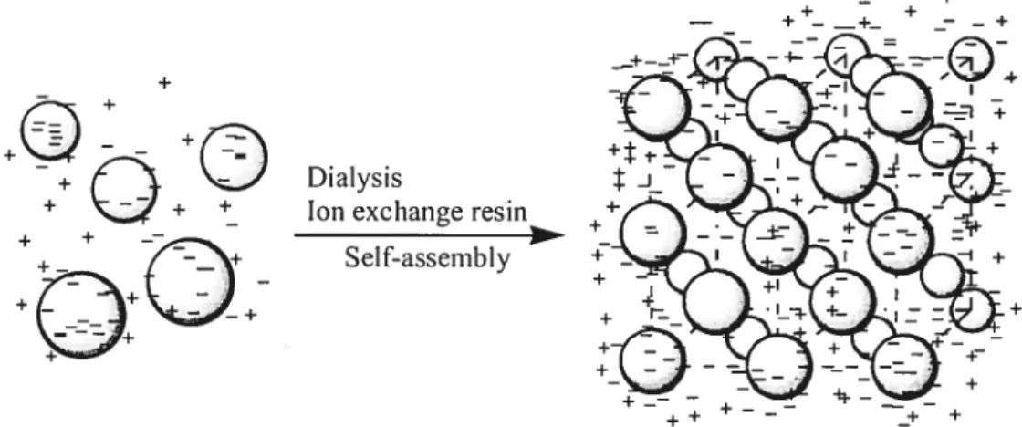
can be exploited to create ordered assemblies. SpecificaÏly, suspended coïloidal spheres bearing high surface charge densities spontaneously assemble into solvated crystalline arrays when the solution is rigorously desalted (Figure 1 .4). These rnethods, coupled to the photopolymerization of a monorner solution surrounding the CCA, have been exploited by Asher and co-workers to create polyrnerized colloidal ciystalline anays (PCCA) for optical filtering, switching, and sensing applications.153 However, the thermosensitive arnphiphilic soft hydrogel microspheres cannot be ordered by using such approaches, since these particles are too buoyant in water to sediment at an appreciable rate. Filtration or slow evaporation methods are equalÏy inappropriate because of die extreme solvation of the particles, which would collapse upon solvent
removal. Lyon and co-workers have dernonstrated that Ïightly cross-linking hydrogel
PNIPAM could forrn colloidal crystalline arrays through concentration by
centrifugation.5557 Their resuits showed that as the effective volume fraction of the microgel crystals (q5) was decreased by reducing the polymer concentration, the lattice constant increased and the crystals eventually melted into a fluid phase at eff = 0.49. When the temperature was raised above the LCST for the component particles, the effective volume fraction became remarkably small as weIl and the array became a milky-white, disordered, and free-flowing solution (Figure 1.5). They suggested that particÏes were compressed together to form c;ystalline aiays by studying the light diffraction behaviors of samples centrifuged at different temperatures and by differential interference contrast microscopy.
/
__
o sedimentation
_____
figure 1.3. Assernbly of hard particles by slow particle sedirnentation.
+
+
+(Y
—
\J Dialysis
+ —— ÷ ÷ Ion exchange resin
+ + —
+— + Self-assembty
+
Figure 1.4. Assernbly of charged polystyrene (PS) by electrostatic repulsion (copied and modified from original in ref 51).
z
Above ____ LCST / 4 Below LCSTfigure 1.5. Assembly of lightly cross-linked, thermosensitive, uncharged and soft particles.
1.3. Polymerized CCA (PCCA)
h is welI-known that the seÏf-assembly of monodispersed charged particles, such as
Ps,5
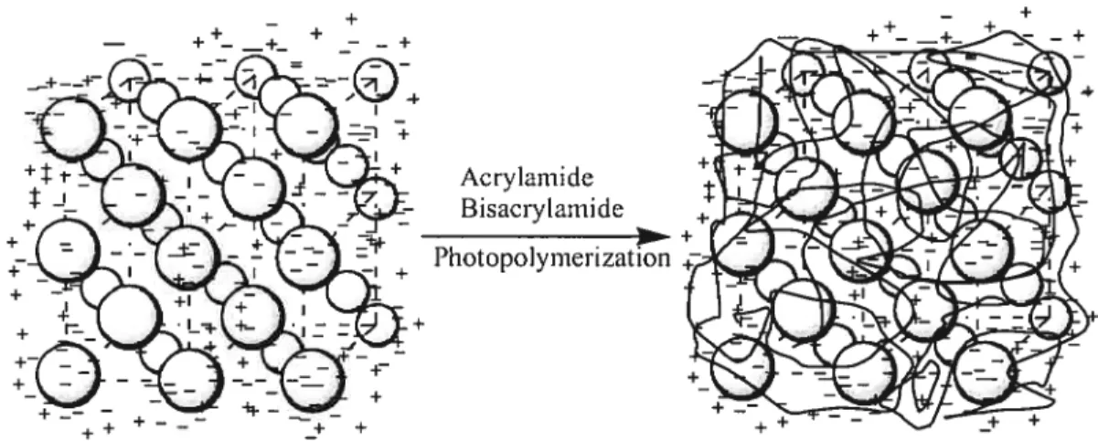
.53.62 PNIPAM,2 and poly(methyl methacrylate)44 produces three dimensionally periodic structures. This phenomenon is primarily due to the electrostatic interactions of charges at the surface of the particles, coupled to the diffuse counterion cloud (figure1.4). 1f a monomer solution surrounding the CCA is photopolymerized (Figure 1.6), a PCCA is generated, as bas been reported by Asher and co-workers for use in optical fi Iteri ng, switch i ng, and sensing applications.5l-5362 When charged poly(styrene—co— styrene scilfonate) CCAs were embedded in PNTPAM matrix, the periodic lattice was
found to change with temperature, while PCCAs fabricated in a PAAm matrix did not
show such a phenomenon. Recently, it was found that Iightly cross—linked
thermosensitive particles, displaying LCSTs, could form CCAs spontaneously via a simple temperature cycling process.5557’6364 The CCAs formed diffracted a wide range ofwavelengths, depending on the concentration and the sizes of the particles. 1-Iowever, such “soft” CCA lattices ai-e relatively unstable. For instance, heating the sample above its LCST qctantitatively breaks the ordered lattice and so does dilution. Therefore, it appears that PCCAs possess much better thermal, chemical and mechanical stability than CCAs, as demonstrated by Hu’s work on PCCA films.64 However, the control of the CCA ordered structure during the matrix formation step remains an issue, especially in the case of soft CCAs. Lyon and co-workers studied the behaviors of microcomposite
hydrogel films composed of lightly cross-linked PNIPAM particles and PNIPAM
matrix.63 However, they could not preserve the ordered structure of the CCAs upon photopolymerization and the PCCAs they obtained did not show any opalescent properties.
1.4. Biotin-avidin interaction 1.4.1. Biotin
+
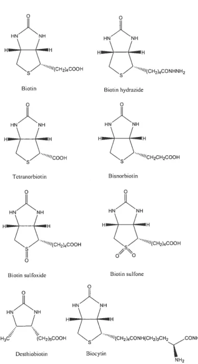
Biotin, or hexahydro-2-oxo-1H-thieno[3,4-d]imidazole-4-pentanoic acid, is a water-soluble vitarnin belonging to the B-complex, which is found in small quantities in ail living ceils. It exists in many isomeric forms, but only D-(+)-biotin and its derivatives are biologically active.
Biotin is synthesized in various bacteria and higher plants.65’66 However, several microorganisms as well as higlier animals are not capable of synthesizing biotin and their needs in this vitamin are met by dietary intake.67 In nature, biotin is found either free or covalently bound to proteins or peptides.68 The structural formulations of biotin and its main derivatives are shown in Figure 1.7.
+
figure 1.6. Fabrication of PCCA of charged polystyrene (copied and modified fi’om original in ref. 53).
HN NH “°“CH2CH2COOH HN H H3 CH2)5COOH Desthiobiotin “fltCH2)4CONHtCH2)3CH2çCONH2 N H2
Figure 1.7. Structural formulations ofbiotin and its main derivatives.
The richest dietaiy sources of biotin include liver, kidneys, heart, pancreas, poultry, egg yolk and milk. Smaller amount is found in plants, rnainly in seeds. The dietary biotin intake in hurnan being in Western countries has been estimated to be 35 to 70 ig/day, which seems to be absorbed almost completely.68 Biotin is involved in the biosynthesis of fatty acids, gluconeogenesis, energy production, and the metaboÏisrn of the amino acids L-leucine, L-isoleucine and L-valine.69 Recent research resuits indicate
Biotin Biotin hydrazide
“U/COOH
Tetranorbiotin Bisnorbiotin
Biotin sol fo\lde Biotin sulfone
that biotin plays an important role in gene expression and that presurnably it may also play a role in DNA replication.69’7° Low biotin intake has been studied to resuit in severe biochemical disorders in animal organisms, such as inhibition of protein and RNA synthesis, reduced carboxylase activity and antibody production. It has indicated that biotin is involved in several animal syndromes, such as the avian fatty liver and kidney syndrome (FLKS) and the “blue siime” disease of salmonids.67’657’ Thus, the detection ofthe biotin level is very important for diagnosis.
1.4.2. Avidin and streptavidin
The early history ofthe protein avidin is closely related to the discovery, isolation, and synthesis of biotin. Although avidin accounts for only 0.05 % of the proteins of egg white, its presence was betrayed by an unusual dermatitis in rats fed with dried egg white as the sole source ofprotein.
Avidin (Figure 1.8)72 is a basic glycoprotein with its isoelectric point at pH 10. It is very soluble in water and sait solutions, and it is stable over a wide range of pH and temperature. Avi din purifled on carboxymethyl cellulose and then ciystallized appears to contain two or three components when chrornatographied on Amberlite CG-50 resin. It crystallizes from strong sait solution between pH 5 and 7, but it has
not
yet been crystallized in the isoelectric region. In contrast, streptavidin has no carbohydrate, and it has an acid isoelectric point. Therefore, it is rnuch less soluble in water and can be crystallized from water or from 50 % isopropanol. Sorne basic properties of avidin and streptavidin are listed in Table 1The gene for streptavidin has been cloned and sequenced with the ultirnate objective that it can be used in general expression systems for detecting and isolating fusion proteins.7374 It codes for a sequence of 159 amino acids, some 30 residues longer than avidin and longer than that expected from molecular weight measurements. It was found that subunits of both low and high molecular weight are present and that the srnallest one, the main constituent of rnost commercial preparations, is the result of the processing at both the N and C termini to give “core” streptavidin of 125-127 residues. It bas a much higher solubility in water than the solubility of unprocessed precursor. This core is identical at 33 ¾ of its residues to avidin, including the four tiyptophan
residues involved in the biotin-binding site. It also resembles avidin in its predicted secondary structure, predominantly
fi
strands and bends, and in other features (Table 1 .2). Some commercial preparations contain unprocessed streptavidin, and published purification rnethods based on iminobiotin columns yields this as the main product.73’74Table 1.2. Properties of avidin and streptavidin (copied and rnodified
from original in refs. 73 and 74).
Avidin egg . . Avidin egg
Property Streptavidin
white yolk
Amino acid residues 128 125-127 —
Subunit size (Da)
From sequence 15,600 13,400 —
SDS gels 16,400 14,500 19,000
Subunits 4 4 4
Isoelectric pH 10 5-6 4.6
28O (M’cnï’) 24,000 34,000 —
Ar733(+ biotin) (Mcnij 24,000 8,000 7,000
Fluorescence
7’max (fliTi) 338 (328) — —
r(nsec) 1.8(0.8) — —
Ku biotin (M) (pH 7.25) 0.6x10¹⁵ 4x10¹⁴ 1.7x10¹²
t112 (days) 200 2.9 0.07
8280 1S absorbance coefficient at wavelength of 280 nm. Ar233 is the
difference between in the presence and absence of biotin at wavelength of 233 nm. 2max is the wavelength of the maxmum of fluorescence peak. t is
fluorescence lifetime. Kd is dissociation constant. t112 is half life of
Two differences between streptavidin and avidin are of some importance. Streptavidin contains no carbohydrate, and it has a slightly acid isoelectric point (pH 5-6), which much minimizes non-specific absorption to nucleic acids and negatively charged ceil membranes. Avidin contains carbohydrate, the heterogeneity of which is the probable cause ofthe poor quality of avidin crystals.
1.4.3. Biotin-avidin binding
The main common features of biotin binding by both avidin and streptavidin (Table 1.2) can be summarized briefly. Avidin and streptavidin are stable tetramers with 2-fold symmetry, which can bind up to four molecules of biotin with the binding sites being arranged in two pairs on the opposite faces of the moÏecuÏe.7374 The stability is greatly enhanced by biotin binding, since the total free energy of binding is about 330 kJ/mol for each tetramer (Ka= 101 M’).73’74 The dissociation constant for biotin is 50
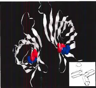
low that it can be estimated only from the ratio of the rate constants of binding and exchange using radioactive-labelled techniques. Both avidin and streptavidin are tetramers of four identical subunits which could be folded into an eight-stranded antiparallel fl-barrel (Figure 1.8). Within each tetramer, biotin binding sites are located in a deep pocket in the core of the tetramer by hydrogen bonding and van der Waals
Figure 1.8. Ribbon diagram of chicken egg white avidin (see inset for
interactions. The binding sites consist of tetramer residues and a loop of the adjacent tryptophan (Trp 110) subunit. Moreover, the binding pocket is partly closed in its outer rim by a residue Trp 110 of a neighboring subunit. Once bound, biotin is almost completely buried in the protein core, with the exception of the valeryl side-chain carboxylate group which is exposed to the solvent. Two Trp residues (Trp 70 and Trp
97) are in close contact with biotin. The binding is accornpanied by a red shift of the
Trp spectrum and by a decrease in fluorescence; both can be used as the basis for quantitative assays.7375 The spectral changes in the tryptophan residues are accompanied by a rernarkable reduction in their accessibility to reagents sucS as N
bromosuccinirnide. In avidin, the tiyptophans of each subunit are protected when biotin is bound. In contrast, fluorescence quenching by oxygen is flot dirninished in the avidin biotin complex; if any, the rate constant for quenching increased.76 The binding of biotin can 5e blocked or weakened by oxidation of any of some tiyptophan residuse of avidin and streptavidin. For example, the binding constant is reduced to be Ka i09 M1 using periodate as oxidation agent,77 or the binding can be weakened by the dinitrophenylation of what appears to be a single lysine residue.77 Reaction of any one of two lysines or three tryptophans led to inactivation, and blocked or weakened further biotin binding. It is predicted that the modification on biotin can lead to weakening the binding as well.
Ï .4.4. Biotinylation techniques
Table 1.3. Commercially available biotinylation agents.
Biotinylation agent The analogs
Target groups: Amine
‘N
Succinimidyl—6—(biotinaniido) hexanoate;
H—H N—Hydroxysciccinmide ininiinobiotin.
Ç,-
Scilfo-succinimido biotin acidS u I fo-sLICCinjnijdyl —6—( bioti namido)—hexanoate;
N-Hydroxysuccinimido biotin 6-Biotinamidocaproylaniido)caproic acid N-hydroxy sciccinimide este r;
Sol fo—succinimidyl—2—(biotinamido)ethyl— I ,3—dithio proliionate.
Target groups: Thiol
N-Biotinyl-N-(3-maleimidopropionyl )-L-Iysine;
H 4,4-Maleimidomethyl)cyclohexane carboxyamido) butane;
s (H2C)4 N. (cH)5 Biotinyl-3-maleimidopropionamidyl-3,6-dioxaoctatie diamine. Maleimido biotin HNIH
X
ç
...,,,,, H ° s (H2C)4yN/ N-( 6-tB ioti namido)hexyl )-3 -(2-pyridylthio) proiionate N-Jodoacetyl-N-biotinylhexylenediamine Target groups: Aldehydes and carboxyls,5j
Biotin—c—aminocaproyl HN JH N-Aminoxymethylcarbonylhydrazino biotin. H s Biotinyl hydrazide B iotin-PEO3 -Amine; Biotin-PEOI2-Biotin. 5—t Biotinamido) pentylamine HrBiotinytation of amines. Biotinylation of amine groups was popularized with the succinimidyl esters of biotin (NH$-biotin) in DMSO or DMF (Figure 1 .9). Non-sulfo agents may be preferred for soluble proteins if organic solvents are acceptable, because hydrolysis can be better controlled. They also allow intracellular labeling. NHS-biotins prefer to react with primary amine at pH 7-9, which is an ideal reagent for antibody and DNA biotinylation. The sulfosuccinimidyl ester of biotin (sulfoNHS-biotins), a more water soluble biotinylation reagent, is extensively used as topological probe to label proteins in the outer membrane surface, or when the use of organic solvents should be avoided.78-80
C (D
D
Figure 1.9. Biotinylation of amine groups ofproteins.
Biotinytation of thiots. $ulfbydroxyl biotinylation is useful for the detection or modification of sites containing SH groups or the study of SH dependent structures (Figure 1.10). It also works when SH groups are introduced into proteins, peptides and nucleotides, which are thiol-modified for similar goals. In classical biotinylation
H2Q
applications, the maleimido-biotin or its more soluble maleimido-Ic-biocytin and maleimido-PEO3 -biotin are recommended, due to their quick, quasi-stoichiornetric and very specific reactivity. Maleimide reacts specifically with free sulphydiyls at pH 6.5-7.5, allowing more defined labeling of proteins and avoiding undesired amine modification on proteins.8183 HNNH II \) (‘ H o CD HN’NH II’ H o H o Figure 1.10. Biotinylation ofthiols groups ofcells.
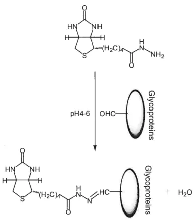
BiotinyÏation of aldehydes and carboxyÏs. Aldehydes generated by periodate oxidation of vicinal diols, and carboxyls activated by EDAC (N-ethyl-N’-(3-dimethylaminopropyl)carbodiimide hydrochloride), can be biotinylated by using biotin hydrazides (Figure 1.11). Biotinylation could be used in glycoproteins, polysaccharides
and sialic acids, steroids, glycolipids, LDL (fi-Lipoprotein), and nucleic acids. Biotin hydrazide, a classical carbohydrate reactive biotinylation reagent, reacts with aldehydes at pH 4-6 giving a stable CH=N-NH- bound allowing the labeling of glycoproteins through their glycan. Biotin hydrazide also reacts with carboxyls in the presence of EDAC.8486
1.5. Objectives
pH4-6 I Œ
H20
The main objective of this thesis is to design and prepare multi-responsive polymeric systems, which canbe post-functionallzed by bio-rnolecules for eventual bio
sensing applications. In this project, we have the following specific research objectives:
(1) Thermosensitive microspheres with controlled sizes will be synthesized by
emulsion polymerization. The variation and control of the sizes of the
microspheres wi 11 be studied.
(2) These microspheres will be used to fabricate CCAs and the packing
mechanism of CCAs will be studied by microscopy and light diffraction technique. The ordered microspheres (CCA) will be irnmobilized in both nonthermosensitive and thermosensitive matrices to make PCCA films by photopolymerization. The thermo-optical behaviors of PCCA films will be investigated. 1H G) o o 1 o CD C,)
(3) Thermosensitive microspheres with advanced core-sheli structure will be prepared by soap-free emulsion polyrnerization. The core-sheil structure and the shrinking behaviors will be studied.
(4) To build a model sensor, biotin, a small biornolecule, is to be attached on the microspheres. Fluorescence technique will be employed to study the interaction between (strept)avidin and biotin on the microspheres. Furthermore, biotin will be attached onto a nonthermosensitive matrix of PCCA films. The interaction of avidin to the biotinylated PCCA will be studied.
The resuits for the first two subjects will be presented and discussed in chapter 2 and the results of the third subject wiIl be included in chapter 3. These two chapters are presented in the form of research papers. The resuits for the last part will be the subject ofchapter 4.
1.6.
References
(1) Hoffman, A. S.; Stayton, P. S. Mactomot. Symp. 2004, 207, 139-152.
(2) McCormick, C. L., editor, Stiinuli-responsive water soluble and amphthilic polymers. ACS symposium series, vol. 780. Washington, DC: The American
Chemical Society; 2001.
(3) Laschewsky, A.; Lôtsh, D.; Seeboth, A.; Storsberg, J.; Stumpe, J. Proceedings of the European coating conferences, Berlin, Germany. Vol. Smart Coating ni 2004;
p. l-16.
(4) Krôger, R.; Menzel, H.; Hallensleben, M. L. Macroinot. Chem. Phys. 1994, 195, 2291.
(5) Kotk, .; Rathi, R. C.; Kopeek, J.; Macromolecittes 1997, 30, 5553. (6) Laschewsky, A.; Rekaï, E. D. Macromol. Rapid Commun. 2000, 21, 937. (7) Desponds, A.; Freitag, R. Langmuir 2003, 19, 6261.
(8) Ivanov, A. E.; Eremeev, N. L.; Wahlund, P. O.; Galaev, I. Y.; Mattiasson, B. Potyiner2000, 43, 3219.
(9) Akiyama, H.; Tamaoki, N. I Potyin. Sci., Polym. Chem. A 2004, A42, 5200. (10) Pieroni, O.; Fissi, A.; Angelini, N.; Lenci, F. Acc. Chem. Res. 2001, 34, 9.
(11) Anton, P.; Heinze, J.; Laschewsky, A. Langinuir 1993, 9, 77.
(12) Anton, P.; Laschewsky, A.; Ward, M. D. Polym. BttÏÏ. 1995, 34, 331. (13) Mulfort, K. L.; Ryu, J.; Zhou,
Q.
Polymer 2003, 44, 3185.(14) Isaksson, J.; Tengstedt, C.; Fahirnan, M.; Robinson, N.; Berggren, M. Adv. Mater. 2004, 16, 316.
(15) Martin, T. J.; Prochakza, K.; Munk, P.; Webber, S. E. Macromolecules 1996, 29, 6071.
(16) Robinson, D. N.; Peppas, N. A. Macromotecules 2002, 35, 3668.
(17) Gohy, J. F.; Varshney, S. K.; Jérôrne, R. Macromolecules 2001, 34, 3361.
(1$) Katono, H.; Sanui, K.; Ogata, N.; Okano, T.; Sakurai, Y. Polymer 1 1991, 23, 1179.
(19) Katono, H.; Maruyarna, A.; Sanui, K.; Ogata, N.; Okano, T.; Sakurai, Y. J. Control. Release 1991, 16, 215.
(20) Sasase, H.; Aoki, T.; Katono, H.; Sanui, K.; Ogata, N.; Ohta, R.; Kondo, T.; Okano, T.; Sakurai, Y. MakromoÏ. Chem. Rapid Commun. 1992, 13, 577.
(21) Bokias, G.; Staikos, G.; Ilipoulos, I.; Audebert, R. Macromotecules 1994, 27. 427. (22) Bekturov, E. A.; Bimendina, A. Adv. Polym. Sci. 1981, 41, 99.
(23) Staikos, G.; Bokias, G.; Karayanni, K. Potymer Inter. 1996, 41, 345. (24) Ito, S.; Hirasa, O.; Yamauchi, A. Kobitnshi RonbunsÏzu 1989, 46, 427. (25) Platé, N. L.; Lebedeva, T. L.; Valuev, L. I. Folyinerl 1999, 31, 21. (26) Schild, H. G. Prog. PoÏym. Sel. 1992, 17, 163.
(27) Hoffman, A. Controlled Dritg DeÏivery. Challenges and Strategies. Park, K., Ed.: American Chemical Society: Washington, DC, 1997; pp 485-498.
(28) Park, T. G. Biomaterials 1999, 20, 517.
(29) Ichikawa, H.; Fukumori, Y. I Contrat. Release 2000, 63, 107. (30) Kim, S. W.; Bae, Y. H.; Okano, T. Pharmn. Res. 1992, 9, 283.
(31) Miyata, T.; Asami, N.; Uragami, T. MacromolecuÏes 1999, 32, 2082. (32) Waler, J. P.; Asher, S. A. Anal. Chem. 2005, 77, 1596.
(33) Kawaguchi, H.; Fujimoto, K. Bioseparation 1998, 7, 253.
(34) Kayaman, N.; Kazan, D.; Erarsian, A.; Okay, O.; Baysal, B. M. I Appt. Folym. Sci. 1998, 67, 805.
(35) Umeno, D.; Kawasaki, M.; Maeda, M. Bioconjugate Chem. 1998, 9, 719. (36) Chen, L.; Kopecek, J.; Stewart, R. J. Biocoiugate Chem. 2000, 11, 734. (37) Stile, R. A.; Burghardt, W. R.; HeaÏy, K. E. Macroniotecules 1999, 32, 7370. (38) Stile, R. A.; Healy, K. E. Bio,nacromoÏecuÏes 2001, 2, 185.
(39) Bergbreiter, D. E.; Case, B. L.; Liu, Y. S.; Caraway, J. W. MacromoÏecïttes 199$, 31, 6053.
(40) Bergbreiter, D. E.; Liu, Y. S.; Osburn, P. L. I Am. Chem. Soc. 1998, 120, 4250. (41) Nagayama, H.; Maeda, Y.; Shimasaki, C.; Kitano, H. MacromoÏ. Chem. Phys.
1995, 196, 611.
(42) Hiltner, P. A.; Krieger, I. M. J. Phys. Chem. 1969, 73, 2386.
(43) Dinsmore, A. D.; Crocher, J. C.; Yodh, A. G. Curr. Opin. CoÏloid Interface Sci. 1998, 3, 5.
(44) Pusey, P. N.; van Megen, W. Nature 1986, 320, 340.
(45) Cheng, Z. D.; Zhu, J. X.; Russe!, W. 3.; Chaikin, P. M. Phys. Rev. Lett. 2000, 85, 1460.
(46) Velev, O. D.; Kaler, E. W. Adv. Mctter. 2000, 12, 531.
(47) Jiang, P.; Hwang, K. S.; Mittieman, D. M.; Bertone, J. F.; Colvin, V. L. I Am. Chem. Soc. 1999, 121, 11630.
(48) Yablonovitch, E.; Gmitter, T. J. Phys. Rev. Lett. 1989, 63, 1950. (49) Lee, W.; Pruzinsky, S. A.; Braun, P. V. Adv. Mater. 2002, 14, 271.
(50) Vlasov, Y. A.; Deutsch, M.; Norris, D. J. AppÏ. Phys. Lett. 2000, 76, 1627.
(51) Reese, C. E.; Mikhonin, A. V.; Kamenjicki, M.; Tikhonov, A.; Asher, S. A. I Am.
CÏiei,i. Soc. 2004, 126, 1493.
(52) Weissman, J. M.; Sunkara, H. B.; Tse, A. S.; Asher, S. A. Science 1996, 274, 959. (53) Asher, S. A.; Alexeev, S. A.; Goponenko, A. V.; Sharma, A. C.; Lednev, I. K.;
Wilcox, C. S.; Finegold, D. N. I Am. Chem. Soc. 2003, 125, 3322.
(54) Hellweg, T.; Dewhurst, C. D.; Bruckner, E.; Kratz, K.; Eimer, W. CoÏÏoid PoÏym. Sel. 2000, 278, 972.
(55) Debord, J. D.; Eustis, S.; Debord, S. 3.; Lofye, M. T.; Lyon, L. A. Adv. Mater. 2002, 14, 658.
(57) Lyon, L. A.; Debord, J. D.; Eustis, S.; Debord, S. B.; Jones, C. D.; McGrath, J. G.; Serpe, M. J. I Phys. Chem. 3 2004, 108, 19099.
(58) Spiy, R. J.; Kosan, D. J. AppÏ. Spectrosc, 1986, 40, 782.
(59) Chutinan, A.; Noda, S. Puys. 3ev. 3. Condens. Mater. 2000, 62, 448$.
(60) Ye, Y. H.; LeBlanc, F.; Hache, A.; Truong, V. V. Appi. Phys. Lett., 2001, 78, 52. (61) Ye, Y. H.; Mayer, T. S.; Khoo, L. C.; Divliansky, I. B.; Abrams, N.; Mallouk, T.
E. I Mater. Chem., 2002, 12, 3637.
(62) Holtz, J. H.; Asher, S. A. Nature 1997, 389, 829.
(63) Nayak, S.; Debord, J. D.; Lyon, L. A. Langinuir 2003, 19, 7374. (64) Hu, Z.; Lu, X.; Gao, J. Adv. Mater. 2001, 13, 1708.
(65) Baldet, P.; Alban, C.; Douce, R. Methods Enzyinol. 1997, 279, 327.
(66) Katakeyarna, K.; Kobayashi, M.; Ykawa, H. Methods Enzymol. 1997, 279, 339. (67) Bonjour, J. P. in: Machiin, L. J. (Ed), Handbook of Vitamins, Marcet Dekker, New
York, 1984, p 403.
(68) Zempleni, J.; Mock, D. M. J. Nutr. Biochein. 1999, 10, 128.
(69) Viiches-Flores, A.; Fernandez-Mejia, C. 3ev. Invest. Clin. 2005, 57, 716.
(70) (a) Whitehead, C. C. Ann. N. Y. Acad. Aci. 1985, 447, 86; (b) Bonjour, J. P. Ana. N. Y Acad. Aci. 1985, 447, 97.
(71) Watkins, B. A. Br. I Nutr. 198$, 61, 99.
(72) http://www.chemistry.unirne1b.edu.au/staff/rn1gee/research/P SI/mclean.html (73) Green, N. M. Methods EnzymoÏ. 1990, 184, 51.
(74) Green, N. M. Methods Enzymol. 1970, 18, 418.
(75) Lin, H. J.; Kirsch, J. F. Methods Enzymol. 1990, 62, 287. (76) Maliwal, B. P.; Lakowicz, J. Biophys. Chem. 1985, 19, 337. (77) Green, N. M. Adv. Protein Chem. 1975, 29, 85.
(7$) Miller, B. T.; Collins, T. J.; Rogers, M. E.; Kurosky, A. Peptides 1997, 18, 1585. (79) Veenman, C. L.; Reiner, A.; Honig, M. G. I Neurosci. Meth. 1992, 41, 239. (80) Petrie, C. R.; Adams, A. D.; Stamm, M.; Van Ness, J.; Watanabe, S. M.; Meyer, R.
B. Bioconjugate Chem. 1991, 2, 441.
($1) Sutoh, K.; Yamamoto, K.; Wakabayashi, T. I Mol. Biol. 1984, 178, 323. (82) Bayer, E. A.; Zalis, M. G.; Wilchek, M. Anal. Biochem. 1985, 149, 529.
(83) Bemier, M.; Nadiv, O.; Kole, H. K. Biochemistiy 1995, 34, 8357.
(84) Musso, G. F.; Scarlato, G. R.; Ghosh, S. S. Biocoiîtgate Chem. 1992, 3, 88. ($5) Allart, B.; Lehtolainen, P.; Yla-Herttuala, S.; Martin, J. F.; Selwood, D. L.
Biocoizjugate Chem. 2003, 14, 187.
($6) Blanchard, C. Z.; Chapman-Smith, A.; Wallace, J. C.; Waldrop, G. L. J. BioÏ. Chem. 1999, 274, 31767.
2.
Preparation and Thermo-Responsive Light Diffraction
Behaviors of Soft Polymerized Crystalline Colloidal Arrays*
2.1. Abstract
We report the formation and light diffraction behavior of polymerized crystalline coïloidal arrays (PCCA) based on lightly cross-linked thermosensitive particles of poly(N,N-diethylacrylamide-co-N-ethylacrylarnide-co-2-hydroxyethyl methacrylate) in both thermosensitive and non-thermosensitive matrices. The formation process of the crystalline colloidal arrays (CCA) and the temperature dependent light diffraction behavior of PCCAs are investigated and rationalized by the use of Flory’s polymer solution and rubber elasticity theories. The light diffraction behaviors of the PCCAs studied are found to display one and two temperature transitions in non—thermosensitive and thermosensitive matrices, respectively. The effects of ionic strength and pH on PCCA films have also been investigated.
2.2. Introduction
Spatial periodicity in the dielectric function of an optical material can yield both allowed and forbidden directions in which electromagnetic waves of certain energies may propagate. These photonic bandgap effects arise from coherent interferences by multiple scattering when the wavelength of the electromagnetic waves are comparable with the periodicity length of the lattice similar to X-ray diffraction by atomic crystals. Such effects have been applied to design chemical
-7 . . 3 . . 4 . 5
and biological sensors, optical wavegmdes, optical switches, and optical filters. It is well-known that the self-assembly of monodispersed charged particles such as polystyrene (P S),2’4’6 poly(N-isopropylacrylamide) (PNIPAM),7 and poly(methyl
*
Paper accepted bySofi Matter, November 2006. Authors: Yitong Chen, Julien E. Gautrot, Zhanyong Li, and X. X. Zhu, Soft Matter, 2007,2, 1-10.
mathacrylate)8 produces three-dimensionally periodic structures. This phenomenon is primarily due to the electrostatic interactions of charges on the particle surface, coupled to the diffuse counterion cloud.
It was found that lightly cross-Iinked thermosensitive particles with lower critical swelling temperatures (LCST) could spontaneously form a crystalline colloidal array (CCA) via a simple temperature-cycling process.9’4 The formation mechanism of such soft thermosensitive particles stiil remains unclear. Lyon and co workers have demonstrated that a lightly cross-linked PNIPAM couÏd form CAAs after concentration by centrifugation.9’ Their resuits showed that, as the effective volume fraction of the microgel coïloidal crystaïs (cft) decreased (by reducing the
polymer concentration), the lattice constant increased and the crystal eventually “melted” into a fluid phase at cCf= 0.49. Similarly, when the temperature was raised above the LCST of the particles, their effective volume fraction decreased resulting in the “melting” of the CCA. Lyon and co-workers suggested that such phenomena could be rationalized by the introduction of compression forces between particles, acting on the equilibrium state of the CCA. However, such “soft” CCA lattices are relatively unstable and cannot directly be used in the fabrication of devices such as
sensors. Asher and co-workers used polymerized CCAs (PCCAs) based on
poly(styrene-co-styrene sulfonate) CCAs embedded in PNIPAM and polyacrylarnide (PAAm) matrices.2’4’6’7 The main advantage of PCCAs is that they possess rnuch better thermal, chemical and mechanical stability than CCAs (Figure 2.1). However, the control of the ordered structure of CCA during matrix formation rernains an issue, especialÏy in the case of soft CCAs. Lyon and co-workers reported that the order was lost upon photopolymerization of a NIPAM matrix embedding PNIPAM CCAs.’2
If a simple method for the preparation of PCCAs with soft particles in a soft matrix can be developed, such hydrophilic composite materials may reveal very promising properties for biosensing applications. A good fundamental understanding of such systems is key to their potential applications. To the best of our knowledge, there have been only a very limited number of reports on this subject. Asher and co workers reported that PCCAs constituted of charged PNIPAM in PAAm can be used
as optical switch, triggered by temperature changes, on the nanosecond scale.4 The diffractive intensity of these PCCAs was increased by heating, while the diffracted wavelength remained unchanged. Hu and co-workers successfully cross-linked PNIPAM particles with epichiorohydrin and divinylsulfone (DVS) to produce “hydrogel opals”, which responded to changes in temperature and solvents.13 Lyon and co-workers studied the deswelling behavior of microcomposite hydrogel films composed of lightly cross-linked PNIPAM particles in PNIPAM rnatrix,’2 where the ordered structure of the particles was not preserved upon photopolymerization.
Here, we report here the preparation of CCA of lightly crosslinked
thermosensitive particles via a simple temperature-cycling process. We chose to use polyacrylamides in conjunction with HEMA to provide free hydroxyl groups that
can be post-functionalized. The temperature and concentration effects were
investigated to gain further understanding of the formation mechanism of CCAs. PCCAs of uncharged thermosensitive CCAs in both non-thermosensitive and
thermosensitive polymer matrices have been prepared. The shrinkage upon
photopolymerization of the matrix and its dependence on the concentration of particles forming the CCA have been studied. The effect of temperature on the PCCAs of thermosensitive particles in non-thermosensitive matrices has been investigated and rationaÏized by the use of Flory’s polymer solution and rubber elasticity theories.’517 In the case of PCCAs with thermosensitive matrices, two volume phase transitions (VPT), corresponding to the matrix and the particles
forming the CCA, respectively, were found to affect the light diffraction
phenomenon. Finally, the effects of pH and ionic strength on the different PCCA films formed were investigated.
2.3. Experimental section
2.3.1. Materials
Ah chemicals were purchased from Aldrich and used without further
purification unless otherwise stated. Distilled water was purified by a Millipore water purification system. 2-hydroxyethyl methacrylate (HEMA) was distilled under
reduced pressure prior to use. NIPAM was recrystallized in acetone. N,N
diethylacrylamide (DEA) and N-ethylacrylamide (EA) were prepared according to the literature.’8 N,N’-methylenebisacrylamide (BA) was recrystallized from methanol.
2.3.2. Synthesis of thermosensitive microspheres
In a typical procedure, the thermosensitive microspheres were prepared by emulsion polymerization in a tri-neck flask equipped with a mechanical stirrer (625 rpm). The monorners (3 3.3 mmol DEA, 22.2 rnrnol EA, and 23.7 mmol HEMA), BA (1.2 mmol) and sodium dodecylsulfate (SDS, 2.2 mmol) were added to 1000 ml of stirred milli-Q water followed by nitrogen purging for 30 minutes at 70 °C. Polymerization was initiated by the addition of a solution of potassium persulfate (KPS, 1.8 rnrnol in 15 ml of Milli-Q water) to the vigorously stirred mixture. The reaction was stopped after 4 h and allowed to cool down to room temperature. Filtration of the rnilky suspension through a 2.0 trn Millipore IsoporeTM membrane filter (Sigma), followed by centrifugation and dialysis for 14 days in a cellulose sack (Sigma, MW > 12,000) afforded pure microparticle solutions at a concentration of
31.1 wt%.
2.3.3. Preparation ofPCCAs
ccAs were prepared via a simple temperature cycling between 10 and 45 °c of
a soltttion containing the thermosensitive microspheres (LCST around 20 °C) at the
desired concentration. In a typical procedure, 2 g of a CA latex at the desired
concentration, 100 mg of the monomer (AAm, N,N-dimethylacrylamide (DMA),
DEA, or NIPAM), 10 mg BA (cross-linker), and 50 pi diethoxyacetophenone
(DEAP, 10 vol% in DMSO) were placed in a chamber gasket (0.5 mm thickness, Molecular Probe) covered with a quartz siide at 5 °c, and subsequently irradiated under UV light (365 nm) for 10 h. The resulting films were soaked in Milli-Q water for 7 days (medium changed daily) to remove chemical residues, and as to swell the
Monomer. RA Photoinitiator, UV
Figure. 2.1 Preparation of PCCAs (hypothetical packing structure).
2.3.4. Dynamic light scattering
Dynamic light scattering (DLS) measurements were performed on a Brookhaven BI-2005M light scattering instrument set up with a Science / Electronics temperature controller and a 532 nm green JDS laser. Samples were prepared by suspending 0.1 ml of CCA latex in 20 ml of Milli-Q water, followed by filtration through a 2-im filter. To ensure data reproducibility, measurements were taken 25 minutes afier the sample temperature was stabilized. At each temperature, eight measurements were taken to determine the average diameter of the spheres, and each temperature cycle was repeated at least 3 times per sample. The standard deviation was estimated to be about 5 % as confirmed by multiple measurements obtained using the same sample afier several heating and cooling cycles. In a DLS experiment the scattering light intensity autocorrelation function gi(t) is measured, which is related to the field autocorrelation function g1(t)=1+ g2(t). In the case of purely translational motion, g.(t) =e’ and = q2D, where r is the relaxation time and D the diffusion coefficient, were used.
From D, a hydrodynamic diameter Rh is calculated using Stokes-Einstein relation D kT / (3JT7?Rh), which holds for non-interacting spheres. T denotes the temperature and ,
the viscosity of the solvent. In the case where a relaxation time distribution G(ln r) must be used, g1Q)=
f’
G(lnr)elltdlnr , which is obtained from gE(t) by Quadratic2.3.5. Visible light diffraction
CCAs and PCCAs diffract light in accordance with the Bragg’s law
m). =2ndsin6 (1)
where m is the order of diffraction, À the wavelength of incident light, n the refractive index of the suspension, d the interplanar spacing, and O the glancing angle between the incident light and the diffracting crystal planes, oriented parallel to the crystal surface. Diffraction measurements were taken on a home-assernbled spectrophotometer (Gamble Technologies USB2000) with wavelength coverage between 350-1000 nm and equipped with a tungsten halogen light source and a
R200-7 VIS/NIR reflection probe (Ocean Optics). Diffraction spectra were
normalized to allow easy comparison. The prepared PCCA films were fragrnented to obtain homogeneous samples and the spectra were recorded in a UV cuvette. The temperature was controlled using a circulating water bath. Temperature of the sample was measured by means
of
a thermocouple immersed in the solution (+ 0.1 °C).2.4. Resuits and discussion
2.4.1. Synthesis and characterization of CCAs
Lightly cross-linked copolymers of DEA, EA and HEMA have been
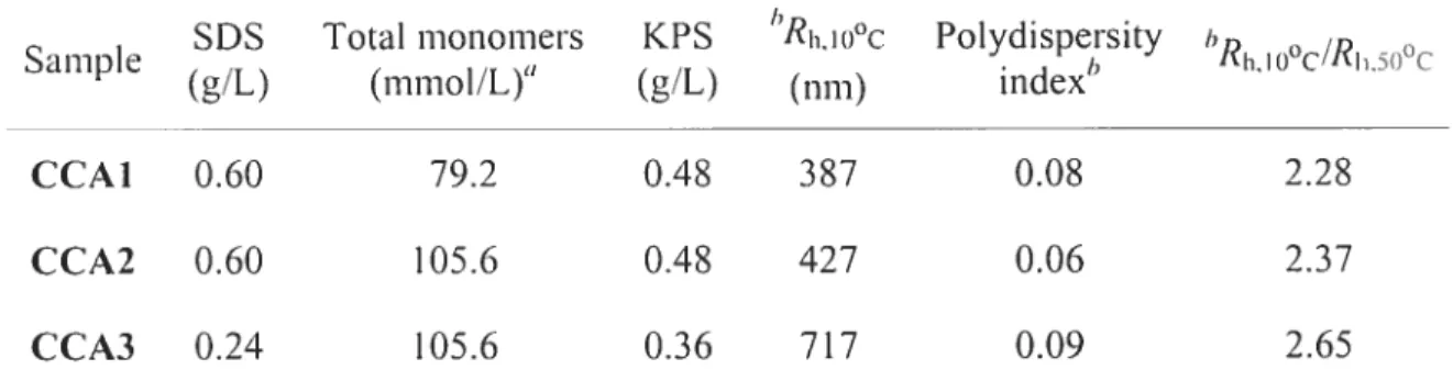
synthesized by emulsion polymerization (Table 2.1). EA was used to raise the LCST while the use of HEMA allows the introduction of a hydroxyl group that could be further functionalized for sensing applications. The particle size was controlled by varying the concentrations of SDS and monomers (Table 2.1).
Table 2.1 Emulsion polymerization conditions for the preparation of microgel particles.
SDS Total monomers KPS hRh,Iooc Polydispersity 1)
Sample . I) Rh,Io0c/RI.5o0c
(g/L) (mmol/L) (g/L) (nm) mdcx
CCAI 0.60 79.2 0.4$ 387 0.0$ 2.28
CCA2 0.60 105.6 0.4$ 427 0.06 2.37
CCA3 0.24 105.6 0.36 717 0.09 2.65
tfeed composition of 1-IEMA : DEA EA 0.3 0.42 0.28 (molar ratios) cross-linked with 1.5 iiiol% oCBA:
hR1110 O (hydrodynamic diameter at 10 °C), polydispersity index and RIlloo(/RI5t)oc of the
particles measured by DLS.
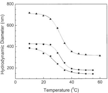
figure 2.2 shows the change ofthe hydrodynamic diameter ofthe particles as a function of temperature. Light diffraction behaviors of particles of various sizes were studied as a function of temperature and particle concentration. A dilute solution of microgel particles was first concentrated via centrifugation followed by dilution of the sample to the desired concentration. As shown in Figure 2.2, the particle volume typically changes by a factor larger than 8 upon increasing temperature above its LCST. In order to generate the CCAs, the temperature of the latex samples was cycled between 10 and 45 oc and diffraction spectra were taken before and after this process and recorded at 10 °c (Figure 2.3A). Before temperature cycling, the spectrum is broad and featureless, whereas after this process the thermosensitive particles spontaneously “crystallize” into a brightly iridescent material displaying a narrow and symmetrical diffraction peak with half
width of 150 nm, a phenomenon also observed by Lyon and co-workers for PNIPAM CcAs.9’2 Since the “crystallization” of the microspheres depends on their size and concentration, the large volume phase transition observed by DLS confers sufficient freedom to the particle to rearrange and adopt a thermodynamically favored configuration upon cooling below their LCST, thus forming better defined ccAs possessing sharper light diffraction features.
800 C 600 G) k G) N . 400 E G) C > -D
o
u.’ 200 ‘ . > B —•• I .——-—--II—-.. 0 20 40 60 Temperature (°C)Figure 2.2 Hydrodynamic diameters ofcolloidal particles making CCAs 1
(.),
2(.),
and 3 (Â) as a [onction oftemperature (measured by DLS).The light diffraction spectra of a 7 wt% solution of CCA1 were taken at difTereit temperatures (Figure 2.3B). The diffraction peak increases slightly while raising temperature from 5 to 10 °C, presumably due to the increase of the refractive index ofthe particles with the temperature, before decreasing again above 15 °C. M 40 °C, the diffraction peak disappears completely. In the CCAs, the particles are believed to be closely packed and compressed. The particle size decreases with increasing temperature, with no change in the interparticle distance (d). When their hydrodynamic diameter measured by DLS becomes smaller than d, the particles can move freely and the cc is disrupted. The diffraction disappears above LCST, in contrast to the observation by Asher and co-workers for the CA of a charged
copolymer, poly(NIPAM-co-2-acrylamido-2-methyl- 1-propanesulfonic acid),
showing a diffraction peak even above the LCST of this material.6 Indeed, in their case, the assembÏy of the charged particles was stabilized by electrostatic repulsions and therefore did not require annealing. The formation of CcAs made of uncharged thermosensitive particles is a reversible process, which needs temperature cycÏing to be performed to improve the ordering ofthe soft particles.
A ing /10°C,after I
/
annealing cycling 400 500 600 700 800 900 10°C B I \\ 5°C //‘ N 1/ 15°C 400 500 600 700 800 900 Wavelength (nm) oFigure 2.3 (A) Light diffraction spectra of CCAT (7 wt4) at 10 C
before and after temperature cycling; (B) light diffraction spectra of
CCAI (7 wt%) at different temperatures after temperature cycling.
In order to understand the CCA formation from soft microspheres, diffraction spectra were obtained at different concentrations for CCA1 at
io
oc (figure 2.4A). The diffraction peak undergoes a blue shift with increasing concentrations. The increased compression between the particles at higher concentrations reduces d andE C -c 3) C a) G) > (‘3 C o o (‘3 D
therefore blue-shifts the diffraction peak. Such an increased compression helps
in
the formation of a more ordered packing structure, which may explain the obvious narrowingin
the diffraction spectrum as the concentration increases. The diffraction wavelengths L), taken as the mass center of the spectra, are plotted as a function of particle concentrationin
figure 2.4B for CCAs 1, 2, and 3.400 500 600 700 800 900 Wavelength (nm) B 1000 800 600 400 — 0.0 . \ . 0.1 0.2 0.3
Particle Weight Fraction
Figure 2.4 (A) Light diffraction spectra of CCA1 at different weight fractions
in
water; (B) diffraction wavelength of CCAs 1(.),
2(.), and
3 (À) as a function ofweight fraction ofthe particles.It is apparent that CCAs formed by larger particles show diffraction peaks at higher wavelengths. Therefore, it is possible to obtain photonic materials with diffraction wavelengths ranging ftom infrared to ultraviolet by simply adjusting the
size and concentration of the particles. The interparticle distance calculated by using Bragg’s law (n = 1.333) and assuming a fCC crystalline structure (as vas obtained ror PN J PAM ) is smaller than the hycirodynamic diameter of the particles at 10 oc and further decreases with increasing concentrations. The interparticle distance reaches a plateau at a certain concentration (‘- 10 wt%), indicating the compression
limit of the CCA, where the interparticle distance is comparable to the hydrodynamic diameter measured above LcST. At very low concentrations (< 3
wt%), the particles are too far apart, and do not form ccAs. When particle
concentration is between ca. 3 and 10 wtYo, the particles are compressed and form
ccAs, below their LCST. 2.4.2.Preparation of
PCCAs
It is \veIl-known that PccAs constituted of charged PS in PAAm hydrogel matrices do not display temperature sensitivity although they can be used as biological and chemical sensors.21720’21 PcCAs formed by charged PS in PNIPAM, however, display a bIne shift of their diffraction wavelength with increasing temperatures.2’ Asher and co-workers reported that PCCAs constituted of charged PNIPAM in PAAm
can be Ltsed as fast optical switches triggered by temperature changes.4 PCcAs of
uncharged thermosensitive particles in non—thermosensitive and thermosensitive matrices like PAAm and PNIPAM have been less studied, most probably due to their diffïculty of their preparation and the complexity of their properties, but were found to display interesting properties for applications as biocompatible sensors.’2’’3 Lyon and co-workers reported the deswelling behavior of PNIPAM microgel composite films
upon heating and a loss of the ordered CCA structure during photopoÏymerization.’2 We
found that it is critical to keep the reaction temperature below the LCST of the material to maintain the order during the preparation of the cc with soft thermosensitive microspheres. Moreover, the swollen state ofthe CA below LCST allows the diffusion
of the monomers within its bulk, forming an interpenetrated polymer system upon photopolymerization.
AAm, DMA, DEA and NIPAM, which are ail water-solttble, were used separately as the monomers and BA as the cross-linker to embed CCAI. The light diffraction spectra of CCA/monomer solutions (%CCA), PCCAs after photopolyrnerization %PccA),
and PCCAs after washing and swelling in milli-Q water (220) were measured at 20 oc
(Figure 2.5). Data obtained for the different samples are gathered in Table 2.2. %PCCA
was found to be srnaÏler than 2CCA due to polyrnerization shrinkage. After washing and
swelling in water, the diffraction wavelength (220) increases, relative to both %CCA and 2PCCA• It is possible to calculate the shrinkage of the matrix by using the following
2.4 6,20,2 I equation:
Shrinkage (V0 —V) / JÇ = 1
—(IQc’A ‘2ccA) (2)
where V0 and V are the volumes before and after polyrnerization, respectively.
From these resuits (Table 2.2), it is apparent that both the concentration of the starting
cc
and the nature of the matrix have an important effect on the shrinkage of the film. In other composite materials, increasing amounts of fillers have also been shown to reduce polymerization shrinkage.22’23Table 2.2 Optical properties of PccAs in non-thermosensitive and thermosensitive matrices prepared at different CCA 1 concentrations.
Sample Matrix CCCA ‘PCCA “CCA 220 A% Shrinkage
(
wt%) (nm) (nrn) (nni) (nm)° PCCAJ PAAm 5 480 658 711 50.2 0.61 PCCA2 PAAm 10 466 599 685 85.3 0.53 PCCA3 PAAm 18.9 430 508 592 105.0 0.39 PCCA4 PDMA 18.9 447 509 578 106.6 0.32 PCCA5 PNIPAM 5 441 670 691 243.6 0.71 PCCA6 PNIPAM 10 417 598 642 216.9 0.66 PCCA7 PNIPAM 18.9 486 508 575 218.8 0.12 PCCA8 PDEA 18.9 485 508 572 190.6 0.132 1 3
Figure 2.5 Typical diffraction spectra of CCA1 (1), PCCA3 just after photopolymerization (2), and PCCA3 washed and equilibrated in water (3) at 20 oc.
2.4.3. PCCAs with non-thenïiosensitive matrices
The ccAs used in this study show thermo-optical properties related to the LCST of the soft particles. PCcAs in both thermosensitive and non-therniosensitive matrices
also show thenno-optical properties. The case of CcAs embedded in non
thermosensitive matrices will first be discussed to distinguish the contributions of the CCA and the matrix from the opalescent phenomena.
Figure 2.6A shows the diffraction spectra of PCCA3 at different temperatures. The peaks are naiiow and symmetrical with haif width of 110 nrn, suggesting a well
ordered particle packing, and a well-defined periodic length. it is apparent that the diffraction spectra are blue-shifted with increasing temperature. The mass centers of the
diffraction spectra versus temperature forPccAs 1, 2 and 3 are plotted in Figure 2.63. The position of the inflexion conelates wefl with the VPT temperature or LCST of the
particles, suggesting that the shrinkage of the particles in the cc can drag the matrix
along, reduce the interpianar distance, and blue-shift the diffraction spectrum. The shrinkage is apparent to the naked eye. Such an efficient dragging phenomenon is the
400 500 600 700 800 900 Wavelength (nm)