LA FORCE DE RÉACTION AU SOL
VERTICALE MAXIMALE COMME TÉMOIN
D’EFFETS FONCTIONNELS ET
STRUCTURAUX CHEZ DES MODÈLES CANINS
D’ARTHROSE : POTENTIEL ENVERS LE
DÉVELOPPEMENT THÉRAPEUTIQUE
par
MAXIM MOREAU
Département de biomédecine vétérinaire Faculté de médecine vétérinaire
Thèse présentée à la Faculté de médecine vétérinaire en vue de l’obtention du grade de
philosophiae doctor (Ph.D.) en sciences vétérinaires
option pharmacologie
Décembre, 2014 © Maxim Moreau, 2014
i
Résumé
Les modèles animaux d’arthrose permettent d’évaluer le potentiel d’agents thérapeutiques en phase préclinique de développement. Le présent ouvrage tient compte du chien comme modèle d’arthrose naturelle (chez l’animal de compagnie) ou expérimentale (par sectionnement chirurgical du ligament croisé crânial). Au sein des expérimentations, la force de réaction au sol verticale maximale, mesurée lors de l’analyse cinétique de la locomotion, est proposée comme témoin d’effets fonctionnels et structuraux sur ces modèles d’arthrose.
Sur un modèle canin d’arthrose naturelle, le seuil de changement minimal détectable a été déterminé. Les changements au dysfonctionnement locomoteur peuvent désormais être cernés en s’affranchissant de la marge d’erreur inhérente à la mesure de la force verticale maximale. Il en découle l’identification de répondants lors d’essais cliniques entrepris chez le chien arthrosique. Une analyse rétrospective a, par la suite, déterminé un taux de répondants de 62.8% et d’une taille d’effet de 0.7 pour des approches thérapeutiques actuellement proposées aux chiens arthrosiques. Cette analyse détermina également que la démonstration d’une réponse thérapeutique était favorisée en présence d’un fort dysfonctionnement locomoteur.
Sur un modèle canin d’arthrose par sectionnement chirurgical du ligament croisé crânial, la force verticale maximale a démontré une relation inverse avec certains
ii
types de lésions arthrosiques évaluées à l’aide d’imagerie par résonance magnétique. Également, la sensibilité de la force verticale maximale a été mise en évidence envers la détection d’effets structuraux, au niveau de l’os sous-chondral, par un agent anti-résorptif (le tiludronate) sur ce même modèle.
Les expérimentations en contexte d’arthrose naturelle canine permettent de valider davantage les résultats d’essais cliniques contrôlés utilisant la force verticale maximale comme critère d’efficacité fonctionnelle. Des évidences cliniques probantes nécessaires à la pratique d’une médecine basée sur des faits sont ainsi escomptées. En contexte d’arthrose expérimentale, la pertinence d’enregistrer le dysfonctionnement locomoteur est soulignée, puisque ce dernier est en lien avec l’état des structures. En effectuant l’analyse de la démarche, de pair avec l’évaluation des structures, il est escompté de pouvoir établir la répercussion de bénéfices structurels sur l’inconfort articulaire.
Cet ouvrage suggère qu’une plateforme d’investigations précliniques, qui combine le modèle canin d’arthrose par sectionnement chirurgical du ligament croisé crânial à un essai clinique chez le chien arthrosique, soit un moyen de cerner des bénéfices structuraux ayant des impacts fonctionnels. Le potentiel inférentiel de ces modèles canins d’arthrose vers l’Homme serait ainsi favorisé en utilisant la force verticale maximale.
iii
Mots-clés : Analyse cinétique démarche, essais cliniques, médicament, recherche préclinique, douleur, boiterie
iv
Abstract
Animal models of osteoarthritis are useful to evaluate the potential of osteoarthritis therapeutics at the preclinical stage of development. In this thesis, the dog is used as a model of naturally-occurring (i.e. companion animal) and experimentally induced (i.e. by surgical transection of the cranial cruciate ligament) osteoarthritis. The peak of the vertically-oriented ground reaction force, which is measured during kinetic gait analysis, is proposed to be an indicator of structural and functional benefits in these models of osteoarthritis.
In a canine model of naturally-occurring osteoarthritis, the threshold of the minimal detectable change in peak vertical force was determined. An improvement in the locomotor disability can now be identified according to the measurement error (noise) of the peak vertical force. This allows the identification of responders when the peak vertical force is used as an outcome measure of functional benefits. A retrospective analysis later determined that current therapeutic approaches provided a responder rate of 62.8% with an effect size of 0.7 in dogs with naturally-occurring osteoarthritis. This analysis also determined that the therapeutic response is favored in cases of severe locomotor disability.
v
In a canine model of osteoarthritis induced by surgical transection of the cranial cruciate ligament, the peak vertical force demonstrated an inverse relationship with different types of structural changes, as evaluated upon magnetic resonance imaging. The sensitivity of the peak vertical force to detect structural benefits on the subchondral bone was also shown in this model using an antiresorptive agent (i.e. tiludronate).
The experiments conducted in dogs with naturally-occurring osteoarthritis further validate findings from clinical trials in which the peak vertical force is used as an outcome measure of functional benefits. The practice of an evidence-based medicine is then expected. The experiments conducted in dogs with surgically-induced osteoarthritis support the recording of the locomotor disability, being in line with the level of the structural changes. By performing gait analysis in addition to structural evaluations, it is expected to establish the impact of structural benefits on joint discomfort
This thesis suggests that a platform for preclinical investigations, which combines the canine model of osteoarthritis induced by surgical transection of the cranial cruciate ligament and a clinical trial in dogs with naturally-occurring osteoarthritis, offers the opportunity to discern structural benefits having functional impacts. A better prediction of outcomes for human clinical trials is expected by using the peak vertical force.
vi
Keywords : Kinetic gait analysis, clinical trials, drugs, preclinical research, pain lameness
vii
Table des matières
Résumé ... i
Abstract ... iv
Table des matières ... vii
Liste des tableaux ... xvi
Liste des figures ... xviii
Liste des sigles et des abréviations ... xxi
Remerciements ... xxx
Introduction ... 1
CHAPITRE 1. RECENSION DES ÉCRITS ... 6
1 Le mouvement locomoteur ... 7
1.1 La locomotion terrestre ... 7
1.1.1 Étude cinétique de la locomotion terrestre ... 7
1.1.2 Le mouvement locomoteur et les forces de réaction au sol ... 9
1.2 Article I: The kinetic measurements of gait for osteoarthritis research in dogs and cats ... 11
1.3 Abstract ... 13
1.4 Résumé ... 13
1.5 Introduction ... 14
1.6 Nature of GRF ... 15
1.6.1 Weight and force ... 15
1.6.2 Action and reaction forces ... 16
1.7 Ground reaction force patterns ... 16
1.7.1 Vertical ground reaction force ... 20
1.7.2 Craniocaudal and mediolateral ground reaction forces ... 21
1.8 Ground reaction vector ... 21
1.9 Center of force ... 25
1.10 Impulse, linear momentum and power ... 26
viii
1.12 Ground reaction force acquisition setting ... 28
1.13 Ground reaction force measurement in experimental model of osteoarthritis ... 32
1.13.1 Joint inflammatory pain ... 32
1.13.2 Structural changes of osteoarthritis ... 33
1.14 Ground reaction force measurement in naturally-occurring osteoarthritis 35 1.15 Conclusion ... 37
1.16 References (Article I) ... 39
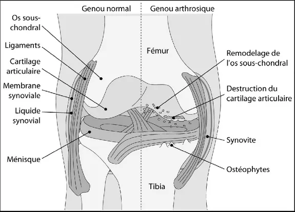
2 La physiopathologie de l’arthrose ... 47
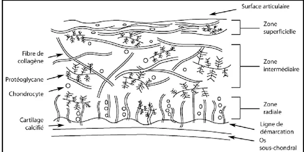
2.1 Le cartilage articulaire en condition d’homéostasie ... 49
2.1.1 Organisation structurelle ... 49
2.1.1 Composants extracellulaires ... 51
2.1.2 Le chondrocyte ... 54
2.1.3 Rôles et propriétés du cartilage articulaire ... 56
2.2 La dégradation du cartilage articulaire lors d’arthrose ... 57
2.2.1 Implication de la sollicitation mécanique excessive ... 59
2.2.2 Implication des composants de la matrice extracellulaire ... 60
2.2.3 Implication des intervenants pro-inflammatoires ... 64
2.2.4 Conclusion ... 70
2.3 L’os sous-chondral ... 71
2.3.1 Organisation structurelle ... 71
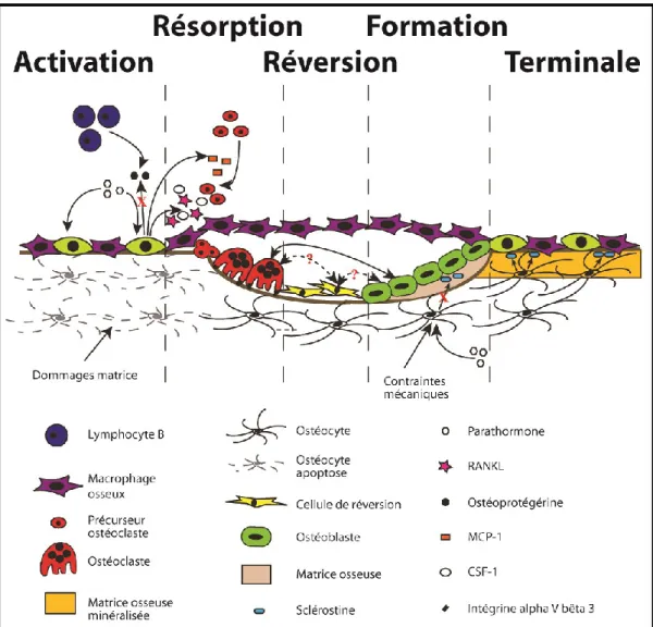
2.3.2 Le remodelage de l’os sous-chondral ... 73
2.3.3 Le remodelage de l’os sous-chondral lors d’arthrose ... 80
2.3.4 Conclusion ... 85 2.4 Synovite ... 86 2.4.1 Membrane synoviale ... 86 2.4.2 Changements inflammatoires ... 87 2.5 Ostéophytose ... 88 3 L’expérience sensorielle ... 89
ix
3.1 Le système somatosensoriel ... 90
3.2 La douleur ... 91
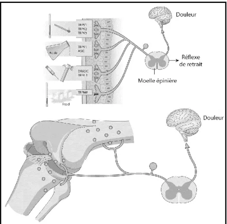
3.3 La nociception ... 92
3.3.1 Transduction d’un stimulus nocif ... 92
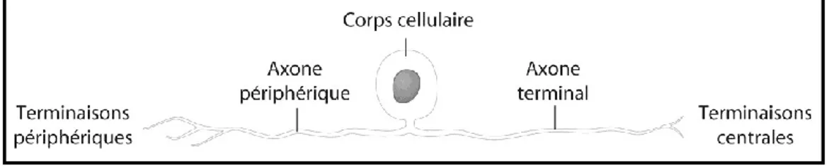
3.3.2 Afférences sensorielles primaires ... 95
3.3.3 Les récepteurs nociceptifs ... 98
3.3.4 Potentiel d’action ... 99
3.3.5 Neurone afférent secondaire ... 101
3.4 Arthrose et plasticité du système somatosensoriel ... 103
3.4.1 Sensibilisation périphérique ... 105
3.4.2 Sensibilisation centrale ... 106
3.4.3 Conclusion ... 108
3.5 Ressenti douloureux et l’os sous-chondral ... 108
4 Instruments métrologiques de l’atteinte fonctionelle en contexte d’arthrose naturelle chez le chien ... 111
4.1 L’analyse télémétrique du mouvement locomoteur ... 111
4.2 Appréciations à l’aide d’un tiers ... 112
4.3 Concepts métrologiques appliqués à la force verticale maximale ... 114
4.4 Biais de mesure ... 118
4.4.1 Effet de l’exercice intense sur la mesure de la force verticale maximale du chien arthrosique ... 119
4.4.2 Article II: Influence of changes in body weight on peak vertical force in osteoarthritic dogs: A possible bias in study outcome ... 119
5 Modèles animaux d’arthrose expérimentale ... 139
5.1 Modèles par injection intra-articulaire d’agents délétères ... 140
5.2 Modèles par altération fonctionnelle ... 140
5.2.1 Modèle canin d’arthrose par sectionnement chirurgical du ligament croisé crânial ... 141
5.3 Modèle par altération physiologique ... 142
x
6 Modèle canin d’arthrose naturelle ... 144
6.1 La force verticale maximale comme témoin d’effets thérapeutiques en contexte d’arthrose naturelle chez le chien ... 144
6.1.1 Article III: Effects of feeding a high omega-3 fatty acids diet in dogs with naturally occurring osteoarthritis ... 146
6.1.2 Article IV. Brachystemma Calycinum D. Don effectively reduces the locomotor disability in dogs with naturally occurring osteoarthritis: A randomized placebo-controlled trial ... 169
6.1.3 Article V. A medicinal herb-based natural health product improves the condition of a canine natural osteoarthritis model: A randomized placebo-controlled trial ... 202
7 Modèle félin d’arthrose naturelle ... 242
7.1 Article VI: Kinetic peak vertical force measurement in cats afflicted by coxarthritis: Data management and acquisition protocols ... 243
7.1.1 Abstract ... 245
7.1.2 Introduction ... 246
7.1.3 Material and methods ... 248
7.1.4 Results ... 253
CHAPITRE 2. EXPÉRIMENTATIONS ... 271
8 Modèle canin d’arthrose naturelle : La force verticale maximale et la détection d’effets thérapeutiques ... 272
8.1 Préambule ... 272
8.2 Abrégé méthodologique et hypothèses ... 274
9 Modèle canin d’arthrose par sectionnement chirurgical du ligament croisé crânial : Relations entre la force verticale maximale et les dommages structuraux275 9.1 Préambule ... 275
9.2 Abrégé méthodologique et hypothèses ... 276
10 Article VII. A posteriori comparison of natural and surgical destabilization models of canine osteoarthritis ... 277
xi
10.2 Introduction ... 280
10.3 Materials and Methods ... 282
10.3.1 Dog model of experimental OA ... 282
10.3.2 Dog model of naturally-occurring OA ... 285
10.3.3 Statistical analyses ... 287
10.4 Results ... 290
10.4.1 Dog model of experimental OA ... 290
10.4.2 Dog model of naturally-occurring OA ... 296
10.5 Discussion ... 298
10.5.1 Dog model of experimental OA ... 298
10.5.2 Dog model of naturally-occurring OA ... 304
10.6 Conclusion ... 307
10.7 Conflict of Interests ... 309
10.8 Acknowledgements ... 309
10.9 References (Article VII) ... 310
11 Modèle canin d’arthrose naturelle : Analyse de répondants selon la force verticale maximale ... 324
11.1 Préambule ... 324
11.2 Hypothèses ... 325
11.3 Méthodologie ... 325
11.3.1 Essais cliniques sélectionnés ... 325
11.3.2 Sujets ... 331 11.3.3 Analyse de la démarche ... 331 11.3.4 Période de retrait ... 332 11.3.5 Analyses statistiques ... 332 11.4 Résultats ... 333 11.4.1 Taux de répondants ... 333 11.4.2 Régression binomiale ... 337
12 Modèle canin d’arthrose par sectionnement chirurgical du ligament croisé crânial : La force verticale maximale et la détection d’effets structuraux ... 338
xii
12.1 Préambule ... 338
12.2 Abrégé méthodologique et hypothèses ... 339
13 Article VIII. Tiludronate treatment improves structural changes and symptoms of osteoarthritis in the canine anterior cruciate ligament model ... 340
13.1 Abstract ... 342
13.2 Introduction ... 344
13.3 Materials and methods ... 347
13.3.1 Animals ... 348
13.3.2 Surgical transection of the anterior cruciate ligament (ACL) ... 349
13.3.3 Treatment ... 349
13.3.4 Pain and functional evaluations ... 350
13.3.5 Macroscopic grading ... 353
13.3.6 Histological grading of cartilage and synovial membrane ... 354
13.3.7 Analysis of synovial fluid ... 355
13.3.8 Immunohistochemistry ... 355
13.3.9 Histomorphometry ... 357
13.3.10 Statistical analysis ... 358
13.4 Results ... 360
13.4.1 Pain and functional outcomes ... 360
13.4.2 Synovial fluid ... 363 13.4.3 Cartilage ... 363 13.4.4 Immunohistochemistry ... 364 13.4.5 Histomorphometry ... 366 13.5 Discussion ... 367 13.6 Conclusions ... 373 13.7 List of abbreviations ... 374 13.8 Competing interests ... 374 13.9 Authors’ contributions ... 374 13.10 Acknowledgements ... 376
xiii
CHAPITRE 3. DISCUSSION GÉNÉRALE ... 386
14 Modèle canin d’arthrose naturelle ... 387
14.1 La force verticale maximale et la détection d’effets thérapeutiques lors d’essais cliniques contrôlés ... 387
14.1.1 Retour sur les hypothèses ... 388
14.1.2 Le changement minimal détectable (à un intervalle de confiance de 95 %) comme critère de discernement d’un effet thérapeutique ... 389
14.1.3 Procédurier statistique lors de l’analyse de la force verticale maximale en contexte d’essais cliniques contrôlés ... 393
14.2 Le mouvement locomoteur et la force verticale maximale ... 394
14.3 L’activité quotidienne comme modalité thérapeutique chez le chien arthrosique ... 397
14.4 Le mouvement locomoteur comme source de biais potentiel envers la force verticale maximale ... 399
14.5 Analyse de répondants selon la force verticale maximale ... 400
14.5.1 Retour sur les hypothèses ... 400
14.5.2 Le taux de répondants ... 401
14.5.3 Relation binomiale ... 401
14.6 Hiérarchisation de l’approche thérapeutique contre l’arthrose ... 402
14.7 Effet nocebo selon la force verticale maximale ... 404
14.8 Effet placebo selon la force verticale maximale ... 407
14.8.1 Article IX. Does a placebo effect really occur in dogs afflicted by hip osteoarthritis as measured by force platform gait analysis? ... 407
14.9 Validation de la force verticale maximale comme critère d’efficacité lors d’essais cliniques contrôlés chez le chien arthrosique ... 422
14.9.1 La validité de contenu ... 423
14.9.2 La validité de critères ... 423
14.9.3 La validité interne ... 425
14.9.4 La validité externe ... 428
xiv
14.9.6 Stabilité ... 429
14.10 La force verticale maximale en contexte d’essais cliniques contrôlés : Conclusion ... 430
15 Modèle canin d’arthrose par sectionnement chirurgical du ligament croisé crânial ... 431
15.1 La force verticale maximale et le dysfonctionnement locomoteur ... 431
15.2 Le sectionnement chirurgical du ligament croisé crânial et l’altération à la dynamique de l’articulation ... 432
15.3 La force verticale maximale et les dommages structuraux à l’aide d’imagerie par résonance magnétique ... 434
15.3.1 Retour sur les hypothèses ... 434
15.3.2 Les lésions focales au cartilage ... 435
15.3.3 Les lésions de la moelle osseuse ... 436
15.3.4 Les ostéophytes ... 437
15.3.5 L’effusion articulaire ... 438
15.3.6 Le volume de cartilage et l’atteinte aux ménisques ... 439
15.4 Mécanistique de la relation entre le dysfonctionnement locomoteur et les dommages structuraux ... 441
15.5 La force verticale maximale et les dommages macroscopiques au cartilage 443 15.5.1 Retour sur les hypothèses ... 443
15.6 La force verticale maximale et la plasticité du système somatosensoriel 445 15.7 La force verticale maximale et la détection d’effets structuraux ... 447
15.7.1 Retour sur les hypothèses ... 447
16 Conclusion générale ... 452
Références bibliographiques ... 458
Annexe I. ... xxxiv
Annexe II. ... xxxv
xv
Annexe IV. ... xxxvii Annexe V. ... xxxviii Annexe VI. ... xxxix Annexe VII. ... xl Annexe VIII. ... xli
xvi
Liste des tableaux
Tableau I. Concept de validation intégré à l’erreur de mesure ... 117 Tableau II. Modèles d’arthrose par altération fonctionnelle d’un membre ... 141 Tableau III. Summary of the diet’s content based on dry matter basis ... 152 Tableau IV. Baseline characteristics of the dogs ... 156 Tableau V. Baseline characteristics of the dogs stratified per group ... 180 Tableau VI. Selected studies that reported statistically significant changes (i.e.
improvement) in peak vertical force following different therapeutic approaches in dogs afflicted by osteoarthritis. ... 190 Tableau VII. Ingredients includes in each natural health products formulations ... 212 Tableau VIII. Baseline characteristics of the dogs stratified per group ... 218 Tableau IX. Statistics for the peak vertical force measured before and after ten
minutes of treadmill exercise in six cats with coxarthritis according to three methods of outcome management at D1. ... 254 Tableau X. Statistics for the forces measured before and when trotting on an
inclined plane (13°) in six cats with coxarthritis according to three methods of outcome management at D42 ... 256 Tableau XI. Statistics for the peak vertical force measured before and after
stair climbing in six cats with coxarthritis according to three methods of outcome management at D84 ... 257 Tableau XII. Sample size and effect size consistent with a 5% change in peak
vertical force measured in six cats with coxarthritis according to three methods of outcome management ... 258 Tableau XIII. Correlation analyses of the change in peak vertical force
measurement and magnetic resonance imaging over the different phases of functional impairment before (Baseline) and following cranial cruciate ligament transection in five dogs ... 294
xvii
Tableau XIV. Regression analyses between the recording of the peak vertical force and macroscopic measurement of cartilage thinning at eight weeks following cranial cruciate ligament transection in 25 dogs ... 296 Tableau XV. Characteristics of peak vertical force measurement in 40
privately-owned dogs affected by naturally-occurring osteoarthritis ... 297 Tableau XVI. Pain and functional outcomes before and after anterior cruciate
ligament transection in dogs ... 362 Tableau XVII. Histomorphometry of the calcified cartilage and subchondral
xviii
Liste des figures
Figure 1. Sagittal and frontal views of a dog in a standing position ... 18
Figure 2. Typical curves of the ground reaction forces ... 19
Figure 3. Ground reaction vector ... 22
Figure 4. Progression of the ground reaction vector ... 24
Figure 5. Patterns of footfalls ... 30
Figure 6. Schématisation des éléments caractéristiques d’arthrose ... 48
Figure 7. Schématisation de la structure et des principaux composants du cartilage articulaire ... 50
Figure 8. Phases du processus de remodelage de l’os sous-chondral ... 77
Figure 9. Schématisation de l’afférence nociceptive périphérique vers les centres supérieurs du système nerveux central ... 94
Figure 10. Schématisation d’un neurone afférent primaire ... 95
Figure 11. Schématisation de l’afférence nociceptive de l’os sous-chondral .. 110
Figure 12. Schématisation de la relation entre attribut, variable et mesure .. 118
Figure 13. Scatter plot of the changes (D30 minus D90) in body weight (BW) against changes in peak vertical force (PVF) values ... 128
Figure 14. Individual dot plot of peak vertical force recorded in privately-owned dogs with naturally occurring osteoarthritis after 7 and 13 weeks of feeding either Control diet or a Veterinary therapeutic diet rich in omega-3 ... 158
Figure 15. Individual changes in peak vertical force recorded in privately-owned dogs with naturally occurring osteoarthritis after 13 weeks of feeding either Control diet or a Veterinary therapeutic diet rich in omega-3 ... 159
Figure 16. Temporal evolution of the CSOM recorded in privately-owned dogs with naturally occurring osteoarthritis after 7 and 13 weeks (W) of feeding either Control diet or a Veterinary therapeutic diet rich in omega-3 ... 160
xix
Figure 17. Flow chart of the study enrolment, randomization, follow-up and
analysis ... 181
Figure 18. Mean (standard deviation) peak vertical force recorded in dogs having received either Brachystemma calycinum D don (BCD) or a placebo ... 183
Figure 19. Individual changes in peak vertical force after 6 weeks of treatment with Brachystemma calycinum D don (BCD) or a placebo ... 184
Figure 20. Temporal evolution of the locomotor activity recording over a 6-week period (42 days) in dogs receiving either treatment with Brachystemma calycinum D don (BCD) or a placebo ... 186
Figure 21. Temporal evolution of the case-specific outcome measures of disability (CSOM) over a 6-week period in dogs receiving either treatment with Brachystemma calycinum D don (BCD) or a placebo ... 188
Figure 22. Flow chart of the study enrolment, allocation, follow-up and analysis ... 217
Figure 23. Peak vertical force ... 219
Figure 24. Changes in peak vertical force ... 220
Figure 25. Locomotor activity recording ... 222
Figure 26. Case-specific outcome measures of disability ... 224
Figure 27. Schematic view of the data reported in A) the dog model of experimental osteoarthritis and B) the dog model of naturally-occurring osteoarthritis ... 284
Figure 28. Averaged peak vertical force values measured before (Baseline) and four, eight and 26 weeks after cranial cruciate ligament transection in dogs ... 291
Figure 29. Individual peak vertical force values measured before (Baseline) and four, eight and 26 weeks after cranial cruciate ligament transection in dogs ... 292 Figure 30. Significant correlation (rs -0.99, p<0.001) for the differences of
xx
gradient recalled echo images (T1w-GRE) scores during the remission phase (week 26 minus week four), with the concurrent difference in peak vertical force measurement ... 293 Figure 31. Individual changes in peak vertical force measured at week four in
40 privately-owned dogs receiving a placebo in randomized controlled trials ... 297 Figure 32. Changements individuels de force verticale maximale selon le
groupe thérapeutique ... 335 Figure 33. Changements individuels de force verticale maximale chez les chiens
ayant reçu un contrôle négatif selon le groupe thérapeutique ... 336 Figure 34. Schematic representation of the study design ... 348 Figure 35. Kinetic gait analysis ... 361 Figure 36. Immunohistochemistry ... 365 Figure 37. Histomorphometry ... 366
xxi
Liste des sigles et des abréviations
% BW: Percentage of body weight – Pourcentage de poids corporel %: Percentage – Pourcentage
: Pearson correlation coefficient – Coefficient de corrélation de Pearson
g: Micrograms – microgrammes
∆: Delta
∫F: Function – Fonction
°: Degree – Degré
Θ: Thêta angle – Angle théta σ: Standard deviation – Écart-type µm: Micrometers – Micromètres
95% CI: 95% confidence intervals – Intervalles de confiance à 95% ACL: Anterior cruciate ligament – Ligament croisé antérieur ADAM: A disintegrin and metalloprotease
ADAMTS: A disintegrin and metalloproteases with thrombospondin motifs AMPA: Alpha-amino-3-hydroxy-5-méthylisoazol-4-propionate
ANCOVA: Analysis of covariance – Analyse de covariance ANOVA: Analysis of variance – Analyse de variance AINS : Anti-inflammatoire non stéroïdien
AP1: Activator protein one ARN: Acide ribonucléique
xxii ASIC: Acid-sensing ion channels ATP: Adénosine triphosphate
AUC: Area under the curve – Aire sous la courbe avertical: Vertical acceleration – Accélération verticale b: Regression y-intercept – Intercepte avec l’axe des Y BCD: Brachystemma calycinum D don
BDNF: Brain-derived neurotrophic factor – Facteur neurotrophique issu du cerveau
BMLs: Bone marrow lesions – Lésions de la moelle osseuse BMU: Basic multicellular unit – Unité multicellulaire de base BPs: Bisphosphonates – Biphosphonates
BW: Body weight – Poids corporel CBPI: Canine brief pain inventory
CCL: Cranial cruciate ligament – Ligament croisé crânial
CCLT: Surgical cranial cruciate ligament transection – Sectionnement chirurgical du ligament croisé crânial
CGRP: Calcitonin gene related peptide cm: Centimeters – Centimètres
CoD: Coefficient of dispersion – Coefficient de dispersion COF: Center of force – Centre de la force
COM: Center of mass – Centre de la masse COX: Cyclooxygenase – Cyclooxygénase
xxiii
CRCHUM: Centre de recherche du Centre Hospitalier de l’Université de Montréal
CSF-1: Macrophage colony-stimulating factor one
CSOM: Case specific outcome measure – Mesure d’évaluation spécifique au patient
CTR: Control group – Groupe contrôle
D: Day – Jour
d: Perpendicular distance from the center of rotation to the force vector – Distance perpendiculaire au centre de rotation vers le vecteur force DAAI: Daily averaged active intensity – Intensité d’activité quotidienne
moyenne
DATI: Daily averaged total intensity – Intensité totale quotidienne moyenne DDAP: Daily duration of active period – Durée quotidienne de période
d’activité
DDR2: Discoidin domain receptor two
DHA: Docosahexaenoic acid – Acide docosahexaénoïque
DMOAD: Disease-modifying OA drug – Agent structuro-modulateur dans l’arthrose
DRASIC: Dorsal root acid sensing ion channel Dt: Time derivative – Dérivée de temps
EDA: Electrodermal activity – Conductance électrique du derme EP: Récepteurs membranaires aux prostaglandines E2
xxiv ES: Effect size – Taille de l’effet
F: Force
FRS: Forces de réaction au sol
g0: Standard gravitational acceleration on earth – Accélération gravitationnelle standard sur terre
GABA: Acide γ-aminobutyrique GDU: Gelatin digesting unit
GREPAQ: Groupe de Recherche en Pharmacologie Animale du Québec GRF: Ground reaction forces
GRV: Ground reaction vector – Vecteur de réaction au sol HCPI: Helsinki chronic pain index
Hz: Hertz
I: Impulse – Impulsion
ICC: Intra-class coefficient of correlation – Coefficient de corrélation intra-classe
ICE: Interleukin – 1 beta converting enzyme IGF-1: Insulin-like growth factor one
IL-1: Interleukine-1 bêta
iNOS: Isoforme inductible de l’enzyme oxyde nitrique synthase Kcal: Kilocalories
kg: Kilograms – Kilogrammes km : Surface area to weight ratio
xxv
LACIME: Laboratoire de communications et d’intégration de la microinformatique
LOAD: Liverpool osteoarthritis in dogs LOX: Lipoxygenase – Lipoxygénase
m/s: Meter per second – Mètre par seconde
M: Masse
m: Meters – Mètres
m: Regression slope – Pente de régression MAP: Mitogen-activated protein
MCP-1: Monocyte chimoattractant protein one
MDC: Minimal detectable change – Changement minimal détectable
MDC95: Minimal detectable change at the 95% confidence level – Changement minimal détectable à l’intervalle de confiance à 95% MIA: Monosodium iodo-acetate
MITF: Microphthalmia-associated transcription factor mm: Millimeters – Millimètres
MMPs: Matrix metalloproteinase – Métalloprotéinases matricielles
MRI: Magnetic resonance imaging – Imagerie par résonance magnétique
N: Newton
NF-kappa B: Nuclear factor-kappa B ng: Nanograms – Nanogrammes
NHPs: Natural health products – Produits de santé naturels Nm: Newton meters – Mètres Newton
xxvi NMDA: N-méthyl-D-aspartate
NOS: Nitric oxide synthase – Enzyme oxyde nitrique synthase NOx: Nitrites et nitrates
NRS: Numerical rating scale – Échelle de cotation numérique
NSAIDs: Non-steroidal anti-inflammatory drugs – Agents anti-inflammatoires non stéroïdiens
NY: New York NK-1 : Neurokinine un
OA: Osteoarthritis – Arthrose
OARSI: OsteoArthritis Research Society International Omega-3: Omega-3 fatty acids – Acides gras oméga-3 OMERACT: Outcome Measures in Rheumatology ON: Ontario
p: Linear momentum – Quantité de mouvement linéaire p: Momentum – Quantité de mouvement
P: Power – Puissance P: Probability – Probabilité
P2X: Ionotropic ligand-gated purinergic receptors PAR-2: Protease activated receptor two
pg: Picograms – Picogrammes PGE2: Prostaglandine E2
PNF: Peak normal force – Force normale maximale
xxvii
PVF: Peak vertical force – Force verticale maximale QC: Québec
RANKL: Receptor activator of nuclear factor kappa-B ligand
RCTs: Randomized controlled trials – Essais cliniques randomisés rs: Spearman coefficient – Coefficient de Spearman
RUNX2: Runt-related transcription factor two s: Seconds – Secondes
SC: Subcutaneous – Sous-cutané SD: Standard deviation – Écart-type
SEM: Standard error of measurement – Erreur standard de mesure
SPGR: Three-dimensional spoiled gradient recalled sequence with fat suppression
t: Time - Temps
T1w-GRE: T1-weighted three-dimensional fast gradient recalled echo T2w-FS: T2-weighted fast spin echo sequence with fat saturation TGF-beta: Transforming growth factor beta
TIMPs: Tissue inhibitor of metalloproteinases TLN: Tiludronate
TNF-alpha: Tumor necrosis factor-alpha – Facteur nécrosant tumoral alpha TNF-R1: Tumor necrosis factor receptor one – Récepteur au facteur
tumo-nécrosant alpha
TRAP: Tartrate-resistant acid phosphatase TREK-1: TWIK1-related K + channel
xxviii TRP: Transient receptor potential
TRPM8: Transient receptor potential melastatin eight TRPV1: Transient receptor potential vanilloid one uPA: Urokinase-type plasminogen activator
USA: United States of America – États-Unis d’Amérique v: Velocity – Vitesse
VAS: Visual analog scale – Échelle visuelle analogique
VEGF: Vascular endothelial growth factor – Facteur de croissance de l'endothélium vasculaire
VOL: Volume
VTD: Veterinary therapeutic diet
W: Watts
W: Week – Semaine
xxix
À ma fille et mes fils,
Puissiez-vous trouver à même cet ouvrage une parcelle d’inspiration au dépassement. Sachez que ce qui importe n’est pas tant l’arrivée mais bien l’ascension.
Pour Anne,
Je te suis d’une infinie reconnaissance pour ton support infaillible. Nulle autre que toi n’aura été un si parfait complément durant ce parcours.
xxx
Remerciements
L’auteur voudrait remercier :
Docteur Éric Troncy
Cet homme d’exception, qui par son esprit de meneur, sa capacité remarquable d’intégration scientifique et son dévouement incomparable, a suscité (et maintenu) chez moi un désir de dépassement et d’accomplissement.
Docteur Bertrand Lussier
Pour son encadrement manifeste lors de ce programme de doctorat et pour avoir rendu mémorables nos séjours à l’étranger.
Docteur Jean-Pierre Pelletier
Pour son appel téléphonique en 2003 qui a donné l’envol au développement d’une expertise et d’une profonde satisfaction personnelle.
Docteure Stéphanie Keroack
Pour avoir teinté ce programme de doctorat d’expériences professionnelles des plus enrichissantes.
xxxi
Mes amis les Docteurs Martin Guillot et Jérôme del Castillo
Deux comparses dotés d’un niveau de savoir que je ne peux qu’espérer un jour atteindre.
Mon ami le Docteur Christian Bédard
Pour nos distractions entretenues à mon bureau dans l’underground.
La subvention de formation des Instituts de recherche en santé du Canada en évaluation et traitement des troubles de la mobilité et posture (Programme MENTOR).
Le Fonds de recherche du Québec – Santé.
1
La force de réaction au sol verticale maximale comme témoin d’effets fonctionnels et structuraux chez des modèles canins d’arthrose: Potentiel envers le développement thérapeutique
Introduction
Le squelette du mammifère forme avec les muscles et les articulations le système myo-arthro-squelettique. Les interactions biomécaniques entre les différents composants de ce système permettent l’exécution du mouvement et l’action locomotrice. L’arthrose est une pathologie du système myo-arthro-squelettique qui afflige de manière fulgurante aussi bien l’humain (Lawrence et al., 2008; Cross et al., 2014) que le chien (Canis domesticus) (Johnston, 1997). La présence de dommages arthrosiques altère les propriétés structurelles et le rôle fonctionnel des tissus de l’articulation synoviale, ce qui mène à une perte d’aisance dans l’exécution des mouvements articulaires. Au niveau d’une articulation sollicitée lors de la locomotion, la présence d’arthrose pourra induire une irrégularité de la démarche qualifiée de dysfonctionnement locomoteur (DeCamp, 1997; Constantinou et al., 2014). Ce dysfonctionnement est principalement attribué à un signe clinique prépondérant lors d’arthrose, soit le ressenti douloureux au niveau de l’articulation.
Le développement d’agents anti-arthrosiques (aux effets structuro-modulateurs et/ou analgésiques) requiert des modèles animaux afin d’évaluer le potentiel thérapeutique d’un candidat d’intérêt et ce, en contexte d’arthrose, qu’elle soit d’étiologie naturelle ou expérimentale. Le modèle à préconiser doit être en mesure
2
de produire fidèlement ce qui sera observé ultérieurement lors d’essais cliniques, particulièrement au regard des effets attendus envers les dommages structuraux et le dysfonctionnement locomoteur.
Le présent ouvrage tient compte du chien comme espèce modèle d’intérêt pour l’ensemble des investigations réalisées en contexte d’arthrose d’étiologie naturelle (chez l’animal de compagnie) ou expérimentale (par sectionnement chirurgical du ligament croisé crânial). Les travaux de recherche, présentés au chapitre Expérimentations, ont comme point commun l’usage de la force de réaction au sol verticale maximale mesurée lors de l’analyse cinétique de la locomotion. La force verticale maximale est proposée comme témoin d’effets thérapeutiques face au dysfonctionnement locomoteur et aux dommages structuraux encourus chez deux modèles canins d’arthrose.
Le Chapitre 1 sera dédié à une recension des écrits avec, en premier lieu, une description du mouvement locomoteur. L’Article I traitera des forces de réaction au sol chez des modèles canins et félins d’arthrose; suivront aussi des notions concernant la physiopathologie de l’arthrose, l’expérience sensorielle et l’instrumentation métrologique. L’Article II traitera de l’influence du gain de poids envers la force verticale maximale chez le chien arthrosique. Finalement, un survol des différents modèles d’arthrose sera effectué. Le modèle canin d’arthrose naturelle sera présenté et supporté par trois essais cliniques contrôlés qui ont utilisé la force verticale maximale comme critère d’efficacité (Articles III, IV, V). Le
3
modèle félin d’arthrose naturelle sera également présenté en mettant en lumière l’acquisition et la gestion des données de force verticale maximale chez ce modèle (Article VI).
Le Chapitre 2 sera dédié aux expérimentations sur le modèle canin d’arthrose naturelle et sur celui par sectionnement chirurgical du ligament croisé crânial. En premier lieu, les travaux de recherche seront voués à déterminer la marge d’erreur inhérente à la mesure de la force verticale maximale de même que le seuil de changement minimal détectable sur le modèle canin d’arthrose naturelle (Article VII). L’établissement de ce seuil permettra de distinguer les changements au dysfonctionnement locomoteur (amélioration ou détérioration) en s’affranchissant de la marge d’erreur inhérente à la mesure de la force verticale maximale. Ce seuil servira, ultérieurement, à cerner l’impact de l’activité quotidienne par l’analyse télémétrique du mouvement locomoteur réalisée chez le chien arthrosique. Également, ce seuil permettra de définir ce qu’est un répondant thérapeutique dans un contexte d’essais cliniques effectués chez le chien atteint d’arthrose naturelle. Une analyse rétrospective sera par la suite effectuée afin de déterminer la présence d’un lien entre la fréquence de répondants et différents facteurs, tels que le niveau de dysfonctionnement locomoteur initial et le type de traitement administré.
En second lieu, le lien entre le dysfonctionnement locomoteur, représenté par la force verticale maximale, et la sévérité des différents types de dommages
4
structuraux sera investigué sur un modèle canin d’arthrose par sectionnement chirurgical du ligament croisé crânial (Article VII). Ces travaux de recherche s’avèrent possibles et particulièrement stimulants suite à la récente caractérisation des dommages structuraux à l’aide d’imagerie par résonance magnétique, soit les atteintes au cartilage articulaire (volumétrie et lésions, Annexe II), les lésions à l’os sous-chondral (Annexe III) de même que la taille des ostéophytes et le degré d’effusion articulaire (Annexe IV).
La sensibilité de la force verticale maximale envers la détection d’effets structuro-modulateurs d’une plante thérapeutique au niveau du cartilage articulaire a été récemment démontrée sur le modèle canin d’arthrose par sectionnement chirurgical du ligament croisé crânial (Annexe V). Dès lors, il est proposé, en troisième lieu, de mettre en évidence la sensibilité de la force verticale maximale envers la détection d’effets structuro-modulateurs au niveau de l’os sous-chondral, tel que transmis par un agent anti-résorptif, le tiludronate, chez ce même modèle (Article VIII).
Le Chapitre 3 discutera du modèle canin d’arthrose naturelle et de celui par sectionnement chirurgical du ligament croisé crânial. Il y aura présentation d’arguments critiques envers un effet placebo présumé selon la force verticale maximale (Article IX). Un retour sera effectué sur l’ensemble des travaux de recherche puisqu’ils se proposent d’être inscrits dans une quête de conformité avec la règle des trois « R » qui consiste à Remplacer, Réduire et Raffiner l'emploi d'animaux en recherche (Russell, 1995). Cette thèse s’intègre dans un désir de
5
présenter la force verticale maximale comme témoin d’effets fonctionnels et structuraux chez des modèles canins d’arthrose. Il est proposé que le développement d’agents anti-arthrosiques, tant pour l’humain que pour l’espèce canine puisse être favorisé.
Des annexes sont présentées à la fin de cet ouvrage. Le lecteur est invité à prendre connaissance de l’Annexe I qui décrit les outils de mesure de la force verticale maximale. Le lecteur est également invité à prendre connaissance d’articles complémentaires, présentés aux Annexes II-VIII, qui abordent divers sujets en lien avec ceux discutés au sein de cette thèse.
6
7
1 Le mouvement locomoteur
Le mouvement se définit comme étant le déplacement d'un corps par rapport à un point fixe situé dans l'espace. La locomotion, pour sa part, est la faculté qu’a un organisme vivant de se mouvoir dans son entier afin d’assurer son déplacement. Du mouvement locomoteur découle un changement de la géométrie du corps et une modification de l’emplacement de son centre de masse, dont la trajectoire définit l’orientation du déplacement dans l’espace.
1.1 La locomotion terrestre
La locomotion d’un organisme est possible dans un milieu terrestre, aquatique et aérien. La locomotion terrestre requiert un contact avec le sol. Elle s’effectue chez l’arthropode à l’aide de membres locomoteurs (communément appelés pattes) du système appendiculaire, qui sont constitués de segments s’articulant entre eux. La locomotion terrestre peut également s’effectuer chez l’apode par un phénomène de reptation.
1.1.1 Étude cinétique de la locomotion terrestre
L’analyse du mouvement locomoteur fascine l’Homme, tel qu’en témoigne l’œuvre la salle des Taureaux (grotte de Lascaux, France). Cette peinture à base de pigments sur des parois rocheuses révèle l’attention particulière de l’Homme envers la locomotion du quadrupède et ce, dès la préhistoire, soit vers 18 000 - 15 000 avant J.C. Cette représentation picturale d’une faune en mouvement démontre une qualité exceptionnelle. Ainsi, l’illustration du mouvement locomoteur au sein de cette
8
œuvre pariétale est d’une exactitude considérable, dépassant même la qualité des représentations datant de l’âge moderne, soit de la préhistoire jusqu’à l’époque Muybridge (1887, voir ci-dessous) (Horvath et al., 2012).
L’apparition d’instruments spécialisés a permis un gain considérable envers l’approfondissement des connaissances en ce qui entoure l’analyse du mouvement locomoteur. Il est possible de dénoter les travaux d’Eadweard Muybridge (1830-1904) comme étant déterminant envers l’étude du mouvement. Cet homme, décrit comme étant le père du cinéma, utilisa la décomposition photographique comme témoin rigoureux et objectif du mouvement locomoteur. Il confirma dès lors la présence d’une phase de non appui chez le cheval au galop, théorie précédemment émise par Étienne-Jules Marey (1830-1904, voir ci-dessous) (Sadoul, 1966).
Les années 1870-1890 furent fastes en découvertes et s’inscrivirent comme un moment clé envers la compréhension du mouvement locomoteur. Il est intéressant de préciser que l’exactitude dans les diverses représentations artistiques du mouvement locomoteur (peinture, sculpture) fut grandement améliorée suite aux travaux d’Eadweard Muybridge. À ce moment, ce dernier illustra les phases consécutives du mouvement locomoteur de différents quadrupèdes (Muybridge, 1979, Intégrale réimprimée). Par conséquent, l’époque Muybridge (1887) explique l’amélioration significative de la qualité de la description du mouvement locomoteur (Horvath et al., 2012).
9
Les éléments se rapportant à l’évaluation cinématique, c'est-à-dire la trajectoire d’un point et sa vitesse à un instant donné, sont sous la gouverne de forces qui régissent le mouvement. Tel est le constat d’Étienne-Jules Marey, un médecin et physiologiste français. Ce dernier conçoit un dynamomètre inscripteur pouvant enregistrer la force générée par un sujet stationnaire et non immobile (Marey, 1883). Cet outil est le prototype de ce qui sera plus tard appelé plateforme de force par Herbert Elftman dont ce dernier décrivit le fonctionnement en 1938 (Elftman, 1938). Dès lors, il fut possible de quantifier les forces générées par le déplacement du centre de masse d’un corps en mouvement et ce, dans les trois plans orthogonaux. L’analyse cinétique du mouvement locomoteur voyait ainsi le jour en décrivant les forces qui interviennent dans le mouvement locomoteur.
1.1.2 Le mouvement locomoteur et les forces de réaction au
sol
Sir Isaac Newton (1642-1727) présenta ses trois lois du mouvement en 1687 dans un ouvrage intitulé Principia Mathematica Philosophiae Naturalis (Newton, 1687). En accord avec la troisième loi du mouvement de Newton, pour chaque action d’une force, il y a une réaction. La réaction est de magnitude égale mais de direction opposée à l’action causale.
10
En contact avec le sol, le membre locomoteur exerce des forces orientées selon trois plans (orthonormés X, Y et Z) dont la direction et l'intensité varient à chaque instant en fonction de la progression de la locomotion (Kirtley, 2006; Whittle, 2007). L'analyse des forces de réaction au sol cible, en fonction du temps, l'évolution des forces générées lors de la locomotion terrestre (Kirtley, 2006; Whittle, 2007).
11
1.2 Article I: The kinetic measurements of gait for
osteoarthritis research in dogs and cats
Cet article, publié dans The Canadian Veterinary Journal, présente la mesure des forces de réaction au sol chez le chien et le chat. Sous forme de synthèse, cet article décrit la nature des forces de réaction au sol de même que différents calculs dérivés. Le but de cet article était de présenter les principes fondamentaux qui régissent la mesure des forces de réaction au sol en contexte d’arthrose naturelle et expérimentale chez le chien et le chat.
Cet article est présenté avec la perspective que la mesure des forces de réaction au sol reflète le dysfonctionnement du membre locomoteur. La capacité fonctionnelle du membre s’avère être particulièrement limitée en présence d’arthrose d’étiologie naturelle ou expérimentale.
M. Maxim Moreau a proposé ce travail ainsi que son contenu, puis rédigé cet article présentement publié (The Canadian Veterinary Journal, 55:1057-1065, 2014) et a effectué l’ensemble des travaux d’infographie. L’article a par la suite été dûment révisé et bonifié par l’expertise de chacun des coauteurs.
12 The Canadian Veterinary Journal
55:1057-1065, 2014
The Kinetic Measurements of Gait for Osteoarthritis
Research in Dogs and Cats
Maxim Moreau1,2,Bertrand Lussier1-3,Laurent Ballaz4, 5 and Eric Troncy1,2,
1Osteoarthritis Research Unit, Université de Montréal Hospital Centre, Notre-Dame Hospital, Quebec, Canada
2GREPAQ, Department of Veterinary Biomedical Sciences, Faculty of Veterinary Medicine, Université de Montréal, Saint-Hyacinthe, Quebec, Canada
3Department of Clinical Sciences, Faculty of Veterinary Medicine, Université de Montréal, Saint-Hyacinthe, Quebec, Canada
4Centre de Réadaptation Marie-Enfant Research Center, Sainte-Justine University Hospital, Montréal, Quebec, Canada
5Département de Kinanthropologie, Université du Québec à Montréal, Montréal, Québec, Canada
13
1.3 Abstract
Over the last two decades the measurement of ground reaction forces (GRF) has been extensively used in dogs and cats to gain insights on normal locomotion, discrepancies under pathologic conditions and biomechanical changes following surgical procedures. Ground reaction forces have become a well established outcome measure of pain-related functional impairment in animals affected by experimental and naturally occurring osteoarthritis. This paper comprehensively reviews the nature of the GRF and presents arguments regarding its measurement in osteoarthritis research.
1.4 Résumé
Au cours des deux dernières décennies, la mesure des forces de réaction au sol (FRS) a été largement utilisée chez les chiens et les chats afin de mieux comprendre la locomotion normale, les anomalies en conditions pathologiques et les changements biomécaniques suivant une procédure chirurgicale. Les FRS sont devenues un critère d'évaluation bien connu de la limitation fonctionnelle liée à la douleur chez l’animal atteint d’arthrose expérimentale et naturelle. Le présent manuscrit dresse un aperçu de la nature des FRS et présente les arguments qui supportent son usage dans un contexte de recherche sur l'arthrose.
14
1.5 Introduction
Animals move from one point to another by means of sequential and coordinate motion of jointed appendages called limbs. Every time a limb interacts with the ground, the animal’s body is subjected to ground reaction forces (GRF) in response to muscular and inertial forces the limb exerts (1).
Ground reaction forces combined with the downward effect of gravity are external forces acting on a moving body (2). The study of GRF involves the field of kinetics, which addresses forces associated with movement (3). Over the last two decades the measurement of GRF has increased in dogs and cats, particularly to gain insight on normal locomotion and discrepancies under pathologic conditions (4). In companion animals, GRF are commonly measured using a force platform, accounting prominently for a large fraction of the overall publications in this field (4). Furthermore, when coupled to kinematic analyses, the measurement of the GRF also provides input for a complete description of the entire mechanical processes of locomotion (5).
Osteoarthritis is a highly prevalent musculoskeletal condition, which involves structural changes and disability at the level of the affected joint (6). The GRF have become well-established outcome measures of functional impairment in dogs and cats affected by osteoarthritis. This comprehensive review aims to describe the nature of the GRF and derivatives and to present the fundamentals regarding their
15
measurement in dogs and cats affected by experimental and naturally occurring osteoarthritis. The authors propose the use of GRF as an outcome measure, which reflects pain-related functional impairment in the context of osteoarthritis.
1.6 Nature of GRF
1.6.1 Weight and force
There is a clear distinction between mass and weight. The mass of an object, denoted as m and expressed in kg refers to the amount of matter contained in it. The weight of an object is the force directed downward in the vertical axis (Fvertical) in response to the gravitational acceleration of the body’s center of mass (COM) (Equation 1) (3).
(1) Weight of an object = m × g0 = Downward Fvertical where g0 is the standard gravitational acceleration on earth.
Weighing an animal accurately isn’t an easy task as the reading changes according to the animal’s movement. When the mass of the body is constant, fluctuation in the measurement of the body weight involves additional acceleration (downward or upward) of the whole body. For a moving object, the net Fvertical corresponds to the weight of the body and additional force due to inertia (Equation 2) (1).
16
(2) Net downward Fvertical = (m × g0) + (m × avertical)
where avertical is the acceleration (m/s2) of the COM in the vertical axis. The
direction of the additional acceleration (downward or upward) governs the net Fvertical, which could therefore be higher or lower than the weight of the moving animal.
1.6.2 Action and reaction forces
Each time an object hits the ground or another object, 2 forces are in opposition: the action and the reaction forces. The relationship between an action and its reaction is given by Newton's third law of motion (3). Hence, for each action, there is a reaction equal in magnitude and opposite in direction. For a body in contact with the ground, the downward Fvertical generates the same reaction back on the body. This reaction force is referred to as GRF. When vertical displacement of the COM occurs, the effect of inertial force has to be considered (Equation 3).
(3) Net downward Fvertical = (m × g0) + (m × avertical) = Upward GRFvertical
1.7 Ground reaction force patterns
The gait of a quadruped is defined as a manner of moving which can differ according to sequence and rhythm of footfalls and to the number of support limb(s) in each stage of 1 cycle of footfalls (7). Whatever the type of gait involved, a series of rhythmic, alternating movements of the body results in the forward progression of the COM along a horizontal trajectory (8). The linear movement of the COM is
17
disrupted by the alternation of footfalls. In humans as well as in dogs and cats, the COM executes a sinusoidal displacement in the 3 orthogonal plans from which the direction and the magnitude of the net acceleration govern the presence of GRF in causal relationships (2). For an object having complex shape and different densities such as an animal’s body, direct measurement or calculation is required to precisely determine limb inertial parameters and thereby the COM. In dogs, although the COM is in close relationship with the length of the neck (7), the common position of the COM is illustrated in Figure 1 as previously described (9).
18
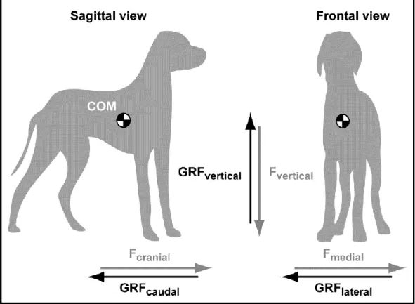
Figure 1. Sagittal and frontal views of a dog in a standing position
The three orthogonal force (F) vectors are paired with their oppositely directed ground reaction forces (GRF). The center of force (COM) is located according to previously published scheme (9) and is only indicative of its exact position. The Fmedial refers to the right limbs.
Movements of the COM give rise to Fcraniocaudal, Fmediolateral and Fvertical (Figure 1). As a result, GRFcraniocaudal, GRFmediolateral and GRFvertical are produced during the stance phase. Typical GRF versus time curves are illustrated in Figure 2. The GRF vectors are expressed herein as positive when directed upward, cranially and medially.
19
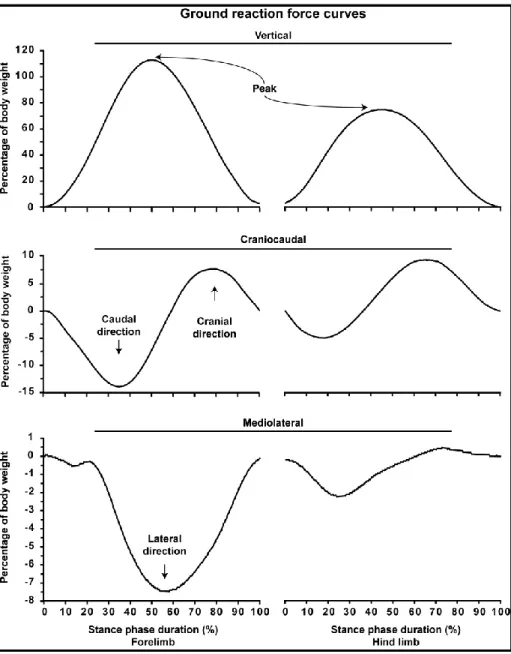
Figure 2. Typical curves of the ground reaction forces
Measurements are from a 27-kg golden retriever dog crossing the force plate at a trotting gait velocity (2.0 m/s). Ground reaction forces are considered positive when directed upward, cranially and medially. The stance phase is expressed for the forelimb and hind limb to ease their distinction. Peaks denote the points of maximal values of the fore and hind limb GRFvertical.
20
1.7.1 Vertical ground reaction force
At the initiation of the stance phase, the GRFvertical begins to be measured as the mass of the dog in motion is gradually supported by the limb (Figure 2). Soon after, a maximal point is attained which is the highest product of the mass of the body and the net vertical acceleration of the COM. This point is referred as the peak of the GRFvertical (i.e. peak vertical force). The GRFvertical then decreases until toe-off, which defines the end of the stance phase. In some situations the pelvic limb begins the stance phase when the thoracic limb is still in contact with the ground. This overlap explains why the GRFvertical doesn’t fall to zero when the thoracic limb has left the ground.
For a dog standing still on his 4 limbs, the body weight is expected to be supported at 30% by each thoracic limb and at 20% by each pelvic limb (10). The COM being closer to the thorax contributes to this imbalance in pelvic-to-thorax weight distribution when standing (10). As illustrated in Figure 2Erreur ! Source du renvoi introuvable., the GRFvertical of the thoracic limb reaches a maximal point (i.e. 113% of body weight), which is in accordance with the literature (11) (see Equation 3). Hence, the body absorbs a high level of forces in the vertical axis reaching more than 3 times the one observed at the stance (i.e. 30% versus 113% of body weight).
21
1.7.2 Craniocaudal and mediolateral ground reaction forces
The pattern of the GRFcraniocaudal involves successive caudal and cranial components Figure 2. The GRFcaudal is in fact generated as a reaction to the force applied in the direction of movement (i.e. cranially or forward). Hence, as a result of the Fcranial, a GRF is generated and directed caudally to the dog (i.e. backward) which decelerates the dog’s motion. During the second half of the stance phase, the GRFcranial propels the dog forward.
A third force vector is also depicted during the gait of dog. Figure 2 illustrates the GRFmediolateral, which involves medial and lateral components. Although the pattern of GRFmediolateral grossly mimics those reported in the literature (12), there is no clear waveform established for GRFmediolateral in dogs and cats due to inconsistent results (10,12).
1.8 Ground reaction vector
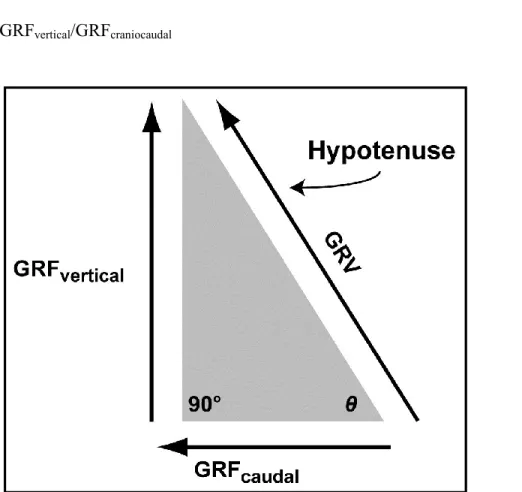
The three GRF components are directed in opposite directions by 90° (Figure 1). The resultant GRF, which is the net effect of the three orthogonal GRF components, is called the ground reaction vector (GRV). In the sagittal plane, a right-angled triangle is formed by the GRFcraniocaudal (adjacent side) and the GRFvertical (opposite side) of the angle θ (Figure 3) (1). The hypotenuse, which defines the magnitude and orientation of the GRV in the sagittal plan, can be resolved according to Pythagoras theorem (Equations 4 and 5) (1).
22
(4) Magnitude of the GRV (sagittal plan) = Square root (GRFvertical2 + GRFcraniocaudal2)
(5) Direction of the GRV (i.e. angle θ at the vertex) = Tangent-1 GRFvertical/GRFcraniocaudal
Figure 3. Ground reaction vector
The concept of sagittal ground reaction vector (GRV) is illustrated using a right angled triangle according to Pythagoras theorem. In the sagittal plane, vertical ground reaction force (GRFvertical) and GRFcraniocaudal (caudal component) are
orthogonal, giving the magnitude of the GRV as the square root of GRFvertical2 +
GRFcraniocaudal2. The angle θ refers to the direction of the GRV obtained by the
23
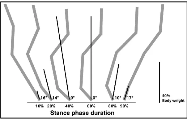
The sagittal GRV derived from the measures of a typical dog are illustrated at selected time points in Figure 4. As shown, the direction and the magnitude of the forelimb sagittal GRV evolve over time. At 60% of the stance phase, the sagittal GRV is perpendicular to the GRFcraniocaudal while being directed cranially thereafter. This force vector diagram can be used to determine where the path of the GRV travels according to anatomical structures or joints at specific points of the stance phase (i.e. caudal to the brachialis muscle, through the carpal joint). In cats, the GRF vector was used to investigate walking strategy and muscle activity under different conditions (13). The GRV can also serve to calculate moment of force, which is the tendency of a force to rotate an object. A moment of force is denoted as M and expressed in newton-metres (Nm) (Equation 6) (3).
(6) M = F × d = GRV × d
24
Figure 4. Progression of the ground reaction vector
Illustration of the progression of the ground reaction vector (GRV) for a 27-kg dog crossing the force plate at a trotting gait velocity (2.0 m/s). The measures are for the forelimb. The degrees indicate the direction of the GRV at specific points of the stance phase. The length of the lines indicates the magnitude of the GRV. The direction and magnitude of the GRV are calculated according to the ground reaction force value presented in Figure 2. In the background, forelimb bone position is detailed in gray as previously illustrated and is only indicative of its exact position. The length of the bar indicates 50% of body weight.
25
1.9 Center of force
The center of force (COF) is a coordinate pair (x,y) located within the surface of any parts of the body which are in contact with the ground. The COF corresponds to the average location where all the Fvertical act. When an animal is moving, the distribution of the Fvertical within the paw is modified and the COF changes accordingly. The COF can also be depicted as a trajectory according to its displacement during the stance phase. In humans, the path of the COF moves in a curvilinear fashion from heel to toes (14). In the dog, little is known about the COF trajectory. However, the Fvertical appears to show a distribution pattern among the pads in the dogs (15). The COF can serve to determine limb positioning, and serve to precisely define where the GRV takes its origin (1,16).
26
1.10 Impulse, linear momentum and power
The area under any force versus time curve represents the impulse denoted as I and expressed in N × s or in kg × m/s. Impulse is the time integral of a force (Equation 7) (3).
(7) I = ∫Fdt
where t is the time (s).
The linear momentum of a moving object is the product of the mass and velocity and is denoted as p and expressed in N × s or in kg × m/s (Equation 8) (3).
(8) p = m × v
where v is the velocity (m/s).
According to the impulse-momentum relationship, the impulse for a given time interval represents the change in linear momentum (equation 9) (17).
(9) I = ∆p = m × ∆v
It is common to report the impulse of the measured GRFvertical and GRFcraniocaudal which reflects the transfer of momentum from a moving limb to the ground. As illustrated in Figure 2, the forelimb undergoes craniocaudal transfer of linear momentum which is higher in the caudal direction. Conversely, the hind limb has a net linear momentum in the cranial direction. The overall net momentum (i.e. fore
27
and hind limbs) has to be null in the craniocaudal axis to maintain a constant gait velocity, otherwise the dog undergoes acceleration or deceleration (16).
During gait, muscles generate or absorb power due to their ability to perform concentric or eccentric activities (3). Power, denoted as P and expressed in watt (W) or in Nm/s is the product of the force and its velocity (Equation 10) (1).
(10) P = F × v
Using Equation 9, the net power output of a limb can be resolved by multiplying the sum of the GRF (i.e. GRFcraniocaudal, GRFmediolateral and GRFvertical) by the velocity of the animal (18).
1.11 Ground reaction force measurement devices
Force transducers are typically designed to measure the strain in a material under load. An electrical output proportional to the applied force is then generated and amplified. The most common type of device used to measure the GRF exerted by the body during locomotion is the force platform, which consists of a steel plate with force transducers at each corner.
The force platform allows the measurement of GRF using either strain gauges or piezoelectric crystals as a sensing element (3). As GRF have components in the 3 orthogonal planes, the force platform should have sensing elements arranged in a manner to capture all oriented strains for a complete measurement of the GRF