Discipline ou spécialité :
ED SEVAB : Pathologie, Toxicologie, Génétique et Nutrition
Unité de Recherche :
UMR 1331 INRA-ToxAlim
En vue de l'obtention du
DOCTORAT DE L'UNIVERSITÉ DE TOULOUSE
Délivré par :
Université Toulouse 3 Paul Sabatier (UT3) Paul Sabatier
THÈSE
Présentée et soutenue par :
Amaranta CARVAJAL CAMPOS
Titre:
Characterization of Aspergillus section Flavi: molecular markers as tools to
unmask cryptic species
Directeurs de Thèse :
Dr. Olivier PUEL
Ingénieur de Recherche, INRA, Toulouse
Directeur de thèse
Dr. Isabelle OSWALD
Directeur de Recherche, INRA, Toulouse
Directrice de thèse
Jury :
Dr. Sabine SCHORR-GALINDO
Professeur, Université de Montpellier, Montpellier
Rapporteur
Dr. Joëlle Dupont
Professeur, MNHN, Paris
Rapporteur
Dr. Herve GRYTA
Maître de conférence, UPS, Toulouse
Examinateur
Dr. Catherine BRABET
Ingénieur de Recherche, CIRAD, Montpellier
Examinatrice
ACKNOWLEDGMENTS
Becoming a PhD candidate means to start a journey, an intellectually and personal one. Intelectually it forces the person to learn, to become flexible, while increasing his/her skills to survive in the laboral world. It is not a smooth trip, it comes with ups and downs, yet it is a path that teaches you to become a better professional. For me, besides of the aforementioned, it also opened the door into the world of toxic mold and their impact in Human Health. At a personal level it has also been quite an adventure, sometimes more a roller coaster. This thesis gave me the opportunity to experience a new culture and to learn a new language, which is not necessary a piece of cake. Despite the difficulties during this period, I have been supported by a bunch of amazing people, that have helped me intellectually and personally. Find in these acknowledgements my most sincere gratitude to all of you, and if I forgot someone, I apologize to you in advance.
This Doctoral Thesis was sponsored by an scholarship from the Ecuadorian Government entity SENESCYT (from it initials in Spanish, Secretaria de Educación Superior, Ciencia, Tecnología e Innovación).
Firstly, I would like to express my sincere gratitude to my advisor Dr. Isabelle Oswald, Director of the Biosynthesis and Toxicity of Mycotoxins Team, who gave me the opportunity to perform my thesis at Toxalim INRA and welcome me in the team. Isabelle, I am really grateful to you for the opporunity and also for all your advice and support during this process. I have learnt a lot from you.
I would also thank Mr. Bernard Salles, former Director of UMR 1331 Toxalim INRA, Toulouse for letting me perform my research in the facilities.
I would also like to express my special appreciation and gratitude to my advisor Dr. Olivier Puel. I would like to thank you for encouraging my research and for your support during these years, I known that has not always being an easy task, and I appreciate all your efforts. I am grateful for your sense of humor, your taste in music, your open door whenever I needed, for letting me got lost and for guiding me to find the way out, for the practical and intelectual help and advice. I must also thank Dr. Sophie Lorber for always being there, I sincerely appreciate all your advice, your help in the lab, your smile, your support, intellectual and personal, and your guide in this proccess. It has been a pleasure learning from you. I would also thank Dr. Jean-Denis Bailly and Ms. Sylviane Bailly, I really appreciate a lot your advice during these years, I have learnt a lot from both of you, you have helped me to understand fungi world and how complicated they can be, in addition I have felt your support at different crucial moments, thanks for that and your cheerful spirit.
I would also thank the members of my thesis committee, Dr. Hervé Gryta, Dr. Vessela Anatovasova-Penichon, Dr. Jean-Denis Bailly and MSc Sylviane Bailly for all your advice and suggestions regardless the work performed during these years.
I would also thanks the members of the jury, my “rapporteurs”, Pr. Schorr-Galindo and Pr. Dupont, and my “examinateurs”, Dr. Gryta and Dr. Brabet, for accepting to read, judge, comment and provide advice for this manuscript and for being part of the jury.
I thanks as well Soraya Tadrist for all her help in the lab, we have sweat together, and without you finishing my experiments would have been even harder. I am grateful that we learnt to get along, and for the nice moments and laughs that we shared. As well I want to express my gratitude to Joëlle, I have learnt a lot from you, your enthusiasm, your professionalism and your character have been an example during these years. I cannot forget all the people that are part of the team, and wich plays a keyrole in my journey even though we do not necessarily work together. Thank you Phillipe for having your door always open and your smile all the time, I felt that I could always count on you, and that is priceless. In addition I want to say thanks to Sylvie, Anne-Marie and Arlette, you are a really good example of proffesionalism, enthusiastic and approachable people, I enjoyed the moments we shared. Selma, thanks for your help and advice in the lab and with French, you gave me a good push at the begining. I am also deeply grateful for the opportunity to known, shared and learnt from the different people that have passed by the team, Immourana, Pascal, Joanna, Delphine, Gisella, Leticia, Viviane, Delphine, Abdula, Su, Ophelie, Crisitine, Claire, Yannick, Tarek, Christian.
I am also really grateful to Dr. Catherine Brabet and Lethicia Manizan for our colaboration, which has lead to an important part of the work presented in this manuscript. Our collaboration is a good example of the importance of sharing between scientific groups.
I would also thank Sarah, Jeannette, Vincent, Vu, Pierre, Aurore and Lolita…. Each one of you are an important part in my journey, the first three for the administrative and computational stuff, and the good spirit and the laughs, Vu for your company in the latest hours and for being so nice and kind (eventhough I am the most desorganized person of the team), and the last three for keeping me alive and in good health by feeding me with amazing food. Thanks for doing your jobs professionally, with enthusiasm and with a smile, you made my doctoral experience easier and more enjoyable.
During this period of time, and in the lab, I have the opportunity to met several women to whom I really grateful… I have shared with all of you unforgetable moments….
Isaura, I have not enough words to express my gratitude to you. At the begining you guided me in this strange and complex country, providing me with the bases of the language, and the clues to not get lost in Toulouse. You welcome me not only in your office, you had not option, but also as a friend. I appreciate enourmously the way you share your knowledge, you are an incredible lab teacher. Thanks for always been in a good mood, even if you are feeling blue. We have shared several moments, mostly good ones, and English does not let me express to you how glad I am to met you and to have your friendship, thanks a lot for coping with my moody character and for being there!! You are an amazing friend. Te quiero chiqui!!!
Christelle, habibie, you were also forced to share your office with me, so you could not scape, as you were obligated to get along with me. Thanks for being there, for your support, your frienship, for the fun, for your crazyness, for letting me destroy your French, for sharing Toulouse, Poland, Leban, Ghent and the long days hanging out with me. I am really glad to met you and to say that you are my friend for lifetime. Also I am grateful with all your familly for making me feel at home.
Bianca, my darling, thanks for everything, you frienship means a lot to me, thank you for the laughs, for your support in the good and bad times, for all the experiences shared. Joya, thanks a lot for your friendship, your good mood, your smile, your kindness. Alix, thanks for the French lessons, for the rides to the lab, for the laughs, the cakes, the carefree spirit, the remarks, for the carnival, Halloween and the game nights. Laura C., thank you for you help, your patience, your kindness, your jokes, your draws, your French, your crazyness, the moments shared and most important for your friendship, you mean a lot to me. Rhoda, thank you for your advice, your sense of humor, your fiendship, your skills with languages, your cupcakes. Manon, thanks for the laughs, the crazyness, the French lessons, your opinions, your friendship, your sense of humor. Chloé, thanks for the French, laughs, and support. Laura S., thank you for putting me in perspective, for your jokes, for your crazyness and lovely spirit, for the music, the gifs, the tiny dances.
Outside the lab, I also want to thank several people. Thanks Ana L., Sebas A. and Micaela for being there and listened to me, and for gave me good advice, intellectually and personally. Francisca Hervas for your help. Pierre T. thanks for your wife (just a joke), your sense of humor and jokes, your French lessons, and for welcome me in your home. Ana P. thanks for all, you are always there, you are my soulmate. Sofy thanks for always being there, does not matter the km between us, you are always there to make me feel safe. Thanks to the clan of correziens and others for the great and funny experiences, for the French free lessons, for showing me another part of France, basically for letting me hang out with you and appreciate even more your country, especially to Melina, Hugo and Julien. To Pablo and Marian for the good memories and delicious food. To Sebastian D. for the friendship.
Last but not the least, I am deeply grateful for the continuous support, insight and patience of my family. Aita, thank you for always being there, I really appreciate how you handle my roller coaster of emotions. Thank you for being by best friend, my dad, a guide, for pushing me to follow my dreams, for recall me that life is a journey and we have to enjoy and learn from it… and to keep going, does not matter if the road looks impossible, it always will bring you to a new place. Thank you ñaño and mamu for being there, for recall me all the time that we can be apart geographically, but we have always each other. I love you, and I cannot be more grateful for having you as my family.
CONTENT
ACKNOWLEDGMENTS ... 3 CONTENT ... 6 PUBLICATIONS ... 8 COMMUNICATIONS ... 8 FIGURE LIST ... 9 TABLE LIST ... 12 ABREVIATIONS LIST ... 13 01 INTRODUCTION ... 16 1.2 FUNGI OVERVIEW ... 191.3 FILAMENTOUS FUNGI AND THEIR SECONDARY METABOLITES ... 21
1.4 WHAT IS A SECONDARY METABOLITE? DEFINITION, FUNCTION AND MYCOTOXINS ... 22
1.5 BIODIVERSITY OF ASPERGILLUS SECTION FLAVI: FOLLOWING THE TRACES OF CRYPSIS ... 33
1.6 DIVERSITY IN THE SECTION ... 37
1.6.1 Basal species ... 39
1.6.2 Clades of species of major economical interest ... 43
1.7 SECONDARY METABOLITES IN SECTION FLAVI ... 55
AIM OF THE STUDY ... 74
02 EXPERIMENTAL WORK ... 76
2.1 CHAPITRE 1: Molecular Flavi Tool ... 77
2.1.2 Polyphasic approach, ways of determining section Flavi species ... 78
2.1.3 Objective... 82
2.1.4 Material and methods : Creating the database of Aspergillus section Flavi, “THE FLAVI TOOL” 82 GENES ... 82
Fungal cell function genes ... 83
Reproduction genes ... 86
Genes linked to aflatoxin pathway ... 87
ISOLATES ... 89
PHYLOGENETIC APPROACH: Alignment, Model Selection and Phylogenetic Inference ... 89
2.2 CHAPITRE 2: Biodiversity of Aspergillus isolates potentially aflatoxigenic recovered from peanuts in Côte d’Ivoire ... 95
2.3 CHAPITRE 3: Aspergillus korhogoensis, a novel aflatoxin producing species from the Côte d’Ivoire ... 125
2.4 CHAPITRE 4: Identification of Aspergillus section Flavi in French maize ... 155
03 GENERAL DISCUSSION, PERSPECTIVES AND CONCLUSION ... 169
3.1 GENERAL DISCUSSION ... 170
3.2 CONCLUSION ... 183
PUBLICATIONS
ARTICLES Carvajal-Campos, A., Manizan, A.L., Tadrist, S., Akaki, D.K., Koffi-Nevry, R., Moore, G.G., Fapohunda, S.O., Bailly, S., Montet, D., Oswal, I.P., Lorber, S., Brabet, C., Puel, O. (2017). Aspergillus korhogoensis, a Novel Aflatoxin Producing Species from the Côte d’Ivoire. Toxins,
9(11), 353.
Manizan, A.L., Carvajal-Campos, A., Akaki, D.K., Koffi-Nevry, R., Montet, D., Oswald, I.P., Lorber S., Puel, O., Brabet, C. Biodiversity of Aspergillus isolates potentially aflatoxigenic recovered from peanuts in Côte d’Ivore. (Manuscript in preparation)
Makhlouf J., Carvajal-Campos A., Querin A., Tadrist S., Puel O., Lorber S., Oswald I.P., Hamze M., Bailly J.D., Bailly S. Biodiversity of Aspergillus section Flavi in spices marketed in Lebanon. (Manuscript submitted)REVIEW
Carvajal-Campos, A., Oswald, I.P., Lorber, S., Puel, O. Biodiversity of Aspergillus section Flavi: following the traces of crypsis. (Manuscript in preparation)
COMMUNICATIONS
ORAL Carvajal-Campos A., Manizan A.L., Tadrist S., Akaki, D.K., Koffi-Nevry, R., Moore, G.G., Fapohunda, S.O., Bailly, S., Montet, D., Oswal, I.P., Lorber, S., Brabet, C., Puel, O. Diving into Guinean Gulf Aspergillus section Flavi diversity: description of a new aflatoxin producing species. 1 st International MycoKey conference, Ghent, Belgium (14-17, September, 2017). Carvajal-Campos A., Manizan A.L., Tadrist S., Akaki, D.K., Koffi-Nevry, R., Bailly, S., Montet, D.,
Oswal, I.P., Lorber, S., Brabet, C., Puel, O. Description of a new aflatoxigenic species of Aspergillus section Flavi from Guinean Gulf. 39th mycotoxins workshop, Bydgoszcz, Poland (19-21, June, 2017).
Caceres I., Carvajal-Campos A., Oswald I.P., Puel O., Bailly J.D. Des remèdes naturels pour barrer la route aux toxines des moisissures. Tête à tête avec des jeunes chercheurs, Musée de
Toulouse, Toulouse, France (3, April, 2016).
POSTER
Orlando, B., Bailly, S., El Mahgubi A., Carvajal-Campos, A., Puel, O., Oswald, I.P., Bailly, J.D. Occurrence and identification of Aspergillus of the Flavi section and Aflatoxins emergence in French maize. 10th conference of The World Mycotoxin Forum, Amsterdam, The Netherlands (12-14, March, 2018)
FIGURE LIST
01 INTRODUCTION
Figure 1. Fungi classification………..20
Figure 2. Biosynthetic pathway of secondary metabolites………..22
Figure 3. Art as evidence of mycotoxins contamination……….27
Figure 4. Pre-harvest contamination and main factors affecting mold contamination………..….29
Figure 5. Cycle of fungi contamination………...29
Figure 6. Post-harvest contamination and main factors affecting mold contamination……….30
Figure 7. Global distribution of the main mycotoxins……….….32
Figure 8. Aspergillum structure……….…..33
Figure 9. Reproductive cycle in Aspergillus ………...36
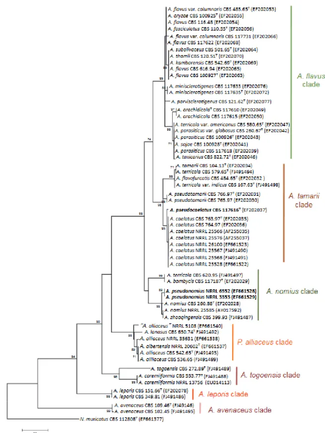
Figure 10. Maximum parsimony phylogenetic tree based on β-tubulin gene………...38
Figure 11. Colonies of Aspergillus avenaceus………..…..39
Figure 12. Colonies of Aspergillus togoensis clade………..…..40
Figure 13. Colonies of Aspergillus alliaceuss clade………..…..41
Figure 14. Colonies of Aspergillus leporis clade………..….42
Figure 15. Colonies of Aspergillus bertholletius………..….43
Figure 16. Aspergillus flavus clade……….…47
Figure 17. Colonies of Aspergillus flavus clade SBG ………48
Figure 18. Aspergillus parasiticus clade ….………..51
Figure 19. Colonies of Aspergillus mottae MUM10.231……….51
Figure 20. Aspergillus tamarii clade ………...53
Figure 21. Aspergillus nomius clade ………55
Figure 22. Aflatoxin gene cluster arrangement ………..57
Figure 23. Biosynthetic pathway of aflatoxin in Aspergillus flavus ………59
Figure 24. Aflatoxins structure………..61
Figure 25. Biotransformation pathways for AFB1………...62
Figure 26. Sterigmatocystin structure……….…63
Figure 27. Versicolorin A and versicolorin B structures………..64
Figure 28. Cyclopiazonic acid structure………...65
Figure 29. Cyclopiazonic cluster………..66
Figure 30. Kojic acid structure………..67
Figure 31.Schematic diagram showing the genes in the cluster………..68
Figure 33. Biosynthetic pathway of aflatrem in Aspergillus flavus……….…...69
Figure 34. Aflavinine structure………...70
Figure 35. Aflavarin structure………..70
Figure 36. Comparison of cluster 39 among Aspergillus species……….71
Figure 37. Tenuazonic acid structure………..72
Figure 38. Ochratoxin A structure……….73
02 EXPERIMENTAL WORK 2.1 CHAPITRE 1: MOLECULAR FLAVI TOOL Figure 1. Genes used during the study, and their main classification……….……...83
Figure 2. Scheme of reproduction genes………..…….….87
Figure 3. Scheme of the aflF-aflU genomic region in different strains of Aspergillus section Flavi….. 88
Figure 4. Phylogenetic inference process……….…. .90
2.2 CHAPITRE 2: Biodiversity of Aspergillus isolates potentially aflatoxigenic recovered from peanuts in Côte d’Ivore Figure 1. Macroscopic comparison of some strains on CYA and MEA……….107
Figure 2. Microscopic comparison of some strains ….……….108
Figure 3. PCR-DGGE β-tubulin profile of Aspergillus strains………109
Figure 4. PCR-DGGE β-tubulin profile of Aspergillus strains………..….110
Figure 5. Phylogenetic tree of Aspergillus section Flavi based on ITS data………..111
Figure 6. Phylogenetic tree of Aspergillus section Flavi based on benA + cmdA combined data……. 113
2.3 CHAPITRE 3: Aspergillus korhogoensis, a novel aflatoxin producing species from the Côte d’Ivoire Figure 1. Phylogenetic tree of Aspergillus section Flavi based on concatenated sequences from genomic loci………..130
Figure 2. Amino acid sequence alignment for the Mat1-1 locus in examined strains representing several Aspergillus species………..131
Figure 3. Amino acid sequence alignment for the Mat1-2 locus in examined strains representing several Aspergillus species ……….132
Figure 4. Comparision between cultures of Aspergillus korhogoensis sp. nov. and other species from
the A. flavus clade……….………135
Figure 5. Comparision between sclerotia of A. korhogoensis and other species from A. flavus clade………136
Figure 6. Conidial heads of A. korhogoensis MACI254 (100X)………136
Figure 7. Conidiophores of A. korhogoensis MACI254 (400X)………136
Annex 1. Maximum likelihood phylogenetic tree of Aspergillus section Flavi………153
2.4 CHAPITRE 4: Identification of Aspergillus section Flavi in French maize Figure 1. European distribution of maize crops ………. 159
Figure 2. No identified strains by morphological exam……….……… 162
Figure 3. Sclerotia of bizarre strains ………. 162
Figure 4. ITS 4-5 BI tree……… 164
Figure 5. BenA-cmdA BI tree………. 165
03 GENERAL DISCUSSION AND PERSPECTIVES Figure 1. Predicted areas to become tropical………..………..….…….. 173
Figure 2. Bayesian topology for concatenated tree using benA, cmdA and mcm7……… 180
Figure 3. Bayesian topology for concatenated tree using benA, cmdA and rpb1………..… 180
Figure 4. Bayesian topology for concatenated tree using benA, cmdA and preA……… 181
TABLE LIST
01 INTRODUCTION
Table 1. Principal mycotoxins and producing species, frequent sources and effects ………..25
02 EXPERIMENTAL WORK
2.1 CHAPITRE 1: MOLECULAR FLAVI TOOL
Table 1. Screening methods to determine fungal species……….78 Table 2. Primers and annealing temperatures used to amplify multiple genomic regions within Aspergillus species………..81
2.2 CHAPITRE 2: Biodiversity of Aspergillus isolates potentially aflatoxigenic recovered from peanuts in Côte d’Ivore
Table 1. Primer sequences ……….102 Table 2. Isolates and accession numbers deposited in GenBank.………103 Annex 1. Aspergillus isolates used in the study……… 121
2.3 CHAPITRE 3: Aspergillus korhogoensis, a novel aflatoxin producing species from the Côte d’Ivoire Table 1. Principal secondary metabolites produced by Aspergillus korhogoensis………..133 Table 2. Aspergillus isolates used in this study……….140 Table S1. Primers used to amplified multiple genomic regions within Aspergillus species……….…….149 Table S2. Isolates examined and accession numbers deposited in GenBank………..…150
2.4 CHAPITRE 4: Identification of Aspergillus section Flavi in French maize
ABREVIATIONS LIST
Amds Acetamidase gen AF(s) Aflatoxin(s)
AFB Aflatoxin B
AFG Aflatoxin G
AflP O-methyltransferase A gene aflF-aflU Aflp-AlfU region
BenA β-tubulin gen
BI Bayesian Inference
BIC Bayesian Inference Criterion
bp Base pairs
BP Bootstrap percentages
CAST Council for Agricultural Science and Technology
CC Climate change
CmdA Calmodulin gen CPA Cyclopiazonic acid
CYA Czapek Yeast Autolysate Agar DNA Deoxyribonucleic acid
EU European Union
FB Fumonisin B
FAO Food and Drug Administration
IARC International Agency for Research on Cancer ITS Nuclear Ribosomal Internal Transcribed Spacer MAT Mating type loci
MEA Malt Extract Agar
Mcm7 Minichromosome maintenance protein
ML Maximum Likelihood
NRPS Non-ribosomal Peptide Synthase PCR Polymerase chain reaction PDA Potato Dextrose Agar PKS Polyketide Synthase PP Posterior probability Ppga Pheromone precursor ppgA PreA Pheromone receptors preB PreB Pheromone receptors preA OMST O-methylsterigmatocystin
OTA Ochratoxin A
Rpb1 RNA polymerase II, largest subunit
SBG Strains of Aspergillus flavus clade with small sclerotia that synthetize AFBG
ST Sterigmatocystin
TeA Tenuazonic acid
VERA Versicolorin A VERB Versicolorin B
WHO World Health Organization YES Yeast Extract Agar
01
1.1 BACKGROUND
Quality of food supplies has always being an issue in human societies. Having access to proper food supplies is necessary to avoid potential risk to human and animal health. Some fungi, especially from Ascomycota, are capable to synthesize a plethora of products as part of their metabolism, some of them toxic to humans and vertebrates, named mycotoxins. The ubiquitous presence of fungi in staples cannot be avoided, thus, their presence become a potential health risk for humans and livestock.
Mycotoxin contamination of staples is an important risk to public health because these compounds produce detrimental effects on vertebrates and humans. Since their discover, several studies have been performed to identify the principal mycotoxins depending on the geographical areas, the minimal doses of their toxicity, the fungi responsible of their production, and to develop strategies to control them in order to avoid their effects on human health, as well on animal health and to reduce economic losses. Once the amount of mycotoxins exceeds the levels permitted by the regulations, it is hardly recommended to eliminate staples from the food chain. However, in some regions worldwide, especially those including developing countries, monitoring and policies against mycotoxin presence in food and feed for human and animal consumption unfortunately are not well regulated. As consequence, the risk of entrance of mycotoxins in the food chain is high (Bhatnagar et al. 2002).
Aspergillus section Flavi is one of the most economically important groups of molds; their detrimental effects are an important public health issue, furthermore the stability of its taxonomy is of practical concern (Geiser et al. 2007; Pildain et al. 2008). The section encloses species able to produce several mycotoxins, among them, aflatoxins are a major concern because their deleterious effects in vertebrates (IARC 2003). Due to their physiological requirements, these species grow principally in tropical and subtropical regions worldwide. In these areas, they are a problem because harvest and storage conditions are not always the most appropriated ones to avoid mold development and mycotoxins production, besides, environmental conditions generally contributes to their production. Resulting in two main issues, the first is the risk on human and animal health, and the second, staples that are contaminated cannot be exported, which affects negatively some countries’ economies because they are based on exportation. In fact, staples contamination of mycotoxins lead to great economic losses. In temperate regions, the importance of section Flavi is linked to the importation of contaminated raw material, as well as the possibility of the colonization of harmful species due to climate change, which could result in new niches for these species (Perrone et al. 2014).
The present introduction is divided in three parts: the first introductive part encloses a general overview of molds and the principal mycotoxins; the second part includes an overview of Aspergillus section Flavi, and the third part that summarizes the principal secondary metabolites yield by them.
1.2 FUNGI OVERVIEW
Fungi is a diverse eukaryotic kingdom containing an estimated of 3.5 to 5.1 million organisms, from unicellular to macroscopic multicellular, that inhabit a wide range of ecological niches worldwide (O’Brien et al. 2005). These organisms play a key role in nutrient cycle as decomposers, and include saprophages, symbionts and pathogens. As heterotrophs fungi fed from others organism by extracellular digestion, yielding enzymes that able them to digest and absorb nutrients. Fungi development requires certain elements that are used in their primary and secondary metabolisms, principally sources of carbon and nitrogen, and in a lesser extent potassium, phosphorus and magnesium, among others trace elements. Additionally, environmental factors, such as pH, light, temperature and water availability, are crucial for their development (Dix 2013; Dighton 2016).
Due to fungi diversity of life history strategies, several species are widely studied and applied in biotechnological industries to produce enzymes, medicines, biocontrol agents, natural fertilizers, natural pigments, cosmetics, alcoholic drinks, and food (Galagan et al. 2005; Schoch et al. 2009; Dupont et al 2016; Blackwell 2011; Jayasiri et al. 2015; Bill and Gloer 2016). Taking into account their diversity, the number of infectious species is low, yet those species have detrimental effects in organisms’ health, including plants, animals and humans, and can be a worldwide threat for food security. Similarly, during the last decades, novel diseases produced by fungi have been discovered, and in some cases host population infected have decreased in alarming numbers, almost disappearing (Fischer et al. 2013). Summarizing, several fungi are economically important organisms, making their study mandatory (Mitchell 2010).
Fungi reproductive cycles include sexual and asexual reproduction, both mechanisms are mediated by spores (conidia, ascospores etc.), which have reproductive and dispersal functions. Some species present only sexual or asexual cycles while others a combination of both reproductive mechanisms. A holomorph fungus present both types of reproduction, an anamoph fungus presents the asexual type and a teleomorph fungus the sexual type. A fungus can have strains in anamorph state and others in teleomorph state; phenotypically they might be different and hence, be classified under different names. Moreover, some fungi show different anamorphic states, like some species of
Neurospora, Fusarium and Botrytis, which show strong differences between their micro- and macroconidia (Webster and Weber 2007; Dix 2013).
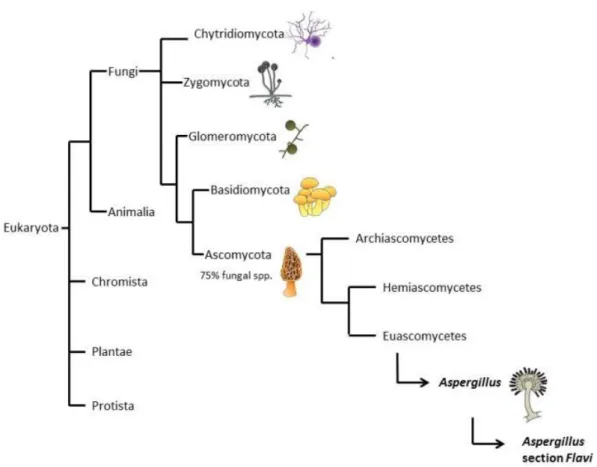
Fungi are divided in ascomycetes, basidiomycetes, zygomycetes, and chytrids (Figure 1); the first two, Basidiomycota and Ascomycota, contain most species, including the most important to humankind. Ascomycota contains approximately 33.000 described species, including most lichens known and about 90% of pathogenic fungi (ca 400 species). Ascomycota fungi are characterized by their reproductive structures, ascus, nevertheless most species produce also asexual spores (Pitt and
Hocking, 2009), in fact they are most commonly found in their conidial state, and for some species, the sexual reproduction seems to be lost; this phenomenon seems to have occurred several times in the evolution of the group. The conidial states of Aspergillus and Penicillium are generally arranged in phialides, and their arrangements are generally used as diagnostic state. In Aspergillus, the conidiophore tip is swollen, forming the vesicle, and phialides start directly on its surface (uniseriate) or present a palisade of sterile cells, metulae, followed by phialides (biseriate) (Raper and Fennell 1965). Penicillium lacks vesicles, and the conidiophore tip has directly a monoverticillate arrangement or series of metulae followed by philiades, the levels of ramification could be from one to several series of metulae (Raper and Thon 1968). The cell walls in Ascomycetes are composed mainly by chitin and glucans and in general the septum is incomplete, forming a central pore that result in coenocytic mycelia (Webster and Weber 2007). Ascomycota probably arose around 500 to 900 million years ago; it is subdivided in three main groups, Archiascomycetes, Hemiascomycetes (yeasts), and the large Euascomycetes (molds) (Mitchell 2010).
Figure 1: Fungi classification. The figure shows the relationships of fungi with other groups of Eukaryotes. In addition, it shows the main groups with Fungi, and the division of Ascomycota, and the
Climate change presents a challenge for fungal relationships, between different fungi, between fungi and other organisms, and between fungi and their ecosystems. Resulting in shifts in community composition, creating new ecological niches, therefore, producing changes in the symbiotic associations between fungi and other organisms, changes in the distribution patterns of species, and the increment of detrimental effects caused by harmful fungi (Jayasiri et al. 2015). Climatic model predictions suggest that climatic conditions will vary over the next two decades, atmospheric concentrations of CO2 are expected to double or triple (from 350 to 700 or 900-1000 ppm) and the regional cycles are going to change, some areas will become drier, and global temperatures will increase by approximately 2-5 °C (Medina et al. 2014). These environmental changes would result in homeostatic stress in crops. Hence, environmental changes would modify the agricultural cycles, affecting mycobiota composition in soils and crops, and the mycotoxinogen species, therefore the mycotoxins yield (Medina et al. 2014).
1.3 FILAMENTOUS FUNGI AND THEIR SECONDARY METABOLITES
Filamentous fungi are considered as the main producers of mycotoxins. It is a paraphyletic group, enclosing the Ascomycota phylum and some species from Mucorophyta (zygomycetes). Nevertheless, Ascomycota species are the most diverse and the most important at economical level, as they are linked to staple spoilage during harvesting or storage processes. In fact, the genera Aspergillus, Fusarium and Penicillium are considered as the main source of mycotoxins (Pitt and Hocking 2009).
Aspergillus and Penicillium genera are important to humankind not only because of their detrimental effects, but also because of their use in biotechnology; enzymes and other compounds synthesized as part of their primary and secondary metabolisms are used, as well as a direct inoculation of fungi on foodstuff. Aspergillus and Penicillium have been used in food production for several centuries in fermentation processes to produce beverages, sauces and in the cheese industry. Likewise, proteases, amylases, lipases and pectinases are important in the manufacture of dairy, bakery, distillery and brewery products, juices and leather, and in the starch industry. Furthermore, they have been used to synthesize antibiotics, such as penicillins and cephalosporins that comprise around the 50% of antibiotics production worldwide (Kavanagh 2017); or griseofulvin used as anti-tumoral and in dermatology (Banani et al. 2016).
1.4 WHAT IS A SECONDARY METABOLITE? DEFINITION, FUNCTION AND MYCOTOXINS
In order to cope with their environment, fungi have developed the ability to produce several extracellular chemicals, called secondary metabolites, which are not essential in the primary metabolism of fungi (i.e. growth, reproduction, respiration), and not required for their survival when growth in laboratory conditions. These compounds are low-weight molecules (< 1000 Daltons) produced by their secondary metabolism, which encloses the molecular pathways that are not essential for the survival of the organism (Bennet 1987; Bennet and Klich 2003). These molecules are diverse in their chemical nature, including polyketides, non-ribosomal peptides, terpene, indole terpenes and hybrids (Figure 2). These organisms are capable to produce a large number of these compounds, and their secondary metabolic profile will vary depending on the genetic information (presence of secondary metabolic gene clusters), environmental conditions (mainly nutrients and water availability), and community composition (Brakhage 2013; Bills and Gloer 2016).
Figure 2: Biosynthetic pathways of secondary metabolites. In blue the groups of secondary metabolites. In black the main mycotoxins produced by these pathways. In orange the enzymes associated with
each pathway; NRPS: non-ribosomal peptide synthetase, PKS: polyketide synthetase, TC: terpene cyclase, DMAT: dimethyl allyl transferase.
Polyketides are the most diverse group of secondary metabolites, including polyphenols, polyenes and macrolides. Due to their diversity, they exert different biological activities, some of them exploited in industrial processes. Fungal polyketides are synthesized by type I polyketide
synthases (PKSs) that are multidomain proteins linked to eukaryotic fatty acid synthases. Polyketides result from the metabolization of acetate, and are formed by the consecutive polymerization of ketide groups (CH2-CO). The main difference between fungal polyketides and fatty acids is that polyketides are formed by PKSs able to use other carboxylic acids, rather than acetyl-coenzyme A as substrate. In addition, the oxidation state is variable and β-carbon is not necessary fully reduced in polyketides synthesis, for which the ketoacyl CoA synthase (KS), acyltransferase (AT) and acyl-carrier (ACP) domains are essential. Fungal PKSs are considered ‘iterative PKSs’ because they present just one module for the addition of methylmalonyl CoA, so the processes of addition require repeated biosynthetic reactions. Fungal PKSs are divided in three groups: non-reduced, partially reduced and highly reduced synthases. In addition, five of the six mycotoxins regulated to date by the EU, aflatoxins, ochratoxin A, patulin, fumonisins and zearalenone belongs to this group (Keller et al. 2005; Cano et al. 2016).
Terpenes are yielded by fungi, bacteria and plants. Plant terpenes are the best known, and are essential for plant growth, development, and interactions with their environment. Terpenes play a main role in interaction with pollinators and predators (i.e. herbivores), in the protection against photo-oxidative stress, in thermoregulation, among others (Tholl 2006). Aristolochenes, carotenoids, gibberellins and trichothecenes are some important terpenes characterized in fungi. Terpenes are formed by the combination of dimethylallyl pyrophosphate (DAMPP) and isopentenyl diphosphate (IPP), this reaction is catalyzed by isoprenyl diphosphate synthases, which belong to the family of phenyl transferases. Based on their chemical structures terpenes are classified as: (i) monoterpenes or geranyl diphosphate that are rarely yield by fungi, (ii) triterpenes that are mainly produce by plants, (iii) sesterterpenes, tetraterpenes or carotenoids linked to the defense against UV radiation, and (iv) sesquiterpenes that enclose the tricothecenes family known as an important group of mycotoxins (Cano et al. 2016).
Non-ribosomal peptides (NRPs) are compounds not involved in the primary metabolism. As their name suggest, their synthesis does not include proofreading mechanisms, making their structure highly variable, in fact, at the moment several hundreds of substrates of NRPs have been identified in comparison to the 20 amino acid involved in protein synthesis (Finking and Marahiel 2004). The synthesis of NRPs is catalyzed by non-ribosomal peptide synthetases (NRPSs), which have functions similar to those of enzymes catalyzing ribosomal peptides. When compared, fungal NRPs are reported to achieve bigger sizes than bacteria NRPs, and could be explained because their synthesis in fungi is generally catalyzed by one NRPS. These enzymes have a dual function, working as temperate and as biosynthetic machinery; actually, they are organized in different modules that integrate amino acids into the polypeptide chain. The synthesis of NRPs requires the presence of at least three domains: (i) the A-domain that determines the amino acid to be included and activates
the amino acid or the hydroxyl acid; (ii) the T or PCP domain, a thiolation or peptidyl-carrier protein, which transports the activated units between active sites of the domains; and (iii) the C-domain, a condensation domain, where the formation of the peptide bond (C-N) occurs between the polypeptide chain and the new amino acid. Other domains could also play a role in the synthesis, by adding special features, like non-proteinogenic amino acids, fatty acids, carboxylic acids, among others (Pang et al. 2016).
Indole alkaloids are mainly synthesized from tryptophan and DAMPP, but sometimes they include other amino acids as precursors. The steps of biosynthesis are yet to be elucidated, for some known compounds three processes were described that include steps of tryptophane prenylation catalyzed by DMATS, followed by the methylation of dimethylallyl tryptophan, and finally, a series of oxidation steps, which can be catalyzed by NRPSs. Some other enzymes can be involved in the biosynthetic pathways, like oxidases, methylases and prenyl transferases (Keller et al. 2005).
The development of genomic, transcriptomic, and proteomic is unmasking processes linked to compound synthesis in fungi. In fact, the genome characterization of several species has enabled to elucidate the biosynthetic pathways of several mycotoxins and the processes occurring in fungal cells, including an increase of knowledge of the biology of harmful fungi. For instance, pathogenic ascomycetes present more genes for polyketides, peptides, terpenes and other secondary metabolites than those non-pathogens such as Neurospora crassa (Desjardins 2006). Likewise, genome studies have shown that secondary metabolic yield depends on global transcriptional factors, encoded by unrelated genes with a specific biosynthetic pathway (e.g. VeA and LaeA), and on specific enzymes for each biosynthetic pathways that differ from primary metabolism enzymes. In Ascomycetes, biosynthetic pathways of secondary metabolites are often clustered together, which makes them different from other eukaryotes. The purpose of secondary metabolites is still not completely understood, however it is believed that they confer selective advantages to fungi under natural conditions, especially under stress conditions (e.g. environmental stress, nutrient availability, interspecies competition, predator defense) (Magan and Adred 2007; Fox and Howlett 2008, Schwab and Keller 2008; Brakhage 2013).
As aforementioned, several secondary metabolites are beneficial to humankind and are used in pharmacology, food industry, cosmetics, energy and construction (Bhatnagar et al. 2002). Some others, known as mycotoxins, are toxic and could exert deleterious effects on vertebrates, including humans (Peraica et al. 1999). Mycotoxins are amply studied due to their detrimental effects on vertebrates’ health and their impact in agriculture and economy. Nowadays, over 1000 secondary metabolites are described, ca 400 are considered mycotoxins, 30 are considered as important mycotoxins for their effects, and from them just 7 are legally regulated by the European Union.
Best-known mycotoxins include aflatoxins, ergot alkaloids, fumonisins, ochratoxin, patulin, trichothecenes and zearalenone (Bennet and Klich 2003; Cano et al. 2016) (Table 1).
Table 1: Principal mycotoxins and producing species, frequent sources and effects (AFSSA 2009; CAST 2003, Bbosa et al. 2013). In red the mycotoxin and their principal producer.
MYCOTOXIN TYPE MAIN PRODUCERS CONTAMINATED
PRODUCTS EFFECTS CHEMICAL NATURE Aflatoxins B1, B2, G1, G2 Aspergillus flavus, A. parasiticus, A. nomius, Several spp. in A. section Flavi Cereals: maize, wheat, rice, sorghum; spices, sunflower, nuts, almonds, pistachio, coconut, cotton, dried fruit Hepatoxic, Carcinogenic, Immunotoxic, Teratogenic, Acute toxicity Polyketide Trichotecenes T-2 Toxin and HT-2 F. tricinctum, F. langsethiae, F. sporotrichioides, F. poae, F. equiseti Cereals: wheat, maize, rice, soy, beans and barley
Genotoxic, Immunotoxic, Reprotoxic, Neurotoxic Terpene Deoxynivalenol Fusarium graminearum, F. culmorum, F. sporotrichoides, F. langsethiae, F. tricinctum, F. poae, F. solani, F. equiseti Cereals: wheat, maize, rice and sorghum Immunotoxic, Digestive problems, Haematopoietic Terpene Fumonisis B1, B2, B3 F. verticillioides, F. proliferatum
Cereals: maize, rice, sorghum Carcinogenic, Neurotoxic Polyketide Ochratoxin A Penicillium verrucosum, Penicillium nordicum A. ochraceus, A. carbonarius Cereals, cacao, coffee, wine, grape juice and spices
Nephrotoxic, Immunotoxic, Teratogenic Polyketide Zearalenone F-2 Toxin F. graminearum, F. culmorum, F. crookwellense
Cereals: maize, soy, sorghum, wheat, rice and oat
Reprotoxic, Immunotoxic
Polyketide
Patulin
P. expansum,
Byssochlamys nivea Apples, pears and derivates juices Neurotoxic, Genotoxic, Cytotoxic Polyketide Ergot alkaloids Claviceps purpurea, C. paspali, C. africana, C. fusiformis
Rye, wheat and triticale
Neurotoxic, Digestive problems, Vasoconstriction
1.4.1 Mycotoxins
Mycotoxins are classified based on fungus that produce them, chemical structure and/or mode of action. Their degradation is a challenge because most are heat-stable and form toxic compounds while degradation processes are applied. Generally this compounds are hydrophobic (except for the fumonisins), allowing them to accumulate in lipophylic tissues in plants and animals (Hussein and Brasel 2001).
Mycotoxicosis symptoms depend on several variables that interacts synergistically, including the mycotoxin chemical nature, the exposure time (duration and dose), the organism that intakes the mycotoxin (species, sex, age, health, diet), and the mixed effects of mycotoxin with other xenobiotics. The effects exerted on vertebrates could be chronic (low doses, long periods of time) or acute toxic (high doses, short periods of time), mutagenic, teratogenic, carcinogenic, nephrotoxic, hepatotoxic, immunotoxic and estrogenic. The main target organs depend on the mycotoxin and the organisms that ingest it, and include the liver, kidney, lungs and the nervous, digestive, endocrine and immune systems (Bhatnagar et al. 2002). In general, more than one mycotoxin is found in staples, and a mix of them is thus usually ingested. The interaction between mycotoxins can produce different effects in the organism: antagonists, additive or synergic, which are linked to the mycotoxin nature, the decontamination pathway, of the host species, the time of exposure, and the doses and ratio of mycotoxins (Peraica et al. 1999; Alassane-Kpembi et al. 2017).
Mycotoxins have being around humans for as long as agriculture was developed, or even before, when recollection started as mechanisms for food storage (Richard 2007). Some episodes of mycotoxicosis can be traced in the literature, myths, and arts. For instance, they could be tracked in the Bible, as part of the Seven Plagues of Egypt or in the Dead Sea Scrolls (Richard 2007). Withal, the decline of Etruscan civilization (5th century B.C.) could be related to fusariotoxins (toxin T2 and ZAE) (Yiannikouris and Jouany 2002). Howbeit, the episodes of hallucinations of “Saint Antony’s fire” or ergotism (11th century), produced by alkaloids of Claviceps purpurea on rye, might be the best-known example of mycotoxicosis in ancient times (Figure 3). During the Middle Ages outbreaks were common, some registered epidemic episodes occurred during 8th and 15th century A.D.; also some more recently episodes are also suggested to be caused by ergotism, like witchcraft in Salem, USA, and Finnmar, Norway. Symptoms of ergotism include delirium, prostration, acute pain, abscess and gangrene of the limbs, and sometimes death (Peraica et al. 1999; Richard 2007; AFSSA 2009). Likewise, “Shoshin-kakke” or “yellow rice disease” is another well-known example of mycotoxicosis outbreak, this disease that causes acute cardiac beri-beri, was reported in Japan, affecting especially the colder regions. This illness is caused by the exposure to citreoviridin, a Penicillium citreonigrum mycotoxin. The fungus contaminated rice during the storage processes due to poor conditions and
practices. Once these conditions were controlled, the disease disappeared from the region, and has not been reported lately (Udagawa and Tatsuno 2004). Although fungal contamination occurred and relations with some diseases were perceived, the awareness of mycotoxins and their toxic effects arose for the first time in London (England) in 1962. Poultry presented a strange disease, “turkey X syndrome”, which killed at least 10,000 birds. Interestingly, while tracing the origin of the illness, it was discovered that peanuts used to feed poultry were contaminated by secondary metabolites, aflatoxins, named after Aspergillus flavus (Bennet and Klich 2003); years later, cyclopiazonic acid was also proved to interfere in this outbreak (Richard 2008). Other compounds were also recognized as mycotoxins and their study became under scope. Mycotoxins are found in a wide variety of staples, use as animal and human food, principally cereals (maize, wheat, rye, rice, etc.), oligenosus seeds (peanuts, cotton, nuts, pistachios, etc.), and spices (Bath et al. 2010).
Figure 3: Art as evidence of mycotoxins contamination. Above: Paint exemplifying an ergotism outbreak. Down left: “Saint Anthony’s hallucinations” by Mathias Grünewald (effects of hallucinations associated to ergotism).
1.4.2 How mycotoxins enter in the chain food and their distribution
Occurrence of mycotoxin contamination can be divided in pre-harvest (crops and recollect) and post-harvest steps (mainly storage). The fungi that contaminate staples will vary depending on the geographical region, but also of the agriculture methods, crop cycles, harvesting and storage conditions. Humans’ exposure to mycotoxins can occur by direct intake of staples of vegetal origin contaminated by mycotoxins, or by the intake of contaminated animal products. There is also the risk of dermal, respiratory and maternal exposure routes (Bath et al. 2010; IARC 2015).
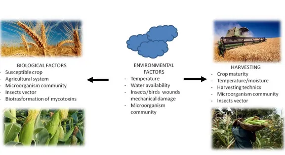
In general, mycotoxigenic fungi are divided in two main groups, one enclosing species more prone to colonize staples and yield mycotoxins in crops (principally Fusarium species), and another group more prone to colonize and yield mycotoxins during storage (principally Aspergillus and Penicillium), yet, some fungi are able to colonize during both steps, like A. flavus (AFSS 2009; Antonissen et al. 2014). For example, Fusarium generally colonized grains before harvest, where moisture is high, whereas maize and peanuts are generally colonized in post-harvest, where temperatures and drought are more suitable for Aspergillus section Flavi colonization (Bryden 2012). As aforementioned, fungi reproduce by spores that are dispersed principally by the wind and insect vectors. Once the spores reach a suitable nutrient source, suitable environmental and atmospheric conditions, such water availability, humidity and drought conditions, pH, and temperature, they will germinate (Figure 4). Besides, these environmental conditions can trigger stress and predisposition of cultivars, helping fungi developpment. The optimal for these variables depend on the fungus, for example, Aspergillus glaucus requires approximately 10% less of water availability than A. flavus. Substrate is also important; some fungi are generalist, while others have more constrained niches. Generalist fungi might prefer a substrate that suits better their nutrimental and physiological requirements, like A. flavus that will prefer maize (though it colonized several commodities), whereas some Fusarium species prefer cereals with small grains (Dierkman and Green 1992; AFSSA 2009; Pinnoti et al. 2016).
Figure 4: Pre-harvest contamination and main factors affecting mold contamination. Modified from Paterson and Lima 2010.
Insects have a key role in fungi contamination; they not only disperse spores, but also damage raw products, allowing an easier colonization by fungi. In fact, insects wound maize kernels, and transport spores of Aspergillus and Fusarium, resulting in disturbance of the natural barrier (leaves protecting kernels) (Aiko and Mehta 2015) (Figure 5).
Agricultural practices are part of the factors that interact in pre-harvest contamination, some of the main practices include the variety of plants used (more or less sensitive), the type of crop rotation and soil tillage. Some techniques used to diminish fungi contamination includes growing resistant crop varieties, management of crop rotation, the type of soil tillage, chemical and biological control of plant diseases, and insect control (Rychlik et al. 2014).
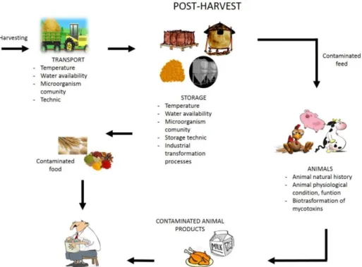
Post-harvest contamination includes steps from crop maturation to feed and food consumption. Abiotic conditions, such as water availability, temperature, oxygen availability, are more easily controlled than the pre-harvest steps, facilitating the control of mold growth. Nevertheless, methods to storage following all the requirements are expensive, and easier to obtain in products that have higher markets, making this process not always achievable for small production or in some countries (Dierkman and Green 1992; Magan et al. 2003; Paterson and Lima 2010). Grain storage is a good example of how mycotoxins can contaminate feed and food supplies. In general, a community of microorganisms, most of them innocuous, colonizes grains. Mycotoxigenic fungi can be good competitors, and under proper temperatures and water availability, they can develop and produce mycotoxins. Insects play a similar role in the storage processes as well (Magan et al. 2003; Paterson and Lima 2010) (Figure 6).
Figure 6: Post-harvest contamination and main factors affecting mold contamination. Modified from Paterson and Lima 2010.
Once the mycotoxins are present in feed and food commodities, they can contaminate the whole food chain. As said above, humans can ingest mycotoxins in two ways, by direct consumption
of contaminated vegetal commodities or by ingesting contaminated animal products, and in general are contaminated with more than one mycotoxin. A study case of nephropathy in Bulgarian pigs and chickens was caused by cocktail of mycotoxins, including ochratoxin A, penicillic acid, FB1 and an uncharacterized metabolite (Bryden 2012).
In animals, ruminants have a gastric system with a rich microbiota that facilitates the degradation of mycotoxins, whereas monogastric species, like pork and poultry, are especially sensible to mycotoxicosis because intestinal microbiota is less diverse. Poultry is less prone to biotransform toxins to less toxic compounds before the intestine absorbs them. For ruminants it has been documented that rumen function is nevertheless affected negatively by the presence of mycotoxins, as well some biotrasformation can produce toxic products, which can be excreted thus making it available, like the case of AFM1 (Hussein and Brasel 2001; AFSSA 2009). The major problem for livestock and poultry associated with ingestion of mycotoxins is the poor animal performance, which can be difficult to diagnose and quantify because of the diversity of life histories, physiological status, biotransformation pathways, detoxification mechanisms and the intra- and inter variability of species that ingested them. Similarly, the type and level of mycotoxin in feed, the time of the exposure, and the interaction between mycotoxins are also a problem (Bryden 2012; Alassane-Kpembi et al. 2015).
In addition, there are the ‘masked’ or conjugated mycotoxins that occur in vegetal food supplies, and are often linked with livestock and poultry feed intake, resulting in a decrease in their performance. These types of mycotoxins are the result of biotransformation processes occurring in plants (Bryden 2012; Pierron et al. 2016). Some examples are zearalenone-4-glucoside, a conjugate of zearalenone, and deoxynivalenol-3-glucoside a conjugate of deoxynivalenol. There is some evidence that OTA and fumonisins can also be conjugated in plants (Bryden 2012). The discovery of these mycotoxins has put them under scope.
1.4.3 Impact of mycotoxins
The presence of mycotoxins in staples is a major concern, not only for public health, but also for their economic impact. Food commodities losses due to mycotoxin contamination represent above 25% of spoiled food (FAO 2003). For instance, only in the United States the Food and Drug Administration (FDA) has estimated that the losses exceed $900 million per year (CAST report 2003). Due to its impacts, food security associated to mycotoxin contamination is a major issue worldwide; public health commissions all over the world try to ensure safe and healthy feed and food for animals and humans (Stoev 2013). In developed countries, food security is carried out better than in developing countries, in which food quality monitoring and the infrastructure to avoid mycotoxin
contamination are more difficult to settle. Nevertheless, regulation of mycotoxins reduced the intake in those countries that have proper regulation and monitoring, and increase exportation standards around the world. On the other hand, it could result in a higher risk of consumption of mycotoxins by human and animal populations of developing countries, as the best quality staples are exported, whereas the poor quality ones remain for domestic consumption (Wild and Gong 2009; Stoev 2013).
As aforementioned, the global distribution of mycotoxins is not homogenous, the conditions in each region will favor the development of certain fungi over others, thus favor some mycotoxins over others. In addition, climate change is shifting distribution and prevalence of some fungi, and thereby, mycotoxin distribution. Streit et al. (2013) determined the presence of the main mycotoxins (aflatoxins, zearalenone, deoxynivalenol, fumonisins and ochratoxins) around the world for a period of eight years. Their results showed that most of the samples (72%) were mycotoxin positive, and 38% showed a multicontamination (more than one mycotoxin). In addition, they determined that the percentages of each mycotoxin were more or less stable during the years, with the exception of aflatoxins, their level increased between 2005 and 2009 in tropical regions. Another study, that also screened mycotoxin presence in long term, showed the risk of mycotoxin contamination depending of the geographical distribution worldwide (Figure 7).
Figure 7: Global distribution of the main mycotoxins. Survey performed by Biomin 2017 based on more than 3715 samples and 14244 analyses in 54 countries. Afla: aflatoxins, ZEN: zearalenone, DON: deoxynivalenol, T-2: T-2 toxin, FUM: fumonisins and OTA: ochratoxin A. Moderate risk: 0-25% of samples above
risk threshold; High risk: 26-50% of samples above risk threshold; Severe risk: 51-75% of samples above risk threshold; Extreme risk: 76-100% of samples above risk threshold.
1.5 BIODIVERSITY OF ASPERGILLUS SECTION FLAVI: FOLLOWING THE TRACES OF CRYPSIS
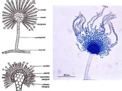
Aspergillus is a group of filamentous ascomycetes that encloses some of the most widespread fungi, containing approximately 350 recognized species. It encloses species of high economic importance for their compound production. Some species are used in biotechnology (enzymes, organic acids, bioactive metabolites), other species are harmful and considered as foodborne contaminants (food spoilage and mycotoxin contamination) or as causal agents of human mycoses (pulmonary, otomycosis, keratitis) (Kocsubé et al. 2016), and others use as model species to understand eukaryotic cell biology and molecular processes (i.e. A. nidulans) (Whiteway and Bachewich 2017). This genus is endowed with a diagnostic morphological trait reminding the holy water sprinkler, the ‘aspergillum’, which consists on a conidiophore that ends in a spherical vesicle bearing phialides and metulae that generate chains of conidia (Dyer and O’Gorman 2012) (Figure 8). The classification of Aspergillus has undergone several modifications over the past years using different approaches aiming to group the growing number of species according to its phylogenetic relationships (Scheidegger and Payne 2003). Aspergilli classification was traditionally based on morphological traits, and has nowadays extended to include the secondary metabolic profile and molecular approaches. This review will focus on the Flavi section of the Circumdati subgenus. This section bears a particular interest since it includes human pathogens, important mycotoxin producers, especially of aflatoxins, as well as safe enzyme producers commonly used in the food industry.
Figure 8. Aspergillum structure. Figures on left represent the mature conidiophore with primary sterigmata (above) and secondary sterigmata (below); right figure microscopic photo of Aspergillus flavus.
1.5.1 A compendium of section Flavi
Aspergillus section Flavi is mainly composed of saprophytic molds occurring in diverse ecological niches and playing a keystone role in the first steps of the nutrient cycle (Cotty et al. 1994; Rodrigues et al. 2012). Among their general phenotypical characteristics are the conidial heads yellow-green to brown shades, uniseriate or biseriate and the production of black sclerotia (Varga et al. 2011; Houbraken et al. 2014). The characteristic secondary metabolites in the group include aflatoxins (AF), paspaline, kojic acid, aspergillic acid, and cyclopiazonic acid (CPA) (Frisvad and Samson 2000). These molds grow better under environmental conditions of humidity (around 0.85 to 0.99 aw) (Medina et al. 2015; Yogendrarajah et al. 2016) and temperatures ranging from 28 to 42 °C and several grow faster at 37 °C (Varga et al. 2011). The environmental humidity and temperature preferences make the species within the section Flavi suitable to grow in the tropics and subtropics over the world, yet some of them are able to grow in temperate regions, like A. flavus. Furthermore, climate change affects principally these two environmental variables, creating new niches in areas that in the past were not suitable for Aspergillus section Flavi species development, which could favor their colonization into temperate regions (Perrone et al. 2014).
Extrolites produced by these fungi make the section interesting for studying purposes. Some species, such as A. flavus and A. parasiticus have an impact on human and animal health as well as on international economy, as they are able to produce aflatoxins, especially aflatoxin B1 (AFB1), considered as mycotoxins of high health risk due to their carcinogenic, mutagenic and teratogenic potential (IARC 2012). In addition, species belonging to this section are also able of producing a wide range of other mycotoxins such as CPA, aflatrems, versicolorins, sterigmatocystin, ochratoxin A (OTA), etc. Albeit, other species are not toxinogenic and are used in biotechnology for producing enzymes and organic compounds commonly used in several industrial processes (Houbraken et al. 2014). For example, A. oryzae and A. sojae synthesize kojic acid, a secondary metabolite used in the production of soy sauce, a market with estimated shares of billions of dollars worldwide (Chang et al. 2007a).
Although there are some morphological characters and secondary metabolites that allow identification at species level, when cryptic species are present they become insufficient for taxonomical differentiation. In addition, morphological analyses to discriminate among isolates can be tricky, because mold phenotype is affected by environmental and nutritional conditions, creating overlapping phenotypical traits (Chang et al. 2007a). Inclusion of molecular analyses to the methods aforementioned is crucial for species identification and to clearly define relations within the Flavi section (Samson et al. 2014). However, finding differences at the molecular level can be challenging since species of this group shares several conserved traits. For instance, Aspergillus flavus and A.
parasiticus share approximately 97-99% nucleotide identity of their genomes (Chang et al. 2007a). ITS gene is usually used as a barcode gene to differentiate fungal species, but in this section, it is highly conserved, making it almost uninformative (Varga et al. 2011; Houbraken et al. 2014). Even though, there are challenges to characterize these organisms at species level, it is important to keep a practical taxonomic system because it is the base to the development of regulations to favor food safety and control (Geiser et al. 2007; Godet and Munaut 2010).
Over the last two decades, Aspergillus section Flavi has suffered several modifications in their composition; currently 26 species have been described based on a polyphasic approach, which includes phylogenetic, morphological and secondary metabolites analyses. The section is constituted by Aspergillus flavus, A. oryzae, A. parvisclerotigenus, A. minisclerotigenes, A. parasiticus, A. sojae, A. arachidicola, A. novoparasiticus, A. sergii, A. transmontanesis, A. mottae, A. nomius, A. pseudonomius, A. bombycis, A. tamarii, A. pseudotamarii, A. caelatus, A. pseudocaelatus, A. bertholletius, A. coremiformiis, A. togoensis, A. leporis, A. hancockii, A. alliaceaus, A. lanosus, and A. avenaceus. From these, the genome (strain = GenBank assembly accession numbers) of A. flavus (NRRL 3357 = EQ963472, AF70 = ASM95283v1), A. oryzae (RIB40 = GCA_000965245.1), A. parasiticus (SU-1= GCA_000956085.1, 68-5 = GCA_001576805.1), A. nomius (NRRL 13137 = GCA_001204775.1), A. bombycis (NRRL 26010 = GCA_001792695.1), A. hancockii (FRR 3425 = GCA_001696595.1), and A. arachidicola (CBS 117610 = GCA_002749805.1) have been sequenced.
1.5.2 Reproduction in Aspergillus section Flavi
Most Aspergillus fungi are only known in an asexual state (64%) (Dyer and O’Gormann 2011), nevertheless, there is evidence that cryptic reproduction occurs in some species. In ascomycetes, sexual identity and later stages of sexual development are partially regulated by the MAT locus, conformed by two idiomorphs MAT1-1 and by the Mat1-2 genes, encoding a protein with a α-box motif and a protein of the high mobility group (HMG), respectively (Ramirez-Prado et al. 2008; Dyer and O’Gorman 2011). In heterothallic species, only one of the idiomorphs is present, whereas in homothallic both idiomorphs are present and they occur in the same loci or in different chromosomes (Dyer and Kück 2017). The section Flavi is mainly composed by heterothallic species, and asexual reproduction seems to occur more frequently. Sexual reproduction is reported only for six species and from them only one species is homothallic, A. alliaceus (Horn et al. 2011; Dyer and Gorman 2012). It has been hypothesized that homothallic type can be the ancestral state in this section because A. alliaceus is a basal species (Ramirez-Prado et al. 2008). The presence of both idiomorphs in most analyzed species, however, suggests that heterothallic type could be the ancestral trait in Aspergillus (Ramirez-Prado et al. 2008). Sexual forms in this section are clustered in
the genus Petromyces, erected to include the teleomorph of A. alliaceus. Later on, sexual states of A. flavus, A. parasiticus, A. nomius and A. oryzae were incorporated, based on morphological evidence, like cleistothecia structure, and to maintain the monophyly of the group (Moore et al. 2009; Horn et al. 2009). Another important trait in the section is the production of sclerotia, which occur in several species. In asexual species, it has been hypothesized that sclerotia aid species to cope with adverse environmental conditions and predators, which is supported by the type of secondary metabolites produced (McAlpin and Wicklow 2005; Cary et al. 2015b), while in sexual species they also play a role in the formation of cleistothecia (Horn et al. 2009).
Figure 9. Reproductive cycle in Aspergillus (Image representing A. nidulans) (reprinted from Casselton and Zolan 2002)