Direction des bibliothèques
AVIS
Ce document a été numérisé par la Division de la gestion des documents et des archives de l’Université de Montréal.
L’auteur a autorisé l’Université de Montréal à reproduire et diffuser, en totalité ou en partie, par quelque moyen que ce soit et sur quelque support que ce soit, et exclusivement à des fins non lucratives d’enseignement et de recherche, des copies de ce mémoire ou de cette thèse.
L’auteur et les coauteurs le cas échéant conservent la propriété du droit d’auteur et des droits moraux qui protègent ce document. Ni la thèse ou le mémoire, ni des extraits substantiels de ce document, ne doivent être imprimés ou autrement reproduits sans l’autorisation de l’auteur.
Afin de se conformer à la Loi canadienne sur la protection des renseignements personnels, quelques formulaires secondaires, coordonnées ou signatures intégrées au texte ont pu être enlevés de ce document. Bien que cela ait pu affecter la pagination, il n’y a aucun contenu manquant.
NOTICE
This document was digitized by the Records Management & Archives Division of Université de Montréal.
The author of this thesis or dissertation has granted a nonexclusive license allowing Université de Montréal to reproduce and publish the document, in part or in whole, and in any format, solely for noncommercial educational and research purposes.
The author and co-authors if applicable retain copyright ownership and moral rights in this document. Neither the whole thesis or dissertation, nor substantial extracts from it, may be printed or otherwise reproduced without the author’s permission.
In compliance with the Canadian Privacy Act some supporting forms, contact information or signatures may have been removed from the document. While this may affect the document page count, it does not represent any loss of content from the document.
A Study of Rat Chromosome 8 by Congenies:
Mapping and Dissecting Quantitative Trait Loci into
Opposite Blood Pressure Effects
par
Anita Ariyarajah
Biologie Moléculaire
Mémoire présenté à la Faculté des études supérieures en vue de l'obtention du grade de Maîtrise
en Biologie Moléculaire
octobre 2007
©
Anita Ariyarajah,2007Université de Montréal Faculté des études supérieures
Ce mémoire intitulé:
A Study of Rat Chromosome 8 by Congenies:
Mapping and Disseeting Quantitative Trait Loei into
Opposite Blood Pressure Effeets
présentée par :
Anita Ariyarajah
a été évalué par un jury composé des personnes suivantes:
Dr. Christian Deschepper président-rapporteur Dr. Alan Deng directeur de recherche Dr. Danielle Malo membre du jury
RÉSUMÉ ET MOTS CLÉS FRANÇAIS
L'hypertension essentielle est une maladie complexe affectée par des facteurs génétiques et environnementaux. Il est estimé que des facteurs génétiques contribuent entre 30% et 50% de la variation de la pression artérielle (PA) observée. L'identification des gènes peut apporter une meilleure compréhension sur la pathophysiologie de la maladie:
Les souches de rats consanguins sont utilisées dans cette étude sont le rat hypertendu Dahl sensibles au sel (S) et le rat normotendu Lewis (LEW). Mon étude se concentre sur le chromosome 8 (C8) du rat. Une étude antérieure des liaisons génétiques suggère qu'une région du C8 contient un locus pour le trait quantitatif (QTL) de la PA. L'objectif de cette étude consiste à vérifier si ce QTL existe et de le localiser à une région précise sur C8. Des lignées congéniques, obtenues par le croisement du rat S et du rat LEW, de façon à remplacer des régions du C8 du rat S, adjacentes et non chevauchants, par les régions homologues du rat LEW, permettent d'associer un changement potentiel de la PA à une région sur le C8. Les mesures du PA ont été faites par télémétrie. Des deux lignées congéniques produites, les allèles LEW d'une des souches baissent la PA (p<0.001). Par contre, les allèles LEW de la deuxième souche congénique augmentent la PA
(p<O.OOI). Cette étude prouve qu'il existe sur la C8 deux QTLs de la PA, adjacents, mais séparés, avec des effets opposés ce qui n'a pas été observé dans les études antérieures de liaisons.
Mots-clés: Hypertension essentielle; rat Dahl sensibles au sel; locus pour trait quantitatif; modèle animal; lignées congéniques; facteurs génétiques; chromosome 8
ENGLISH ABSTRACT AND KEYWORDS
Essential hypertension is a complex disease with genetic and environmental factors.
It is estimated that 30 to 50 % of the variations observed in blood pressure (BP) is caused
.
by genetic factors. The identification of the genes that affect BP is crucial for a full comprehension of the pathophysiology of the disease.My study focuses on rat chromosome 8 (C8). According to previous linkage studies, C81ikely contains one quantitative trait locus (QTL) for BP. The purpose ofmy study is to determine whether such a QTL exists, and if so, to assess its effect on BP. Our basic strategy is to use congenic strains. Our congenic strains were constructed from inbred strains with contrasting effects on BP regulation, namely the hypertensive Dahl Salt-Sensitive (S) strain and the normotensive Lewis (LEW) strain. Non-overlapping, adjacent C8 regions of the S rat were replaced with homologous regions of the LEW rat. BP was measured by telemetry. Of the two congenic strains produced, LEW alle1es of one congenic strain lower BP (p<0.001). In contrast, LEW alleles of the other increase BP (p< (0.001). Thus, two BP QTLs on C8, adjacent to each other, but with opposite effects exist on C8. The effect of the second, novel QTL has not been reported in previous linkage studies.
Keywords: essential hypertension; salt-sensitive Dahl rat; quantitative trait locus; animal model; congenic strain; genetic factors; chromosome 8
TABLE OF CONTENTS
RÉSlTMÉ ET MOTS CLÉS FRANÇAIS ... III ENGLISH ABSTRACT AND KEYWORDS ...••... IV INDEX OF TABLES ... XI INDEX OF FIGURES ... XII ABBREVIATIONS ... XIII DEDICA TION ... XVI ACKNOWLEDGEMENTS ... XVII
REVIEW OF THE LITERATURE ... 1
CHAPTER 1 ... 1
HYPERTENSION: CLINICAL FEATURES OF HYPERTENSION ... 1
1.1 THE CARDIOV ASCULAR SySTEM ... 1
1.1.1 Hemodynamics
0/
Circulation ... 31.2 DEFINITION OF HYPERTENSION ... 3
1.2.1 Types o/Hypertension ... 5
1.2.1.1 Primary or Essential Hypertension ... 6
1.2.1.1.1 Monogenic F orm of Hypertension ... , ... 6
1.2.1.1.2 Po1ygenicForm of Hypertension ... 8
1.2.1.2 Secondary Hypertension ... 8
1.3 TRADITIONAL RISK FACTORS FOR HYPERTENSION ... 10
1.3.1 Saltsensitivity ... ... ... 10
1.3.3 Obesity ... 12
1.3.4 Insulin Resistance ... ... 12
1.3.5 Gender, Age and BP ... 13
1.4 HIGH BP AS A RISK FACTOR FOR MORBfDITY AND END-ORGAN DAMAGE ... 14
1.5 FACTORS REGULATING ARTERIAL BP ... 15
1.5.1 Neural Regulation of BP ... 17
1.5.1.1 The Nervous System ... 17
1.5.1.1.1 Innervation of the Vasculature ... 18
1.5.1.1.2 Arterial Baroreceptors ... 19
1.5.1.1.3 The Cardiovascular Center ... 20
1.5.2 Neurohumoral Mechanisms - The Endocrine System ... 22
1.5.2.1 Hormones ... 22
1.5.2.1.1 Renin-Angiotensin-Aldosterone System ... 22
1.5.2.1.2 Adrenergic and Dopaminaerigic Receptors and Actions ... 25
1.5.3.1.3 Vasopressin (Antidiuretic Hormone) ... 27
1.5.2.1.4 Nitric Oxide ... 28
1.5.2.1.5 Atrial andSrain Natriuretic Peptides ... 30
1.5.2.1.6 Endothelial Mechanisms and Endothelin ... 31
1.5.2.1.7 Arachidonic Acid Metabolites ... 32
1.5.2.1.8 Kinins ... 33
1.6. ANTI-HYPERTENSIVE DRUGS ... 34
CHAPTER 2 ... 35
THE SEARCH FOR GENES CAUSING HYPERTENSION ... 35
2.1.1 BP as a Quantitative Trait ... 36
2.1.2 Studying Quantitative Traits ... 36
2.2 STRATEGIES FOR SEARCHING FOR GENES OF COMPLEX TRAITS ... 37
2.2.1 Mapping ... 3 7 2.2.1.1 Genetic Maps ... 37
2.2.1.1.1 Homologous Recombination ... 38
2.2.1.2 Genetic Markers ... 39
2.2.1.3 Linkage Analysis ... 40
2.2.1.4 Statistics: The LOD Score ... 41
2.2.2 Physical Maps ... ... 42
2.2.2.1 Radiation Hybrid Mapping ... .43
2.2.2.2 Physical Mapping Using Genome Sequences ... .44
2.2.3 Co-segregation analysis ... 44
2.2.4 Candidate gene approach ... 45
2.2.5 Animal models ... 45
2.2.5.1 Development of the Rat Model.. ... .47
2.2.5.2 The Rat as an Animal Model in the Study ofHypertension ... 48
2.2.5.2.1 The New Zealand Genetically Hypertensive Rat (GH) ... 51
2.2.5.2.2 The Spontaneously Hypertensive Rat (SHR) ... 51
2.2.5.2.3 The Dahl Salt-Sensitive Hypertensive Rat (S) ... 51
2.2.5.2.4 The Milan Hypertenive Rat (MHS) ... 53
2.2.5.2.5 The Lyon Hypertensive Rat (LH) ... 54
2.2.5.2.6 The Sabra Hypertensive Rat (SBH) ... 54
2.2.5.3 Advantages ofusing the Rat as a Genetic Model in Hypertension ... 54
2.3 PREVIOUS STUDIES ON RAT CHROMOSOME 8 ... 56
CHAPTER 3 ... 60
EXPERIMENTAL METHODS USED TO STUDY RAT CHROMOSOME 8 ... 60
3. 1 OBJECTIVE OF MY STUDY ... 60
3.2 MATERIALS AND METHODS ... 60
3.2.1.Rat Strains Used-Dahl Salt -Sensitive vs. Salt-Resistant Rats ... 60
3.2.2 Lewis Strain ... 61
3.2.3 Strategies used in the Study of the Rat Model ... 61
3.2.4 Congenic Strains ... 61
3.2.4.1 Baekerossing and Speed Congenies ... 63
3.2.4.1.1 How Chromosome 8 Congenies StraÏns in this Study were Seleeted.65 3.2.5 Genome-wide scanning ... ... 66
3.2.6 Choice of Markers: ... 67
3.2. 7 Microsatellite markers ... 67
3.3 SAMPLE PREPARATION AND DNA ANALYSIS ... 68
3.4 MOLECULAR SEPARATIONS USED IN THIS STUDY ... 69
3.4.1 DNA Gel Electrophoresis ... 69
3.4.2 Autoradiography ... 70
3.5 MEASURING BP- TELEMETRY ... 73
3.6 STATISTICAL ApPROACH ... 74
CHAPTER 4 ... 76
RESULTS ... 76
4.2 TELEMETRY DATA ... 80
CHAPTER 5 ... 84
DISCUSSION ... 84
5.1 THE PRESENCE OF Two QTLs WITH OPPOSING EFFECTS ... 84
5.1.1 Epistasis ... 85
5.1.2 Overall Phenotype Versus lndividual QTLs ... 86
5.2 PHYSIOLOGICAL ANAL YSIS -MEASURING BP ... 87
5.2.1 Tail Cuff ... 87
5.2.2 Telemetry ... 88
5.2.3 lndwelling Catheter ... 88
5.3 GENE-ENVIRON MENT INTERACTION ... 89
5.4 THE GENETIC BACKGROUND AND ITS EFFECTS ... 90
5.5 OTHER LIMITATIONS AND POTENTIAL SOURCES OF ERROR ... 95
5.6 THE QTL REGIONS ... 96
5.6.1 Reproducibility of the Negative BP QTL ... 96
5.6.2 Comparative Mapping and Potential Candidate Genes ... 99
5.6.2.1 Ion Channels ... 99
5.6.2.1.1 Potassium Channel (Kenj5) ... 99
5.6.2.1.2 FXYD2 Prote in-Sodium Potassium Pump Aetivity Modifier ... 100
5.6.2.2 Cytoehrome P450 Enzyme CyplAl.. ... 101
5.6.2.3 Alpha B Crystallin ... 103
5.6.2.4 Neuropeptide Y Family Member- GPR72 ... 103 5.6.2.5 Nieotinie Cholinergie Receptors ... l04 5.6.2.6 Dopamine Reeeptor 2 ... 1 05
5.7 PROPOSALS FOR FUTURE RESEARCH ... 107
5. 7.1 QTL-QTL Interactions ... 1 07 5.7.1.1 Double Congenies within the Same Chromosome ... 108
5.7.1. 2 Double Congenies Using Different Chromosomal Segments ... 108
5.7.1.3 Construction of Congenies Using Other Contrasting Strains ... 108
5.7.1.4 Narrowing the QTL Regions Using Congenies ... 109
5.7.2 Functional and Positional Candidate Genes ... 1 09 5.7.2.1 Analysis of DifferentiaI Gene Expression ... 109
5.8 CONCLUSION ... 111 REFERENCE LIST ... XVIII
Index of Tables
Table 1 -Classification of hypertension ... 5
Table 2-Hypertension and end organ damage ... 15
Table 3- Physiologie actions of angiotensin II ... 25
Table 4- Advantages of the rat as a model for genetic hypertension ... 54
Table 5- BP QTLs localized in the Dahl salt sensitive rat.. ... 55
Table 6 Chromosome Markers used to make congenic Strains ... 66
Table 7 -New microsatellite markers constructed for rat chromosome 8 ... 79
Table 8 -Advantages and disadvantages in the three princip le methods for measuring BP in the rats ... 89
Index of Figures
Figure 1 - The Cardiovascular System ... 2
Figure 2 - Factors Contributing to Essential Hypertension ... 7
Figure 3 - Various Ways Certain Conditions Cause Secondary Hypertension ... 8
Figure 4 - Factors Affecting Mean BP ... 16
Figure 5 - How the Cardiovascular Center Regulates Blood Pressure ... 21
Figure 6 - Components of the RAAS system and their main effects on the cardiovascular system ... 24
Figure 7 - Physiological Effects ofNorepinephrine and Epinephrine ... 26
Figure 8 - Vasopressin release and physiological effects ... 27
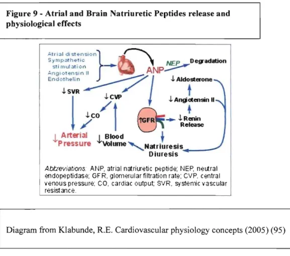
Figure 9 - Atrial and Brain Natriuretic Peptides release and physiological effects ... 30
Figure 10 - LOD score and QTL location-Garrett 1998 ... 56
Figure Il - LOD score and QTL location-Garrett 2000 ... 57
Figure 12 - The Congenic ... 62
Figure 13 - Speed Congenic- ... 63
Figure 14 - Comparison of Traditional and Speed Congenic Methods ... 64
Figure 15 - Microsatellite example ... 67
Figure 16 - DNA gel electrophoresis ... 71
Figure 17 - Autoradiography ... 72
Figure 18 - Timeline for data collection ... 74
Figure 19 - Map of Congenic Strains Constructed ... 77
Figure 20 - BP readings comparing the two parental strains, S and LEW ... 80
Figure 21 - Comparisons ofMAP, DAP and SAP ofS rats with those of the congenics .... 82
Figure 22 - Summary including Statistical Analysis ... 83
Figure 23 - Deducing the Effect QTLs Using Congenics ... 94
Figure 24 - Localizations oftwo quantitative trait loci (QTLs) and comparative mapping .97 Figure 25 - Comparative Genomics ... 112
ABBREVIATIONS
ADH
antidiuretic hormoneACE
angiotensin converting enzymeANP
atrial natriuretic peptideANS
autonomie nervous systemATI
angiotensin 1ATH
angiotensin IIAVP
vasopressinBNP
brain natriuretic peptideBP
blood pressurebp base pair (s)
CS
chromosome 8CSS.Ll
chromosome 8 S-LEW 1CSS.L2
chromosome 8 S-LEW 2cGMP
cyclic guanosine monophosphateCHD
coronary heart diseasecM
centimorgancNOS
constitutive nitric oxide synthaseCNS
central nervous systemCO
cardiac outputcR
centiRaysCVC
cardiovascular centerCVD
cardiovascular diseaseDAP
diastolic arterial pressureDBP diastolic blood pressure DNA deoxyribonucleic acid (s)
dNTP deoxyribonucleotide triphosphate DIR donor/recipient ET-l endothelin Gb gigabases GDB genome database
UR
heart rateiNOS inducible nitric oxide synthase KCl potassium chloride
LEW Lewis strain
LOD logarithm of odds
MAP mean arterial pressure MgClz magnesium chloride O1mHg millimeters of mercury NaCI sodium chloride
NE norepinephrine
NO nitric oxide
NTS nucleus tractus solitarius PCR polymerase chain reaction PGI2 prostacyclin
PNS parasympathetic nervous system QTL quantitative trait locus/loci
R Dahl salt-resistant strain
S Dahl salt-sensitive strain
SAP
systolic arterial pressureSBP
systolic blood pressureSHR
spontaneously hypertensive ratSNP
single nucleotide polymorphismSNS
sympathetic nervous systemSV
stroke volumeSVR
systemic vascular resistanceU units
WHO
world health organizationDEDICATION
This work is dedicated to my parents, who have supported me throughout my life and have been pillars of strength when 1 really needed them. In loving memory of my grandmother whose favourite saying was 'Hitch your wagon to the highest stars' and in memory of Roselle Cossette, who was like an aunt to me, whose passing has left many lives with one less ray of sunshine.
ACKNOWLEDGEMENTS
l would like to thank first and foremost my supervisor, Dr. Alan Deng for giving me the opportunity to work in his laboratory and to participate in new scientific discoveries. Aiso for his advice on how to structure this thesis.
Another thank: you goes to Mr. Eric Regener, for his tremendous help in proof-reading and organizing my writing and to Dr. Madeleine Ravaoarinoro for her help with the french abstract.
The lab has been a motivating and inspirational experience: The people of the Lab: Julie Dutil my inspiration and mentor, of whom l am in awe, Myriam Moujahidine,
kindness itself. Ana Palijan, may you always challenge aU things with your forthright ways and may you al ways be a beam ofjoy and laughter, Raphaëlle Lambert whose calmness and insightfulness inspired me. Vikki Eliopoulos, a joy to be around, Kalyani Prithiviraj whose thoughtfulness and kindness will be remembered, Myrian Grondin and Sophie Charron.
Finally l would like to thank: Julie Roy and Annie Ménard, without whom the lab would cease functioning.
l would like to thank you aIl for helping me throughout my time in the lab and for enduring my flawed French. It has been a wonderfuIIeaming experience.
l would aiso like to thank Mme Vivianne Jodoin for gui ding me through the technicalities of the program.
REVIEW OF THE LITERATURE
Chapter 1
Hypertension: Clinical features of hypertension
1. 1
The Cardiovascular System
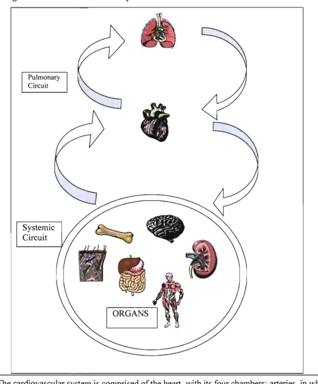
The cardiovascular system is an organ system whose function is to propel and transport blood and its constituents throughout the body. The components of the cardiovascular system are: the blood, the heart and blood vessels(67).
Blood consists of liquid plasma and ceIls. It is the medium by which substances such as nutrients, hormones, gases (oxygen, carbon dioxide) and metabolic wastes are transported to and removed from tissues. The vascular system is made up of arteries, veins, and capillaries (blood vessels), which carry blood to and from aIl tissues (Figure1).
The heart pumps blood at a high pressure through the vascular system. It sends blood to the lungs for oxygenation and via the aorta, distributes oxygenated blood to the tissues of the body.1t is a four-chamber pump, with the right side receiving deoxygenated blood from the body at low pressure and pumping it to the lungs (the pulmonary
circulation) and the left side receiving oxygenated blood from the lungs and pumping it at high pressure around the body (the systemic circulation)(20,95). It can also help stabilize body temperature, oxygen concentration, ionic composition, osmolarity and pH as part of homeostasis(119,124).
Figure 1 - The Cardiovascular System Pulmonary Circuit Systemic Circuit ORGANS
The cardiovascular system is comprised of the heart, with its four chambers; arteries, in which blood moves away from the heart; veins, in which blood retums to the heart; and a system of capiUaries, which transport blood between smaU arteries and smaU veins. The cardiovascular system is further divided into the pulmonary circuit, which delivers deoxygenated blood to the lungs, and the circulatory circuit, which delivers oxygenated blood to aU of the organs and tissues.
1.1.1 Hemodynamics of Circulation
The primary determinants of systemic blood pressure (BP) are cardiac output (CO) and peripheral vascular resistance. CO is the amount of blood ejected per minute by the left ventricle into the aorta or by the right ventricle into the pulmonary trunk. It is determined by plasma volume, cardiac stroke volume, heart rate and myocardial contractility. It is calculated as CO (mLlmin)= Stroke Volume (SV) (mLibeat) x Heart Rate (HR)
(beats/min), where SV the amount ofblood ejected by a ventricle during each systole. SV is related to the stretch of the heart before it contracts, the forcefulness of the contraction and the resulting pressure before ventricular ejection begins. Peripheral vascular resistance is a function of the balance of humoral vasoconstriction and vasodilation, adrenergic activity and arteriole smooth muscle tone. CO and peripheral vascular resistance are the primary determinants of systemic BP. Given the se two factors, mean arterial BP is calculated as CO x peripheral vascular resistance.
Extremely complex physiological networks and other factors underlie BP regulation and hypertension susceptibility. Arterial BP is generated by the left ventricle ejecting blood into the systemic vasculature, which acts as a resistance to CO. With each ejection ofblood (ventricular systole), the aortic blood volume increases, stretching the wall of the aorta. As the heart relaxes (ventricular diastole), blood flows from the aorta into distributing arteries that distribute the blood to the various organs. Within the organs, the arterial vasculature undergoes extensive branching and the vessel diameters decrease. The smaller arteries and arterioles serve as the chief resistance vessels. Through changes in their diameter, they serve to regulate systemic vascular resistance and organ blood flow. In hemodynamic terms, the mean arterial pressure (MAP) can be described by MAP (CO x SVR)
+
CVP, where SVR = systemic vascular resistance, and CVP central venous pressure. Therefore, increases in CO, SVR or CVP willlead to increases in MAP (95,119,124).1.2
Definition
of
hypertension
The general term "blood pressure" was coined in the early 1700s by the man who tirst measured it, the Reverend Stephen Hales ofEngland (75,112). The expression "blood pressure" (BP) refers to the arterial pressure in systemic circulation. The movement of
blood throughout the organism depends on the strength of the heartbeat and on the pressure gradient in the cardiovascular system. BP is dependent on cardiac pressure and vascular resistance. BP is highest in the aorta and progressively decreases as the blood flows further away from the heart. So the pressure gradient is such that there is a progressive lowering of BP as the blood travels from large arteries to small ones, then from the capillaries back through the veins, until it returns to the heart atria through the vena cava. BP is measured in millimetres of mercury (mmHg).
The cardiac cycle consists oftwo parts: systole (contraction of the heart muscle in the ventricles) and diastole (relaxation of the ventricular heart muscles). When the
ventricles contract, they force the blood out of the heart into the arteries. The left ventricle empties into the aorta (systemic circuit) and the right ventricle into the pulmonary artery (pulmonary circuit). The he art valves open and close to limit flow to a single direction. During cardiac diastole BP fluctuates between a maximum value and a minimum value (diastolic blood pressure (DBP) or diastolic arteriai pressure (DAP)). The systolic blood pressure (SBP) or systolic arterial pressure (SAP) is a measure of the BP within the artery at the moment when the heart contracts (ventricular contraction) and ejects blood towards the arteries. DBP corresponds to the BP when the heart relaxes and the aortic valve closes (BP between ventricular contraction). Mean arterial pressure (MAP) is the average between DBP and SBP. MAP is a measure that reflects the BP to which small blood vessels are continuously submitted to.
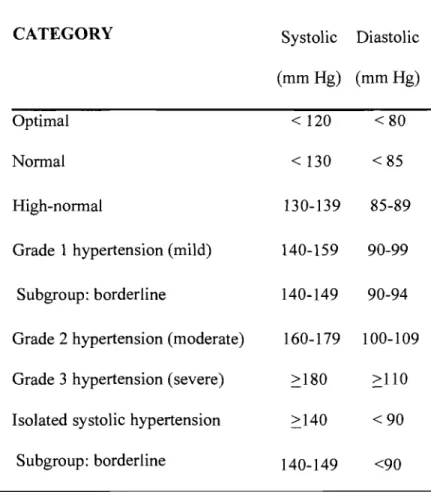
When the values for these pressures are chronically elevated, the condition is called hypertension. Hypertension is defined in general as a chronic elevation in BP in the arteries, when compared to values that are considered normal. In general, hypertension is defined as a BP systolic value ~ 140 mmHg or diastolic value ~ 90 mmHg (i.e. values exceeding
140/90 mmHg), or both. The official criteria on threshold values for hypertension vary, but the criteria set by the World Health Organization (WHO) are widely referred to (Table 1) (75).
Elevated BP is a common and powerful predisposing factor for stroke, coronary disease, cardiac failure and peripheral artery disease, imposing a 2-3 fold increased risk of one or more ofthese atherosclerotic sequelae. The risk ratio imposed by hypertension is greatest for cardiac failure and stroke, but in Western countries coronary disease is the most common and lethal hazard (88).
Table 1 -Classification of hypertension
CATEGORY Systolic Diastolic
(mm Hg) (mm Hg)
Optimal < 120 < 80
Nonnal < 130 < 85
High-nonnal 130-139 85-89
Grade 1 hypertension (mild) 140-159 90-99
Subgroup: borderline 140-149 90-94
Grade 2 hypertension (moderate) 160-179 100-109 Grade 3 hypertension (severe) ~180 ~110
Isolated systolic hypertension 2:.140 <90 Subgroup: borderline 140-149 <90
Table adapted from Hansson, L. Hypertension management in 2002: where have we been? where might we be going? Am. J Hypertens. 15, 101S-107S (2002). (75)
1.2.1 Types of Hypertension
Hypertension affects 15-30% of the human population, including 50 million
Americans. The two main categories of hypertension are primary hypertension, also known as essential hypertension, and secondary hypertension. The vast majority of patients have essential hypertension, which is a fonn with no identifiable underlying cause(95).
Unknown genetic and environmental factors are thought to play a role in the pathogenesis of essential hypertension. Secondary hypertension, which accounts for only 5-10% of cases is due to definable causes such as renal vascular disease, chronic renal failure, renal artery disease, thyroid disease pregnancy and endocrine abnonnalities such as, primary
1.2.1.1 Primary or Essential Hypertension
Despite many years of research, there is no unifying hypothesis to account for the pathogenesis of primary hypertension. There is a natural progression of this disease that suggests that early elevations in blood volume and CO might initiate increased resistance in the systemic vasculature. In hypertensive patients, the vascular endothelium produces less nitric oxide and the vascular smooth muscle is less sensitive to the actions of this powerful vasodilator. There is also an increase in endothelin production, which can enhance
vasoconstrictor tone. Many mechanisms may operate to initiate and sustain hypertension. Treatment of patients with primary hypertension generally involves
pharmacological intervention (2) to modify factors such as angiotensin II, sympathetic activity and calcium entry into cells so as to reduce arterial pressure. These treatments do not target the underlying disease. Nevertheless, treatment of hypertension with
antihypertensive drugs is important, because hypertension increases the risk for coronary artery disease, strokes, renal disease and other disorders. The three broad classes of drugs used to treat primary hypertension are diuretics (to reduce blood volume), vasodilators (to decrease systemic vascular resistance), and cardioinhibitory drugs (to decrease CO). Essential hypertension is further subdivided into a monogenic form and polygenic forms.
1.2.1.1.1 Monogenic Form of Hypertension
In the monogenic form of essential hypertension, the phenotype is due to lesions or mutations in a single gene, leading to eg., overproduction of mineralocorticoids. This form is also known as the Mendelian form of hypertension. The monogenic form of hypertension is only responsible for only 5% of cases (41,81). Mendelian forms of hypertension incIude Liddle's syndrome, glucocorticoid remediable aldosteronism, apparent mineralocorticoid excess and pseudohypoaldosteronism type II. The examples previously listed affect the homeostasis of salt and water reabsorption. Even though Mendelian forms of hypertension are more rare and severe than essential hypertension, there is a tremendous effort to better understand their etiology in the hope that it would lead to cIues about the pathophysiology of essential hypertension (51,55,185,186,199).
Figure 2 - Factors Contributing to Essential Hypertension Excess sodium intake Reduced nephron number
[i]
Genetlc
GJ
Factors
(e.g. alcobol, smoking, salt etc.)
Hypertension
Four factors resulting in hypertension:
1) Genetic factors working individually 2) Environmental factors working individually 3) The interaction of genetic factors with environmental factors 4) The interaction of different genetic factors.
8
1.2.1.1.2 Polygenic Form of Hypertension
Polygenic hypertension is caused by the interaction of many genes, as opposed to the monogenic form, which is due to a single gene. It is also influenced by environment factors and the interaction between environmental factors and genetic factors (Figure 2). 90-98% of aIl patients with primary hypertension suffer from polygenic hypertension (178).
For example, it has long been known that hypertension aggregates in certain families. Familial clustering of high BP could be the consequence of inheritance of a BP gene or genes, household environment, or a combination ofboth. Familial aggregation studies have shown that clustering occurs early in life since siblings living apart do not become dissimilar and spouses do not become more similar even though they share the same environment. Phenotyping hypertension is remarkably complex because of the difficulty of accurately measuring BP and the vast variety of factors regulating BP. For example, BP is partly determined by the pumping action of the heart, so it is inherently pulsative in character. What is more, an individual 's BP varies by time of day, se as on and other circumstances such as stress and emotions (137).
1.2.1.2 Secondary Hypertension
Figure 3 - Various Ways Certain Conditions Cause Secondary Hypertension
Hypertension
1 1Î
Cardiac Output • HypervolemiafGnal artery stenos/s fGnaJ d/seas& Il)'pera.'doslerorusm hypers&crflt.<rm of ADH aor.lC coarctation prflgl1ancy (prfleclampsia) • Stress - sympalhfllrc acIJVa/.'O!l • Pheochromocytoma - I/lcroased calocholamines 1
Î
Systemic Vascular Resistance • Stress - sympatltelic aC/Na/Ion • Atherosclerosis • Renal artery disease• I!Icreaslrd angio/ensin /1 • P,heochromocytoma . I!Icroasfld catflcholam,nes • Thyroid dysfur.ction • Diabetes • Cerebral ischemia
Disease conditions increase either cardiac output or systemic vascular resistance or both, and thereby increase blood pressure.
Secondary hypertension is the direct result of another disease or condition such as obesity, thyroid, or adrenal dysfunction. These conditions increase either SVR or CO or both, which in tum results in hypertension (Figure 3). The most common conditions resulting in secondary hypertension are as follows.
9
Renal artery disease: Renal artery disease causes stenosis, which is the narrowing of the vessellumen. The reduced lumen diameter increases the pressure drop along the length of the diseased artery, which reduces the pressure at the afferent arteriole in the kidney. Reduced arteriolar pressure and reduced renal perfusion stimulate rennin release by the kidney. This increases circulating angiotensin H (ATH) and aldosterone. These honnones increase blood volume by enhancing renal
reabsorption of sodium and water. Increased ATH causes systemic vasoconstriction and enhances sympathetic activity. Chronic elevation of ATH promotes cardiac and vascular hypertrophy. The net effect of these renal mechanisms is an increase in blood volume that augments CO. Therefore, hypertension caused by renal artery stenosis results in an increase in both SVR and in CO.
Primary hvperaldosteronism: This is the increased secretion of aldosterone, which generally results from adrenal adenoma or adrenal hyperplasia. Increased circulating aldosterone causes renal retention of sodium and water, so blood volume and
arterial pressure increase. Furthennore, the adrenal medulla secretes more catecholamines (epinephrine and norepinephrine). Activation of the sympathetic nervous system also increases circulating A TU, aldosterone, and
vasopressin, which can increase SVR. Prolonged elevation of A TH and
catecholamines can lead to cardiac and vascular hypertrophy, both of which can contribute to a sustained increase in BP.
Stress: Physical or psychological stress leads to activation of the sympathetic
nervous system, which causes increased release of norepinephrine from sympathetic nerves in the heart and blood vessels. This leads to increased CO and increased SVR.
Sleep Apnea: Sleep apnea is a disorder in which people repeatedly stop breathing for short periods oftime (10-30 seconds) during their :;leep. lndividuals suffering from sleep apnea have a higher incidence of hypertension.
Hyperthvroidism: This results from an excess ofthyroid hormone induces systemic vasoconstriction, an increase in blood volume, and increased cardiac activity, all of which can lead to hypertension.
Pheochromocytoma: This occurs when catecholamine secreting tumours in the adrenal medulla give rise to high levels of circulating eatecholamines, both
epinephrine and norepinephrine. Thus, the tumours th(:mselves produce epinephrine and norepinephrine. The presence of these hormones k~ads to systemic
vasoconstriction and cardiac stimulation, both ofwhicfl contribute to significant elevations in BP.
Aortic coarctation: This is the narrowing of the aorta, typically just distal to the left subclavian artery, is a congenital defect that obstructs ,:lOrtic outflow, leading to elevated BP proximal to the coarctation (i.e., elevated arteriai pressures in the head and arms). Reduced systemic blood flow and reduced renal blood flow lead to an increase in the release ofrenin and an activation of the
renin-angiotensin-aldosterone system (RAAS). This in tum elevates bloüd volume and BP. Although the aortic arch and carotid sinus baroreceptors are exposed to higher than normal pressures, the baroreceptor reflex in blunted due to structural changes in the walls of vessels where the baroreceptors are located. AIso, baroreceptors become
desensitized to chronic elevation in pressure and become "reset" to the higher pressuree 95,124).
1.3 Traditional risk factors for hypertension
1.3.1 Salt sensitivity
"Hence, if too much saIt is used, the pulse hardens" Huang Ti 2600 BCE. This ancient wisdom of Chinese doctors shows us for how long sodium has been associated with circulatory effects (130), and thus it is maybe the most studied risk factor predisposing for
hypertension. Large studies, like Intersalt (46-48,174-176), have linked sodium and BP convincingly. Moreover, primitive people with diets low in salt have no hypertension. However, the response ofBP to variations in dietary salt intake is quite heterogeneous. In sorne hypertensive individuals, decreases in BP as a result of !;alt restriction are clinically significant, while in others little or no change in BP occurs, whereas in others, BP may actually increase with salt restriction(193). Salt-sensitive patknts are also more likely than salt-resistant patients to exhibit left ventricular hypertrophy, microalbuminuria, and
metabolic abnormalities. These effects naturally make salt-sensitive individuals attractive subjects for genetic studies(l75,181 ,194, 196-198).
An individual's sensitivity to dietary salt may also be related to ethnic background, gender, and other environmental factors. For example, the decline in the glomerular filtration rate with age is more marked in individuals of Africm origin. Also, in a study of more than 400 hypertensive subjects, seven missense mutations were found in the gene coding for the ~-ENaC subunit of the amiloride-sensitive sodium channel, most ofthem in patients of African-American descent. Sorne of the ENaC pol:rmorphismsmight be
associated with greater ENaC activity in vivo and contribute to ethnic differences in sodium retenti on and the risk of low-renin hypertension (180).
The identification of several molecular mechanisms lillking dietary salt and hypertension confirm that saltsensitivity and BP are not determined by a single gene but rather by the combined action of a number of genes. Each of these genes may affect one or more channels, transporters, or enzymes associated with the neural, hormonal, vascular, and renal mechanisms affecting BP. For example, salt-sensitive hypertension may be associated with changes not only in vascular mechanisms but also in the renal Na+/Ca2+ exchanger protein (93). A high-salt diet may affect cellular mechanisms not only in vascular smooth muscle, but also in the endothelium. Sorne studies have ShOWIL that an increase in aortic basal tone in Cl2-Na+ -K+ -ATPase deficient mice that is depend(mt on the endothelium(93). AIso, homozygous II-HSD2-deficient mice that exhibit impaired renal sodium excretion may have an additional endothelial dysfunction, as demonstra:~ed by enhanced
norepinephrine-induced vascular contraction, which may contribute to hypertension(93).
It is thought that changes in BP normally serve as a physiological response whose purpose is to maintain sodium balance and extracellular volume within normallimits. Impairment of the mechanisms responsible for the pressure-natriuresis relationship will cause BP levels to ri se in order to achieve the adjustments in urinary sodium excretion
required to maintain homeostasis. The BP regulatory system i:; thereby reset at a higher BP lev el (158).
1.3.2 Alcohol
Heavy alcohol intake has been found to associate with hypertension (120) .Up to now, no signalling mechanism has been discovered that could c1early explain this link. Among the male Caucasian population, SBP and DBP increase with the quantity of alcohol consumed. In male African-Americans on the other hand, there is a greater increase in diastolic pressure for comparable quantities of alcohol consumed. This tendency is also observed in women, but the increase in BP is less pronounced (5,116,120,140).
1.3.3 Obesity
Studies of the genetic epidemiology of obesity have suggested that the predisposition for development of obesity is partly geneticaUy determined
(5, Il,77,171,174). Obesity is a leading risk factor for essential hypertension. Obese people have also more left ventricular hypertrophy than lean individu ais. Those who weigh the most have, on the average, the highest BP of a given age group. Losing weight reduces BP and left ventricular hypertrophy. However, it has also been loug known that not aU obese individuals are hypertensive and vice versa. AdditionaUy, in Sl)me populations, such as Mexican-Americans and African-Americans, obesity seems t(l have less effect on the development of hypertension than in Caucasians (193).
1.3.4 Insulin Resistance
Insulin resistance syndrome, also known as metabolic syndrome or "syndrome X", is a condition which is characterized by a combination of pathologies including
hypertension, diabetes, abdominal obesity, and dyslipidemia, :unong others. This syndrome is very significant when considering the genetic makeup of the hypertensive population (13,149-155,164).
Insulin is a polypeptide hormone secreted by the pancreas. Its main purpose is to regulate the levels of glucose in the body antagonistically with glucagon through negative feedback loops. It also exhibits vasodilatory properties.
Insulin resistance is characterized by a blunting of the body's normal reaction to the regulating influence of insulin, resulting in an over-production of insultin to compensate. It
Îs one of the causes of diabetes and is the main cause of obese individuals being diabetic. In the hypertensive patients with insulin resistance, the reduction in MAP, after a glucose load, was blunted, when compared to insulin-sensitive hypertt::nsive patients and normotensive patients (172). In normotensive and insulin-sen!:itive hypertensive individuals, insulin may stimulate sympathetic activity without elevating MAP
(117).Though it is known that diabetes often appears simultaneously with hypertension, the mechanism or mechanisms, causing syndrome X have yet to l:'e discovered.
1.3.5 Gender, Age and BP
Men generally have a higher mean BP than women, regardless of age. As for SBP, men also have higher levels than women during early adulthood. Above the age of sixt y, the opposite becomes true (138). DBP, on the other hand, tends to be slightly higher in men than in women regardless of age.
The prevalence of hypertension increases progressively with age in both men and women. However, for BP there is a sexual dimorphism (14,160). For women, although long-term studies such as the Framingham(78,90) have not documented a rise in BP with menopause, other studies have found significantly higher SBP and DBP in postmenopausal compared to pre-menopausal women, even after adjustment for age and body mass index (160). According to these studies, postmenopausal women are twice as likely to have hypertension than pre-menopausal women. This menopause-œlated increase in BP has been attributed to a variety of factors, including estrogen withdrawal, over-production of
pituitary hormones, weight gain, or perhaps a combination ofthese and other yet undefined neurohumorai influences.
Androgen is thought to play a role in the sex differenœs in BP, though its effects are more pronounced in men. One possible mechanism may b~ the blunting of the pressure-natriuresis relation (138). Female sex hormones and their receptors may also be implicated in BP differences between men and women. Though no significant associations have been detected between estrogen receptor genes and BP among women, a genetic association study by the Victorian Family Heart Study investigators found that men înheriting the "a" allele on the estrogen receptor a gene had significantly higher SBP levels (5 mm Hg) than men with other genotypes (49,138).
Increased age is an important risk factor for higher BP (160), since it may reflect the accumulation of different factors predisposing for hypertension as weIl as factors related to normal aging, such as pre-programmed senescence or just general wear and tear (167,170).
1.4 High BP as a Risk Factor for Morbidity and End-Organ Damage
Hypertension is one of the most important risk factors for cardiovascular diseases (66). The risk of coronary heart disease and stroke increases with increased BP. Patients who do not control high BP face a reduced life span, because hypertension can cause certain organs to deteriorate over time. Sorne of the most com.TIon complications of hypertension are listed in Table 2.For the heart, for example, high BP contributes to 75% ofall strokes and heart attacks, according to the National Heart, Lung, and Blood Institute. Compared with normal individuals, hypertensive people can have as high as ten times the risk of stroke and five times the risk of a heart attack, depending on the severity of the hypertension. The risk for developing congestive heart failure is also significantly higher with increased BP. People whose high BP has led to enlargement of the left side of the h,~art (left ventricular
hypertrophy) remain at risk for strokes, heart attacks, sudden death, and heart failure even after their BP is under medical control.
Hypertension is also implicated in kidney dysfunction and in calcium retenti on problems. High BP causes 30% of aIl cases of kidney failure, a rate that is second only to diabetes. Animal studies have indicated that when heart cells ,~nlarge in response to high BP, they undergo molecular changes that cause an abnormal release of calcium, a mineraI crucial for healthy heart contractions. This defect appears to be irreversible. Hypertension also increases the elimination of calcium in urine, which in tum may lead to loss ofbone mineraI density (136,188). This is a significant risk factor for fractures, particularly in elderly women.
Cardiovascular disease, (CVD) shares most of the saIne risk factors as hypertension, su ch as age, smoking, dyslipidemia, particular personality characteristics (aggressive or type A personality), diabetes and obesity. For example, excess alcohol consumption predisposes to acute cardiac events, whereas it has been suggested that moderate alcohol consumption protects against CVD morbidity (89). On the other hand, left ventricular hypertrophy, a form of CVD, is a common complication ofh~lpertension. It is a powerful
risk factor for coronary artery disease and sudden death, independent of alcohol consumpti one 191 ).
Table 2-Hypertension and end organ damage
Cardiac
Coronary heart disease
Myocardial infarction (heart attacks) Sudden death
Cerebrovascular
Cerebral thrombosis / hemorrhage Hypertensive encephelopathy
Renal
Microalbuminuria
Chronic renal insufficiency Other
Carotid artery stiffening Arterial aneurysms
Left ventricular hypertrophy
Diastolic dysfunction Congestive heart failure
Dementia and reduced cognitive function Stroke
Retinopathy
From L. Hasson article "Assessment of hypertensive organ danage" (118)
1.5 Factors Regulating Arlerial BP
BP and blood flow is part of the homeostatic system regulated by feedback
mechanisms. Information on BP and blood flow is picked up by sensory receptors, mainly baroreceptors and chemoreceptors( 16), and transmitted to higher brain regions such as the cerebral cortex, the limbic system and the hypothalamus (123,200). These regions transmit regulatory responses along sympathetic nerves (cardiac accelerator nerves and vasomotor
nerves) and opposing parasympathetic nerves (vagus nerves). The endocrine system is also implicated in the complex homeostasis ofBP.
The following scheme (Figure 4) summarizes the general factors that regulate CO and SVR.
Figure 4 - Factors Affecting Mean BP
IvlAP
/ ,
CO SVR/ ,
SV HR/ ,
Preload Inotropv/ ,
.Blood Ven ous Volume Complianœ
L
Renal l'la' and H20 .... -~ handling F Zl,:tors TisSLl~ F Zl ,::torsMean arterial pressure is regulated by changes in cardiac output and systemic vascular resistance. See text for details.
Adapted from Klabunde, 2005 (95).
MAP is regulated by changes in SVR and CO. The SVR depends on the vascular anatomy, in vascular, tissue and neurohumoral factors. CO, on the other hand is the product of stroke volume (SV) and heart rate (HR), while SV is a function ofventricular preload (defined as the initial stretching of the cardiac myocytes prior to contraction) and inotropy. Ventricular preload is dependent on venous compliance and blood volume, which itself relies on sodium and water handling by the kidneys. Neurohumoral factors control CO indirectly, by directly affecting HR, inotropy, venous compliance and sodium and water regulation in the kidneys. Genes that regulate any component ofthis scheme will modify MAP.
SVR is determined by the anatomy of the vascular network. Generally, vascular structure remains relatively constant; however, certain pathological conditions, such as vascular thrombosis can affect the number ofperfused blood vessels. Furthermore, other pathological conditions can result in changes in the relative number of parallel and series resistance elements of the vascular network.
The most important mechanism for changing SVR involves changes in vessel lumen diameter. In chronic hypertension, vessel radius is often reduced due to thickening of the vessel wallleading to a reduction in lumen size. Vascular factors su ch as nitric oxide, endothelin, and prostacyclin also influence vessel diameter. Furthermore, myogenic mechanisms intrinsic to the vascular smooth muscle can also dter vessel diameter. Tissue factors (e.g., adenosine, potassium ion, hydrogen ion, histamine) are chemicals released by parenchymal cells surrounding blood vessels and can significantly alter vessel diameter. In general, tissue factors are involved with regulating organ blood flow more so than systemic arterial pressure; however, any change in vessel tone will affect both organ blood flow and systemic arterial pressure. Finally, neurohumoral mechanisms play a very important role in regulating SVR and BP (2,95).
1.5.1 Neural Regulation of BP
1.5.1.1 The Nervous System
The, nervous system is the system of cells, tissues, and organs that regulates the body's responses to internaI and external stimuli. The nervous system is divided into two parts, the central nervous system (CNS) and the peripheral neJVOUS system (PNS).
The central nervous system (CNS) represents the largest part of the nervous system, including the brain and the spinal cord. The peripheral nervou; system consists of the nerves and neurons that extend outside the CNS , such as the nerves in the limbs and organs.
The peripheral nervous system is divided into the somatic nervous system and the autonomie nervous system (ANS). The somatic nervous system includes aIl the neurons connected with the muscles, sense organs and skin. It consists of afferent fibers that receive information from external sources and efferent fibers that are responsible for muscle
contraction. It is responsible for receiving external stimuli and. for the voluntary control of body movements through the action of skeletal muscles.
The ANS is the part of the nervous system that controls homeostasis. It does so mostly by controlling cardiovascular, digestive and respiratory functions. The ANS is further divided into the sympathetic nervous system (SNS) and the parasympathetic nervous system (PNS), which typically function in opposition, complementing each other.The SNS is involved in actions requiring quick responses, while the PNS is involved
in actions that do not require immediate reaction. The SNS act3 via adrenergic receptors, a class of guanosine nucleotide-binding regulatory prote in -coupled receptors that are targets of catecholamines. Adrenergic receptors specifically bind theil' endogenous ligands, the catecholamines epinephrine and norepinephrine and are activated by these. The PNS acts via muscarinic receptors. Receptors that are membrane-bound acetylcholine receptors are more sensitive to muscarine than to nicotine. Those, for which the contrary is true, are known as nicotinic acetylcholine receptors.
1.5.1.1.1 Innervation
of the Vasculature
The ANS is controlled from the medulla, located in the: brainstem above the spinal cord. The medulla receives sensory input from various system:lc and central receptors (e.g., baroreceptors and che more cep tors) as weIl as signaIs from other brain regions (e.g., the hypothalamus). Autonomic outflow from the brainstem is divided principally into sympathetic and parasympathetic (vagal) branches. Efferent fibers ofthese autonomic nerves travel to the heart and blood vessels where they modulate the activity of these organs.
Cardiac function is altered by neural activation. Symp~cthetic stimulation increases heart rate, inotropy (contractility), and conduction velocity, whereas parasympathetic stimulation of the heart has opposite effects. Sympathetic and parasympathetic effects on heart function are mediated by beta-adrenoceptors and muscarinic receptors, respectively.
Blood vessels and nerves travel primarily along the same pathways in the body. Sympathetic adrenergic nerves travel along arteries and are found in the adventitia (outer walls of a blood vessels). Varicosities, which are small enlargl!ments along the nerve fibers, are the site ofneurotransmitter release. Capillaries receive no mnervation. Activation of vascular sympathetic nerves causes vasoconstriction of arteries and veins mediated by alpha-adrenoceptors. The release of acetylcholine (ACh) from parasympathetic nerves has a direct vasodilatory action. ACh release is often coupled to nitric oxide formation and guanylyl cyclase activation.
The SNS, the fast-acting component of the ANS, play~: a major role in the
regulation of arterial pressure. The norepinephrine-releasing, ~;ympathetic adrenergic nerves that innervate the heart and blood vessels are postganglionic efferent nerves. Within the medulla are located sympathetic excitatory neurons that have ,ignificant basal activity, which generates a level of sympathetic tone to the heart and v:lsculature even under basal
conditions. The sympathetic neurons within the medulla recei'/e input from other neurons within the medulla (e.g., vagal neurons), from the nucleus tractus solitarius (which receives input from peripheral baroreceptors and chemoreceptors), and from neurons located in the hypothalamus. Together, these neuronal systems, with the PNS, regulate outflow to the heart and vasculature(2,91 ,95,123,124,183).
1.5.1.1.2 Arterial Baroreceptors
Baroreceptors are pressure sensors that furnish the mo:;t immediate input to the negative feedback systems that regulate arterial BP. Arterial baroreceptors are located in the carotid sinus at the bifurcation of external and internaI carotids and in the aortic arch. The aortic arch baroreceptors are innervated by the aortic nene, which then combines with the vagus nerve, traveling to the brainstem. Arterial baroreceptors are sensitive to stretching of the walls of the vessels in which the nerve endings lie. Incr,!ased stretching augments the firing rate of the receptors and nerves, and recruits additional afferent nerves.
The carotid sinus receptors respond to pressures ranging from 60-180 mmHg. The receptors within the aortic arch have a higher threshold pressure and are less sensitive than the carotid sinus receptors. Therefore, the carotid sinus receptors are the dominant type of arterial baroreceptor. Maximal carotid sinus sensitivity occurs near the normal mean arterial pressure. This "set point" changes during hypertension, heart jàilure, and other disease states. Receptors are sensitive to the rate of pressure change a!; well as to the steady or mean pressure. Therefore, at a given mean arterial pressure, d,!creasing the pulse pressure (SBP minus OBP) decreases the baroreceptor firing rate. This is important during
conditions such as hemorrhagic shock in which both pulse pœssure and mean pressure decrease. The combination of reduced mean pressure and reduced pulse pressure reinforces the baroreceptor reflex (2,16,91).
1.5.1.1.2.1 How Baroreceptors Respond to a Sudden Change in BP
Baroreceptors function as "sampling areas" for many homeostatic mechanisms involved in maintaining BP. A decrease in arterial pressure (rnean, pulse or both) results in decreased baroreceptor firing. The "cardiovascular center" wilhin the medulla responds by increasing sympathetic outflow and decreasing parasympathe1ic outflow. Under normal physiological conditions, baroreceptor firing exerts a tonic inhibitory influence on
20
sympathetic outflow from the meduIla. Therefore, hypotension results in a disinhibition of the medullary centers. These autonomic changes cause vasoconstriction (increased SVR), tachycardia and positive inotropy. The latter two changes increase CO. The increases in CO and SVR then lead to a partial restoration ofBP.1.5.1.1.3 The Cardiovascular Center
The cardiovascular center (CVC)(119) is located in thE: meduIla oblongata. It
controls the neural and honnonal negative feedback systems that regulate BP and blood flow. Groups ofneurons within the CVC regulate BR, contractility ofventricles and blood vessel diameter. The CVC receives infonnation from the cerehral cortex, limbic system, and hypothalamus, as weIl as from sensory receptors: proprioc:eptors, baroreceptors, and chemoreceptors. The response signaIs flows along sympathetic and parasympathetic neurons of the ANS.
Neural control ofBP occurs via negative feedback loo])s (Figure 5) that occur as two types ofreflexes: baroreceptors and chemoreceptors. The two most important baroreceptors are the carotid sinus reflex and the aortic reflex. The carotid sinus reflex helps regulate BP in the brain, while the aortic reflex helps regulate systemic BP.
The chemoreceptors monitor the chemical composition of the blood. They consist of the carotid and aortic bodies, which are located close to the carotid sinus reflex and the aortic reflex, respectively. They detect changes in blood level of oxygen, carbon dioxide, and hydrogen ions
Figure 5 - How the Cardiovascular Center Regulates Blood Pressure
\,
Some stimulus dlsrupts homeostasls by
Decreasing Blood pressure
Baroreceptors in arch of Borta and carotid sinus are stretched less
,n
:JL
--
n
-
D
-
:
-'-
~
-"
~
::
e
=
~
::"
~
-'
~~I:t:
01~ Relum 10 homeostasis
CV center ln "\ when increased
medulla oblongata cardiac oulput and
•
and adrenal medulla /
,.-./ / increased vascular resistance bring blood pressure back to normat
Output Increased decreased parasympalhellc sympalhetic. stImulation Increased secretion of epinephrine and noreplnephnne Irom ,/ adrenal medulla Etrector. Increased Constriction stroke of blood
volume and vessels
heart rate increases
lead to systemic
Increased vascular
cardiac reslstance
ouI put (CO) (SVR)
i .
/ '
Increased blood pressure
Negative feedback regulation ofblood pressure via baroreceptor reflexes
22
1.5.2 Neurohumoral Mechanisms - The Endocrine System
The heart and vasculature are regulated, in part, by neural (autonomie) and humoral (cireulating) factors. Neural mechanisms primarily involve sympathetic adrenergic and parasympathetic cholinergie branches of the autonomie nervous system. In general, the sympathetic system stÏmulates the heart and constricts blood vessels, resulting in a rise in arterial BP. The parasympathetic system depresses cardiac function and dilates selected vascular beds. Humoral mechanisms, on the other hand directly or indirectly alter cardiae funetion, vaseular funetion, and arterial pressure. Important humoral systems include circulating catecholamines, the renin-angiotensin system, vasopressin (antidiuretic hormone), atrial natriuretic peptide, and endothelin.
1.5.2.1 Hormones
A hormone is a substance, usually a peptide or steroid, conveyed by the bloodstream to effect physiological activity, such as growth or metabolism. They are primarily derived from vascular tissues, but are produced also in the adrenal gland, kidney and brain. Hormones play important roi es in the physiology and pathophysiology of sodium and water balance.
1.5.2.1.1 Renin-Angiotensin-Aldosterone System
The renin-angiotensin-aldosterone system (RAAS) (Figure 6) plays an important role in regulating blood volume, BP, and cardiac and vascular function. Pathways for the RAAS have been found in a number of tissues. The main output ofthe RAAS is
angiotensin II (ATII), a vasoconstrietor involved in salt excretion and control of
baroreceptor functions, among others. A TIl is derived from angiotensinogen, a circulating protein present throughout the body, in a two-step process.
ln the first step, angiotensinogen undergoes proteolytic c1eavage by renin to form the deeapeptide angiotensin 1 (ATI). The most important site for renin release is the kidney. Sympathetic stimulation, renal artery hypotension, and deereased sodium delivery to the distal tubules stimulate the release ofrenin by the kidney. The second step involves further cleavage of two amino aeids from A TI, produeing the octapeptide A TIl. One mechanism for this cleavage is angiotensin-converting enzyme (ACE), which is almost entirely
localized within the vascular endothelium, particularly in the lungs. In addition to ACE-dependent A TIl generation, non-ACE pathways for A TIl generation have also been identified, such as the chymotrypsin-like serine protease, chymase.(113) Chymases are found in mast cells in many tissues and species. Chymase is thought to be responsible for >80% of tissue A TIl formation in the human heart and >60% of that in the arteries.( 54,131)
ATH has several very important functions (19)(Table 3), inc1uding vasoconstriction, dipsogenesis, increased cardiac contractility and the release of catecholamines from nerve endings, aldosterone from the adrenal gland and vasopressin from the posterior pituitary resulting in renal sodium and water absorption. It constricts resistance vessels (via ATIl receptors) thereby increasing SVR and BP. It acts upon the adrenal cortex to release aldosterone, which in tum acts upon the kidneys to increase sodium and fluid retention. It
stimulates the release ofvasopressin (antidiuretic hormone, ADH) from the posterior pituitary, which acts upon the kidneys to increase fluid retention. It stimulates thirst centers within the brain. It facilitates norepinephrine (NE) release from sympathetic nerve endings and inhibits NE re-uptake by nerve endings, thereby enhancing sympathetic adrenergic function. It stimulates cardiac hypertrophy and vascular hypertrophy.
Due to the far-reaching effects of the RAAS pathway, therapeutic manipulation of this pathway is very important in treating hypertension and heart failure. ACE inhibitors and ATIl receptor blockers are used to decrease arterial pressure, ventricular after-Ioad, blood volume and hence ventricular preload, as weIl as to inhibit or reverse cardiac and vascular hypertrophy.
Angiotensinogen
1 + - - "
Angiotensin l Chymase..
1 Angiotensin II 1/~
AT 1 Receptor 1 1 AT 2 Receptor Vasoconstriction Aldosterone secretion '" Sympathetic tone '"Cardiac and vascular hypertrophy '" Renin Secretion ~ Apoptosis '" Bradykinin Inactive Fragments NO
l'
Renin catalyzes the cleavage of angiotensinogen into the inactive angiotensin I, which is
subsequently converted to the active hormone angiotensin II by both ACE and chymase. ACE also degrades bradykinin into inactive fragments, preventing bradykinin induced increases in the
vasodilators NO and Prostacyclin. Most known effects of Angiotensin II are due to the activation of two of the Angiotensin II receptors, namely AT! and ATz (19).
Table 3- Physiologie actions of angiotensin II
Vaseular Vasoconstriction - increased total peripheral resistance by direct actions on the contractile elements via enhancement of norepinephrine release from nerve terminaIs innervating the blood vessels, increased
sympathetic discharge, increased medulIary epinephrine release Remodeling - enhanced migration; proliferation and hypertrophy of vascular smooth muscle celIs, cardiomyocytes and fibroblasts; increased matrix formation; indirect stimulation of cardiomyocyte proliferation and hypertrophy by theincreased cardiac afterload due to volume eXj)ansion secondary to sodium retenti on
RenaI/ Adrenal Antinatriuresis - prevention of the excretion of excessive amounts of sodium in the urine; direct proximal tubule effect to stimulate sodium reabsorption and indirect effect via stimulation of aldosterone release and subsequent sodium reabsorption in distal tubule.
Hemodynamic - renal vasoconstriction; increased renal sympathetic tone
Brain Sympathostimulation and attentuation of the baroreceptor reflex Dipsogenesis - Thirst Stimulation
Stimulation of salt appetite
Stimulation of vasopressin and oxytocin secretion
Pituitary Synergism with corticotropin-releasing hormone adrenocorticotropin Gland secretion
1.5.2.1.2 Adrenergic and Dopaminaerigic Receptors and Actions
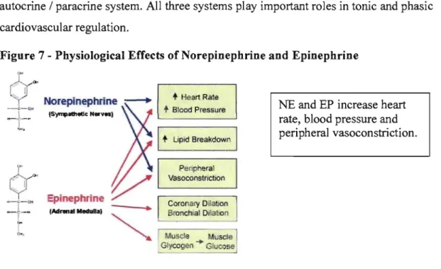
NE and epinephrine (EP) are endogenous catecholamines released by the
postganglionic sympathetic nerve terminaIs and the adrenal gland. They interact with celI surface receptor molecules in various target organs. This interaction starts a cascade of membrane and intracellular events, which result in altered cellular activity (Figure 7). Adrenergic receptors and dopaminergic receptors are coupled to G proteins and activate cells through alterations in intercelIular calcium, cyclic nucleotides, inositol phosphates, and protein phosphorylation. The activation of adrenergic receptors increases HR and the strength of cardiac contraction, and causes cardiac and vascular hypertrophy,
bronchodilation, vasoconstriction, sedation, and analgesia. Adrenergic receptor inhibition results in vasodilation, decreased heart rate, and strength of contraction and relaxation of prostate smooth muscle. AlI these actions are important in the treatment of diseases such as hypertension, congestive heart failure, and angina.
J
26
The three endogenous catecholamines in humans, NE, EP and dopamine act as the chemical effectors of the SNS, adrenomedullary hormonal system and DOPA-dopamine autocrine / paracrine system. AlI three systems play important roles in tonic and phasic cardiovascular regulation.
Figure 7 -Physiological Effects of Norepinephrine and Epinephrine
Çr
'"'
<>, ~ 1 --<:-Qo H _ . 1-
,
q
'
----.-0, 1 __ j_Hr
0.. Noreplnephrlne Epinephrine (Ad ... Medulla, + Heart Rate+
Sioad Pressure Caronary Dilatlon Branchial DllatlonNE and EP increase heart rate, blood pressure and peripheral vasoconstriction.
Syrnpathetic adrenergic nerves are found in the heart where they innervate the sinoatrial and atrioventricular nodes, conduction pathways, and myocytes. Sympathetic adrenergic fibers are also found innervating arteries and veins in the peripheral vasculature. These adrenergic nerves release NE, which binds to specific receptors in the target tissue. Parasympathetic cholinergic nerves derived from the vagus nerves also innervate the heart. Acetylcholine (ACh) released by these fibers binds to muscarinic receptors in the target tissue. The vasculature in sorne organs of the body is innervated by either parasympathetic cholinergic fibers or by sympathetic cholinergic fibers. These nerves release ACh, which binds to muscarinic receptors on the smooth muscle and/or endothelium.
In the heart, NE released by sympathetic nerves preferentially binds to
adrenoceptors causing positive inotropy, chronotropy, and dromotropy. In blood vessels, NE binds adrenoceptors to cause smooth muscle contraction and vasoconstriction. (Figure 7) NE can also bind to adrenoceptors, which causes vasodilation (this can be observed during alpha adrenoceptor blockade), Circulating EP binds to the adrenoceptors to cause vasodilation in sorne organs. NE regulates its own release by stimulating adrenoceptors, which inhibit its release, or inhibiting other adrenoceptors, which facilitate its release.
In the heart, ACh is released by cholinergie nerves, which bind to a cholinergie receptor (M2 muscarinic receptor). This produces negative inotropy, chronotropy, and dromotropy in the heart. Prejunctional M2 receptor activation inhibits NE release and is one mechanism by which vagal stimulation overrides sympathetic stimulation in the heart. In blood vessels, M2 receptors on the vascular endothelium are coupled to the formation of nitric oxide (NO), which causes vasodilation; however, ACh causes smooth muscle contraction through a smooth muscle M3 receptor wh en formation of NO is blocked. Sympathetic cholinergie nerves that release ACh and cause vasodilation innervate sorne arterial blood vessels, for example in skeletal muscle. Neurotransmitter binding to the adrenergic and cholinergie receptors activates signal transduction pathways that cause the observed changes in cardiac and vascular function. Drugs are available for blocking adrenergic and cholinergie receptors. For example, beta-blockers are used in the treatment of angina, hypertension, arrhythmias, and heart failure. Alpha-blockers are used in treating hypertension( 19).
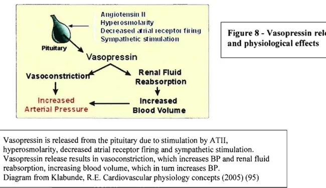
1.5.3.1.3 Vasopressin (Antidiuretic Hormone)
A nu iotensill Il
Hyp el 051110 1.11 ity
1
Decl etlsed '-'Iial leceptOi fil illU Sy III 1 hlt h etic st illllll.1tio Il Vasopressin
vasoconstrictio~
Renal Fluid.. Reab.orption Increased Arterial Pressure Increased Blood Volume ,
Figure 8 - Vasopressin release and physiological efTects
Vasopressin is released from the pituitary due to stimulation by ATII,
hyperosmolarity, decreased atrial receptor firing and sympathetic stimulation. Vasopressin release results in vasoconstriction, which increases BP and renal fluid reabsorption, increasing blood volume, which in turn increases BP.
Diagram from Klabunde, R.E. Cardiovascular physiology concepts (2005) (95)
Vasopressin (AVP), also known as antidiuretic hormone, is a peptide hormone formed in the hypothalamus, then transported via axons to the posterior pituitary and released from there. A VP has two principal sites of action: the kidney and the blood
![Figure 2 - Factors Contributing to Essential Hypertension Excess sodium intake Reduced nephron number [i] Genetlc GJ Factors](https://thumb-eu.123doks.com/thumbv2/123doknet/2079915.7023/25.915.118.785.175.1034/figure-factors-contributing-essential-hypertension-reduced-genetlc-factors.webp)