Regulation of vimentin by SIP1 in human epithelial breast tumor cells
S Bindels1, M Mestdagt1, C Vandewalle2, N Jacobs3, L Volders1, A Noël1, F van Roy2, G Berx2, J-M Foidart1 and C Gilles1
1Laboratory of Tumor and Developmental Biology, Center for Biomedical Integrated Genoproteomics, University of Liège, CHU Sart-Tilman, Liège, Belgium
2Department for Molecular Biomedical Research, VIB-Ghent University, Ghent (Zwijnaarde), Belgium
3Laboratory of Anatomopathology, Center for Biomedical Integrated Genoproteomics, University of Liège, CHU Sart-Tilman, Liège, Belgium
Abstract
The expression of Smad interacting protein-1 (SIP1; ZEB2) and the de novo expression of vimentin are frequently involved in epithelial-to-mesenchynial transitions (EMTs) under both normal and pathological conditions. In the present study, we investigated the potential role of SIP1 in the regulation of vimentin during the EMT associated with breast tumor cell migration and invasion. Examining several breast tumor cell Unes displaying various degrees of invasiveness, we found SIP1 and vimentin expression only in invasive cell Unes. Also, using a model of cell migration with human mammary MCF10A cells, we showed that SIP1 is induced specifically in vimentin-positive migratory cells. Furthermore, transfection of SIP1 cDNA in MCF10A cells increased their vimentin expression both at the mRNA and protein levels and enhanced their migratory abilities in Boyden Chamber assays. Inversely, inhibition of SIP1 expression by RNAi strategies in BT-549 cells and MCF10A cells decreased vimentin expression. We also showed that SIP1 transfection did not activate the TOP-FLASH reporter system, suggesting that the β-catenin/ TCF pathway is not impUcated in the regulation of vimentin by SIP1. Our results therefore impUcate SIP1 in the regulation of vimentin observed in the EMT associated with breast tumor cell migration, a pathway that may contribute to the metastatic progression of breast cancer.
Keywords: vimentin ; SIP1 ; cell migration
Introduction
During their metastatic conversion, epithelial cells acquire the ability to invade the surrounding tissue and disseminate into secondary organs. There is mounting evidence that the acquisition of migratory and invasive properties by epithelial cells is associated with the gain of mesenchymal characteristics and the loss of epithelial features, a phenomenon referred to as epithelial-to-mesenchymal transition (EMT) (Savagner, 2001; Thiery, 2002; Gotzmann et al., 2004; Gilles et al., 2004).
A growing number of zinc-finger transcription factors has now been implicated in the regulation of EMT phenomena. Among them is SIP1 (Smad Interacting Protein-1) or ZEB2, a large zinc-finger protein that, together with δEF1, belongs to a small family of transcriptional repressors. It has originally been identified as a factor binding the Smad proteins, implicated in the signaling by TGF-β (Verschueren et al., 1999). The
δEF1/ZEB family comprises two vertebrate prototypes (δEF1/ZEB1 and SIP1/ZEB2) characterized by two
zinc-finger clusters separated by a homeodomain. The N-terminal and C-terminal zinc-zinc-finger clusters are made of four and three zinc fingers, respectively. Each zinc-finger cluster recognizes a CACCT motif on the DNA but the full-length SIP1 molecule has been shown to bind a bipartite element composed of one 5'-CACCT and one 5'-CACCTG sequence (Remacle et al., 1999; Verschueren et al., 1999). A mechanism by which SIP1 might contribute to EMT processes is through its ability to downregulate E-cadherin. Indeed, SIP1 has been shown to bind the E-cadherin promoter and to downregulate the expression of this cell adhesion protein (Comijn et al., 2001; van Grunsven et al., 2003). E-cadherin is a transmembrane glycoprotein that mediates homotypic cell-cell contacts between epithelial cells and, thereby, largely contributes to the cohesive architecture of normal
epithelia. The cytoplasmic part of E-cadherin is associated with the actin cytoskeleton via its cytoplasmic binding partners, the catenins (α-, β- and γ- catenin). Under particular conditions, when β-catenin is not sequestered in the junctional E-cadherin complexes, it can translocate in the nucleus where it acts as a transcriptional coactivator through its binding with the members of the TCF/LEF-1 (T-cell factor/lymphoid enhancer factor) transcription factor family (Bienz, 2005). Downregulation of E-cadherin expression and reorganization of E-cadherin-based adhesion junctions are considered hallmarks of EMT processes and have largely been implicated in EMT associated with the acquisition of a migratory/invasive phenotype by epithelial tumor cells (Comijn et al., 2001; Van Aken et al., 2001; Peinado et al., 2004).
Besides the disorganization of E-cadherin-based junctional complexes, the de novo expression of vimentin is also frequently associated with EMT processes and with the metastatic conversion of epithelial cells. Vimentin is a type-Ill intermediate filament normally expressed in cells of mesenchymal origin (Steinert and Roop, 1988). However, numerous data have now demonstrated that vimentin can also be expressed in epithelial cells when they become involved in physiological or pathological processes requiring epithelial cell migration. Vimentin has indeed been described in migratory epithelial cells involved in embryonic and organogenesis processes, in placentation, wound healing and tumor invasion (Ramaekers et al., 1983; Guarino, 1995; Gilles and Thompson, 1996; Gilles et al., 1999, 2003). Also, vimentin-specific antisense cDNA or oligonucleotide transfection in vimentin-expressing cell lines was shown to reduce their in vitro invasiveness or migration, strongly emphasizing a functional contribution of vimentin to epithelial cell invasion/migration (Hendrix et al., 1997; Gilles et al., 1999; Singh et al., 2003). Accordingly, impaired wound healing has been observed in vimentin knockout mice (Eckes et al., 1998, 2000). Furthermore, a direct or indirect interaction of vimentin with microfilaments and microtubules and more particularly with molecules such as plectin or integrins has been described (Svitkina et al., 1996; Maniotis et al., 1997; Homan et al., 1998; Goldman et al., 1999; Wu et al., 1999; Gonzales et al., 2001; Tsuruta and Jones, 2003; Helfand et al., 2004; Kreis et al., 2005). A role of vimentin in the mechanical transduction of signals from the cell surface to the nucleus and in the overall reorganization of the cytoskeleton associated with cell motility and migration has therefore been suggested (Hendrix et al., 1996; Gilles et al., 1999; Eckes et al., 2000; Helfand et al., 2004).
Because both SIP1 and vimentin expression are clearly associated with EMT events, we thus examined in the present study the implication of SIP1 in the induction of vimentin expression associated with epithelial cell migration and invasion. We also explored the potential role of the β-catenin/TCF pathway in this regulation.
Results
Vimentin expression correlates with SIP1 expression in migratory /invasive breast cell lines
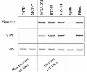
In order to examine the relationship between SIP1 and vimentin, we first analysed their expression in two non-invasive (MCF-7, T47D) and three non-invasive (MDA-231, BT549, Hs578T) epithelial breast cancer cell lines. The invasive phenotype of these breast cancer cells, as assessed by modified Boyden chamber assays, as well as their vimentin and E-cadherin expression status have been characterized in previous studies which established a clear association between high invasive properties and EMT traits such as vimentin expression and lack of E-cadherin expression (Thompson et al., 1992; Sommers et al., 1994; Nawrocki et al., 2001). Examining SIP1 in this set of breast cell lines, we observed that invasive breast cancer cell lines showed both SIP1 and vimentin mRNA expression in contrast to non-invasive cells (Figure 1). SIP1 and vimentin mRNA expression was also analysed in primary airway human fibroblasts versus primary human differentiated epithelial cells. Cells isolated from airway tissues were used because of the possibility to obtain a clear epithelial differentiation, characterized by the presence of ciliated cells, when cultivated in appropriate culture conditions. In support of the results obtained with the tumor cell lines suggesting that the invasive cell lines have gained mesenchymal characteristics, a weak expression of vimentin and no detectable expression of SIP-1 was found in differentiated lung epithelial cells whereas both mRNA were strongly co-expressed in fibroblasts (Figure 1).
Figure 1 Expression of SIP1 correlates with vimentin expression in human breast cancer cell lines. RT-PCR analyses of vimentin and SIP1 were performed on five human tumor cell lines (T47D, MCF-7, MDA-231, BT549 and Hs578T) as well as in primary fibroblasts (Fibro.) and differentiated primary epithelial (Epith.) cells isolated from human airway tissues. 28S rRNA analysis is shown as control.
Figure 2 Expression of SIP1 correlates with vimentin expression in migratory MCF10A cells. (a) Single or double visualization (merge) of vimentin (in red) and EGFP expression driven by the vimentin promoter (in green) in VP-EGFP MCF10A cells plated in the migration assay. DAPI staining is in blue. The white line schematically represents the limit between the vimentin-positive and the vimentin-negative cell population. Bar = 80µm. (b, c) RT-PCR analyses for SIP1 and vimentin were performed on migratory (mig) and stationary (stat) subpopulations sorted by FACS for GFP expression of two clones (#11 and #12) of VP-EGFP MCF10A plated in the migration assay in complete growth medium (FCS + EGF) (b) or in FCS-free, EGF-containing growth medium (c). 28S rRNA analysis is shown as control. Quantification of RT-PCR analyses of vimentin and SIP1 normalized for the 28S rRNA values in three independent migration and FACS sorting experiments, as described in (b, c), is shown. Data are expressed as fold induction in the migratory subpopulation relative to the stationary subpopulation (P<0.05).
Next, we studied SIP1 expression in relation to vimentin expression in a migration assay that we had previously used to demonstrate the transient expression of vimentin during epithelial cell migration (Gilles et al., 1999, 2003). Briefly, in this migration assay, MCF10A human breast cells are plated at high density in a glass ring and migrate as an outgrowth after the removal of the ring. In this assay, vimentin expression has been shown to vary in relationship with the migratory status of the cells. Using video microscopy, we indeed previously
demonstrated that the subpopulation of cells at the periphery of the outgrowth is involved in an orientated migration and undergoes an EMT process characterized by the de novo expression of vimentin and a
relocalization of E-cadherin and β-catenin (Gilles et al., 1999, 2003). In contrast, the stationary cells in the area initially delimited by the ring do not express vimentin and display a typical honeycomb pattern of E-cadherin and β-catenin staining. In the present study, we used two clones (#11 and #12) of MCF10A cells stably transfected with a plasmid containing the vimentin promoter controlling the expression of the enhanced green fluorescent protein (EGFP) gene (VP-EGFP MCF10A cells as previously described in Gilles et al., 1999). The VP-EGFP MCF10A cells were plated in the migration assays and sorted by FACS to physically separate the GFP-positive and the GFP-negative populations (Figure 2a). RT-PCR performed on these two separated cell populations clearly showed co-expression of SIP1 and vimentin mRNA in the migratory subpopulation (Figure 2b).
The effect of EGF on vimentin and SIP1 co-expression was also investigated in this assay. We indeed previously showed that EGF, which is present in the growth medium of the MCF10A cells, induces vimentin expression in migratory MCF10A cells even in the absence of serum. Similar to our observations in the presence of complete growth medium (containing FCS and EGF) (Figure 2b), a strong co-expression of vimentin and SIP1 mRNA was also found in the migratory subpopulation of the two clones of VP-EGFP MCF10A plated in EGF-containing, serum-free medium (Figure 2c).
Smad interacting protein-1 regulates vimentin expression
Because of the correlation between vimentin and SIP1 expression in both the breast cancer cell line panel and
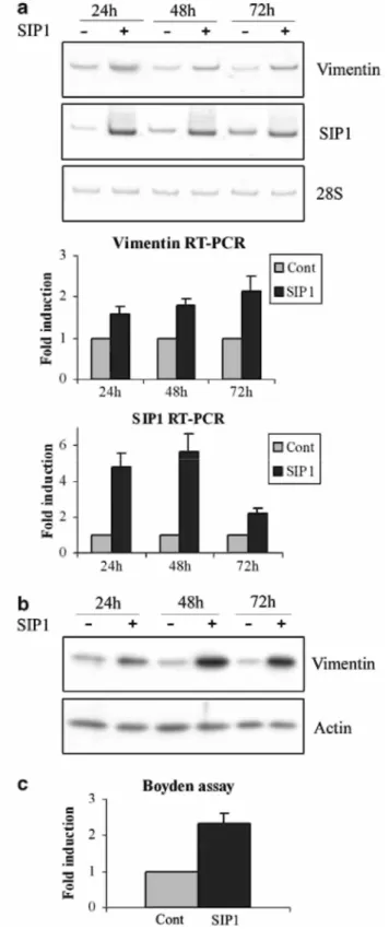
the MCF10A migration assay, we examined whether SIP1 could regulate vimentin expression. We first observed that transient transfection of SIP1 cDNA for 24, 48 or 72 h clearly induced vimentin expression both at the mRNA (Figure 3a) and at the protein (Figure 3b) levels. This correlated with increased migratory properties in the Boyden chamber assay (Figure 3c).
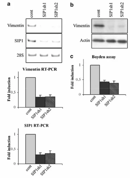
Inversely, we investigated the effect of an inhibition of SIP1 on vimentin expression using RNAi strategies both on MCF10A cells and on vimentin-positive BT549 cells. Because an efficient diminution of SIP1 could not be achieved in MCF10A cells by transient transfection of SIP1 siRNA, these cells were transduced with two lentiviral constructs expressing two different SIP1 short hairpin RNA (shRNA) sequences which efficiently diminished SIP1 mRNA levels (Figure 4a). The SIP1 shRNA construct transduction also diminished vimentin expression both at the mRNA (60% decrease compared to control transduced cells) and at the protein level (Figure 4a and b). This was associated with a diminution of migratory properties in the Boyden chamber assay (Figure 4c). Strengthening these data obtained with MCF10A cells, we also showed that transient transfections of two species of SIP1 siRNA in vimentin-positive BT549 cells significantly decreased vimentin mRNA levels (34 and 28% decrease for siRNA 1 and 2, respectively) (Figure 5a). A decrease of vimentin protein expression was also observed (Figure 5b).
Because vimentin has been shown to be a target of the β-catenin/TCF pathway (Gilles et al., 2003) and because SIP1 has been described as a potent transcriptional repressor of E-cadherin (Comijn et al., 2001; van Grunsven et
al., 2003), we examined whether the regulation of vimentin by SIP1 involved the β-catenin/ TCF pathway. A
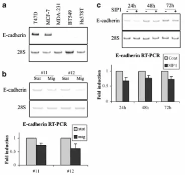
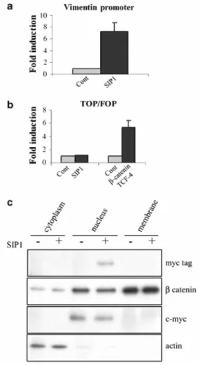
downregulation of E-cadherin may indeed result in increased availability of β-catenin in the nucleus. We thus first confirmed that SIP1 and E-cadherin expression were inversely correlated in the breast tumor cell lines (Figure 6a). This was also seen in the MCF10A migration assay (Figure 6b). Furthermore, transfection of SIP1 cDNA in MCF10A cells decreased the level of E-cadherin mRNA in these cells (Figure 6c). In order to analyse the implication of the β-catenin/TCF pathway in SIP1-induced vimentin expression, we next examined the ability of SIP 1 to transactivate a luciferase reporter plasmid containing wild-type (TOP-FLASH) or mutated (FOP-FLASH) β-catenin/TCF-binding sites as regulatory elements. Although SIP1 cDNA transfection clearly activated the vimentin promoter reporter construct (Figure 7a), it failed to activate the TOP-FLASH reporter system (Figure 7b). This suggests that SIP1 does not activate the β-catenin/TCF pathway. Accordingly, we did not observe any increase of β-catenin in the nuclear fraction (nor in the membranous or cytosolic fractions) of cells transfected with SIPl cDNA though the presence of the myc-tagged SIPl was detected (Figure 7c). Since it has been suggested that the NH2-unphosphorylated form of β-catenin is mainly implicated in the transcriptional activity of β-catenin (Staal et al., 2002), we also examined this particular form of β-catenin. Using a specific antibody against the unphosphorylated β-catenin, we still did not observe any increase of this particular form in the nuclei of SIP1-transfected cells (data not shown).
Figure 3 SIP1 transfection regulates vimentin expression, (a) RT-PCR analyses of vimentin and SIP1 expression in MCF10A cells transiently transfected with SIP1 cDNA for 24, 48 or 72h. Quantification of RT-PCR analyses of vimentin, normalized for the 28S rRNA values, in three independent transfection experiments is shown. Data are expressed as fold induction in SIP1 transfectants relative to the backbone vector transfectants (P<0.05). (b) Western blotting analyses of vimentin expression in MCF10A cells transiently transfected with SIP1 cDNA for 24, 48 or 72h. Actin detection is shown as a control. A representative experiment is shown out of three independent experiments performed, (c) Boyden chamber analyses of the migratory abilities of MCF10A transfected for 48 h with the SIP1 cDNA (SIP1) or the backbone vector (Cont). Data are expressed as fold induction for SIP1 transfectants relative to the backbone vector transfectants (P<0.05).
Figure 4 SIP1 shRNA decreased vimentin expression and migratory abilities of MCF10A cells. MCF10A cells were transduced with lentiviral constructs expressing two different sequences of SIP1 shRNA (SIP1sh1, SIP1sh2) or with a control vector (Cont). (a) RT-PCR analyses of vimentin and SIPl in the SIPl shRNA-expressing cells compared to the control cells. Quantification of RT-PCR analyses of vimentin normalized for the 28S rRNA values in three independent transfection experiments is shown. Data are expressed as fold induction in shRNA-expressing cells relative to the controls (P<0.01). (b) Western blot analyses of vimentin expression in SIPl shRNA-expressing cells. Actin was used as a control. A representative experiment is shown out of three independent experiments performed. (c) Analyses of the migratory abilities of SIPl shRNA-expressing MCF10A cells compared to control cells in the Boyden chamber assay. Data are expressed as fold induction for SIPl shRNA-expressing cells relative to the control cells (P<0.05).
Figure 5 SIP1 siRNA decreased vimentin expression in BT-549 cells. BT549 cells were transfected with either two different siRNA sequences against SIP1 (SIPsi1, SIPsi2) or two control siRNAs (cont1 and cont2). (a) RT-PCR analyses of vimentin and SIP1 in the SIP1 siRNA-expressing cells compared to the control cells. Quantification of RT-PCR analyses of vimentin normalized for the 28S rRNA values in three independent transfection experiments is shown. Data are expressed as fold induction in SIP1 siRNA-expressing cells relative to the controls (P<0.01). (b) Western blot analyses of vimentin expression in SIP1 siRNA-expressing cells. Actin was used as a control. A representative experiment is shown out of three independent experiments performed.
Figure 6 SIP1 expression inversely correlates with E-cadherin expression. RT-PCR analyses of E-cadherin in five breast tumor cell lines (a), in the migratory (mig) versus stationary (stat) subpopulations of VP-EGFP MCF10A cells plated in the migration assay (b) and in MCF10A cells transiently transfected with SIP1 cDNA for 24, 48 or 72h (c). Quantifications of RT-PCR analyses of E-cadherin normalized for the 28S rRNA values in three independent experiments are shown for (b) and (c). Data are expressed as fold induction in the migratory subpopulation relative to the stationary subpopulation for (b) (P<0.05) and in the SIP1 transfectants relative to the backbone vector transfectants for (c) (P<0.05).
Figure 7 No implication of the β-catenin/TCF pathway in the regulation of vimentin by SIP1. (a) Vimentin promoter reporter assay. The human vimentin promoter luciferase reporter plasmid was cotransfected in MCF10A cells either with the SIP1 cDNA expression vector (SIP1) or with the corresponding control vector (Cont). Data are expressed as fold induction in SIP1 transfectants relative to the values obtained in the cells transfected with the backbone control vector (P<0.01). (b) FLASH/FOP-FLASH reporter assay. The TOP-FLASH or FOP-TOP-FLASH reporter plasmid was cotransfected in MCF10A cells with a control vector (Cont) or the SIP1 expression vector (SIP1). The normalized FOP values were subtracted from the normalized TOP values. Data are expressed as fold induction relative to the values obtained in the cells transfected with the control vector (P>0.05). Cotransfection with the β-catenin and TCF-4 expression vectors was performed as a control (P<0.02). (c) Western blotting analyses of β-catenin in cytosolic, nuclear and membranous fractions of MCF10A cells transfected either with the SIP1 expression vector (SIP1) or with the backbone control vector (Cont). The presence of the myc-tagged SIP1 is also shown in the nucleus of SIP1-transfected cells. Endogenous actin and c-myc expression were analysed as controls. A representative experiment is shown out of three independent experiments performed.
Discussion
In the present study, we demonstrate that SIP1 regulates vimentin expression in epithelial cells and emphasize the implication of this regulation in epithelial breast cancer cell migration/invasion. We indeed show that (1) SIP1 expression coincided with vimentin expression in invasive breast tumor cell lines, (2) SIPl and vimentin expression were specifically induced in migratory epithelial MCF10A cells, (3) SIP1 cDNA transfection increased vimentin expression and migratory abilities of MCF10A cells, (4) SIP1-specific siRNA or shRNA diminished vimentin expression in invasive breast tumor cell lines and (5) the activation of the vimentin promoter by SIPl was not associated with the activation of the β-catenin/TCF/LEF signaling pathway.
Upon examination of several human breast cancer cell lines, we found a clear correlation between vimentin and SIPl expression in invasive cell lines. A correlation between vimentin and SIP1 expression was also obvious in the assay of MCF10A cell migration. Previously, this dynamic model allowed to clearly show the induction of vimentin during cell migration, discriminating a sub-population of migratory, vimentin-positive cells from a vimentin-negative, stationary subpopulation within the same cell line (Gilles et al., 1999). Here, we were able to
demonstrate increased SIPl expression specifically in the vimentin-positive migratory subpopulation. In
agreement with previous studies (Sommers et al., 1994; Comijn et al., 2001; Gilles et al., 2003), a lower level of E-cadherin was observed both in the invasive cell lines, as well as in the migratory subpopulation of MCF10A, also attesting the EMT-derived phenotype of these invasive and migratory cells expressing SIP1. In line with our observations, SIP1 expression has been associated with migratory and invasive mechanisms occurring during embryonic development. Indeed, a high level of SIP1 has been detected during the formation of the neural tube and this has been shown to play a key role in the migration of neural crest cells (Eisaki et al., 2000; van
Grunsven et al., 2000; Van de Putte et al., 2003), a system largely described as a physiological archetypal model of EMT (Duband et al., 1995; Tucker, 2004). Also, an increased expression of SIP1 has been described in NMuMG cells which have undergone a TGFβ1-induced EMT characterized by decreased E-cadherin expression and increased N-cadherin expression (Maeda et al., 2005). Our results thus demonstrate a relationship between SIP1 and an EMT-derived phenotype characterized by vimentin expression, and further emphasize the
implication of this relationship in dynamic cell migration.
We further observed that transfection of SIP1 cDNA in MCF10A cells increased vimentin expression both at the mRNA and protein level, which was associated with increased migratory ability. Inversely, the expression of SIP1-specific shRNAs in MCF10A cells clearly diminished vimentin expression as well as their migratory properties. Strengthening these data, a diminution of vimentin expression following SIP1 siRNA transfection was also shown in BT549 breast tumor cells. Up to now, the implication of SIP1 in EMT phenomena associated with tumor cell invasion has been more particularly linked to its ability to repress E-cadherin, as shown in MDCK cells and in hepatocellular and colon epidermoid carcinoma cells transfected with SIP1 (Comijn et al., 2001; Miyoshi et al., 2004; Vandewalle et al., 2005). A repression of E-cadherin by SIP1 was accordingly observed in our cell systems. Overall, an increasing number of zinc-finger transcription factors, including Snail and Slug, both belonging to the Snail family, have been described as E-cadherin repressors and have been implicated in the regulation of EMT phenomena (Nieto, 2002; Come et al., 2004). Forcing the expression of these factors in a variety of epithelial cell systems was found to induce EMT processes characterized by decreased E-cadherin levels and by increased migratory/invasive properties (Cano et al., 2000; Hajra et al., 2002; Nieto, 2002; Come et al., 2004; Peinado et al., 2004). Accumulating data now also show that these factors can modulate the expression of other genes implicated in tumor cell invasion. For instance, they have been shown to downregulate other cell-cell contact molecules but also to upregulate 'mesenchymal' genes including fibronectin, vimentin and members of the matrix metalloproteases (MMP) family (Savagner et al., 1997; Cano et
al., 2000; Hajra et al., 2002; Bolos et al., 2003; Ikenouchi et al., 2003; Yokoyama et al., 2003; Ohkubo and
Ozawa, 2004; Vandewalle et al., 2005). Much less is known regarding the δEF1/ZEB family. Recent studies have nevertheless shown that SIP1-induced EMT in hepatocellular and in colon epidermoid carcinoma cells is also associated with an increased expression of N-cadherin and mesenchymal p120ctn isoforms (Vandewalle et
al., 2005) as well as several members of the MMP family (Miyoshi et al., 2004). Thus, together with our
observations that expression of endogenous vimentin and SIP1 associates with migratory/invasive abilities, these results suggest that SIP1 can contribute to the upregulation of vimentin transcription such as that occurs during epithelial cell migration/invasion.
The mechanism by which SIP1 regulates vimentin remains unclear. SIP1 has been described as a transcriptional repressor downregulating target genes through direct binding of bipartite elements made of one 5'-CACCT-3' and one 5'-CACCTG-3' sequence (Remacle et al., 1999). The fact that vimentin is upregulated by SIP1 and that no such bipartite element could be found in the vimentin promoter sequence suggested that vimentin regulation by SIP1 is most likely indirect. Considering the repressive effect of SIP1 effect on E-cadherin (Comijn et al., 2001; van Grunsven et al., 2003), it was tempting to speculate that SIP1 induction of vimentin could depend on the activation of the β-catenin/TCF pathway. Although it is not always observed (Comijn et al., 2001),
diminished E-cadherin expression could indeed favor translocation of β-catenin in the nucleus where it can trigger the transcription of several target genes through binding with members of the TCF/LEF transcription factor family (Hecht and Kemler, 2000; Giles et al., 2003). Furthermore, vimentin has been shown to be a target of the β-catenin/TCF pathway (Gilles et al., 2003). However, our present results rather suggest that the
β-catenin/TCF pathway is not activated following SIP1 transfection, as also observed by Comijn et al. (2001) in
MDCK cells. Indeed, transfection of SIP1 cDNA into MCF10A cells did not lead to an increase in the TOP-FLASH reporter system though it activated the vimentin promoter. Also, in agreement with our TOP-TOP-FLASH results, we did not observe any accumulation of nuclear β-catenin in SIP1-transfected MCF10A cells. It has also to be noticed that SIP1 downregulation by siRNA in invasive BT549 cells, which do not express E-cadherin, can also modulate vimentin expression. Taken together, these data therefore suggest that regulation of vimentin by SIP1 can be independent of E-cadherin expression and does not necessarily rely in modulations of the
β-catenin/TCF pathway. Nevertheless, many other indirect mechanisms could be involved. Additionally, the
possibility that SIP1 could directly bind to and activate the vimentin promoter (through the binding of a single CACCT motif or another motif similar in sequence) cannot be excluded and is currently under investigation.
In conclusion, our results clearly demonstrate that SIP1 and vimentin expression correlate with a
migratory/invasive phenotype and that SIP1 can regulate vimentin expression in epithelial breast tumor cells. Because of the established functional role of vimentin in cell migration, the upregulation of vimentin by SIP1 has implications for all processes requiring epithelial cell migration including tumor cell invasion.
Materials and methods
Cell culture
All human mammary epithelial cells used were obtained from the American Type Culture Collection (Rockville, MD, USA). MCF10A-VP-EGFP cells were generated previously by stable transfection of MCF10A cells with the VP-EGFP plasmid in which the human vimentin promoter controls the expression of EGFP (Gilles et al., 1999). MCF-7, T47D, MDA-MB-231, BT549 and Hs578T cells were cultured in Dulbecco's modified Eagle's medium (DMEM) containing 10% FCS. MCF10A cells were grown in a 1:3 (v/v) mixture of HAM F12 and DMEM, supplemented with 20 µg/ml of adenine, 5 µg/ml of insulin, 0.5 µg/ml of hydrocortisone, 2 ng/ml of EGF, 5 µg/ml of transferrin, 1.5 ng/ml of triiodothyronin and 10% FCS.
Primary epithelial cells and primary fibroblasts were isolated from human polyps as previously described (Million et al., 2001). Primary epithelial cells were allowed to differentiate, as attested by the presence of ciliated cells, in a transwell chamber coated with collagen type I for 12 days (Million et al., 2001).
In vitro migration assay
In order to sort by FACS the vimentin-expressing migratory cells from the stationary, vimentin-negative cells, we used two clones of MCF10A-VP-EGFP expressing the EGFP reporter gene driven by the vimentin promoter (MCF10A-VP-EGFP #11 and #12 as also described in Gilles et al., 1999). These cells were analysed in a migration assay as described previously (Gilles et al., 1999). Briefly, 5 × 10⁴ cells were seeded in growth medium inside a 6-mm glass ring. At 24h after plating, the glass ring was removed and the cells were covered either with complete growth medium (containing FCS and EGF) or with FCS-free growth medium in order to examine the implication of EGF. In this model, the cells migrate as an outgrowth from the confluent area initially delimited by the ring. We have previously shown that cells at the periphery of the outgrowth are implicated in an oriented migration and express vimentin and GFP. In contrast, the cells in the area initially delimited by the ring are rather stationary and do not express vimentin nor GFP (Gilles et al., 1999). At 48h after the removal of the ring, the cells were collected by trypsinization of 24 migration assays and were sorted by FACS (FACSVantage-SE, Becton Dickinson, Erembodegem, Belgium) into EGFP-positive and EGFP-negative populations, which were then used for RNA extraction and RT-PCR analysis.
Immunofluorescence
Cells cultured in the migration assay on glass coverslips were fixed in paraformaldehyde (4% in PBS) for 10 min and then permeabilized with 0.1% Triton X-100 for 1 min at room temperature. The coverslips were then blocked for 30 min with 3% BSA in PBS. After several washes with PBS, fixed monolayers were incubated for 1h with a monoclonal antibody to vimentin (clone V9, Dako, Glostrup, Denmark) and then exposed to a TRITC-conjugated rabbit anti-mouse antibody (Dako). Finally, nuclei were labeled with 4',6-diamidino-2-phenylindole (DAPI; 1 µg/ml) for 20 min. The coverslips were then mounted with Aquapolymount antifading solution (Agar, UK) onto glass slides and observed under a fluorescence microscope (Olympus, Tokyo, Japan).
Plasmids
The vimentin promoter luciferase reporter vector (VimPro) used in this study has been characterized previously (Gilles et al., 2003). This plasmid consists of the human vimentin promoter cloned into the firefly luciferase reporter plasmid pGL-3 (Promega, Madison, WI, USA).
The expression vector for the SIP1 cDNA encoding a myc-tagged protein has been described previously (Verschueren et al., 1999; Comijn et al., 2001).
An expression vector encoding a mutated form of β-catenin which is less susceptible to degradation was kindly provided by Dr K Orford and Dr S Byers (Lombardi Cancer Center, Georgetown University, Washington, DC, USA) (Orford et al., 1997). Dr HC Clevers (University Hospital, Utrecht, The Netherlands) donated the expression vector encoding human TCF-4 (pCDNA-hTCF4) and the TOP-FLASH and FOP-FLASH plasmids containing, respectively, three wild-type (5'-CCTTTGATC-3': TOP-FLASH) or mutated (5'-CCTTTGGCC-3': FOP-FLASH) copies of the β-catenin/ TCF-binding sites upstream of a minimal c-fos promoter driving the expression of firefly luciferase (Korinek et al., 1997; van de Wetering et al., 1997).
Luciferase reporter assay
Transient transfections were performed with Fugene transfection reagent (Roche, Branchburg, NJ, USA) on 50000 cells plated in a 24-well plate half an hour before the addition of the DNA/Fugene mixture.
For the determination of the vimentin promoter induction by SIP1, each well was supplemented with a mixture containing 20 µl of serum-free DMEM, 0.6 µl of Fugene and 0.2 µg of the promoter reporter construct (either VimPro, TOP-FLASH or FOP-FLASH reporter construct), 0.2 µg of the SIP1 expression vector (or the backbone vector control) and 1.6 ng of the Renilla luciferase reporter phRG-TK (Promega) used as an internal control. For β-catenin/TCF-4 induction, each well was incubated with a mixture containing 20 µl of serum-free DMEM, 0.6 µl of Fugene, 0.15 µg of the firefly luciferase reporter plasmid (either VimPro, TOP-FLASH or FOP-FLASH reporter construct), 0.15 µg of the β-catenin expression vector (or the corresponding backbone vector), 0.15 µg of the TCF-4 expression vector (or the corresponding backbone vector) and 1.6 ng of the Renilla luciferase vector phRG-TK.
At 24h after transfection, the cells were lysed in 100 µl of lysis buffer followed by determination of luciferase activity in 20 µl of lysate with a luminometer using the Dual Luciferase Assay System (Promega). The firefly luciferase activity was normalized to the activity of the Renilla luciferase used as internal control. Results were expressed as fold induction, calculated by dividing the normalized values obtained following cDNA transfection by the normalized values obtained following transfection of the corresponding backbone expression vector. To assess the β-catenin/TCF/LEF activities using the TOP/FOP-FLASH reporter system, the normalized values obtained with the FOP-FLASH reporter plasmid were subtracted from the normalized values obtained with the TOP-FLASH reporter plasmid. Results were then expressed as fold induction relative to the value obtained in the cells transfected with the control vector. Each experiment was performed at least three times in triplicate. Data are expressed as means ± s.e. A one-sample t-test was performed and a P-value <0.05 was considered
significant.
Transient transfections of Smad interacting protein-1 cDNA
To study the regulation of the endogenous vimentin gene by SIP1, 150000 MCF10A cells plated in six-well plates were transiently transfected with the SIP1 expression vector using Lipofectamine 2000 transfection reagent (Invitrogen, Carlsbad, CA, USA). At 24h after plating, transfection was carried out as recommended by the manufacturer by adding, in each well, a mixture containing 500 µl of serum-free medium, 3 µl of
Lipofectamine 2000 and 2 µg of the SIP1 expression vector. As control, cells were transfected with the corresponding backbone vector. At 24, 48 and 72h after transfection, cells were collected for RT-PCR or Western blotting analyses. A control transfection condition using a plasmid encoding GFP (pEGFP-IRESpuro, Clontech, CA, USA) was always performed in parallel to determine the transfection efficiency. All experiments were set up to obtain at least 70% of transfected cells.
Transfection of small interfering RNA
Two 19-nt-specific sequences were selected in the coding sequence of SIP1 to generate 21-nt sense and 21-nt antisense strands of the type (19N) TT (N, any nucleotide). The sense and antisense strands were then annealed to obtain duplexes with identical 3' overhangs. The sequences were submitted to a BLAST search against the human genome to ensure the specificity of the small interfering RNA (siRNA) to the targeted sequence. Two duplexes, which do not recognize any sequence in the human genome, were used as controls. The 19-nt-specific sequences for the two SIP1 siRNAs are as follows: SIP1 Si1, 5'-GGUAAUCGCAAGUUCAAAU-3'; SIP1 Si2, 5'-GAACAGACAGGCUUACUUA-3'. For transfection of the siRNA duplexes, 75 000 cells were plated in six-well plates in 2 ml/six-well of culture medium. At 24h after plating, the cells were transfected by phosphate calcium precipitation by adding in each well 200 µl of a mixture containing the siRNA duplexes (20 nM), 140 mM NaCl, 0.75 mM Na2H-PO4, 6 mM glucose, 5 mM KCl, 25 mM HEPES and 125 mM CaCl2. At 24h after transfection, the cells were extensively washed with PBS and incubated for 48h in culture medium before they were harvested for RT-PCR analyses or Western blotting analyses. The transfection of an FITC-labelled control siRNA
(Eurogentec, Belgium) was also performed in parallel and revealed an uptake of the siRNA in 100% of the cells.
Transduction of the lentiviral vector for SIP1 short hairpin RNA
A SIP1-specific siRNA sequence was designed using selection criteria as described (Brummelkamp et al., 2002; Ui-Tei et al., 2004). A double PCR approach was used to create shRNA expression cassettes containing the H1 promoter and both the sense and antisense shRNA sequences with a loop sequence in between. In a first step, PCR was performed using pSuper plasmid (Brummelkamp et al., 2002) as a template, the H1 promoter primer 5'-CTGCAGGAATTCGAACGCTGACGT CATCAA-3' and the sense shRNA oligonucleotide 5-AAATC TCTTGAATTTAACAATACCCAGCTCCGGGGATCTGT GGTCTCATACAGAACTTATAA-3' (SEC1 = SIP1shl) or 5'-TGTTCTCTTGAAACAAAGGTAACGTTCATGCGGG
second PCR reaction with the same H1 promoter primer and the antisense shRNA oligonucleotide 5'-CCATCGATAAGCTTT TTTTCCAAAAAAGGAGCTGGGTATTGTTAAATCTCT TGAATTTA-3' (SEC1 = SIP1sh1) or 5'-CCATCGATAAGC TTTTTTTCCAAAAAAGCATGAACGTTACCTTTGTTC
TCTTGAAACAA-3' (SEC8 = SIP1sh2). The shRNA expression cassette was cloned in the lentiviral pLV-TH vector (Wiznerowicz and Trono, 2003) using EcoRI and ClaI restriction sites.
For lentivirus production, 1.2 million cells of the packaging cell line HEK293T were seeded in a 25-cm2 flask. After 24h, 3 µg of the pLV-THshRNA construct or empty vector, 3 µg of the packaging plasmid pCMVdR8.91 and 1.5 µg of the envelope plasmid pMD2G-VSVG were first precipitated together and then transfected into the HEK293T cells using the calcium phosphate precipitation method. The DNA was premixed with 50 µl of 2 M CaCl2 and 190 µl TE buffer and then slowly added to 250 µl 2 × HBS. The mixture was put on a shaker for 15 min before it was added to the cells. After 8h, the cells were washed and incubated for 48h in 4 ml fresh culture medium. The virus-containing medium was then harvested and filtered through a 0.45 µm low protein binding filter unit (Millipore, Billerica, MA, USA). Aliquots were stored at -70°C.
Transduction of the MCF10A cells was performed by mixing 50000 cells with 200 µl viral supernatant in a 96-well plate and three replicates of each transduction were made. These mixtures were centrifuged for 1.5h at 32°C and 1500 r.p.m. before putting them in the 37°C incubator. After 24 h, the cells were trypsinized and replicates were pooled in a 24-well plate together with 800 µl fresh viral supernatant. The mixtures were again centrifuged as mentioned above and incubated for 24h before replacing the medium with fresh culture medium.
Transduction efficiencies were determined by measuring EGFP expression using FACS analysis (Epics Altra from Beckman Coulter, Fullerton, CA, USA). Subsequently, the cells were sorted to obtain cell populations with more than 90% EGFP-positive cells.
Western blotting analyses
Total protein extracts were prepared in RIPA buffer (50 mM Tris, pH 7.4, 150 mM NaCl, 1% (v/v) Igepal, 1% (w/v) sodium deoxycholate, 5 mM iodoacetamide, 0.1% (w/v) SDS), containing complete protease inhibitor cocktail (Roche). To examine the subcellular distribution of β-catenin, cytosolic, membranous and nuclear extracts of MCF10A cells transfected with the SIP1 expression vector (or the corresponding backbone vector) for 48h were prepared using the ProteoExtract Subcellular Proteome Extraction Kit (Calbiochem, LaJolla, CA, USA). Protein concentration was determined with the DC protein assay (BioRad, Richmond, CA, USA). In all, 10 µg or 500 ng of total protein were separated on 15% SDS-PAGE gels for analysis of vimentin in MCF10A and BT549 cells, respectively. Total protein extract of MCF10A cells (500 ng) was separated on 10% SDS-PAGE gels for E-cadherin analysis. In total, 4 µg of cytosolic or nuclear extracts and 1 µg of membranous extracts of MCF10A cells were separated on 10% SDS-PAGE to analyse the expression of β-catenin. Proteins were transferred to PVDF membranes (NEN, Boston, MA, USA), which were then blocked with 5% milk (w/v) + 0.1% Tween 20 (w/v) in PBS for 2h. They were then exposed to primary antibodies overnight at 4°C: a monoclonal antibody (mAb) to vimentin (clone V9, Dako), an mAb to E-cadherin (BD Transduction Laboratories, San Jose, CA) or an mAb to β-catenin (either from BD Transduction Laboratories or clone 8E4 from AG Scientific, CA, USA, which is a specific antibody for the unphosphorylated form of β-catenin). The filters were then incubated with a horseradish peroxidase-conjugated swine anti-mouse antibody (Dako). Signals were detected with an enhanced chemiluminescence (ECL + ) kit (Amersham Pharmacia Biotech,
Buckinghamshire, UK). Subsequent detection of actin (using a rabbit antibody to actin, clone A2066, Sigma-Aldrich, St Louis, MO, USA), or c-myc (using an mAb to c-myc, clone 9E10, Sigma) was performed on the same filters as a control. Western blotting analyses were performed on three independent experiments and a representative experiment is shown in the figures.
RT-PCR analysis
Total RNA was extracted with an RNA isolation kit (Roche). RT-PCR was performed using 10 ng of total RNA and the GeneAmp Thermostable RNA PCR Kit (Perkin Elmer, Foster City, CA, USA). Forward and reverse primers (Eurogentec, Seraing, Belgium) were as follows: vimentin primers (forward
5'-GACAATGCGTCTCTGGCACGTCTT-3', reverse 5'-TCCTCCGCCTCCTGCAGGTTCTT-3'), SIP1 primers (forward 5'-AGTCCATGCGAACTGCCATCTGAT-3', reverse 5'- CTGGACCATCTACAGAGGCTTGTA-3'), E-cadherin primers: (forward 5'-CCCATCAGCTGCCCAGAAAATGA A-3', reverse 5'-
CTGTCACCTTCAGCCATCCTGTTT-3'), 28S rRNA primers (forward 5'-GTTCACCCACTAATAGGG AACGTGA-3', reverse 5'-GGATTCTGACTTAGAGGCGT TCAGT-3'). Reverse transcription was performed at 70°C for 15 min. Products were separated on acrylamide gels, stained with SYBR Gold (Molecular Probes, Eugene, OR, USA) and quantified by fiuorimetric scanning (LAS-1000, Fuji, Stamford, CT, USA).
Quantification was performed by normalization of the values obtained for 28 S rRNA amplification. For SIP1 cDNA transfection or RNAi experiments, results were expressed as fold induction calculated by dividing the normalized value of a given condition (SIP1 cDNA transfection, SIP1 siRNA transfection or SIP1 shRNA
transduction) by the normalized value of the corresponding control. Each experiment was performed at least three times. Data are expressed as means ± s.e. A one-sample t-test was performed and a P-value <0.05 was considered significant.
Boyden chamber invasion assay
The migratory properties of MCF10A cells transfected with SIP1 cDNA for 48h or transduced with the
SIP1shRNA lentiviral vector were assessed using the Boyden chamber assay. Cells (100000) were suspended in 300 µl of serum-free medium supplemented with 0.1% BSA and placed in the upper compartment of a 24-well transwell (Costar, NY, USA). The lower compartment was filled with 600 µl of medium containing 10% FCS and 1% BSA. After 6h of incubation at 37°C, the filters were fixed in methanol for 10 min and stained with Giemsa for 30 min. Cells on the upper surface of the filters were wiped away with a cotton swab. Migration was quantified by counting the number of cells on the lower surface of the filters. Experiments were performed at least three times in triplicate. Data are expressed as means ± s.e. A one-sample t-test was performed and a P-value <0.05 was considered significant.
Acknowledgements
We thank Dr H Clevers for the TOP-FLASH, the FOP-FLASH and TCF-4 expression vector and Dr K Orford and Dr S Byers for the β-catenin expression vector. This work was supported by grants from the 'Communauté française de Belgique (Actions de Recherches Concertées)', the Commission of European Communities (European Union Framework Programs 5 and 6, BRECOSM), the 'Fonds de la Recherche Scientifique
Médicale', the 'Fonds National de la Recherche Scientifique' (FNRS, Belgium), the 'Fédération Belge Contre le Cancer', the 'coopération C.G.R.I.-F.N.R.S.-INSERM', the 'Fonds spéciaux de la Recherche (University of Liège), the 'Centre Anticancéreux près l'Université de Liège', the Fortis Banque Assurances, the 'Fondation Léon Fredericq' (University of Liège), the D.G.T.R.E. from the 'Région Wallonne', the 'Fonds d'Investissements de la Recherche Scientifique (CHU, Liège, Belgium)', the Interuniversity Attraction Poles Program - Belgian Science Policy (Brussels, Belgium). CG is a Research Associate from the FNRS (Belgium), SB is an FRIA grant holder from the F.N.R.S., MM is a Research Fellow from the F.N.R.S, and GB is a postdoctoral researcher with the FWO (Belgium).
References
Bienz M. (2005). Curr Biol 15: R64-R67.
Bolos V, Peinado H, Perez-Moreno MA, Fraga MF, Esteller M, Cano A. (2003). J Cell Sci 116: 499-511. Brummelkamp TR, Bernards R, Agami R. (2002). Science 296: 550-553.
Cano A, Perez-Moreno MA, Rodrigo I, Locascio A, Blanco MJ, del Barrio MG et al. (2000). Nat Cell Biol 2: 76-83. Come C, Arnoux V, Bibeau F, Savagner P. (2004). J Mammary Gland Biol Neoplasia 9: 183-193.
Comijn J, Berx G, Vermassen P, Verschueren K, van Grunsven L, Bruyneel E et al. (2001). Mol Cell 7: 1267-1278. Duband JL, Monier F, Delannet M, Newgreen D. (1995). Acta Anat (Basel) 154: 63-78.
Eckes B, Colucci-Guyon E, Smola H, Nodder S, Babinet C, Krieg T et al. (2000). J Cell Sci 113: 2455-2462. Eckes B, Dogic D, Colucci-Guyon E, Wang N, Maniotis A, Ingber D et al. (1998). J Cell Sci 111: 1897-1907. Eisaki A, Kuroda H, Fukui A, Asashima M. (2000). Biochem Biophys Res Commun 271: 151-157.
Giles RH, van Es JH, Clevers H. (2003). Biochim Biophys Acta 1653: 1-24.
Gilles C, Newgreen D, Sato H, Thompson EW. (2004). Rise and Fall of Epithelial Phenotype In: Savagner P (ed). Eurekah.com and Kluwer Academic/Plenum Publishers: New York, (Chapter 2).
Gilles C, Polette M, Mestdagt M, Nawrocki-Raby B, Ruggeri P, Birembaut P et al. (2003). Cancer Res 63: 2658-2664. Gilles C, Polette M, Zahm JM, Tournier JM, Volders L, Foidart JM et al. (1999). J Cell Sci 112: 4615-4625. Gilles C, Thompson EW. (1996). Breast J 2: 83-96.
Goldman RD, Chou YH, Prahlad V, Yoon M. (1999). FASEB J 13(Suppl 2): S261-S265.
Gonzales M, Weksler B, Tsuruta D, Goldman RD, Yoon KJ, Hopkinson SB et al. (2001). Mol Biol Cell 12: 85-100. Gotzmann J, Mikula M, Eger A, Schulte-Hermann R, Foisner R, Beug H et al. (2004). Mutat Res 566: 9-20. Guarino M. (1995). Histol Histopathol 10: 171-184.
Hajra KM, Chen DYS, Fearon ER. (2002). Cancer Res 62: 1613-1618.
Hendrix MJ, Seftor EA, Chu YW, Trevor KT, Seftor RE. (1996). Cancer Metast Rev 15: 507-525. Hendrix MJ, Seftor EA, Seftor RE, Trevor KT. (1997). Am J Pathol 150: 483-495.
Homan SM, Mercurio AM, LaFlamme SE. (1998). J Cell Sci 111: 2717-2728. Ikenouchi J, Matsuda M, Furuse M, Tsukita S. (2003). J Cell Sci 116: 1959-1967.
Korinek V, Barker N, Morin PJ, van Wichen D, de Weger R, Kinzler KW et al. (1997). Science 275: 1784-1787. Kreis S, Schonfeld HJ, Melchior C, Steiner B, Kieffer N. (2005). Exp Cell Res 305: 110-121.
Maeda M, Johnson KR, Wheelock MJ. (2005). J Cell Sci 118: 873-887. Maniotis AJ, Chen CS, Ingber DE. (1997). Proc Natl Acad Sci USA 94: 849-854.
Million K, Tournier F, Houcine O, Ancian P, Reichert U, Marano F. (2001). Am J Resp Cell Mol Biol 25: 744-750. Miyoshi A, Kitajima Y, Sumi K, Sato K, Hagiwara A, Koga Y et al. (2004). Br J Cancer 90: 1265-1273. Nawrocki RB, Polette M, Gilles C, Clavel C, Strumane K, Matos M et al. (2001). M J Cancer 93: 644-652. Nieto MA. (2002). Nat Rev Mol Cell Biol 3: 155-166. Ohkubo T, Ozawa M. (2004). J Cell Sci 117: 1675-1685. Orford K, Crockett C, Jensen JP, Weissman AM, Byers SW. (1997). J Biol Chem 272: 24735-24738. Peinado H, Portillo F, Cano A. (2004). Int J Dev Biol 48: 365-375.
Ramaekers FC, Haag D, Kant A, Moesker O, Jap PH, Vooijs GP. (1983). Proc Natl Acad Sci USA 80: 2618-2622. Remacle JE, Kraft H, Lerchner W, Wuytens G, Collart C, Verschueren K et al. (1999). EMBO J 18: 5073-5084. Savagner P. (2001). BioEssays 23: 912-923. Savagner P, Yamada KM, Thiery JP. (1997). J Cell Biol 137: 1403-1419. Singh S, Sadacharan S, Su S, Belldegrun A, Persad S, Singh G. (2003). Cancer Res 63: 2306-2311.
Sommers CL, Byers SW, Thompson EW, Torri JA, Gelmann EP. (1994). Breast Cancer Res Treat 31: 325-335.
Staal FJ, Noort MM, Strous GJ, Clevers HC. (2002). EMBO Rep 3: 63-68. Steinert PM, Roop DR. (1988). Annu Rev Biochem 57: 593-625. Svitkina TM, Verkhovsky AB, Borisy GG. (1996). J Cell Biol 135: 991-1007.
Thiery JP. (2002). Nat Rev Cancer 2: 442-454. Thompson EW, Paik S, Brunner N, Sommers CL, Zugmaier G, Clarke R et al. (1992). J Cell
Physiol 150: 534-544.
Tsuruta D, Jones JC. (2003). J Cell Sci 116: 4977-4984. Tucker RP. (2004). Int J Biochem Cell Biol 36: 173-177.
Ui-Tei K, Naito Y, Takahashi F, Haraguchi T, Ohki-Hamazaki H, Juni A et al. (2004). Nucleic Acids Res 32: 936-948. Van Aken E, De Wever O, Correia da Rocha AS, Mareel M. (2001). Virchows Arch 439: 725-751.
Van de Putte T, Maruhashi M, Francis A, Nelles L, Kondoh H, Huylebroeck D et al. (2003). Am J Hum Genet 72: 465-470. Vandewalle C, Comijn J, De Craene B, Vermassen P, Bruyneel E, Andersen H et al. (2005). Nucleic Acids Res 33: 6566-6578. van de Wetering M, Cavallo R, Dooijes D, van Beest M, van Es J, Loureiro J et al. (1997). Cell 88: 789-799.
van Grunsven LA, Michiels C, Van de Putte T, Nelles L, Wuytens G, Verschueren K et al. (2003). J Biol Chem 278: 26135-26145. van Grunsven LA, Papin C, Avalosse B, Opdecamp K, Huylebroeck D, Smith JC et al. (2000). Mech Dev 94: 189-193. Verschueren K, Remacle JE, Collart C, Kraft H, Baker BS, Tylzanowski P et al. (1999). J Biol Chem 274: 20489-20498.
Wiznerowicz M, Trono D. (2003). J Virol 77: 8957-8961. Wu AL, Wang J, Zheleznyak A, Brown EJ. (1999). Mol Cell 4: 619-625. Yokoyama K, Kamata N, Fujimoto R, Tsutsumi S, Tomonari M, Taki M et al. (2003). Int J Oncol 22: 891-898.