Solution Structural Study of BlaI: Implications for the
Repression of Genes Involved in b-Lactam
Antibiotic Resistance
He´le`ne Van Melckebeke
1†, Christelle Vreuls
2,3†, Pierre Gans
1Patrice File´e
2, Gabriel Llabres
3, Bernard Joris
2and
Jean-Pierre Simorre
1*
1Institut de Biologie Structurale-Jean-Pierre Ebel CEA-CNRS-UJF, 41 Avenue Jules Horowitz, 38027 Grenoble Cedex 1, France2Centre d’inge´nierie des
prote´ines, Institut de Chimie B6A, Universite´ de Lie`ge Sart-Tilman B4000, Belgium
3Laboratoire de Physique
Expe´rimentale, De´partement de Physique, Bat B5, Universite´ de Lie`ge, Sart Tilman B4000 Belgium
b-Lactamase and penicillin-binding protein PBP20mediate staphylococcal resistance to b-lactam antibiotics, which are otherwise highly clinically effective. Two repressors (BlaI and MecI) regulate expression of these inducible proteins. Here, we present the first solution structure of the 82 amino acid residue DNA-binding domain of Bacillus licheniformis BlaI which is very similar in primary sequence to the medically significant Staphyloccocal BlaI and MecI proteins. This structure is composed of a compact core of three a-helices and a three-stranded b-sheet typical of the winged helix protein (WHP) family. The protein/DNA complex was studied by NMR chemical shift comparison between the free and com-plexed forms of BlaI. Residues involved in DNA interaction were identi-fied and a WHP canonical model of interaction with the operators is proposed. In this model, specific contacts occur between the base-pairs of the TACA motif and conserved amino acid residues of the repressor helix H3. These results help toward understanding the repression and induction mechanism of the genes coding for b-lactamase and PBP20.
q2003 Elsevier Ltd. All rights reserved.
Keywords: b-lactamase repressor BlaI; NMR spectroscopy; winged helix protein; protein– DNA interaction; antibiotic resistance
*Corresponding author
Introduction
To date, the nosocomial spread of the bacterial strains resistant to antibiotics has become a major public health concern.1,2Many strains have become
resistant to b-lactam antibiotics, by either (i) the expression of a specific hydrolase, the b-lactamase, which inactivates b-lactam antibiotics by hydro-lyzing their endocyclic amide bond, or (ii) the production of an alternative transpeptidase penicillin-binding protein (PBP20), insensitive to
penicillin inhibition.3 Although b-lactamase and
PBP20 have different structures and functions,
their synthesis is regulated by a similar repression mechanism.
The expression of the b-lactamase, BlaZ in Staphylococcus aureus or BlaP in Bacillus licheniformis 749/I, is under the control of three genes: blaI, blaR1 and blaR2. The first two genes are clustered in an operon and form a divergon with the blaZ/ blaP gene (Figure 1A). The blaR2 gene is not linked to the divergon, and it has not been identified yet.4
The BlaR1 membrane-bound protein is a penicillin receptor, which detects the presence of the b-lactam antibiotic outside the cell.5BlaI is a
cyto-plasmic protein, which specifically recognizes DNA operator sequences in the intergenic region between the blaP/blaZ and blaI-blaR1 genes (Figure 1A). In absence of a b-lactam antibiotic, BlaI is bound to its operator sequence as a dimer and acts as a repressor for the blaP/blaZ gene expression. The presence of b-lactam antibiotics leads to the acylation of BlaR15and in turn to the
transmission of an intracellular signal whose final target is BlaI itself. Consequently, BlaI is released from its operator and this leads to a high level of b-lactamase production. The S. aureus and
0022-2836/$ - see front matter q 2003 Elsevier Ltd. All rights reserved.
Supplementary data associated with this article can be found at doi: 10.1016/j.jmb.2003.09.005
†H.V.M. and C.V. contributed equally to this work. E-mail address of the corresponding author: [email protected]
B. licheniformis BlaI repressors are known to form dimers and multimers in solution, and are composed of two domains.6 The N-terminal
domain (BlaI-NTD) is involved in the DNA recog-nition and the C-terminal domain (BlaI-CTD) is responsible for BlaI dimerization. The two domains of the B. licheniformis BlaI repressor can be sepa-rated by papain digestion. BlaI-NTD is still able to bind its operator with reduced affinity, but has completely lost its ability to dimerize.7
In the methicillin-resistant S. aureus strain, the low-affinity penicillin-binding protein PBP20 (or PBP2a or MRSA) is encoded by the mecA gene. Production of this protein is regulated by similar sensory-transducer and repressor proteins as b-lac-tamase production. The presence of MecR2 is also postulated in the induction mechanism of mecA (Figure 1(A)). The S. aureus BlaI and MecI repres-sors are 60% identical when compared to each other and 31– 41% identical when compared to the B. licheniformis BlaI repressor, respectively. The per-centages of highly conserved amino acid residues in the transducers are 30 for the S. aureus BlaR, S. aureus MecR pair, 21 for the B. licheniformis BlaR, S. aureus BlaR pair and 23 for the B. licheniformis BlaR, S. aureus MecR pair.8 To date, no structural
data concerning BlaI, MecI or homologous proteins have been reported.
DNase footprinting experiments with the BlaI and MecI proteins revealed that in the correspond-ing strains the different repressors recognize 22 base-pair long symmetry dyads, which exhibit a high degree of similarity (Figure 1B). Furthermore, S. aureus BlaI and MecI were shown to corepress PBP20 and b-lactamase production.9,10 These
studies suggest that BlaI and MecI share the same
structural properties concerning DNA interaction. The inactivation mechanism is, however, different for the two repressors, allowing a fine regulation process.9,11
To elucidate the BlaI/MecI interaction with its DNA operator sequence, and its mechanism of inactivation during the induction process, the structural study of the B. licheniformis BlaI has been undertaken by heteronuclear NMR spectro-scopy. Here, we report (i) the high-resolution solu-tion structure of the B. licheniformis BlaI-NTD, (ii) the characterization of the BlaI interaction zone with its DNA operator by chemical shift mapping, (iii) a general DNA interaction model describing the interaction of BlaI/MecI with their DNA opera-tors. These results provide important insights about the regulation mechanism of the genes encoding b-lactamase and PBP20.
Results and Discussion
BlaI repressor belongs to the WHP family
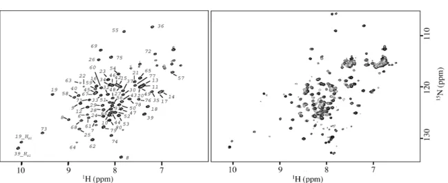
The full-length B. licheniformis BlaI protein was cleaved using papain digestion. The position of the unique cleavage site, unambiguously obtained from N-terminal sequencing and matrix-assisted laser desorption/ionization time-of-flight (MALDI-TOF) mass spectrometry was localized between the residues Ser82 and His83. The full-length BlaI protein as well as the N-terminal domain 1 –82 (BlaI-NTD) were studied by hetero-nuclear NMR. The similarity between the BlaI-NTD and BlaI 15N-heteronuclear single quantum
coherence (HSQC) spectra (Figure 2) demonstrates
Figure 1. Schematic representation of bla and mec operon organization in B. licheniformis and S. aureus. A, The mec and bla regulators, mecR1-mecI and blaR1-blaI, respectively, encode inducer-repressor systems with a high level of sequence similarity. The S. aureus BlaI and MecI repressors are 60% identical when compared to each other and 31– 41% identical when compared to the B. licheniformis BlaI repressor. The percentages of highly conserved amino acid residues in the transducers are 30 for the S. aureus BlaR–S. aureus MecR pair, 21 for the B. licheniformis BlaR–S. aureus BlaR pair and 23 for the B. licheniformis BlaR–S. aureus MecR pair.8B, Alignment of B. licheniformis bla, S. aureus bla,
Figure 2. Comparison of the15N HSQC spectra at 25 8C of BlaI N-terminal domain (BlaI-NTD) and of full-length BlaI.
Left: BlaI-NTD spectrum with assignment obtained for residues 6–81. Right: spectrum of the complete protein BlaI. Except for residue 81, all BlaI-NTD resonances are observed in the same positions, indicating that the N-terminal part of the protein forms an independent and structured domain. The resonances corresponding to the C-terminal domain (83–128) are poorly resolved and are mainly located in a region characteristic of extended structure.
Figure 3. NMR structures of BlaI-NTD (1–82). Secondary structures are colored: H1 in yellow, H2 in orange, H3 in red and the three strands of the b-sheet in blue. Top: stereo view of the ribbon backbone diagram of the lowest-energy structure. Bottom: stereo view of the backbone diagram of the 19 refined low-energy structures. Backbone heavy atoms were used for superimposition.
resonance assignment procedures. The few missing assignments correspond to the N and C-terminal ends (residues 1– 4, and 82).
For structure calculation, a total of 1252 inter-proton, 110 dihedral and 97 chiral restraints were used. The distance constraints contain 596 intra-residue, 205 sequential, 87 medium-range, 145 long-range and 219 ambiguous correlations derived from 2D, 3D, and 4D nuclear Overhauser effect spectroscopy (NOESY) spectra. The dihedral c and f angle restraints were deduced from TALOS, using backbone chemical shift values. At the final stage, 80 conformers were calculated, and the conformers with the lowest-energy functions were selected to be refined. A ribbon diagram of the lowest-energy structure and a superposition of the final ensemble of 19 simulated annealing struc-tures are shown (Figure 3).
Ensemble analysis of BlaI-NTD backbone stereo-chemical quality was done with NMR-PROCHECK.12 Over 99% of the dihedral angles
were found in the most favored and additional allowed regions of the Ramachandran plot. Struc-tural conformations of residues 1– 4 and 79– 82 are poorly defined, due to the lack of assignment for residues 1 –4 and 82, or to the small number of NOE constraints for residues 79– 81. Heteronuclear NOEs (Supplementary Material) confirm that the N and C-terminal parts of the protein are rather mobile. For statistics, we consider only the rigid core of BlaI-NTD (residues 5 –78). For these resi-dues, we obtained an average of 16.4 restraints per residue. A good agreement with the imental restraints is reflected by the low exper-imental energy level. There is no distance restraints violation greater than 0.1 A˚ . The back-bone and heavy-atom root-mean-square deviation (rmsd) values calculated for BlaI-NTD 5 –78 with respect to the mean coordinates are 0.63(^ 0.17), and 1.07(^0.18) A˚ , respectively. A summary of the restraint and structural statistics is presented in
Table 1.
The BlaI-NTD 3D structure consists of a three-stranded b-sheet (S1, Ser23-Asn25; S2, Leu57-Glu62; and S3, Val65-Pro70) packed against three a-helices (H1, Asp9-Lys20; H2, Thr26-Thr36; and H3, Pro41-Lys53) arranged in the order H1-S1-H2-T1-H3-S2-W1-S3. S2 and S3 form an antiparallel hairpin (loop called wing W1: Gly63-Arg64) and
LexA, MarR, FokI, DTXR, and Genesis, all of them being DNA recognition domains. It is interesting to note that the MarR protein is also involved in a mechanism of antibiotic resistance.16Although no
sequence homology could be detected by primary sequence similarity searches, all these proteins pre-sent a central core similar to that of BlaI-NTD. Whereas their helices can be of significantly differ-ent lengths, angles between the three a-helices are conserved. The main differences observed for these proteins are localized in the wing region. Some of the WHP proteins like Genesis have a long and flexible W1 wing, as revealed by hetero-nuclear NOE measurements.17 In the case of
BlaI-NTD, wing W1 is short (residues 63– 64). Hetero-nuclear NOE data (Supplementary Material) show no particular mobility for residues neighboring W1.
A high degree of sequence homology exists between B. licheniformis BlaI and S. aureus MecI, especially for the non-polar core residues. In par-ticular, many of the buried residues forming the core of the structure are identical or similar (BlaI/ MecI) between BlaI and MecI: (H1) Ala10, Val14,
Table 1.Structural statistics
NMR-derived restraints 1362 Interproton restraints 1252 Intraresidue restraints 596 Interresidue restraints 437 Sequential restraints 205 Medium-range restraints 87 Long-range restraints 145
Hydrogen bond restraints 0
Ambiguous restraints 219
Dihedral angle restraints 110
Chiral restraints 97
Average rms deviations from the mean coordinates (A˚ )a,b
Backbone heavy-atoms: 0.63 ^ 0.17
All heavy-atoms: 1.07 ^ 0.18
Ramachandran analysis (%)b,c
Residues in most favoured regions: 85.6 Residues in additional allowed regions: 13.7 Residues in generously allowed regions: 0.2
Residues in disallowed regions: 0.5
a Statistics were made for residues 5–78 (see the text). b
Average for the 19 lowest-energy structures.
c Only non-Gly and non-Pro residues were assessed with
Met15, Ile18, Trp19, (H2) Val/Ile29, Ile/Val30, Glu32, Leu/Ile33, (H3) Ile44, Leu/Ile48, Leu51, (b-sheet) Leu/Ile57, Tyr68, Ile72. Since there is no deletion or insertion necessary to align BlaI and MecI, the secondary structures predicted for MecI have the same length as for BlaI. All these data support the conclusion that MecI also belongs to the WHP family, and that its fold is very close to the BlaI structure.
DNA recognition by the repressors BlaI and MecI
For some of the WHP proteins with a BlaI-NTD similar fold listed above, the protein – DNA com-plex has been investigated by X-ray crystallogra-phy, NMR or computational tools.13 For all of
them, the same DNA interaction mode has been reported. Helix H3, called the recognition helix, is presented to the major groove of the DNA, and makes specific contacts with the base-pairs. The wings (in particular W1) and the protein N-term-inal part also make contacts with the minor groove of the DNA. Unlike this WHP canonical mode of
DNA recognition, the WHP RFX1 makes most of the contacts with the DNA major groove via wing W1.18 The so-called recognition helix H3 overlies
the minor groove, and there is no contact between the N-terminal part of the protein and the DNA.
To investigate the BlaI DNA recognition mode, the BlaI – DNA interaction surface was determined using the NMR chemical shift perturbation method. The method detects protein residues that are interacting directly, or that undergo confor-mational changes upon binding.15N-HSQC spectra
of B. licheniformis BlaI-NTD were recorded with increasing amounts of DNA for both the 30-mer DNA corresponding to the B. licheniformis BlaI operator op1, and a palindromic 24-mer DNA based on the symmetric consensus sequence of the mec and bla operators. Results obtained with the palindromic 24-mer DNA are shown in Figure 4
and are similar to the results obtained with the 30-mer DNA (data not shown).
Since the 15N-HSQC spectra of DNA-free
BlaI-NTD, and of DNA-bound BlaI-NTD share many similarities (Figure 4A), the amide1H and15N
chemi-cal shifts of the N-terminal domain of DNA-bound
Figure 4. Chemical shift mapping of BlaI-NTD in interaction with DNA. A,15N HSQC spectra recorded at 25 8C. Left:
BlaI-NTD spectrum in absence of DNA. Right: BlaI-NTD/DNA complex spectrum. The 24-mer palindromic DNA was added to the sample to reach a protein/DNAduplexratio of 1/2. (B) Graph of the chemical shift variations between the
DNA free BlaI-NTD and the BlaI-NTD/DNA complex versus residue number. The vertical axis corresponds to ðld1Hl £
lgH=gNlþ ld15NlÞ measured on the spectra presented in A. Arrows represent amino proton resonances that have been broadened and not assigned in the complexed form. The absence of a bar indicates the presence of a proline residue or an unmeasured shift due to overlap. The limit between weak and strong chemical shift variations has been fixed to 0.4 ppm.
ing surface located on a single side of BlaI-NTD. This surface can be superimposed nicely with the positively charged region of the protein electro-static surface (Figure 5A). The residues of the BlaI-NTD N-terminal part that are affected upon bind-ing demonstrate the functional importance of the repressor N-terminal part in DNA-binding. This result is consistent with former DNA footprinting studies showing that a BlaI repressor mutant carry-ing a six amino acid deletion (Lys3-Ser8) within its N-terminal region had lost its ability to bind the operator.7 Mutation of lysine 4 to alanine also
severely reduces the BlaI DNA-binding ability.19
The crucial role of the N-terminal domain and the shape of the electrostatic and interaction surfaces are not consistent with RFX1-DNA recognition mode, where there is no interaction between the N-terminal part of the protein and the DNA. On the contrary, they are in good agreement with a canonical WHP – DNA interaction, as described for E2F4.20Furthermore, the distances separating helix
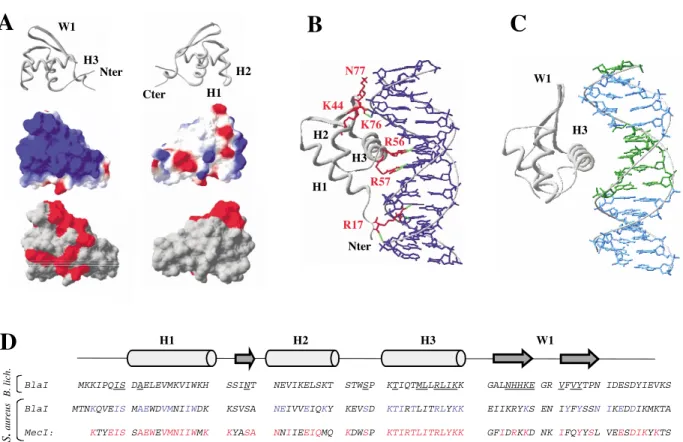
H3, wingW1, and the N-terminal ends that interact with DNA are quite equal for BlaI and E2F4. We therefore propose a model for the interaction between the repressors BlaI and MecI and their operators by comparison with the structure of the E2F4– DNA complex (Figure 5B). In this model, helix H3 interacts with the major groove of the operator. Other DNA –protein contacts occur via wing W1 and in the N-terminal part. Precise pos-ition of the contacts between the amino acid resi-dues of BlaI and MecI and the base-pairs must still be investigated via a high-resolution structure of the DNA – repressor complexes.
Biological implication: insights about the BlaI and MecI repression mechanism
It has been reported that BlaI and MecI could corepress b-lactamase and PBP20 gene expression
in S. aureus.9In the same way, B. licheniformis BlaI
interacts with S. aureus mec operator, as highlighted by gel mobility-shift assay (C. V., unpublished results). To explain this mec/bla corepression, the sequence homology for both the repressors and the DNA operator sequences of S. aureus and B. licheniformis was investigated. The primary structure alignment between B. licheniformis BlaI and S. aureus MecI and BlaI (Figure 5D) shows
(His59, His60, and Lys61, next to W1) facing an also conserved AT base-pair. Furthermore, locating two BlaI-NTD monomers on the two symmetric TACA motifs along the operator places the C-term-inal parts of the two monomers pointing in a con-venient orientation for dimerization contacts. Thus, we propose a repression mechanism, in which the repressor helix H3 makes specific con-tacts with the TACA motif of the operator sequence. This model is able to explain the core-pression of the mec and bla genes.
Although the S. aureus and B. licheniformis BlaI share the same DNA-binding features, they exhibit different induction mechanisms. In S. aureus, the presence of the b-lactam inducer outside the cell results in the proteolytic cleavage of the repressor between residues N101 and F102,21 leading to the
loss of its ability to dimerize. As pointed out above, the BlaI-NTD 3D-structure is not or slightly modified when compared to that of the wild-type BlaI, whereas the decrease of affinity between BlaI and BlaI-NTD in B. licheniformis is about 500 –1000 times.7 The SmtB Zn(II) sensing metallo-regulated
repressor is a WHP whose DNA-binding affinity is in the same range as BlaI – DNA affinity. The binding of a zinc ion to its C-terminal domain pro-motes the disassembly of the SmtB – DNA complex through a loss of affinity of about 1000.22,23 The
binding of a co-activator to the C-terminal domain, as in SmtB, is not the only way to inhibit the repressor activity of a winged-helix transcriptional regulator. For example, in the case of Escherichia coli MarR, the binding of two salicylate groups close to the DNA-binding a-helix inhibits the MarR activity.16 To investigate the role of the
C-terminal domain in BlaI –DNA interaction, a chemical shift mapping experiment was carried out for the dimeric full-length BlaI in the same con-ditions as for the monomeric BlaI-NTD (Supplementary Material). Two interesting obser-vations can be derived from this study. First, a similar chemical shift perturbation pattern is obtained for the residues of the N-terminal domain. Second, no significant modifications of the resonances of the C-terminal domain are observed. This suggests that the interaction mode between BlaI-NTD and the DNA is not influenced by the presence of the C-terminal domain. The BlaI/MecI dimerization C-terminal domain is,
however, essential to obtain a DNA-binding pro-tein of high affinity, probably placing the two DNA-binding domains in a favorable conformation for DNA contacts.
Conclusion
The 3D solution structure of B. licheniformis BlaI DNA-binding domain presented in this study reveals that BlaI is a member of the WHP family. S. aureus BlaI and MecI share also a similar WHP fold. Chemical shift mapping experiments high-lighted that BlaI presents the canonical DNA recognition mode. The C-terminal part of the pro-tein is involved in dimerisation, but not in DNA interaction. The N-terminal part is a winged helix motif, interacting via its N-terminal part, helix H3 and wing W1. A model of the BlaI/MecI DNA
interaction is proposed. In this model, specific contacts occur between the base-pairs of the TACA motif and conserved amino acid residues of the repressors helix H3. These results help under-standing the mechanism by which the BlaI/MecI repressor regulates blaP/blaZ/mecA expression.
Materials and Methods
Plasmids and DNA manipulations
PET22b (Novagen) was used as vector for the over-expression of the BlaI and BlaIGM2 products. The construction of plasmids pET22bblaIWT and pET22b-blaIGM2 has been described.4 The GM2 mutation
(M97V98/IL) is located in the C-terminal domain of BlaI.
For BlaI-NTD production, the GM2 mutant was pre-ferred due to its better over-expression yield.
Figure 5. Comparison of the region involved in the protein/DNA complex for E2F4 and BlaI-NTD. A, Electrostatic surface of BlaI and surface mapping of the region affected by the DNA interaction. On top, ribbon representation of BlaI-NTD structure in a front and back orientation. In the middle, surface electrostatic potential is colored in red (posi-tive) and blue (nega(posi-tive). The calculations were performed using the Swiss-PDB viewer (version 3.7). At the bottom, chemical shift variations ðld1Hl £ lg
H=gNlþ ld15NlÞ superior to 0.4 ppm (see Figure 4B) are indicated in red on the BlaI-NTD surface. B, Structure of E2F4/DNA complex.20Side-chains involved in a hydrogen bond with the DNA are
indicated in red and the H-bonds in green. Helix H3 forms direct H-bonds with the bases of the DNA (Arg56 and Arg57) as well as the N terminus part (Arg17). The winged helix and the turn between H1 and H2 form H-bonds with the phosphate and the sugar of the DNA (Lys44 and Asn77). Note that the residue numbering of E2F4 is quite different from that of BlaI where the H3 helix extends between the residues 41 and 53. C, Interaction model of the com-plex BlaI-NTD/DNA derived from the E2F4/DNA comcom-plex. BlaI-NTD structure was superimposed with E2F4 struc-ture. Totally conserved nucleotides presented in the consensus sequence ofFigure 1are colored in green D, Sequence alignment of B. licheniformis BlaI, and S. aureus BlaI and MecI. Residues indicated in blue correspond to amino acid residues conserved between BlaI of B. licheniformis and S. aureus. Residues indicated in red are conserved between BlaI and MecI of S. aureus. The highlighted residues correspond to the residues affected by the DNA interaction (deter-mined by the B. licheniformis BlaI chemical shift mapping).
Overnight-induced cells were then harvested by cen-trifugation, lysed and purified.4The final yield of
puri-fied 15N-labeled protein was 6 mg per liter of cell
culture and the isotopic labeling was 94% as determined by mass spectrometry.
The uniformly15N/13C-labeled BlaIGM2 sample was
produced according to the same procedure, apart from the fact that M9 minimal medium was replaced by the Silantes OD2-CN medium (Silantes GmbH, Gollier-strasse 70C, D-80339 Munchen). The final yield of puri-fied labeled protein was 20 mg/l of cell culture and the percentage of isotopic labeling was 99%.
Uniformly 15N/13C/2H-labeled BlaIGM2 sample was
produced using the optimized M9 medium in which water had been replaced by2H
2O and with15NH4Cl and 13C glucose concentrations of 18 mM and 10 mM,
respectively. The final yield of purified labeled protein was 4 mg per liter of cell culture and the percentage of deuterium labeling was approximately 75%.
Papain digestion and BlaI-NTD purification
Purified BlaI or BlaIGM2 were dialysed against a buf-fer containing 50 mM Hepes (pH7), 100 mM NaCl, 1 mM EDTA and was digested overnight using 1% mol/mol papain at 28 8C. After this time, the cleavage was complete and the protein digest was applied to an S-Sepharose-Fast-Flow column equilibrated in buffer A (50 mM Hepes (pH7), 100 mM NaCl, 1 mM EDTA, 5% (v/v) glycerol). BlaI-NTD was eluted with 1 M NaCl in the same buffer. The BlaI-NTD containing fractions were pooled, dialyzed exhaustively against 50 mM sodium phosphate (pH7.6), 200 mM KCl, 1 mM NaN3, 0.1 mM
Pefabloc and then concentrated to a final protein concentration of 0.5 mM by ultrafiltration with a Centricon (cut-off 5 kDa, Amicon, Brussels, Belgium).
Amino N-terminal sequencing of BlaI-NTD was carried out using a Procise 492 pulsed liquid phase pro-tein sequencer (Applied Biosystems) with 20–30 pmol of protein.
Band-shift assays
Gel retardation experiments were realized as described.24
NMR sample preparation
NMR samples of BlaI-NTD were prepared at a concen-tration of 0.75 mM in 75 mM sodium phosphate buffer (90% H2O, 10%2H2O), 300 mM KCl, pH 7.6. For the
full-length protein, an extensive microdrop study was
car-BlaIdimer/DNAduplexratios of 0.5, 1.0, 1.5 and 2.0, and a
BlaI-NTD/DNAduplexratios of 0.5, 1.0, 1.5, 2.0, and 2.5.
NMR spectroscopy
For sequential assignment, standard triple resonance experiments were carried out on15N and15N/13C-labeled
samples of BlaI-NTD: HNCA, HNCO, HN(CA)CO, CBCANH, and CBCA(CO)NH for backbone assignment and (H)C(CO)NH-TOCSY, H(CCO)NH-total correlated spectroscopy (TOCSY) for aliphatic side-chain assign-ment. For resonance assignment of aromatics, HSQC and H(C)CH-TOCSY experiments with the 13C carrier
set to 125 ppm, and 2D-NOESY spectrum performed on a sample dissolved in2H
2O were used. An HNCA
exper-iment was performed on a15N/13C/2H-labeled sample of
BlaIGM2 to confirm the assignment of the N-terminal part in the full-length protein.
Inter-protons restraints were derived from NOESY spectra: 2D-NOESY, 3D15N-edited NOESY-HSQC,
regu-lar 3D13C-edited NOESY-HSQC, 3D13C-edited
NOESY-HSQC optimized for CH groups and 4D 13C-edited
HSQC-NOESY-HSQC.26Mixing times were set to 120 ms.
For chemical shift mapping, 15N-HSQC, 13C-HSQC
optimized for aromatics and CT-HSQC were performed at different protein/DNA ratios.
All NMR experiments were performed on Varian INOVA 600 and INOVA 800 spectrometers, both equipped with a triple-resonance (1H, 15N, 13C) probe
and shielded z-gradients. The temperature was set to 25 8C. All triple-resonance experiments used the pulse sequences provided by the Varian Protein Pack.† All data processing, peak picking and peak intensity measurements were performed using the FELIX pro-gram version 2000 (Accelrys).
Structure calculation
The NOE distance restraint calibration was performed using cross-relaxation volumes between atoms of known separation in rigid parts of the molecule. A factor of 35% was added to the resulting upper distance, to account for the inherent uncertainty in the distance calculation. Clas-sical pseudo atoms corrections were applied. The lower distance limit was set to the sum of the van der Waals radii of the two protons. Assignment, restraint gener-ation, and structure calculation were performed itera-tively. Restraints that were violated, resulting from
misassignments or overlapped peaks, were corrected after the first steps of calculation.
The c and f angles calculated by TALOS from back-bone chemical shift values were used as constraints with lower and upper bounds of ^ 208.27Structure
calcu-lation was carried out using the Discover programs (Accelrys) interfaced to INSIGHTII for visualization and analytical purposes. The force-field used was AMBER.28
The structure determination protocol used a simulated annealing calculation starting from randomized Cartesian coordinates to explore the conformational space, and a restrained molecular dynamics calculation to refine each structure as described.29
Data Bank accession numbers
Chemical shift assignments of BlaI-NTD have been deposited with BioMagResBank (accession number 5873). The coordinates of the 19 BlaI-NTD refined struc-tures have been deposited in the Protein Data Bank under the accession code 1P6R.
Acknowledgements
This work was supported by the Belgian pro-gram of Interuniversity Poles of Attraction initiated by the Federal Office for Scientific Technical and Cultural Affaires (PAI no. P5/33), the “Fonds National de la Recherche Scientifique” (FNRS, Cre´dit aux chercheurs no 1.5201.02 and FRFC no. 2.4530.03) and the “Communaute´ Franc¸aise de Belgique” (projet Tournesol 2003, 03/ 013). We thank Martin Blackledge for the gift of protocols for structure calculation and for many useful discussions. We also thank Laurence Blanchard for helpful discussion and protein expression protocols. We thank Edwin De Pauw for mass spectrometry analysis.
References
1. Charlier, P., Coyette, J., Dehareng, D., Dive, G., Duez, C., Dusart, J. et al. (1998). Resistance bacte´rienne aux b-lactamines. Medecine/Sciences, 14, 544–549.
2. Joris, B., Hardt, K. & Ghuysen, J. M. (1994). Analysis of the penA gene of Pseudomonas cepacia 249. New Comput. Biochem. 27, 505–510.
3. Lim, D. & Strynadka, C. J. (2002). Structural basis for the b-lactam resistance of PBP2a from methicillin-resistant Staphylococcus aureus. Nature Struct. Biol. 9, 870–876.
4. File´e, P., Benlafya, K., Delmarcelle, M., Moutzourelis, G., Frere, J. M., Brans, A. & Joris, B. (2002). The fate of the BlaI repressor during the induction of the Bacillus licheniformis BlaP beta-lactamase. Mol. Micro-biol. 44, 685–694.
5. Duval, V., Swinnen, M., Lepage, S., Brans, A., Granier, B., Fransen, C. et al. (2003). The kinetic properties of the carboxy terminal domain of the Bacillus licheniformis 749/I BlaR penicillin-receptor shed a new light on the derepression of b-lactamase synthesis. Mol. Microbiol. 48, 1553–1564.
6. File´e, P., Vreuls, C., Hermann, R., Thamm, I., Frere,
J. M. & Joris, B. (2003). Dimerization and DNA-bind-ing properties of the Bacillus licheniformis 749/I BlaI repressor. J. Biol. Chem. 278, 16482–16487.
7. Wittman, V., Lin, H. C. & Wong, H. C. (1993). Functional domains of the penicillinase repressor of Bacillus licheniformis. J. Bacteriol. 175, 7383–7390. 8. Joris, B., Hardt, K. & Ghuysen, J. M. (1994). Induction
of b-lactamase and low affinity penicillin binding protein 20 synthesis in Gram-positive bacteria.
Bac-terial Cell Wall, 27, 505–515.
9. McKinney, T. K., Sharma, V. K., Craig, W. A. & Archer, G. L. (2001). Transcription of the gene med-iating methicillin resistance in Staphylococcus aureus (mecA) is corepressed but not coinduced by cognate mecA and beta-lactamase regulators. J. Bacteriol. 183, 6862 –6868.
10. Lewis, A. & Dyke, G. H. (2000). MecI represses synthesis from b-lactamase operon of Staphylococcus aureus. J. Antimicrob. Chemother. 45, 139–144.
11. Salerno, A. J. & Lampen, J. O. (1988). Differential transcription of the bla regulatory region during induction of beta-lactamase in Bacillus licheniformis. FEBS Letters, 227, 61–65.
12. Laskowski, R. A., Rullmannn, J. A., MacArthur, M. W., Kaptein, R. & Thornton, J. M. (1996). AQUA and PROCHECK-NMR: programs for checking the quality of protein structures solved by NMR. J. Biomol. NMR, 8, 477–486.
13. Gajiwala, K. S. & Burley, S. K. (2000). Winged helix proteins. Curr. Opin. Struct. Biol. 10, 110–116. 14. Clark, K. L., Halay, E. D., Lai, E. & Burley, S. K.
(1993). Co-crystal structure of the HNF-3/fork head DNA-recognition motif resembles histone H5. Nature, 364, 412–420.
15. Holm, L. & Sander, C. (1993). Protein structure comparison by alignment of distance matrices. J. Mol. Biol. 233, 123–138.
16. Alekshun, M. N., Levy, S. B., Mealy, T. R., Seaton, B. A. & Head, J. F. (2001). The crystal structure of MarR, a regulator of multiple antibiotic resistance, at 2.3 A˚ resolution. Nature Struct. Biol. 8, 710–714. 17. Jin, C. & Liao, X. (1999). Backbone dynamics of a
winged helix protein and its DNA complex at different temperatures: changes of internal motions in genesis upon binding to DNA. J. Mol. Biol. 292, 641 –651.
18. Gajiwala, K. S., Chen, H., Cornille, F., Roques, B. P., Reith, W., Mach, B. & Burley, S. K. (2000). Structure of the winged-helix protein hRFX1 reveals a new mode of DNA binding. Nature, 403, 916–921. 19. Gregory, P. D., Lewis, R. A., Curnock, S. P. & Dyke,
K. G. (1997). Studies of the repressor (BlaI) of beta-lactamase synthesis in Staphylococcus aureus. Mol. Microbiol. 24, 1025–1037.
20. Zheng, N., Fraenkel, E., Pabo, C. O. & Pavletich, N. P. (1999). Structural basis of DNA recognition by the heterodimeric cell cycle transcription factor E2F-DP. Genes Dev. 13, 666–674.
21. Lewis, R. A., Curnock, S. P. & Dyke, K. G. (1999). Proteolytic cleavage of the repressor BlaI of beta-lactamase synthesis in Staphylococcus aureus. FEMS Microbiol. Letters, 178, 271–275.
22. Cook, W. J., Kar, S. R., Taylor, K. B. & Hall, L. M. (1998). Crystal structure of the cyanobacterial metal-lothionein repressor Smtb: a model for metalloregula-tory proteins. J. Mol. Biol. 275, 337–346.
23. Vanzile, M. L., Chen, X. & Giedroc, D. P. (2002). Structural characterization of distinct alpha3N and
for chemical shift and sequence homology. J. Biomol. NMR, 13, 289–302.
28. Pearlman, D. A., Case, D. A., Caldwell, J. C., Seibel, G. L., Singh, U. C., Weiner, P. & Kollman, P. A. (1995). Amber 4.1, Department of Pharmaceutical Chemistry, University of California, San Francisco, CA.
Supplementary material comprising two figures is available on Science Direct