Sex Differences in the Neurokinin B System in the
Human Infundibular Nucleus
Melanie Taziaux, Dick F. Swaab, and Julie Bakker
Netherlands Institute for Neuroscience (M.T., J.B.), Royal Netherlands Academy of Arts and Sciences, Neuroendocrinology Laboratory, 1105 BA Amsterdam, The Netherlands; Netherlands Institute for Neuroscience (D.F.S.), Neuropsychiatric Disorder Laboratory, 1105 BA Amsterdam, The Netherlands; and Groupe Interdisciplinaire de Ge´noprote´omique Applique´e Neurosciences (M.T., J.B.), University of Lie`ge, 4000 Lie`ge, Belgium
Context: The recent report that loss-of-function mutations in either the gene encoding neurokinin B (NKB) or its receptor (NK3R) produce gonadotropin deficiencies in humans strongly points to NKB as a key regulator of GnRH release.
Objectives: We used NKB immunohistochemistry on postmortem human brain tissue to determine: 1) whether the human NKB system in the infundibular nucleus (INF) is sexually dimorphic; 2) at what stage in development the infundibular NKB system would diverge between men and women; 3) whether this putative structural difference is reversed in male-to-female (MtF) transsexual people; and 4) whether menopause is accompanied by changes in infundibular NKB immunoreactivity. Methods: NKB immunohistochemical staining was performed on postmortem hypothalamus ma-terial of both sexes from the infant/pubertal period into the elderly period and from MtF transsexuals.
Results: Quantitative analysis demonstrated that the human NKB system exhibits a robust female-dominant sexual dimorphism in the INF. During the first years after birth, both sexes displayed a moderate and equivalent level of NKB immunoreactivity in the INF. The adult features emerged progressively around puberty until adulthood, where the female-dominant sex difference ap-peared and continued into old age. In MtF transsexuals, a female-typical NKB immunoreactivity was observed. Finally, in postmenopausal women, there was a significant increase in NKB immu-noreactivity compared to premenopausal women.
Conclusion: Our results indicate that certain sex differences do not emerge until adulthood when activated by sex steroid hormones and the likely involvement of the human infundibular NKB system in the negative and positive feedback of estrogen on GnRH secretion. (J Clin Endocrinol
Metab 97: 0000 – 0000, 2012)
B
ecause immunohistochemical studies have failed to show colocalization of estrogen receptor␣ in GnRH neurons in different species [rat (1), sheep (2), primates (3)], estradiol effects on GnRH release are presumed to be mediated indirectly via other steroid-sensitive neuronal systems, which then converge onto GnRH cell bodies or terminals. Human genetic studies demonstrated thatkiss-peptin (4) as well as neurokinin B (NKB) signaling (5) are both potent regulators of GnRH secretion and are there-fore thought to be essential for the onset of puberty and the maintenance of adult reproductive function. In humans, infundibular kisspeptin neurons are sexually dimorphic (6), but no information is available for the NKB system, although it has been shown to be sexually dimorphic in rat ISSN Print 0021-972X ISSN Online 1945-7197
Printed in U.S.A.
Copyright © 2012 by The Endocrine Society
doi: 10.1210/jc.2012-1554 Received March 1, 2012. Accepted September 7, 2012.
Abbreviations: ARC, Arcuate nucleus; AVH, anteroventral hypothalamus; BST, bed nucleus of the stria terminalis; BSTc, BST central part; DAB, 3,3-diaminobenzidine; IBAS, image-based analysis system; INF, infundibular nucleus; ME, median eminence; MPO, medial preoptic nucleus; MtF, male-to-female; NBM, nucleus basalis of Meynert; NKB, neurokinin B; NKB-ir, NKB immunoreactive; NPY, neuropeptide Y; PT, pars tuberalis; TBS, Tris-buffered saline.
O R I G I N A L A R T I C L E E n d o c r i n e R e s e a r c h
J Clin Endocrinol Metab, December 2012, 97(12):0000 – 0000 jcem.endojournals.org 1 Copyright (C) 2012 by The Endocrine Society
(7, 8) and sheep (9, 10). Therefore, in the present study, we first asked whether the NKB system is sexually dimorphic in the human hypothalamus. By using postmortem brain tissues, we mapped the distribution of NKB fibers and cell bodies in the human hypothalamus and investigated the presence of putative sex differences. Next, we determined whether these sex differences are potentially the result of activational actions of sex steroid hormones by analyzing NKB expression at different stages of life (postnatal, pu-berty, adulthood, old age) in both sexes. We also included brain material from male-to-female (MtF) transsexuals who had undergone estrogen treatment in adulthood.
Subjects and Methods
Human brain tissue
Hypothalami of 42 subjects (Table 1) were obtained through autopsies by The Netherlands Brain Bank following the required permissions for brain autopsy and the use of tissue and medical information for research purposes. The subjects were catego-rized in an infant/pubertal period, an adult period, and an elderly period. The MtF transsexual group consisted of four sex-reas-signed and estrogen-treated individuals and one individual who was not orchidectomized but was hormonally treated (see Table 2 for detailed hormonal profiles). A nontreated individual with strong cross-gender identity feelings, which were already present since his earliest childhood, was also analyzed. Exclusion criteria for the subjects were a history of endocrine deregulation (ovar-ian, uterine, or breast cancer; recent abortion; or pregnancy), use of corticosteroids or drugs affecting the hypothalamo-pituitary-gonadal axis during at least the last month before death, and neurodegenerative or psychiatric diseases. The subjects were matched for age, postmortem delay, and the duration of the formalin fixation.
Histology
Hypothalami were formalin-fixed, paraffin-embedded, and cut serially in 6-m coronal sections from rostral to caudal. Ev-ery 100th section was collected on a SuperFrost/Plus (Menzel, Braunschweig, Germany) slide and stained with 0.5% thionin for general orientation. Every 50th section was then collected over the whole length of the hypothalamus for NKB immuno-cytochemistry to map the distribution of NKB-immunoreactive (NKB-ir) cells and fibers. To determine the border of the INF, every 50th section—adjacent to the NKB-stained ones— of the putative INF was mounted and stained for neuropeptide Y (NPY) (11).
NKB and NPY immunohistochemistry
Unless mentioned otherwise, all incubations were carried out at room temperature, and all washes were performed using Tris-buffered saline (TBS; 0.05MTris and 0.9% NaCl; pH 7.6). Sec-tions were deparaffinized and rehydrated. Antigen retrieval was used for NKB (but not NPY) staining by placing sections in ci-trate buffer (0.1Mcitric acid, 0.1Mtrisodium citrate, pH 6.0) in a microwave (10 min at 700 W). Next, sections were saturated in TBS-milk [5% nonfat dry milk (Elk; Campina Melkunie,
Eind-hoven, The Netherlands)] for 1 h to decrease nonspecific bind-ing. Sections were then incubated overnight at 4 C with a rabbit polyclonal anti-NKB antibody (1:2000; Peninsula Laboratories, San Carlos, CA; T-4450) or a rabbit polyclonal NPY anti-body (1:1000; Niepke 26/11/1988; Netherlands Institute for Neuroscience, Amsterdam, The Netherlands) diluted in Super-mix-milk (0.5% Triton X-100, 0.25% gelatin, 5% milk powder in TBS; pH 7.6). After rinsing in TBS-milk and TBS, sections were incubated with a biotinylated goat-antirabbit antibody (1:400; Vector Laboratories, Burlingame, CA) in Supermix (0.5% Tri-ton X-100, 0.25% gelatin in TBS; pH 7.6) for 1 h. The antibody-antigen complex was amplified with the avidin-biotin complex method (1:800; Kit ABC Vectastain Elite PK-6100; Vector Lab-oratories PLC, Cambridge, UK). Finally, sections were incubated in nickel-DAB (3,3-diaminobenzidine) solution (0.5 mg/ml; Sigma Chemical, St. Louis, MO; 0.01% H2O2; 2.33 mg/ml
am-monium nickel sulfate in TBS), rinsed in distilled water, dehy-drated, cleared in xylene, and coverslipped with Entellan (Merck, Darmstadt, Germany).
NKB and NPY specificity test
Specificity of the NKB antibody was confirmed by a solid phase preadsorption test. The synthetic human NKB peptide (Bachem) was dissolved (1g/l) in isoelectric focusing medium (10% glycerol, 10% dimethylformamide, 2.5% Nonidet; Sigma Chemical) and spotted on five separate strips of gelatin (0.2%) -coated nitrocellulose (0.1 m pore size, BA45; Schleicher & Schull, Dassel, Germany), each containing 30g of peptide. Fix-ation of the peptide to the nitrocellulose was performed over-night with 4% formaldehyde in a press block, followed by rinses in distilled water, Tris/HCl (0.05M; pH 7.6), and Su-permix, respectively. The anti-NKB antibody was adsorbed at 1:2000 in Supermix-milk 5% during five successive cycles. Binding of the antibody to the peptide was visualized by stain-ing the spotted strips usstain-ing the ABC method. No stainstain-ing was observed after absorption of NKB to its blocking peptide. Cross-reactivities of the NKB antibody with other tachykinins were previously excluded (11). The specificity of the NPY antibody was previously demonstrated (12, 13).
Estimation of the total NKB-ir volume of the INF Digital images of NPY-stained sections throughout the ros-trocaudal axis of the INF were made using a 5⫻ objective (Plan-Neofluar lenses) on a Zeiss Axioscope microscope mounted with a Sony B/W CCD camera (XC77CE) and connected to an Im-ageProPlus version 5.1 image analysis system (MediaCybernet-ics, Silver Spring, MD). Serial NKB-stained sections (300 m between two successive sections) were digitized at 10⫻ objective. By referring to the NPY staining images, the contour of the INF was manually outlined on the NKB staining images by an inves-tigator (M.T.) who was blind to the nature of the patients. A standard threshold (corresponding to 2-fold the background) was used for discriminating the labeled material from the back-ground and was quantified by a homemade OD program for DAB-nickel staining. To estimate the volume of the DAB-nickel precipitate as a measure of NKB immunoreactivity (cells and fibers together) throughout the INF, the surface of the outlined area and the masking area of NKB immunoreactivity within the outlined area were calculated in each section. All data were stored, and volume estimates of the INF (INF volume) and of total NKB immunoreactivity within the INF (NKB-ir INF
vol-TABLE 1. Clinicopathological data
Netherlands Brain
Bank no. (patient no.) Age
Brain weight (g) Postmortem delay (h:min) Fixation time (d) Clinicopathological information Females (n⫽ 18) Infant/pubertal period (n⫽ 5)
86-027 5 months 735 10:00 40 Sudden infant death syndrome
89-027 6 months 780 ⬍17:00 28 Cardiomyopathy
89-036 1 yr 820 NA 31 Hypoglycemia
87-077 7 yr 1320 ⬍9:45 33 Astrocytoma
87-035 13 yr 1250 ⬍13:00 48 Histiocytic lymphoma, cardiac failure Adult period (n⫽ 7)
01-009 21 yr 975 19:35 65 Myocardial infarction
84-025 23 yr 1300 ⬍10:00 35 Acute myeloid leukemia
01-072 25 yr 1500 ⬍17:00 31 Found dead (epileptic seizure)
99-058 34 yr 1395 72:00 132 Cardiac abnormalities
91-009 36 yr 1348 ⬍71:30 61 Fecal peritonitis
01-023 40 yr 1279 ⬍41:00 54 Pulmonary carcinoma
97-131 43 yr 1345 ⬍92:00 63 Myocardial infarction
Elderly period (n⫽ 6)
97-037 58 yr 1221 6:45 28 Epileptic convulsions after craniotomy
98-036 69 yr 1264 6:15 31 Cardiogenic shock
98-104 74 yr 1207 7:25 31 Necrosis of the intestines
97-156 77 yr 1235 2:40 47 Septic shock; metastasized pancreas
carcinoma
98-016 82 yr 1078 10:45 35 Congestive cardiac failure
98-089 90 yr 1047 7:15 39 Ruptured abdominal aorta aneurysm
Males (n⫽ 18)
Infant/pubertal period (n⫽ 5)
86-041 6 months 800 ⬍6:30 14 Sudden infant death syndrome
88-058 1 yr 1070 ⬍35:35 28 Bacterial meningitis, sepsis
84-016 4 yr 1565 23:55 100 Sepsis 87-057 6 yr 1550 3:30 41 Peritonitis 87-036 14 yr 1640 ⬍41:00 32 Lymphadenopathy Adult period (n⫽ 7) 97-083 22 yr 1334 16:29 26 Hypertrophic cardiomyopathy 02-076 27 yr 1520 NA NA Drowning
98-031 33 yr 1588 46:25 72 Hemothorax after car accident
99-071 39 yr 1400 ⬍16:30 130 Heart infarction
96-253 41 yr 1150 ⬍17:00 1231 Kidney failure
99-141 44 yr 1565 ⬍10:00 149 Myocardial infarction
99-035 44 yr 1434 ⬍41:00 123 Multiorgan failure
Elderly period (n⫽ 6)
98-090 58 yr 1528 41:00 72 Collapsed; unsuccessful resuscitation
08-032 71 yr 1600 8:55 70 Pancreas and rectum carcinoma with
hepatic metastases
06-028 76 yr 1514 19:35 27 Cardiac arrest
97-143 79 yr 1392 6:00 31 Pulmonary embolism
97-116 80 yr 1380 6:56 33 Respiratory insufficiency, lung emphysema
98-189 81 yr 1276 5:20 33 Unspecified respiratory problems
98-033 82 yr NA ⬍89:00 63 Carcinoma of the lung, respiratory
problems MtF transsexuals (n⫽ 5) 98-137 (T1) 26 yr 1500 NA NA Suicide 93-042 (T2) 36 yr 1145 21:43 31 Pneumonia 88-064 (T3) 43 yr 1540 NA NA Neurosarcoma 95-018 (T4) 48 yr 1198 ⬍40:20 36 Cardiac arrest 84-020 (T5) 50 yr 1380 NA 30 Suicide
Nontreated male with cross-gender identity feelings (n⫽ 1)
96-088 84 yr 1433 41 38 Lung carcinoma
ume) were performed by an image-based analysis system (IBAS) conversion program based upon multiplication of the outlined areas and masking area, respectively, by sample frequency cor-rected for section thickness (6m) over the whole length of the INF. The mean⫾SEMnumber of NKB-stained sections quanti-fied per subject was 17.4⫾ 3.4.
Estimation of the total number of NKB-positive neurons within the INF
Every other NKB-stained section (600m between two suc-cessive sections) throughout the INF in the rostrocaudal direc-tion of each subject was used for analysis. Estimates were made using an IBAS (Kontron Elektronik, Chichester, UK), connected to a Sony B/W CCD camera (XC77CE) mounted on a plain objective microscope (Zeiss Axioskop with Plan-NEOFLUAR Zeiss objectives). From the section to be measured, an image covering the INF (⫻2.5 objective) was loaded into the IBAS and displayed on the computer monitor. In this image, the INF was outlined manually (based on the adjacent NPY staining), and over this outlined area a grid was superimposed. From the re-spective grid fields, x and y coordinates were stored, and all individual images were retrieved using a 40⫻ objective on the image analysis monitor. For analysis, 100% of the rectangular fields (each field covering at least 50% of the outlined area) were analyzed. To prevent double counting, only neurons containing a nucleolus (⬃2m diameter) were counted. The number of neurons per section was multiplied by the sampling frequency (the interval distances between individual sections) to obtain an estimation of the total number of NKB-ir neurons in the INF from 8.7⫾ 1.7 sections per subject (mean ⫾SEM).
Statistical analysis
Two-way ANOVAs with the stage of life (infant/pubertal vs. adult vs. elderly period) and the sex (male vs. female) as inde-pendent factors were used to analyze the overall difference in infundibular NKB-ir volume and NKB-positive cells as well as possible confounding factors. A separate one-way ANOVA was performed to compare the volume occupied by NKB immuno-reactivity and the number of NKB-positive cells in the INF among only adult men, adult women, and adult MtF transsex-uals. All ANOVAs were followed when appropriate by post hoc tests using Fisher’s protected least significant difference tests. The Fisher’s exact probability test was used to compare the
pres-ence of NKB innervation in the pars tuberalis (PT) vs. its abspres-ence between adult men and women. Differences were considered significant for P⬍ 0.05.
Results
NKB expression in the human hypothalamus
NKB-ir fibers are widely distributed throughout the human hypothalamus, whereas cell bodies were confined to a few specific nuclei (Fig. 1). NKB immunoreactivity was found in the cytoplasmic compartment of cell bodies, which exhibited varying size depending on their localiza-tion. Small, oval-to-round NKB neurons were numerous in the central and medial portions of the bed nucleus of the stria terminalis (BST) and in the INF/median eminence (ME) complex. The distribution of the neuronal popula-tion in the INF exhibited a rostrocaudal topography, with the majority of NKB-ir cells being found in the middle and caudal part of the INF. Less intensely labeled cell bodies were also found scattered periventricularly throughout the caudal extent of the hypothalamus. A small number of medium-sized round NKB neurons were observed in the nucleus basalis of Meynert (NBM), the anteroventral hy-pothalamus (AVH), the medial preoptic nucleus (MPO), and the posterior BST. Immunoreactive fibers for NKB exhibited varicosities and were abundantly identified throughout the hypothalamus, notably in the MPO, lat-eral septum, AVH, NBM, the ventromedial hypothala-mus, the dorsomedial hypothalahypothala-mus, and in the periven-tricular area, but they were particularly prominent in the BST (central, medial, ventral, and posterior portions; Fig. 2, A and B), the INF/ME complex (Fig. 2D), and the ME, where NKB-ir fibers ran spirally around the capillary ves-sels (Fig. 2E).
Sexual dimorphism of the NKB system
Systematic examination of NKB staining yielded obvi-ous sex differences in the PT and the INF. Although the PT
TABLE 2. Hormonal profile of MtF transsexuals
Netherlands Brain Bank no. (patient no.) Age (yr) Age at beginning of hormonal treatment (yr) Age at
castration (yr) Estrogen treatment
Antiandrogen treatment (cyproterone acetate)
98-137 (T1) 26 21 26 EE 5g 2 dd 50 mg 2 dd
93-042 (T2) 36 36 Not operated Therapeutic doses (at least 5 yr b.d.) 50 mg 1 dd (at least the last 10 months b.d.) 88-064 (T3) 43 36 39 Age 39, EE 50g 2 dd (stopped 3 months b.d.) 50 mg 2 dd (stopped 2 yr b.d.) 95-018 (T4) 48 35 36 Age 35, EE 50g 2 dd. Age 40, EE 50g 1 dd (lasted until death)
50 mg 2 dd (stopped 10 months b.d.) 84-020 (T5) 50 42 44 Age 42, stilbestrol 5 mg 1 dd; after
2 months to 5 mg 2 dd. Age 44, EE 50g 2 dd (lasted until death)
Age 44, 50 mg 2 dd (stopped 2 yr b.d.) NA, Not available; b.d., before death; EE, ethinyl estradiol; dd, dose per d.
of both sexes is totally devoid of NKB immunoreactivity during the infant/pubertal period, 100% of adult men ex-amined in this study displayed a dense NKB innervation in the PT (seven of seven), whereas it was totally absent in adult women (seven of seven). A Fisher’s exact probability test comparing the presence of NKB innervation in the PT vs. its absence between adult men and women revealed a highly significant effect (P⬍ 0.001; see Fig. 3 for repre-sentative pictures). However, in elderly subjects, both sexes showed a high level of NKB immunoreactivity in this region; the same was true in MtF transsexuals (hormon-ally treated or not).
Another robust sexual dimorphism of the NKB system was observed in the INF; adult women consistently dis-played more robust NKB staining and denser NKB inner-vation in the INF than adult men (Fig. 4). This sex differ-ence only appeared in adulthood and continued into old age when NKB immunoreactivity was up-regulated after menopause (Fig. 5A). A two-way ANOVA on NKB-ir vol-ume in the INF revealed an almost significant effect of the
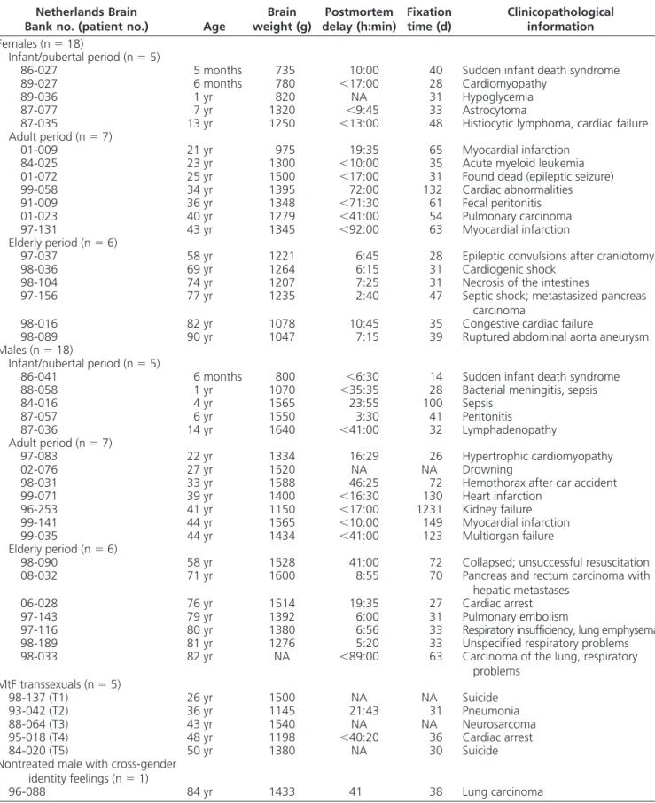
stage of life (F2,30⫽ 3.163; P ⫽ 0.0567), a significant effect of sex (F1,30 ⫽ 19.134; P ⫽ 0.0001), and a significant interaction effect between the two factors (F2,30⫽ 7.771; P ⫽ 0.0019). Post hoc tests confirmed that the volume occupied by NKB immunoreactivity in the INF of adult women is larger relative to adult men (P⫽ 0.032). This female-dominant sexual dimorphism was heightened at menopause, i.e. elderly women have a larger NKB-ir vol-ume in the INF compared with elderly men (P⫽ 0.012) and compared with premenopausal women (P⫽ 0.0455), whereas the NKB-ir volume in the INF in adult men did not differ from elderly men (P⫽ 0.8788). No significant dif-ferences in NKB-ir volume were found in the INF between boys and girls during the infant/pubertal period (P ⫽ 0.749). Likewise, NKB-ir volume in the INF did not sig-nificantly differ between girls and adult women (P ⫽ 0.372), or between boys and adult men (P ⫽ 0.268). A qualitative examination of the neuronal NKB population in the INF showed a hypertrophy of NKB-ir neurons in FIG. 1. Schematic drawing from anterior to posterior (A–H) in a representative female hypothalamus to illustrate the distribution of NKB-ir cells and fibers. NKB-ir cells are represented by closed circles, whereas NKB-ir fibers are represented by dotted lines (single fibers or low density),
continuous lines (moderate density), and crossed lines (high density). AC, Anterior commissure; BSTm, BST, medial part; BSTp, BST, posterior part;
BSTv, BST, ventral part; DMH, dorsomedial nucleus of the hypothalamus; FO, fornix; LS, lateral septum; NTL, lateral tuberal nucleus; OC, optic chiasm; OT, optic tract; PEN, periventricular nucleus; PH, posterior hypothalamic nucleus; PVN, paraventricular nucleus; SCN, suprachiasmatic nucleus; SON, supraoptic nucleus; TMN, tuberomamillary nucleus; VMH, ventromedial nucleus of the hypothalamus.
postmenopausal compared with premenopausal women (Fig. 5F).
An estimation of the total number of NKB-positive cells in this nucleus was also performed. Although the pattern of NKB-positive cells was very similar to that of NKB-ir volume (Fig. 5B), a two-way ANOVA failed to reveal any significant difference between the different factors (all P⬎ 0.10).
The developmental pattern in NKB expression in the INF throughout life is clearly different between men and women. Regression analysis showed that NKB-ir volume in the INF is positively correlated with age in female sub-jects from the infant/pubertal period to the elderly period (r⫽ 0.548; P ⫽ 0.0185; n ⫽ 18), whereas no significant
correlation was observed between NKB-ir volume in the INF and age in male subjects (r⫽ 0.365; P ⫽ 0.1369; n ⫽ 18; Fig. 5C). When the regression analysis was conducted using the total number of NKB-positive cells (Fig. 5D), a just significant negative correlation was observed with age in male subjects (r⫽ 0.473; P ⫽ 0.0473; n ⫽ 18), but no correlation was found in female subjects (r⫽ 0.254; P ⫽ 0.3089; n⫽ 18).
In a separate analysis, we compared the volume occu-pied by NKB immunoreactivity and the number of NKB-positive cells in the INF among adult men, adult women, and adult MtF transsexuals (Fig. 5E). One-way ANOVA revealed that NKB-ir volume in the INF is significantly different between the three groups (F2,16⫽ 3.994; P ⫽ 0.0392). Post hoc tests showed that the volume occupied by NKB immunoreactivity in the INF of adult men (0.021⫾ 0.011 mm3) is significantly lower compared with adult women (0.072⫾ 0.038 mm3; P⫽ 0.0232) and the MtF transsexuals (0.072 ⫾ 0.058 mm3; P ⫽ 0.0351). However, the NKB-ir volume of adult women and that of MtF transsexuals did not differ (P⫽ 0.9919). It is inter-esting to note that the 84-yr-old gender dysphoric non-treated male subject had an NKB-ir volume (0.152 mm3) in the postmenopausal female range (0.120 ⫾ 0.045 mm3). The number of NKB-positive cells was not statis-tically different between adult men and women and MtF transsexuals (one-way ANOVA; all P⬎ 0.5).
Confounding factors
As expected, two-way ANOVA on brain weights re-vealed a significant effect of sex (F1,19 ⫽ 11.660; P ⫽ 0.0019) but no significant effect of stage of life (F2,29⫽ 2.865; P⫽ 0.0732) and no interaction between the two factors (F2,29⫽ 1.272; P ⫽ 0.2955). Although a sex dif-ference in brain weight was detected, such effect was not observed in the volume estimate of the INF (NPY delin-eated volume of the INF), suggesting that the sex differ-ence observed in NKB-ir volume in the INF was not con-secutive to a structural sex difference of the brain area analyzed. Indeed, a two-way ANOVA showed that the volume estimate of the INF is not different at different stages of life (F2,30⫽ 0.087; P ⫽ 0.9166) or between males and females (F2,30⫽ 0.021; P ⫽ 0.8853), and no interac-tion between the two factors was found (F2,30⫽ 1.638; P⫽ 0.2113). Moreover, no effects of postmortem delay or fixation time on NKB immunocytochemical staining (ir volume in the INF and total number of NKB-positive cells in the INF) were observed (all P⬎ 0.10).
Discussion
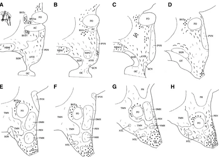
We showed here that the sex difference in NKB expression in the INF reached only significance in adulthood and that FIG. 2. Representative photomicrographs illustrating the main
localization of NKB-ir cells and fibers in the human hypothalamus: BSTm and BSTc (A), BSTv (B), INF (D), and ME (E). Panel C and the
insets of D and E show higher magnifications. Scale bars, 1 mm in A,
D, and E; 0.5 mm in B; 0.05 in C and in the insets. AC, Anterior commissure; BSTm, BST, medial part; BSTv, BST, ventral part; LV, lateral ventricle; IIIV, third ventricle.
MtF transsexuals had a female-typical infundibular NKB system. These results suggest that: 1) in addition to the well-known perinatal period of steroid-dependent brain organization, sex steroid hormones during puberty might also contribute to the emergence of sex differences in adulthood; and 2) the sex-reversal observed in MtF trans-sexuals may reflect, at least in part, an atypical sexual differentiation of the hypothalamus.
Distribution of NKB immunoreactivity in the human hypothalamus
We observed that neurons and fibers expressing NKB immunoreactivity were found predominantly in all parts of the BST and in the INF/ME region, where NKB-ir fibers richly innervated portal capillaries. The neuronal distri-bution is in agreement with the distridistri-bution of NKB mRNA (14) and of preprotachikinin B protein (6) in the human hypothalamus, which strengthens antibody spec-ificity as we showed by the preadsorption procedure. NKB neurons have also been described in the INF/arcuate nu-cleus (ARC) of the monkey (15, 16), sheep (9, 10, 17), goat (18), rat (7, 8, 19), and mouse (20).
Sexual dimorphisms of the human NKB system
A dense plexus of NKB-ir fibers in the PT was consis-tently found in adult men but was completely absent in adult women. This sex difference is only visible in adult-hood, disappears into old age and cannot be attributed to low circulating sex steroid levels given that all orchidec-tomized and estrogen-treated MtF transsexuals displayed a dense NKB innervation similar to that observed in adult men. Similar observations were reported in the rat, in which a male-specific NKB innervation was observed around blood vessels of the external zone of the ME,
com-pared with a more diffuse axonal wir-ing in the female (7). Moreover, the masculine phenotype emerges only in puberty and is activated by nonaroma-tizable androgen (21). Based upon the adult pattern of NKB innervation that we observed in humans, it is likely that androgens stimulate, whereas estrogens inhibit this innervation in the PT in adult-hood. Although the PT can be considered as a gateway uniquely placed to influence hypothalamic-pituitary communication and function (22), there is currently no insight into the function of the male-spe-cific NKB innervation of the PT.
Subsequently, we demonstrated that the NKB-ir volume in the INF is sexu-ally dimorphic in adulthood (women⬎ men). Surprisingly, no such significant sex difference was found in the total number of NKB-positive cells in the INF, suggesting a sexually dimorphic NKB innervation rather than a sex difference in the number of NKB-expressing cells. A female-dominant number of NKB neurons has been identified in the rat (8) and sheep (9, 10) ARC. It is interesting to note that although a sex difference was observed in the number of kisspeptin-immunoreactive neurons in humans, the most prominent sex difference was observed in the den-sity of kisspeptin-immunoreactive fibers (6). The sta-tistical discrepancy observed between our two measures of NKB expression could be partly explained by limi-tations related to the use of postmortem brain tissue, whose conditions at death cannot be tightly controlled [i.e. a long postmortem delay may cause fast axonal transport of the neuropeptide between death and fixa-tion (23)]. It should also be noted that NKB gene ex-pression varies with the rat estrous cycle (19), which could partly explain the lack of significant differences in NKB-positive cells between adult men and women, given that the stage of the menstrual cycle at death was not reported in the patient’s medical folder.
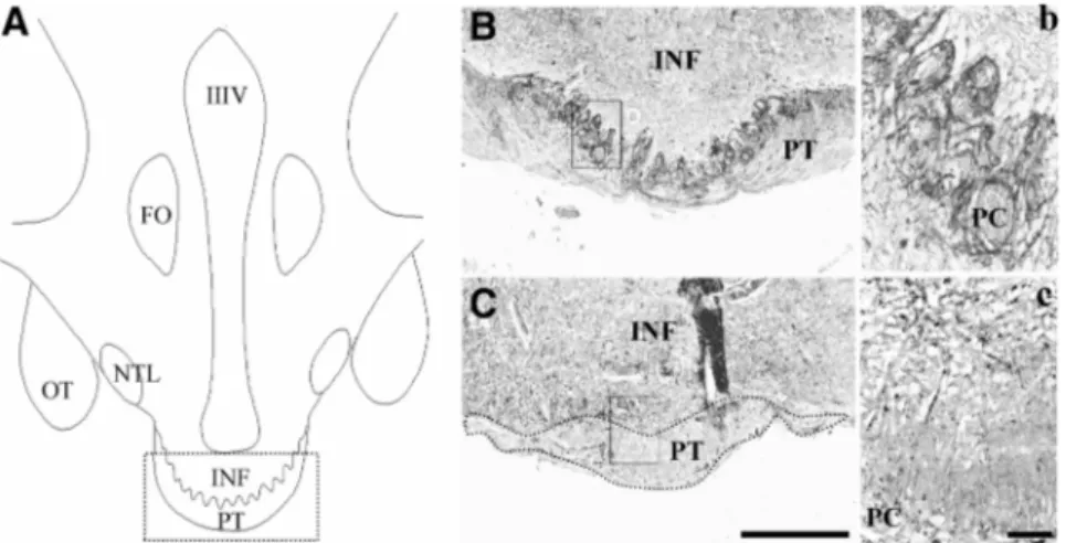
Finally, the sex difference found in NKB-ir volume in the INF between adult men and women continues during the elderly period, where we also observed an increased infundibular NKB-ir volume and a hypertrophy of NKB-ir neurons in postmenopausal compared with premeno-pausal women. Our results thus confirm a previous study showing an increase and hypertrophy of neurons express-ing NKB mRNA after menopause (24). These changes in NKB mRNA were duplicated in perimenopausal, post-menopausal, and young ovariectomized rhesus macaques (25). Importantly, these changes are reversed by estradiol FIG. 3. Sexually dimorphic NKB innervation in the PT. A, Schematic drawing depicting the
location of the human PT. The box in panel A illustrates the area photographed from a 39-yr-old man (B) and a 34-yr-39-yr-old woman (C). The boxes in B and C illustrate the area
rephotographed at higher magnification in panels b and c. The boundaries of the PT were drawn in a dotted line (C). Scale bars, 0.5 mm in A and B; 0.05 in a and b. FO, Fornix; NTL, lateral tuberal nucleus; OT, optic tract; PC, portal capillaries.
replacement (15), suggesting that these changes are likely due to the loss of ovarian estradiol.
The apparent discrepancy between a female-like INF vs. a male-like PT observed in MtF transsexuals is in favor of the “brain-sex theory of transsexualism,” suggesting atypical or-ganizational effects of sex hormones during development in MtF. Although certain aspects of sexual differentiation might have been altered in MtF transsexuals (such as NKB expres-sion in the INF), they were exposed to androgens throughout life, which could still have masculinized other brain struc-tures and functions, such as the NKB innervation in the PT.
Development of the human infundibular NKB system
On the basis of the ontogenetic profile for GnRH re-lease in primates (26), we hypothesized that the pubertal period might be the time when sex differences could de-velop in NKB expression. Indeed, we demonstrated that the NKB system is immature in both sexes during the first years after birth and starts to differentiate progressively from puberty to adulthood, where the female-dominant sex difference appears for the first time and continues over the years. Although it is generally accepted that sexual
differentiation of the neuroendocrine hypothalamus does not proceed be-yond the early postnatal period, pu-berty has been recently recognized as another period of development during which gonadal hormones organize the nervous system (27). Other human hy-pothalamic structures seem to differen-tiate later in life, such as the sexually dimorphic nucleus of the preoptic area [between 4 yr and puberty (28)], the darkly staining posteromedial compo-nents of the BST [around puberty (29)], and the BST central part (BSTc) [in adulthood (30)]. Furthermore, the sex-ually dimorphic NKB innervation in the rat ARC does not become visible before puberty (21). It is likely that the sex difference in human NKB expres-sion also reflects organizational actions of testosterone during the prenatal pe-riod. However, this sex difference is only revealed postpubertally, suggest-ing that it needs to be activated by sex steroid hormones.
Sex reversal of NKB immunoreactivity in MtF transsexuals
Our data revealed a female-like in-fundibular NKB-ir volume in MtF transsexuals who had undergone estrogen treatment and sex reassignment in adulthood. The feminization of the NKB system of MtF transsexuals might be explained by either the presence of a higher estrogen concentration in the blood due to estro-gen treatment or the lack of androestro-gens due to orchidec-tomy. However, there is some evidence against these ex-planations: 1) subject T3 showed a large infundibular NKB-ir volume, despite the fact that estrogen treatment was stopped about 3 months before death; 2) subjects T4 and T5 continued to take estrogens until death but had smaller infundibular NKB-ir volumes than T3; and 3) sub-ject T2, who was never orchidectomized, had the largest infundibular NKB-ir volume of the MtF transsexual group. Additional evidence comes from the 84-yr-old man who also had very strong cross-gender identity feelings but remained untreated, whose NKB-ir volume was in the range of postmenopausal women. In line with previous human postmortem studies showing a female-like BSTc (31, 32) and a female-like third interstitial nucleus of the anterior hypothalamus (33) in MtF transsexuals, our re-sults may suggest an atypical sexual differentiation of the FIG. 4. Representative photomicrographs of the sexually dimorphic NKB immunoreactivity in
the INF between adult men (A) and women (B). The boxes in panels A and B illustrate the area rephotographed at higher magnification in a and b. Scale bars, 0.5 mm in A and B; 0.25 mm in a and c; 0.05 mm in b and d. IIIV, Third ventricle.
hypothalamus in transsexual people. Because the sex re-versal does not seem to be influenced by circulating hor-mone levels in adulthood, the sexual differentiation of the infundibular NKB system is likely due to an organizational action of sex steroid hormones (presumably testosterone) during a critical period of development, which is likely to emerge postpubertally in humans as evidenced by the de-velopmental curve of NKB expression over the different stages of life. Nevertheless, due to the small sample of MtF transsexuals and differences in estrogen levels, we cannot rule out an effect of the hormonal milieu in adulthood on NKB expression. In contrast with rodents where the LH
surge mechanism is irrevocably fixated perinatally by ex-posure to androgen (34), the debate about a “permissive” vs.“deterministic” hypothalamic control of GnRH release is still open in humans (35). Although both sexes can show estrogen-induced LH surges (36), the evocability of the LH surge in men requires prolonged estrogen priming, and the amplitude of the surge is smaller compared with women (37). Nevertheless, the LH surge in MtF transsexuals was male-typical before sex reassignment and almost female-typical afterward, suggesting that long-term estrogen treatment to men could partly feminize the gonadotropin response (38). However, the best evidence of an atypical FIG. 5. Estimation of the volume occupied by NKB immunoreactivity (A) and of the total number of NKB-ir cells (B) in the INF of males and females during the infant/pubertal period (between 5 months and 14 yr), the adult period (between 22 and 44 yr), and the elderly period (between 58 and 90 yr). *, P⬍ 0.05 vs. male from the adult period; #, P ⬍ 0.05 vs. female from the adult period;, P ⬍ 0.05 vs. male from the elderly period. Estimation of the total NKB-ir volume (C) and of the total number of NKB-ir cells (D) in the INF of both sexes from the postnatal period (5 months) to the elderly period (90 yr). The lines represent the regression line for each sex. E, Estimation of the volume occupied by NKB immunoreactivity in the INF of men, women, and MtF transsexuals in adulthood. *, P⬍ 0.05 vs. male. F, Representative microphotographs illustrating the increase and hypertrophy of neurons expressing NKB in postmenopausal women (panel F2, 90 yr old) compared with
premenopausal women (panel F1, 34 yr old) in the INF (40⫻ objective). The boxes in panels 1 and 2 illustrate the area rephotographed at higher magnification (100⫻ objective) in the insets. Scale bars, 0.05 mm in F1 and F2; 0.01 mm in the insets.
sexual differentiation in MtF transsexuals came from the nontreated individual with strong cross-gender identity feelings, who displayed a female-like NKB expression.
Although the rodent model offers advantages to study the neuroendocrine control of steroid feedback on GnRH regulation, the neural mechanisms that govern the pre-ovulatory LH surge in women seem to differ from those in rodents (35). Studies of functioning and anatomical local-ization of neuropeptides involved in GnRH regulation such as NKB in the human brain are critical to validate the animal experimental data and increase our understanding of the human reproductive axis and related neuroendo-crine disorders.
Acknowledgments
We thank A. Sluiter, R. Balesar, U. Unmehopa, B. Fisser, J. J. van Heerikhuize, A. M. Staphorsius, and L. van Keimpema for their technical assistance.
Address all correspondence and requests for reprints to: Mel-anie Taziaux, GIGA Neurosciences, University of Lie`ge, 1 ave-nue de l’hoˆpital (Baˆt. B36), 4000 Lie`ge, Belgium. E-mail: mtaziaux@ulg.ac.be.
This research was supported by a VICI grant of the Neder-landse Organisatie voor Wetenschappelijk Onderzoek (Grant 453-08-003; to J.B.). Brain material was provided by The Neth-erlands Brain Bank (coordinator, Dr. I. Huitinga). J.B. is a senior research associate at the Fonds National de la Recherche Scien-tifique (FNRS). M.T. is a postdoctoral researcher at the FNRS. Disclosure Summary: The authors have nothing to disclose.
References
1. Herbison AE, Theodosis DT 1992 Localization of oestrogen recep-tors in preoptic neurons containing neurotensin but not tyrosine hydroxylase, cholecystokinin or luteinizing hormone-releasing hor-mone in the male and female rat. Neuroscience 50:283–298 2. Lehman MN, Karsch FJ 1993 Do gonadotropin-releasing hormone,
tyrosine hydroxylase-, and-endorphin-immunoreactive neurons contain estrogen receptors? A double-label immunocytochemical study in the Suffolk ewe. Endocrinology 133:887– 895
3. Sullivan KA, Witkin JW, Ferin M, Silverman AJ 1995 Gonadotro-pin-releasing hormone neurons in the rhesus macaque are not im-munoreactive for the estrogen receptor. Brain Res 685:198 –200 4. de Roux N, Genin E, Carel JC, Matsuda F, Chaussain JL, Milgrom
E 2003 Hypogonadotropic hypogonadism due to loss of function of the KiSS1-derived peptide receptor GPR54. Proc Natl Acad Sci USA 100:10972–10976
5. Topaloglu AK, Reimann F, Guclu M, Yalin AS, Kotan LD, Porter KM, Serin A, Mungan NO, Cook JR, Ozbek MN, Imamoglu S, Akalin NS, Yuksel B, O’Rahilly S, Semple RK 2009 TAC3 and TACR3 mutations in familial hypogonadotropic hypogonadism re-veal a key role for neurokinin B in the central control of reproduc-tion. Nat Genet 41:354 –358
6. Hrabovszky E, Ciofi P, Vida B, Horvath MC, Keller E, Caraty A, Bloom SR, Ghatei MA, Dhillo WS, Liposits Z, Kallo I 2010 The kisspeptin system of the human hypothalamus: sexual dimorphism
and relationship with gonadotropin-releasing hormone and neuro-kinin B neurons. Eur J Neurosci 31:1984 –1998
7. Ciofi P, Leroy D, Tramu G 2006 Sexual dimorphism in the organi-zation of the rat hypothalamic infundibular area. Neuroscience 141: 1731–1745
8. Ruiz-Pino F, Navarro VM, Bentsen AH, Garcia-Galiano D, San-chez-Garrido MA, Ciofi P, Steiner RA, Mikkelsen JD, Pinilla L, Tena-Sempere M 2012 Neurokinin B and the control of the gonad-otropic axis in the rat: developmental changes, sexual dimorphism, and regulation by gonadal steroids. Endocrinology 153:4818 – 4829 9. Goubillon ML, Forsdike RA, Robinson JE, Ciofi P, Caraty A, Her-bison AE 2000 Identification of neurokinin B-expressing neurons as an highly estrogen-receptive, sexually dimorphic cell group in the ovine arcuate nucleus. Endocrinology 141:4218 – 4225
10. Cheng G, Coolen LM, Padmanabhan V, Goodman RL, Lehman MN 2010 The kisspeptin/neurokinin B/dynorphin (KNDy) cell pop-ulation of the arcuate nucleus: sex differences and effects of prenatal testosterone in sheep. Endocrinology 151:301–311
11. Goldstone AP, Unmehopa UA, Bloom SR, Swaab DF 2002 Hypo-thalamic NPY and agouti-related protein are increased in human illness but not in Prader-Willi syndrome and other obese subjects. J Clin Endocrinol Metab 87:927–937
12. Coven˜as R, Martin F, Belda M, Smith V, Salinas P, Rivada E, Diaz-Cabiale Z, Narvaez JA, Marcos P, Tramu G, Gonzalez-Baron S 2003 Mapping of neurokinin-like immunoreactivity in the human brainstem. BMC Neurosci 4:3
13. van der Beek EM, Pool CW, van Eerdenburg FJ, Sluiter AA, van der Donk HA, van den Hurk R, Wiegant VM 1992 Fc-mediated non-specific staining of the porcine brain with rabbit antisera in immu-nocytochemistry is prevented by pre-incubation of the sera with proteins A and G. J Histochem Cytochem 40:1731–1739 14. Chawla MK, Gutierrez GM, Young 3rd WS, McMullen NT, Rance
NE 1997 Localization of neurons expressing substance P and neu-rokinin B gene transcripts in the human hypothalamus and basal forebrain. J Comp Neurol 384:429 – 442
15. Abel TW, Voytko ML, Rance NE 1999 The effects of hormone replacement therapy on hypothalamic neuropeptide gene expres-sion in a primate model of menopause. J Clin Endocrinol Metab 84:2111–2118
16. Ramaswamy S, Seminara SB, Ali B, Ciofi P, Amin NA, Plant TM 2010 Neurokinin B stimulates GnRH release in the male monkey (Macaca mulatta) and is colocalized with kisspeptin in the arcuate nucleus. Endocrinology 151:4494 – 4503
17. Goodman RL, Lehman MN, Smith JT, Coolen LM, de Oliveira CV, Jafarzadehshirazi MR, Pereira A, Iqbal J, Caraty A, Ciofi P, Clarke IJ 2007 Kisspeptin neurons in the arcuate nucleus of the ewe express both dynorphin A and neurokinin B. Endocrinology 148:5752– 5760
18. Wakabayashi Y, Nakada T, Murata K, Ohkura S, Mogi K, Navarro VM, Clifton DK, Mori Y, Tsukamura H, Maeda K, Steiner RA, Okamura H 2010 Neurokinin B and dynorphin A in kisspeptin neurons of the arcuate nucleus participate in generation of periodic oscillation of neural activity driving pulsatile gonadotropin-releas-ing hormone secretion in the goat. J Neurosci 30:3124 –3132 19. Rance NE, Bruce TR 1994 Neurokinin B gene expression is
in-creased in the arcuate nucleus of ovariectomized rats. Neuroendo-crinology 60:337–345
20. Navarro VM, Gottsch ML, Chavkin C, Okamura H, Clifton DK, Steiner RA 2009 Regulation of gonadotropin-releasing hormone secretion by kisspeptin/dynorphin/neurokinin B neurons in the ar-cuate nucleus of the mouse. J Neurosci 29:11859 –11866 21. Ciofi P, Lapirot OC, Tramu G 2007 An androgen-dependent sexual
dimorphism visible at puberty in the rat hypothalamus. Neurosci-ence 146:630 – 642
22. Morgan PJ, Williams LM 1996 The pars tuberalis of the pituitary: a gateway for neuroendocrine output. Rev Reprod 1:153–161 23. Dai J, Swaab DF, Buijs RM 1998 Recovery of axonal transport in
24. Rance NE, Young 3rd WS 1991 Hypertrophy and increased gene expression of neurons containing neurokinin-B and substance-P messenger ribonucleic acids in the hypothalami of postmenopausal women. Endocrinology 128:2239 –2247
25. Sandoval-Guzma´n T, Stalcup ST, Krajewski SJ, Voytko ML, Rance NE 2004 Effects of ovariectomy on the neuroendocrine axes regu-lating reproduction and energy balance in young cynomolgus ma-caques. J Neuroendocrinol 16:146 –153
26. Plant TM 2008 Hypothalamic control of the pituitary-gonadal axis in higher primates: key advances over the last two decades. J Neu-roendocrinol 20:719 –726
27. Sisk CL, Zehr JL 2005 Pubertal hormones organize the adolescent brain and behavior. Front Neuroendocrinol 26:163–174 28. Swaab DF, Hofman MA 1988 Sexual differentiation of the human
hypothalamus: ontogeny of the sexually dimorphic nucleus of the preoptic area. Brain Res Dev Brain Res 44:314 –318
29. Allen LS, Gorski RA 1990 Sex difference in the bed nucleus of the stria terminalis of the human brain. J Comp Neurol 302:697–706 30. Chung WC, De Vries GJ, Swaab DF 2002 Sexual differentiation of the bed nucleus of the stria terminalis in humans may extend into adulthood. J Neurosci 22:1027–1033
31. Zhou JN, Hofman MA, Gooren LJ, Swaab DF 1995 A sex difference
in the human brain and its relation to transsexuality. Nature 378: 68 –70
32. Kruijver FP, Zhou JN, Pool CW, Hofman MA, Gooren LJ, Swaab DF 2000 Male-to-female transsexuals have female neuron numbers in a limbic nucleus. J Clin Endocrinol Metab 85:2034 –2041 33. Garcia-Falgueras A, Swaab DF 2008 A sex difference in the
hypo-thalamic uncinate nucleus: relationship to gender identity. Brain 131:3132–3146
34. Karsch FJ, Dierschke DJ, Knobil E 1973 Sexual differentiation of pituitary function: apparent difference between primates and ro-dents. Science 179:484 – 486
35. Plant TM 2012 A comparison of the neuroendocrine mechanisms underlying the initiation of the preovulatory LH surge in the human, Old World monkey and rodent. Front Neuroendocrinol 33:160 – 168
36. Goh HH, Ratnam SS 1988 The LH surge in humans : its mechanism and sex difference. Gynecol Endocrinol 2:165–182
37. Do¨rner G 1988 Neuroendocrine response to estrogen and brain differentiation in heterosexuals, homosexuals, and transsexuals. Arch Sex Behav 17:57–75
38. Gooren L 1986 The neuroendocrine response of luteinizing hor-mone to estrogen administration in the human is not sex specific but dependent on the hormonal environment. J Clin Endocrinol Metab 63:589 –593