Université de Montréal
The effect of calcium homeostasis on HSV-1 propagation
Par
Mayerline Dorsainvil
Département de Microbiologie, Infectiologie et Immunologie Faculté de Médecine
Mémoire présenté en vue de l’obtention du grade de Maitrise ès Science (M.Sc.) en Microbiologie et Immunologie
19 Janvier, 2020
Université de Montréal
Département de Microbiologie, Infectiologie et Immunologie, Faculté de Médecine
Ce mémoire intitulé
The effect of calcium homeostasis on HSV-1 propagation
Présenté par
Mayerline Dorsainvil
A été évalué par un jury composé des personnes suivantes
Guy Lemay Président-rapporteur Roger Lippé Directeur de recherche Louis-Éric Trudeau Membre du jury
Résumé
Au cours d'une infection lytique, le virus de l'herpès simplex de type 1 (VHS-1) doit entreprendre plusieurs étapes de fusion afin de se répliquer et se propager correctement. Ainsi, le virus a évolué afin de tirer avantage de la machinerie cellulaire en utilisant des protéines et facteurs de l’hôte à cet effet. Dans la littérature, les processus sous-jacents à l’entrée du VHS-1 ont été largement élucidés. Cependant, on ne sait toujours pas comment les particules virales nouvellement synthétisées sortent de la cellule hôte et quels facteurs cellulaires sont impliqués dans ce processus. Des résultats publiés par notre laboratoire indiquent que la protéine cellulaire, Extended Synaptotagmin 1 (E-Syt1), a un impact négatif sur la propagation globale du virus lorsqu’inhibée par de l’ARN d’interférence. Conséquemment, la présente étude a pour objectif de confirmer et d'approfondir le rôle d’E-Syt1 sur la propagation virale, en particulier sur la sortie du virus. Étant donné que l’activation d’E-Syt1 est liée à l’augmentation de la concentration de calcium cytoplasmique, nous avons également étudié l'implication du calcium au cours des stades ultérieurs de la réplication virale. Ici, nous avons démontré que la surexpression d’E-Syt1 n’a pas d’effet détectable sur la sortie du VHS-1, mais que le calcium a effet sur la propagation virale. Alors que la séquestration précoce du calcium (4 et 6 heures post-infection) à l'aide de chélateurs réprime la sortie virale, aucun effet significatif a été détecté lorsque les chélateurs ont été ajoutés à un stade avancé de l’infection (12 et 16 heures post-infection). Nos résultats fournissent des données intéressantes sur la nécessité de l’homéostasie du calcium intracellulaire afin que VHS-1 puisse assurer une médiation adéquate de la sortie virale. Ces résultats pourraient conduire à la découverte de nouveaux mécanismes ou protéines cellulaires régulées par le calcium et utilisés par le VHS-1 lors de réplications lytiques virales.
Abstract
During a lytic infection, Herpes Simplex Virus type 1 (HSV-1) must go through multiple steps of fusion to replicate and propagate properly. For this purpose, the virus has evolved consequently by taking advantage of the cellular machinery using host factors and proteins. In the literature, processes underlying HSV-1’s entry have been extensively elucidated. However, it remains unclear how newly synthesized viral particles egress from the host cell, and what cellular factors are implicated in this process. Results published by our laboratory suggest that the cellular protein, Extended Synaptotagmin 1 (E-Syt1), has a negative and global impact on the viral propagation when down regulated by RNA interference. Consequently, this study aims to confirm and deepen our understanding of E-Syt1’s role on HSV-1, particularly during viral egress. Since activation of E-Syt1 is linked to the increase in cytoplasmic calcium concentration, we also investigated calcium involvement during later stages of viral propagation. Interestingly, overexpression of E-Syt1 had no measurable effect on HSV-1 propagation whereas calcium has a dual effect on viral propagation. While early calcium sequestering (4 and 6 hours post-infection) using chelators represses viral egress, no significant effect was detected when chelators were added at later time points (12 and 16 hours post-infection). Our results give interesting insights on how HSV-1 relies on intracellular calcium homeostasis to properly mediate viral egress. These results may lead to the discovery of new mechanisms or cellular proteins that are regulated by calcium and hijacked by HSV-1 during lytic replication.
Table of Contents
Résumé ... i
Abstract... ii
Table of Contents ... iii
List of Figures ... v
List of Tables ...vi
List of Abbreviations ... vii
Acknowledgements ... x
1. Introduction ... 1
1.1. HSV-1 Structure ... 2
1.2. Viral Entry ... 3
1.3. Nuclear Entry ... 6
1.4. Viral Genomic Expression and Replication ... 7
1.5. Capsid Assembly and DNA Packaging ... 9
1.6. Nuclear Egress ... 9
1.7. Tegumentation and Re-Envelopment ... 11
1.8. Cell-to-Cell Spread ... 12
1.9. gM and E-Syt 1 Interaction ... 12
1.9.1. E-Syt1 ... 13
1.9.2. E-Syt1 Structure ... 14
1.9.3. E-Syt1 Activation ... 16
1.10. Calcium and HSV-1 ... 17
1.11. Hypothesis and Approach ... 17
2. Material and Methods ... 18
2.1. Cell line ... 18
2.2. Viruses and Infection ... 18
2.2.1. HSV-1 17+ WT ... 18
2.2.2. HSV-1 V701 ... 19
2.2.3. HSV-1 K26 GFP ... 19
2.3.1. EGFP E-Syt1 WT, mCherry E-Syt1 (7x) and EGFP E-Syt1 ΔC2E constructs. 20 2.3.2. EGFP-N2 ... 21 2.3.3. pCyto-gBFull ... 21 2.4. Transfection ... 22 2.5. Plaque Assay ... 22 2.6. Western Blot ... 23 2.7. Drugs ... 23 2.8. Immunofluorescence ... 23 2.9. qPCR ... 24 2.9.1. Infectivity ratio ... 24 2.10. Viability test ... 25 2.10.1. Drugs ... 25 2.10.2. Transfection ... 25 2.11. Statistics ... 25 3. Results... 26
3.1. Calcium is required for HSV-1 egress ... 26
3.2. Calcium Is Required at Early Time Points During HSV-1 Egress ... 30
3.3. Chelation of Intracellular Calcium Early During Viral Egress Reduces Viral Genomic DNA Replication ... 33
3.4. Mutants of E-Syt1 Do Not Induce Viral Titer Recovery ... 39
4. Discussion ... 45
5. Conclusion ... 49
List of Figures
Fig 1. The structure of HSV-1’s genome ... 2 Fig 2. The two models of HSV-1 entry inside a cell ... 5 Fig 3. Structure of E-Syt1 and mutations used in this study ... 15 Fig 4. Hela cell viability is not affected by the different calcium chelators or ionomycin treatments... 27 Fig 5. Chelation of intracellular calcium inhibits viral egress ... 28 Fig 6. Viral localization inside the cell is not affected by post entry calcium level changes
29
Fig 7. The viral titer is decreased in the supernatant when cytosolic calcium is chelated at early time point during viral egress ... 32 Fig 8. The viral titer is decreased in the cell lysate when cytosolic calcium is chelated at early time point during viral egress ... 33 Fig 9. Chelation of cytosolic calcium reduces the amount of newly synthesized viral DNA in the supernatant ... 35 Fig 10. Chelation of cytosolic calcium reduces the amount of newly synthesized viral DNA in the cell lysate fraction... 36 Fig 11. Changes in the cytosolic calcium level affect the infectivity ratio of newly synthesized viral particles released in the supernatant ... 37 Fig 12. Changes in the cytosolic calcium level affect the infectivity of newly synthesized viral particles released in the cell lysate fraction ... 38 Fig 13. Overexpression of the different constructs of E-Syt1 does not affect cell viability .. 41 Fig 14. The different E-Syt1 constructs are expressed at similar level ... 42 Fig 15. Overexpression of the different E-Syt1 mutants did not inhibit viral egress in the supernatant of infected Hela cells ... 43 Fig 16. Overexpression of the different E-Syt1 mutants did not inhibit viral egress in the cell lysate of infected Hela cells ... 44
List of Tables
List of Abbreviations
HSV-1: Herpes simplex virus type 1E-Syt1: Extended-Synaptotagmin 1 E-Syt2: Extended-Synaptotagmin 2 E-Syt3: Extended-Synaptotagmin 3 Syt: Synaptotagmin
Tcb: Tricalbin
AD: Alzheimer’s disease TNF: Tumour necrosis factor UL: Long unique sequence US: Short unique sequence IRL: Internal long repeat TRL: Terminal long repeat IRS: Internal short repeat TRS: Terminal short repeat TGN: Trans-Golgi network gB: Glycoprotein B gC: Glycoprotein C gD: Glycoprotein D gH: Glycoprotein H gL: Glycoprotein L gM: Glycoprotein M gN: Glycoprotein N
HVEM: Herpesvirus entry mediator HS: heparan sulfate
3-O-HS: 3-O sulfated heparan sulfate PM: Plasma membrane
ER: Endoplasmic reticulum
NMHC-IIA: non-muscle myosin heavy chain IIA NMHC-IIB: non-muscle myosin heavy chain IIB
MT: Microtubule
NM: Nuclear membrane INM: Inner nuclear membrane ONM: Outer nuclear membrane NCP: Nuclear pore complex Nup: Nucleoporin
HCF1: Host cell factor 1
Oct1: Octamer-binding protein 1 H/P: Helicase/primase complex ssDNA: single-stranded DNA dsDNA: double-stranded DNA
SERCA: Sarco/endoplasmic reticulum Ca2+ ATPase PMCA: Plasma membrane Ca2+ ATPase
SMP: Synaptotagmin-like mitochondrial lipid-binding protein PI(4,5)P2: Phosphatidylinositol-4,5-bisphosphate
PLC: Phospholipase C IP3: Inositol triphosphate DAG: Diacylglycerol IP3-R: IP3-receptor
SOCE: Store-operated Ca2+ entry PAA: phosphonoacetic acid IFN: Interferon
Acknowledgements
First, I would like to thank my family who supported me throughout the realization of my master’s degree. They were present during the highs and lows, and always believed in me and my abilities to make great achievements.
Second, I would like to thank my colleagues from the laboratory who are now great friends. Without their helps, advice, opinions, knowledge and amazing energy, I would not have been able to finish my master’s degree. Bita, Mackenzie, Hugo, Elisabeth, Catherine and Kendra, I will be forever grateful that I had the chance to work with such great individuals and scientists. Also, a special thanks for the students in Michel Desjardin’s laboratory, Melanie and Ahmed, who were always there to help me in my experiments.
Third, I would like to thank Johanne for her amazing work as a lab technician. Her devotion to her work and making sure that the lab runs properly was remarkable. Not only my project, but the projects of everyone in the lab were successful because of you. Thank you so much! And for Marie-Josée who takes over so graciously after Johanne’s retirement, I would like to express my sincere appreciation. You always had a positive energy and ready to overcome any challenges. Thank you for your help and support.
Last but not least, I would like to thank the principal investigator of my laboratory, Dr. Roger Lippé. You gave me the chance to work in your laboratory and bloom as the scientist that I am today. Your door was always open when I needed a second look at my results and you always supported me with my thousands of ideas for this project. I will also like to thank you for making sure that there was always a great atmosphere in the lab by encouraging us to make activities (sport, restaurants, etc.) together. Maybe someday I will be as good as you in squash, and I’ll be able to get revenge. It was my pleasure and honour to work with you and I wish you all the best for the lab at Ste-Justine.
1. Introduction
Herpesviridae is a large family of DNA viruses that subdivide into three subgroups; Alphaherpesvirinae, Betaherpesvirinae and Gammaherpesvirinae [1]. Viruses from these three groups have a broad tropism and can infect human and different species of animals. Beta- and Gammaherpesvirinae are comprised of 18 and 32 species of viruses respectively, whose diseases cause mild to severe symptoms, and even death [1, 2]. Viruses like HHV-5 (human cytomegalovirus), HHV-6 (sixth disease), HHV-4 (mononucleosis) and HHV-8 (Kaposi’s sarcoma) are all examples of viruses that belong to those two subclasses [2]. Among all the 37 species of Alphaherpesvirinae, HHV-1 (herpes simplex virus 1), HHV-2 (herpes simplex virus 2) and HHV-3 (Varicella-Zoster virus) are examples of viruses commonly found in the human population. Indeed, it is estimated that 69% (3.7 billion individuals) of the world population is infected with herpes simplex virus type 1 (HSV-1) [3]. This widely spread infection is partly due to the fact that HSV-1 is asymptomatic. Additionally, this virus, as other Herpesviridae, has the capacity to go into latency. For HSV-1, the tropism for latency are trigeminal ganglions (TG) and the central nervous system (CNS) of the infected organism which lead to a lifelong persistent viral reservoir [4, 5]. HSV-1 transmission between organisms occurs through direct contact with the buccal mucosa, skin with or without lesions and genitals. It is important to mention that even during the asymptomatic period, the virus is still infectious as it can replicate and generate a small viral load in the infected individual. During a lytic viral replication, HSV-1 can cause painful open sores around the mouth, nose, face and inside the throat, but it can also cause keratitis in immunocompromised individuals and encephalitis in newborns [6-8]. Recently, it was suggested that brain damages caused by HSV-1 can lead to the development of neurodegenerative diseases like Alzheimer’s disease (AD) [9, 10]. Furthermore, some studies demonstrated that patients who have oral submucous fibrosis, oral leukoplakia and other oral cancers also had a high cell-mediated immune response against HSV-1 [11-13]. In this context, it is clear that HSV-1 can lead to severe health issues, reinforcing the relevance of research aiming to understand cellular and molecular processes underlying HSV-1 propagation.
1.1.
HSV-1 Structure
HSV-1 is an enveloped virus containing a 152 kbp linear double-stranded DNA genome [14]. The genome structure is composed of one long unique sequence (UL) and one short unique sequence (US) [15] (figure 1). Both sequences are flanked by inverted repeats which allow the unique sequences to change orientation during DNA replication. Those inverted repeats are termed internal and terminal long repeats (IRL, TRL) or internal and terminal short repeats (IRS, TRS) (figure 1). Consequently, there are four possible genome orientations that are found in a pool of virus which all seems to be equally infectious [16, 17]. In the virus, essential genes responsible for lytic replication, DNA splicing and packaging are highly conserved [18]. At the IRL and TRL sequences, we find the LAT gene along with the RL1 and RL2 genes encoding for ICP0 and ICP34.5 respectively [19]. While the LAT gene encodes for latency-promoting micro RNAs, ICP0 and ICP34.5 are both implicated in early phases of the viral replication [17]. Similarly to the long repeats, IRS and TRS contain a highly conserved gene, RS1, which encodes for ICP4. ICP4 is also implicated in the early stages of viral replication as it regulates viral transcription [20]. Since both pairs of repeats have identical sequences, those four genes are present twice in the viral genome. The other 76 genes of HSV-1 are located throughout the UL and Us sequences [21].
Fig 1. The structure of HSV-1’s genome
This figure is from a recent publication [22]. The genome structure of HSV-1 is composed of a unique long and unique short sequences (UL and Us) flanked by inverted repeats termed terminal and
internal long repeats (TRL and IRL) and terminal and internal short repeats (TRS and IRS).
During capsid assembly and maturation, this large genome is tightly packed into the mature viral capsid. Capsids are mainly composed of four viral proteins that are highly organized to form the 20 faces of the capsid [23]. Each capsomer is composed of five or six monomers of VP5 linked to six monomers of VP26. Attached to the capsomers, there is a
heterotrimeric complex composed of two monomers of VP23 and one molecule of VP19C [24, 25]. Altogether, they form the 1250Å diameter icosahedral capsid [25]. Surrounding the nucleocapsid, there is a layer of tegument proteins required in many steps throughout the viral propagation. It is unclear where this layer of viral protein is added to the nucleocapsid, but some studies suggested that it mostly occurs as the viral particle pass through the trans-Golgi networks (TGN) and its interaction with endosomes containing tegument proteins [26-29]. To this day, there are about 20 viral proteins that were identified at the tegument, as well as some host cell proteins. As an example, VP1/2, also referred to as pUL36, is an essential tegument protein at the early stage in the infection as it regulates DNA release from the incoming capsid inside the nucleus. A mutant virus lacking this tegument protein shows a significant decrease in de novo viral synthesis [30]. Other viral proteins like VP11, VP12, VP13/14, VP16, VP22or pUL37 have been identified as tegument proteins where their mutation affects viral propagation at different levels [31-33]. The final layer is composed of a host-derived phospholipid bilayer containing multiple glycoproteins implicated in cellular trafficking. Trafficking in the cell implies multiple steps of envelopment and de-envelopment which are tightly regulated by the fusion machinery mainly composed of glycoproteins B, D and H/L [34-36].
1.2.
Viral Entry
Virus trafficking starts by the binding of the viral particle to the cell surface. Reversible bindings occur between viral glycoproteins and specific cellular receptors. Depending on the cell type, different glycoprotein-receptor interactions are required to enable an effective infection. HSV-1 primary infection site occurs at the mucosal epithelium where the viral glycoprotein B and C (gB and gC) interacts with cellular heparan sulfate (HS) [37]. This interaction enables docking of viral particles but is not sufficient to initiate penetration inside the cell [38]. Following this first interaction, the glycoprotein D (gD) interacts with one of the three possible cellular receptors: herpesvirus entry mediator (HVEM) which is part of the tumour necrosis factor (TNF) family, Nectin-1 or 2 and 3-O sulfated heparan sulfate (3-O-HS). They are all expressed by multiple types of human cells and are required to initiate fusion between the viral particle and the cell plasma membrane (PM) [39]. It was demonstrated that deleterious mutation affecting either one of those gD-receptor interactions significantly affects
mechanisms to ensure optimal viral entry. The glycoprotein complex H and L (gH/gL) was also reported to be involved in viral entry [41]. In fact, gH/gL interacts with integrin αVβ3, which was demonstrated to relocate nectin-1 to lipid rafts when overexpressed in cells [42]. Furthermore, one group suggested that gH acts as an activator of gB by binding to it through its domain III to support gB fusogenic activity [43]. Even if gH/gL functions are not clear, it seems to enhance viral entry efficiency. Since HSV-1 entry requires interaction with multiple receptors, it was reported that it occurs at receptor-rich sites (entry hotspot). Some studies demonstrated that gB can also interact with non-muscle myosin heavy chain IIA and B (NMHC-IIA or –IIB), which are receptors and motor proteins that allow the movement on actin filaments [44]. Since the plasma membrane is lined with a layer of cortical actin meshwork, gB-NMHC-IIA or -IIB interactions mediate viral particles to “surf” on the cell surface to reach those entry hotspots [45, 46]. Even if NMHC-IIA and -IIB are mostly expressed in the cytoplasm, exposure of HSV-1 to cells triggers the relocation of those motor proteins at the PM, especially at filopodia [44, 46].
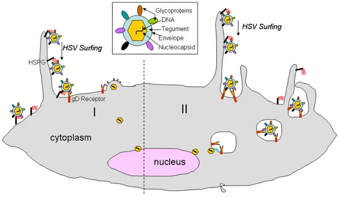
Currently, there are two models describing HSV-1 entry into targeted cells (figure 2). It is important to mention that the type of entry depends on the cell line. In fact, HSV-1 enters Vero or HEp-2 cells by fusion with their PM while it enters Hela, epithelial or keratinocyte cells through a phagocytosis-like mechanism [47, 48]. The first model is the fusion of HSV-1 envelope with cellular PM. In this model, gB, gD, gH/gL are required to support the fusion between the two lipid bilayers [49]. The capsid and tegument proteins are directly released in the cytoplasm where most teguments detach from the capsid except VP1/2, pUL37 and pUS3 [50]. HSV-1 also triggers cytoskeletal network rearrangement such as actin modification to facilitate the passage of viral particles through the PM. It is important to mention that these modifications are specific to entry mechanism employed by the virus [51-53]. Cellular factors are then hijacked by the virus to support the capsid trafficking toward the nucleus. In fact, the capsid circulates in the cytoplasm toward the minus end of microtubules (MT) by interacting with the motor protein dynein along with its cofactor dynactin [54]. This process is defined as being pH-independent. The second entry model is based on a phagocytosis-like mode of entry of the enveloped viral particles, following by the fusion of the viral envelope with the vesicular membrane in the cytoplasm [55]. In this process, the binding of gD to nectin-1 activates local actin rearrangement through the RhoA-GTPase activity, which promotes cellular protrusion
formation and viral particles uptake [47] . It was demonstrated that the interior of these vesicles was rich in nectin-1 and HVEM. Consequently, the low-pH inside those vesicles generated by the vacuolar H+-ATPase induces the exposure of certain gB and gD domains which further promotes the interaction with their respective receptors and enables fusion [55-58] . As the first model, the capsid is then released in the cytoplasm and is translocated toward the nucleus by the same active transport. Alternatively, some groups suggested that HSV-1 release from these vesicles was pH-independent in C10 cells, which supports the idea that the virus uses different entry routes depending on the cell line [59]. Interestingly, Dr. Sodeik's laboratory demonstrated a pH-dependent mechanism in some cells as, in presence of a H+-ATPase inhibitor, endocytosis of enveloped virus was driven by Na+/H+ exchanger (NHE) and p21 kinase activities [60]. Although HSV-1 employs cells specific entry processes, one observation that seems to be commonly accepted is that the formation of these vesicles does not occur through clathrin- or caveolin- derived endocytosis [48, 49, 59-61].
Fig 2. The two models of HSV-1 entry inside a cell
This figure is from a recent publication [62]. I: first entry mode where HSV-1 enters a cell through the fusion of its envelope with cellular PM. II: the second mode of entry is based on a
phagocytosis-like mechanism where the enveloped viral particles are uptaken in a vesicle, followed by the fusion of the viral envelope with the vesicular membrane in the cytoplasm.
1.3.
Nuclear Entry
As other DNA viruses, HSV-1 replicates its viral genome inside the nucleus using its viral replicative machinery along with the cellular replicative machinery. To do so, HSV-1 must bypass the nuclear membrane (NM) barrier and inject its viral genome inside the nucleus. In fact, the mammalian NM is composed of two distinct membranes, the inner nuclear membrane (INM) and outer nuclear membrane (ONM), separated by the perinuclear space. This NM is impermeable to most molecules as it tightly regulates trafficking across the membrane using either passive diffusion of ions and molecules smaller than 10 nm or active transport of larger molecules, both through nuclear pore complex (NPC) [63, 64]. NPC is a transmembrane complex composed of 30 different proteins termed nucleoporins (Nup), organized into an octagonal funnel [65]. In its open conformation, the NPC core allows the passage of molecules up to 39 nm [66]. Consequently, when the 125 nm capsid reaches the nucleus, it interacts with NPC to subsequently release its genome inside the nucleus as the capsid cannot pass through the NM [67]. The capsid docking occurs through the binding of NPC cytoplasmic components Nup358 and Nup 214 and the minor capsid protein pUL25 [50, 68]. It is not clear if the tegument protein VP1/2 also binds directly to NPC, but previous experiments demonstrated that antibodies against this tegument protein inhibit capsid docking [50]. Furthermore, studies also demonstrated that capsid docking was an importin-β and Ran GTPase dependent process [69, 70]. Following the docking stage, the release of the viral genome is initiated, a process that is thought to be energy independent [71]. Even if it remains unclear what triggers this process, a study demonstrated that VP1/2 hydrolysis is required, as its inhibition results in capsid docking with no DNA ejection [72]. The viral DNA exits the capsid through the portal vertex which is aligned with the NPC core. It was demonstrated that there was a high pressure inside the capsid due to the packaging of the large genomic DNA. Consequently, it is thought that this pressure is the driving force that allows the DNA to pass through the NPC [23, 73].
1.4.
Viral Genomic Expression and Replication
To efficiently produce viral progenies, expression of the viral genes is sequentially regulated to ensure synthesis of viral components only when required. To depict this phenomenon, HSV-1 genes have been classified into three classes; the immediate-early genes (α) which are the first to be expressed, the delayed-early genes (β) and the late gene (γ) [74]. The γ class can be subdivided into the leaky-late (γ1) and true-late (γ2), where γ1 are expressed at a low level prior to DNA replication while γ2 are only expressed during viral DNA synthesis [75, 76]. α gene products are involved in β and γ gene expressions as they interact or recruit multiple cellular factors to support viral transcription. ICP0, ICP4, ICP22, ICP27, ICP47 are the viral proteins that are detected at early time points during the infection [77]. As for β gene products, they are mostly implicated in the viral genomic replication into a concatemeric DNA structure. γ genes are expressed at later time points during lytic replication as they required α and β gene products. Their expression results in the synthesis of proteins supporting capsid assembly, DNA encapsidation and nuclear egress [78, 79].
When the viral genomic DNA is inside the nucleus, it is suggested that it rapidly circularizes prior to any viral protein synthesis [17]. It was shown that the cellular DNA ligase was involved in DNA circularization [80]. Subsequently, cellular factors coordinate the assembly of viral DNA around nucleosome [81]. This process is thought to be part of the cellular defence mechanism as the nucleosome-associated-viral DNA is organized into a heterochromatin conformation which prevents gene expression [82]. To alleviate this silencing effect, a trimeric complex composed of two cellular and one viral proteins recruits cellular factors to modify nucleosomes, triggering a conformational switch from heterochromatin to euchromatin [83]. This complex is composed of the tegument protein VP16, which interacts with the host cell factor 1 (HCF1) in the cytoplasm [84]. HCF1 then mediates their translocation inside the nucleus where they associate with octamer-binding protein 1 (Oct1) [84]. This trimeric complex recruits histone modifiers such as LDS1, Set1 and MILL1 at the α gene promoters where the genome is now accessible to cellular transcription factors [85, 86]. Depending on the α gene promoter’s unique response element composition, VP16 recruits the required cellular transcription factors along with the cellular RNA polymerase II to transcribe α genes.
As mentioned earlier, β gene expression results in the synthesis of the viral DNA replication machinery. To this day, there are seven essential proteins involved in DNA replication: ICP8 (UL29), the single-stranded DNA binding protein; pUL30 and pUL42, each forming the catalytic and processivity subunits of the viral DNA polymerase respectively; pUL9 which binds to one of the three replicative origins; and pUL5, pUL8 and pUL52 that comprise the helicase/primase (H/P) complex [87, 88]. Following the synthesis of viral replication proteins, they merge together at what is termed the pre-replicative sites [89]. Those sites are located at the inner nuclear periphery where it is thought that input viral DNA accumulates upon nuclear entry. As DNA replication occurs, those sites, now defined as replicative compartments, grow and remain located at the nuclear periphery [90-92].
DNA replication starts at one of the three origins of replication; one located in the UL sequence (oriL) and the other two located in inverted repeats of the US sequence (oriS). All three replicative origins are palindromic sequences flanked by rich regions [93]. The oriL’s AT-rich regions are flanked by two pairs of pUL9 binding sites termed box I and box III [87]. The oriS’ AT-rich regions are also flanked by pUL9 binding sites, but box I and III are at the 5’end whereas a lower affinity pUL9 box II is at the 3’end [87, 94]. Regardless at which origin pUL9 binds, it interacts with ICP8 to destabilize the AT-rich region and initiate the opening of the viral dsDNA. When the replication fork is generated, the helicase/primase (H/P) complex is recruited to proceed to DNA unwinding and synthesis of DNA primers for the lagging strand [95]. As the ssDNA are formed from the H/P complex activity, ICP8 dissociates from pUL9 to bind to the ssDNA, preventing re-annealing of complementary sequences [95, 96]. The viral DNA polymerase is then recruited to replicate the viral genome on both leading and lagging strands [75]. It was demonstrated that HSV-1 genome is replicated as head-to-tail concatemers through a rolling circle mechanism [17]. Some other studies suggested that DNA recombination might also play a role in the formation of the concatemeric genome [97]. Additionally to the major viral replicative proteins, some other viral proteins were identified as being implicated in DNA replication. Such viral proteins like thymidine kinase (UL23), deoxypuridine triphosphatase (UL50), ribonucleotide reductase (UL39 and UL40), uracil DNA-glycosylase (UL2) and alkaline nuclease (UL12) were all shown to be non-essential for viral replication but provide an advantage in non-dividing cells like neurons [87]. Several cellular factors associated
mostly with DNA repair mechanisms such as RPA, RAD51 or NBS1 are also involved in viral DNA replication as they are located at the replicative compartments [98-100].
1.5.
Capsid Assembly and DNA Packaging
After capsid proteins have been translated in the cytoplasm, they are translocated inside the nucleus near replication compartments. There, VP5, VP26, VP23 and VP19C along with the scaffold proteins pUL26 and pUL26.5 assemble and form circular procapsids [101]. Procapsid angulation then occurs following conformational changes and scaffold protein maturation, where the proteolytic action of pUL26 generates VP22a (from pUL26.5), VP21 and VP24 (from pUL26) [102, 103]. Angular capsids, now defined as capsids, are found in three subtypes in the infected nucleus: A, B and C. Following capsid assembly, newly synthesized viral genome must be cleaved into monomers [104]. Many γ gene products are implicated in those processes to ensure that only a single copy of viral DNA gets encapsidated to form competent viral progenies [105]. To this day, the mechanism underlying DNA cleavage is not clear, but some studies demonstrated that the viral genome is spliced and ligated at conserved sites termed a sequences [106]. This process is thought to occur during DNA packaging by a complex comprised of pUL15, pUL28 and pUL33. DNA packaging is also mediated by pUL6, pUL17, pUL25 and pUL36 as they form the portal vertex (pUL6) and the capsid vertex specific component (CVSC) (pUL17, pUL25 and pUL36) respectively [104]. During DNA packaging, the viral genome gets inserted inside the capsid concomitant to some scaffold proteins displacement [104]. Studies of B capsids showed that their core is filled with VP22a along with some other scaffold proteins. Therefore, it is thought that B capsids are capsids that have not encountered DNA [104, 107]. As for A capsids, it was demonstrated that they are hollowed angular capsids, suggesting that DNA packaging might have started but failed during this process [108]. C capsids are the only type filled with the entire viral genome and eventually result in mature virions [109].
1.6.
Nuclear Egress
After the viral DNA has been packaged inside the capsid, the filled capsid has to exit the nucleus. At this stage, the capsid must bypass the same barriers it encountered during nuclear entry but bring along both the viral DNA and capsid. To do so, capsids have to displace the
nuclear lamina and physically cross the INM and ONM. The most prominent model to describe nuclear egress is the envelopment/de-envelopment hypothesis. In this model, primary envelopment occurs through budding from the INM and was shown to be mediated by the heterodimeric complex pUL31 and pUL34 termed nuclear egress complex (NEC) [110, 111]. NECs are located at the luminal face of the INM and initiate capsid budding by interacting with many viral and cellular factors [112]. One important interaction during nuclear egress is recruitment of the viral kinase pUS3 by NEC [111]. This kinase phosphorylates lamin A/C that is lining the luminal face of the INM [113]. NEC also recruits the cellular protein kinase C which phosphorylates lamin B [114]. Phosphorylation of lamin proteins promotes nuclear lamina's disassembly, along with marginal chromatins displacement which are tethered to it. This rearrangement of the nuclear architecture is thought to give the capsid a better access to the INM [110]. Following capsid docking, NEC interacts with tegument proteins ICP22 and pUL47. Even if the exact function of those tegument proteins is yet to discover, mutations in ICP22 or pUL47 result in reduced nuclear egress of capsids [115, 116].
NEC is the major component that triggers INM curvature and scission of the membrane to form a vesicle. When NEC is overexpressed without other proteins, it mediates vesicle formation at the INM [117, 118]. Therefore, it was suggested that other viral proteins might support capsid budding, but this process was performed mainly by the accumulation of NEC multimers at the site of membrane curvature [119]. As the nuclear membrane-derived vesicle is formed, NEC recruits the cellular endosomal sorting complex required for transport III (ESCRTIII) to scission the membrane and released the primary enveloped virion (PEV) into the perinuclear space [120].
In the envelopment/de-envelopment model, the second step of nuclear egress is the fusion of the PEV with the ONM to release the virion into the cytoplasm. Studies have demonstrated that the fusion machinery glycoproteins gB, gD, gH/L and the glycoprotein gM were found at the INM and associated with perinuclear virions [121, 122]. Additionally, the pUS3 kinase is also implicated in the PEV-ONM fusion as it phosphorylates gB and pUL31 [123, 124]. It is noteworthy that NEC is not implicated in this process. In fact, it dissociates from the PEV following pUL31 phosphorylation resulting in its absence in cytoplasmic viral particles [125].
1.7.
Tegumentation and Re-Envelopment
When the naked nucleocapsids get released in the cytoplasm, tegumentation and re-envelopment must be performed to produce infectious particles. The molecular details describing those processes are yet to be elucidated, but some works suggested that tegumentation occurs through protein-protein interactions [29, 126]. Furthermore, it seems that teguments are incorporated in a sequential manner as some of them remained associated with naked cytoplasmic capsids following nuclear egress while others are added later on during viral egress. It was demonstrated by our laboratory and other groups that US3, UL36, UL37, ICP0 and ICP4 are incorporated onto nuclear capsids before their release into the cytoplasm [111, 127-129]. However, much evidence hints that the majority of tegument proteins are incorporated at the TGN as newly synthesized viral particles were shown to co-localize with TGN markers [26, 29]. Since the Golgi apparatus is the organelle where final post-translational modifications occur, it is not surprising that mature viral proteins are incorporated at this site. The re-envelopment model also suggests that the outer membrane of mature virions is decorated with viral glycoproteins acquired at this site [28]. Although re-envelopment is not precisely understood, some viral factors were shown to promote it. Hence, it was demonstrated that the viral glycoproteins gE/I and gD were involved in re-envelopment as deletion of those glycoproteins generate accumulation of unenveloped capsids in the cytoplasm [130]. Moreover, it was observed that L particles (vesicles containing tegument proteins and glycoproteins, but no capsid or viral genome) undergo secondary envelopment using the same pathway as viral particles [131]. Those data give strong evidence that re-envelopment can be mediated by teguments and glycoproteins regardless of the presence of the capsid [132, 133]. Enveloped nucleocapsids finally leave the TGN by budding out into vesicles. This process was shown to be regulated by the cellular protein kinase D (PKD), as its downregulation results in a lower extracellular viral yield and capsid accumulation at the TGN [134]. Those vesicles containing nucleocapsids are targeted to the plasma membrane by hijacking the cellular secretory pathway for their own transport [135, 136]. They eventually fused with the plasma membrane and release mature virions in the extracellular space where the virus can infect neighbouring cells.
1.8.
Cell-to-Cell Spread
Although HSV-1 can infect neighbouring cells by releasing newly synthesized viral particles into the extracellular space, cell-to-cell spreading was demonstrated to be employed by the virus. Indeed, virion containing vesicles fused at the cell’s periphery PM where tight junctions between neighbouring cells are found. The virus then enters and propagates as previously described in nearby cells [137]. This infection method provides an evolutionary advantage as it allows the virus to expand its infection without being neutralized by antiviral antibodies produced by the host. It is also an essential mechanism to establish latency as viral particles travel from mucosal epithelium to trigeminal ganglions (TG) using this process [138, 139]. During reactivation, HSV-1 then employs the same mechanism to reach back the epithelium cell layer [140].
It was determined that the gE/gI complex was an important factor to mediate cell-to-cell infection [141]. In animal model experiments, viruses lacking this complex did not reach neurons and failed to establish latency. Additionally, ΔgE/gI viruses produced smaller plaques than their wild type counterpart when titrated on a cell monolayer [139, 142, 143]. A heterotrimeric complex comprised of the pUL16, pUL21 and pUL11 was shown to interact with the gE cytoplasmic tail and influence the cell-to-cell mechanism of the virus [144]. gK was also demonstrated to promote cell-to-cell spreading in culture and animal experiments [145]. Interestingly, in in vitro experiments using BHK and Vero cells, gK and the membrane protein UL20 have an anti-fusogenic activity when overexpressed with the core fusion machinery gB,gH/gL and gD. In those studies, formation of syncytia (multinucleated cell) increased when cells were infected with mutated-gK viruses, suggesting that gK-UL20 prevents viral-mediated aberrant cell fusion between surrounding cells [146].
1.9.
gM and E-Syt 1 Interaction
As illustrated earlier, proper HSV-1 trafficking requires multiple steps of fusion between various cellular compartments. While some viral proteins promote fusion, others inhibit this process, which demonstrates that HSV-1 can tightly modulate its own fusogenic machinery. As substantially described in the literature, gM is a transmembrane protein found in many cellular compartments during infection [35]. Indeed, early during infection, it is detected at the NM
while it is detected at the TGN and cellular surface at later times [35, 147]. Although it is defined as a non-essential glycoprotein in culture, gM was shown to interact with the viral fusion machinery as it participates in the translocation of gD and gH/gL to viral assembly site during viral egress [148-150]. Furthermore, mutated gM seems to reduce viral infectivity and promote accumulation of unenveloped nucleocapsid in the cytoplasm [147, 151]. Some other groups also demonstrated that ΔgM-virus generated a lower viral titer in vivo, and that the same virus could not efficiently spread throughout the nervous system [152]. Although protein interactions among viral proteins were substantially elucidated [29], it remains unclear for many processes which cellular protein are hijacked by the virus and how those interactions contribute in efficient viral production. For such purpose, previous experiments in our laboratory tried to unveil cellular proteins interactions with gM, a mediator of viral fusion machinery. Mass spectrometry (MS) and co-immunoprecipitation (co-IP) data demonstrated that not only gM interacts with the viral glycoprotein gN, but also with the cellular protein Extended-Synaptotagmin1 (E-Syt1) [35, 153]. In fact, when the lysate from infected cells were subject to anti-E-Syt1 or anti-gM in co-IP experiments, both proteins were reciprocally detected. Likewise, when cells were only transfected with those two proteins, they co-precipitated together which suggest that their interaction is independent of other viral proteins [153]. Interestingly, published data from our laboratory demonstrated that downregulation of E-Syt1 using small interfering RNA (siRNA) significantly increases the viral titer in the supernatant [153]. In agreement with those data, E-Syt1 knockdown cells had more viral particles at their cell surface (53%) compared to the mock treated cells (30%). Similarly, viral entry was also elevated when E-Syt1 was downregulated [153]. Moreover, increasing numbers of syncytium were observed when E-Syt1 and the related E-Syt3 were both knocked down, again reinforcing the importance of this cellular protein in fusion mechanisms and proper viral propagation [153]. Altogether, E-Syt1 seems to have an inhibitory effect on viral production as lower levels of this protein promotes HSV-1 propagation.
1.9.1.
E-Syt1
As for HSV-1, cellular trafficking and signalling are tightly regulated by the fusion machinery. Indeed, there is a large variety of proteins that play a role in coordinating those important cellular processes in order to maintain cell homeostasis. One important fusogenic
They can be subdivided into 2 groups which comprise vesicle (v or R)-SNAREs located on circulating vesicle membranes and targets (t or Q)-SNAREs located on the targeted fusion sites [156]. As example, VAMPs/synaptobrevins and Synaptotagmins are members of R-SNAREs whereas SNAP-25 and syntaxin 1 are members of Q-SNAREs, all found in mammalian cells [157-159]. One SNARE paralog are the mammalian extended-synaptotagmin (E-Syt) proteins [160]. Their name originates from sequence homologies with the SNARE protein synaptotagmin (Syt). Where Syts only have two C2 domains, the “extended” version, E-Syts, can have three to five C2 domains [161]. Despite their sequence similarities, their functions and localization varies. Human synaptotagmin 1 (Syt1) is found at neuronal axon terminals where they regulate exocytosis of neurotransmitters from vesicles [162, 163]. In contrast, human E-Syts are located at the endoplasmic reticulum-plasma membrane (ER-PM) junction where they induce the tethering of those two membranes without initiating fusion. C2-containing proteins are also found in other organisms like yeast. While yeasts don’t express Syts, they express an ortholog version of E-Syts, tricalbins (Tcb), which perform similar functions [161, 164].
1.9.2. E-Syt1 Structure
As mentioned earlier, E-Syts are transmembrane tethering proteins that allow two membranes to significantly reduce their distance. In humans, this family subdivides into three different isoforms referred as E-Syt1, E-Syt2 and E-Syt3.
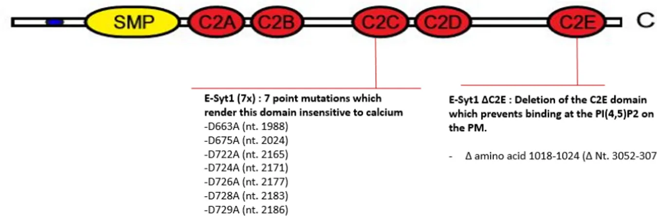
E-Syt1 is 1114 amino acids long and is anchored at the endoplasmic reticulum (ER) through an N-terminal hairpin like structure. This hairpin structure has a 30 hydrophobic amino acids stretch which enables the protein to interact with the phospholipids found at the ER membrane. Due to this hairpin conformation, the N- and C-terminal of E-Syt1 are both oriented toward the cytosolic portion of the cell. Following the hydrophobic stretch, there is a synaptotagmin-like mitochondrial lipid-binding proteins (SMP) domain that has a β-barrel conformation. It was demonstrated that this SMP domain is involved in the dimerization (homodimers of E-Syt1/1 or heterodimers of E-Syt1, 2 or 3) of the different isoforms [165]. Additionally, many studies suggest that SMPs are also implicated in the shutting of multiple types of phospholipids between two membranes through their highly hydrophobic core [166-168].
Downstream of the SMP sequence, there is five C2 domains named C2A, C2B, C2C, C2D and C2E which enable E-Syt1 to perform its functions as a calcium sensor and lipid interactor [55, 160]. The C2C domain functions as the calcium binding subunit where it interacts with the calcium present in the cytosol [169]. As a result of calcium binding, conformational changes in E-Syt1 induces the formation of homodimers which are then translocated to the ER-PM junction [169]. At this site, the C2E domain functions as the lipid binding subunit where it directly binds to phosphatidylinositol-4,5-bisphosphate (PI(4,5)P2) found at the PM [168, 169]. To this day, it is unclear what are the exact functions of C2A, C2B and C2D domains. One group suggested that C2A can also bind to Ca2+, which alleviates its inhibitory effect on the SMP domain to allow the transfer of glycerophospholipids [170].
E-Syt 2 and 3 have a similar structure as E-Syt1. They diverge in their number of C2 domains, where E-Syt 2 and 3 only have three named C2A, C2B and C2C. Nonetheless, their C2 domains performed the same functions as the ones from E-Syt1. In E-Syt2 and 3, C2A is the calcium sensor while C2C binds to the PI(4,5)P2 at the PM [161].
At rest, there are less than 1% of the PM areas where ER-PM junctions are maintained constitutively by E-Syt2/3 at about 20 nm [171, 172]. Those ER portions are referred to as cortical ER. When the cytosolic Ca2+ concentration increases and E-Syt1 gets translocated to those areas, the cortical ER expands, which increases the ER-PM junction’s surface area and the distance between the two lipid bilayers can get as close as 10 nm [173].
The illustration was modified from a recent publication [160]. The figure illustrates the different domains of wild type E-Syt1. The specific mutations used to abrogate the function of E-Syt1 are also illustrated.
1.9.3. E-Syt1 Activation
Calcium is a molecule involved in many cellular processes such as membrane depolarization, signalling pathways or apoptosis [174-176]. Consequently, calcium mobilization is tightly regulated, preventing activation of unnecessary pathways. In mammalian cells, the ER, mitochondria and Golgi apparatus are the compartments where calcium is stored, with ER being the predominant one [177-179]. In order to recruit E-Syt1 at the ER-PM junction, there must be a significant change in the cytoplasmic level of Ca2+. At rest, the cytosolic Ca2+ concentration range from 50 to 100 nM [180]. This low calcium level is tightly maintained by the activity of the sarco/endoplasmic reticulum Ca2+ ATPase (SERCA) and the plasma membrane Ca2+ ATPase (PMCA) [180, 181]. At that state, E-Syt1 is dispersed randomly throughout the ER [161]. Upon an external stimulus via various signaling molecules, phospholipase C (PLC) gets recruited and activated through the activity of G proteins or tyrosine kinase pathways [182-184]. Activated PLC hydrolyzes PI(4,5)P2 from the PM, generating soluble inositol triphosphate (IP3) and diacylglycerol (DAG). Soluble IP3 binds to the IP3 -receptor (IP3-R) found at the ER membrane outer leaflet, which triggers the release of ER-Ca2+ storage in the cytosol. The change of ER-Ca2+ concentration is rapidly detected by the ER-Ca2+ refilling mechanism that induces the translocation of STIM1 at the ER-PM junction. There, STIM1 interacts with Orai1, found at the PM, to actively pump calcium inside the cytosol from the extracellular environment. The large influx of extracellular Ca2+ induces a sustained and significant increase in the cytosolic Ca2+ concentration in the millimolar range which triggers the translocation of E-Syt1 at the ER-PM junction. This large calcium influx not only induces the ER-PM junction expansion, but also supports other cellular processes that require Ca2+ signalling, metabolism and membrane depolarization. The calcium concentration is then actively brought back to resting level through SERCA activity that replenishes the ER-Ca2+ storage and PMCA that pumps Ca2+ outside the cell. This process of extracellular calcium influx is referred as store-operated Ca2+ entry (SOCE) which is independent of E-Syt1 translocation, but is required for E-Syt1 recruitment [170, 172, 173, 185].
1.10. Calcium and HSV-1
Since calcium was demonstrated to participate in cellular trafficking and cytoskeletal rearrangement [186-188], it is not surprising that HSV-1 hijacks this process to its advantage. Indeed, it was observed that intracellular calcium increases upon HSV-1 entry [188]. When calcium elevation was blocked by pharmaceutical drugs targeting ER-Ca2+ release, viral entry diminished significantly [189]. One group even concluded that calcium release is mediated by the interaction between the viral fusogenic machinery and the cellular receptors syndecan-2, nectin-1 and integrin αV [190]. Whilst many studies provided evidence of calcium implication during viral entry, it remains unknown if calcium is required during egress, and if so, at which stage.
1.11. Hypothesis and Approach
Given previous data from our laboratory suggesting the inhibitory effect of E-Syt1 on viral propagation, the main objective of this project was to test the hypothesis that E-Syt1 negatively modulates HSV-1 egress by limiting the trafficking of newly synthesized viral particles toward the PM. We further hypothesize that this regulatory function is linked to E-Syt1’s capacity at sensing calcium and interacting with PI(4,5)P2.
To investigate the regulatory effect of E-Syt1 on HSV-1, full-length E-Syt1 along with calcium sensing and lipid binding deficient mutants were overexpressed in HSV-1 permissive cells. The viral titer was then measured to evaluate the effect of those proteins on viral egress. In parallel, the implication of calcium on HSV-1 egress was also evaluated using two different calcium chelators, EGTA and Bapta-AM, as well as the calcium-release inducer ionomycin. Those drugs were added at different time points during the infection to evaluate at which moment calcium was required. Likewise, viral titers were measured to determine the effect on viral egress.
2. Material and Methods
2.1.
Cell line
Hela (cervical adenocarcinoma) and Vero (African green monkey) cells were obtained from the American Type Culture Collection (ATCC). They were cultured in 1x Dulbecco's Modified Eagle Medium containing 5% bovine growth serum (BGS) and 1% L-Glutamine. Cells were cultured at 37°C with 5% CO2 and passaged when 90% confluent. They were also regularly tested for any mycoplasma contamination, which was absent. In all experiments, HeLa cells were used to produce the virus in different conditions (see below) while the Vero cells were employed to titer viral yields under those conditions.
2.2.
Viruses and Infection
All virus stocks were produced on Vero cells and harvested 48-72 hours post-infection (hpi). The viral titer of the supernatant and cell lysate were subsequently measured by plaque assay.
2.2.1. HSV-1 17+ WT
This virus was obtained from the laboratory of Dr. Sodeik1. It is a 17+ strain which has the wild type viral genome. Prior to the infection, Hela cells were seeded in the appropriate dish for 24 hours at 37°C with 5% CO2. The day of the infection, cells were suspended in culture medium and counted to determine the proper amount of virus corresponding to a multiplicity of infection (MOI) of 5. The viral stock was diluted in Roswell Park Memorial Institute (RPMI) media containing 0.1% bovine serum albumin (BSA) and then added to the cell monolayer. After 1 hour of absorption at 37°C and 5% CO2, complete 1x DMEM was added to the infected cells. Eighteen to twenty-four hpi, the supernatant and the cell lysate were harvested for subsequent analysis. Briefly, the supernatant was simply harvested by collecting the media. Subsequently, the cell lysate was harvested by scraping the monolayer of infected cells in 1x phosphate-buffered saline (PBS) containing a cocktail of protease inhibitors (CLAAAP).
Following a centrifugation at 500x g for 10 min at 4°C, the supernatant was removed and fresh 1x PBS containing CLAAAP was added to pellet. The pellet was then re-suspended and subjected to 3 cycles of freeze-thaw in liquid nitrogen and 37°C water bath. Both fractions were stored at -80°C for subsequent analysis.
2.2.2. HSV-1 V701
This virus was obtained from Dr. Register2. This 17+ strain virus has 2 point mutations in the UL26 protease gene [191]. Those mutations render the virus thermosensitive, which inhibits the egress of newly synthesized viral particles at 39°C but allows it at 31°C. As previously mentioned, Hela cells are seeded 24 hours prior infection. Using a MOI of 2 for the infection, complete 1x DMEM at 39°C was added after 1 hour of absorption. The infected cells were incubated at 39°C for 4 hours and then incubated at 31°C for 14 hours. Eighteen hpi, the supernatant and the cell lysate were harvested for subsequent analysis as describe previously.
2.2.3. HSV-1 K26 GFP
This virus was obtained from the laboratory of Dr. Desai3. It is derived from the KOS strain and has a green fluorescent protein (GFP) fused with the viral protein VP26. VP26 is an accessory component of capsids [192]. It is a late protein which allows the observation of circulating mature capsids inside the cell. As previously mentioned, Hela cells were seeded 24 hrs prior infection in complete 1x DMEM at 37°C on coverslip. Cells were then infected with a MOI of 5 and mounted in Hoechst-Dako 18-24 hpi. The localization of the virus inside the cells were monitored using the fluorescent Zeiss axio-imager Z2 microscope and analyzed with ZenPro software.
2 Department of Biological Chemistry, Merck Research Laboratories, West Point, PA 19486, USA.
2.3.
Plasmids
All the plasmids were produced by transformed E.Coli DH5α strain and isolated with NucleoBond Xtra midi kits (Macherey-Nagel). To confirm that there was no undesirable mutation in the gene of interest, all plasmids were sequenced.
2.3.1. EGFP E-Syt1 WT, mCherry E-Syt1 (7x) and EGFP E-Syt1
ΔC2E constructs
Those plasmids were obtained from Dr. De Camilli4 [193]. The expression of E-Syt1 was confirmed by immunofluorescence and western blot.
The first plasmid, EGFP E-Syt1WT, contains the WT version of E-Syt1 tagged with a GFP protein at its N-terminus. This modification does not affect the proper folding or the ER localization of E-Syt1 in the cell [193].
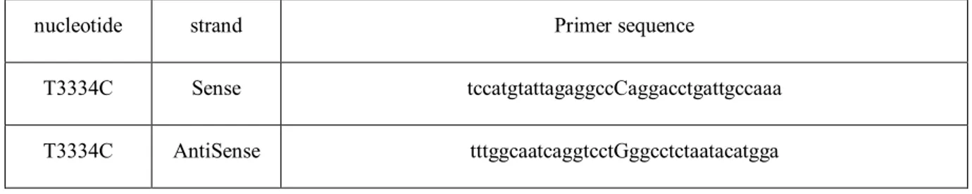
The second plasmid, mCherry E-Syt1 (7x), contains E-Syt1 tagged with mCherry at its N-terminus, which also doesn’t affect the proper folding or the ER localization of E-Syt1 in the cell [193]. This protein has seven point mutations in its C2C domain which abolish its calcium sensing function (figure 3). The seven point mutations in the C2C domains result in amino acid substitutions from aspartic acid to alanine. Upon sequencing, we identified an expected stop codon at nucleotide 3334. A site-directed mutagenesis was performed to eliminate this nonsense mutation. Briefly, the site-directed mutagenesis was performed using the QuikChange II XL Lightning site-directed mutagenesis kit (Agilent) and the primers illustrated in table 1. The qPCR experiment was performed using the following program in a Biometra Thermocycler T-Gradient Thermoblock:
- Initial denaturation: 1 cycle: 95℃, 1 min
- Amplification: 18 cycles: 95℃, 50 sec / 50℃, 50 sec / 73℃, 8.033 min - Final extension: 1 cycle: 68℃, 1 min
4 Department of Cell Biology, Program in Cellular Neuroscience, Neurodegeneration, and Repair, and Howard
Fifty nanograms of the modified mCherry E-Syt1 (7x) construct was transformed into XL10-Gold ultracompetent bacteria and the antibiotic kanamycin was used at a concentration of 50 µg/ml as the selective agent. The plasmid was extracted following the DNA extraction kit protocol (Qiagen) and a second sequencing analysis validated that there was no other unexpected mutation in the sequence.
Table 1. Primer sequences used to remove the stop codon in mCherry E-Syt1 (7x) protein
nucleotide strand Primer sequence
T3334C Sense tccatgtattagaggccCaggacctgattgccaaa
T3334C AntiSense tttggcaatcaggtcctGggcctctaatacatgga
The third plasmid, EGFP E-Syt1 ΔC2E, contains the E-Syt 1 protein tagged with a GFP at its N-terminus. This protein has a deletion of 441 nucleotides (from nucleotides 2871 to 3313) resulting in the absence of the C2E domain. A sequencing experiment validated the absence of this domain, and no other mutation was found in the sequence.
2.3.2. EGFP-N2
This plasmid was obtained from Invitrogen and was used as a DNA control for the transfected-infected cells. The expression of the GFP protein was confirmed by immunofluorescence and western blot.
2.3.3. pCyto-gBFull
This plasmid was obtained from Dr. Desjardin5’s laboratory. This plasmid was used to create the standard curve in qPCR experiments to quantify viral genome copies as it contains the full sequence of gB.
2.4.
Transfection
The transfection agent used in those experiments was Lipofectamine 3000. Twenty-four hours prior to the transfection, Hela cells were seeded in 24- or 6-well plates to obtain a 60-70% confluence. As described in the manufacturer’s protocol, the appropriate amount of DNA and lipofectamine 3000 were diluted in Opti-MEM. The DNA-Lipofectamine complex was added to the culture media containing Hela cells. Depending on the experiment, the transfected cells were either used in subsequent manipulations or mounted in Dako-Hoechst for immunofluorescent analyses 12-24 hours post transfection.
2.5.
Plaque Assay
Viral titers in the supernatant and cell lysate were determined by plaque assays. Twenty-four hours prior the titration, Vero cells were seeded in 6-well plates to obtain a confluent monolayer of cells. Samples containing an unknown amount of viruses were diluted in the appropriate amount of RPMI-0.1% BSA and added to the monolayer of Vero cells. For the supernatant, samples were diluted from 1,000 to 100,000 times the original concentration whereas the cell lysate fractions were diluted from 1,000,000 to 100,000,000 times. One hour after the absorption, 2x complete DMEM mixed with 2% agarose in a 1:1 ratio was added to the infected monolayer. Depending on the virus present in the sample, the infected cells were incubated at 37°C (HSV-1 17+ WT) or 31°C (HSV-1 V701). Three days post-infection (dpi) for the wild-type virus or 5 dpi for the V701 virus, the DMEM-agarose media was removed from the monolayer and the cells were fixed using -20°C cold 100% methanol. A solution of 0.1% crystal violet was subsequently added to the fixed cells to stain them. By taking into account the dilution and the volume plated, the initial viral titer was calculated using the number of plaque forming unit produced by the virus in each sample. The following equation was used to calculate the viral titer in each sample:
2.6.
Western Blot
E-Syt1 expression was monitored by western blot. Thirty micrograms of proteins from the cell lysate of each sample was mixed with 1x sample buffer and 0.1% β-mercaptoethanol. They were then heated at 95°C for 10 min and run on an 8% SDS-PAGE gel at 130 V. The proteins were subsequently transferred on a PVDF membrane (Bio-Rad) and blocked for an hour in a 5% milk/ 1x PBS/ 0.1% Tween20 (Sigma) solution. The primary antibody, anti-E-Syt1 (Bethyl laboratory, 1:1000), Anti-GFP (Roche, 1:1000) or anti-Gamma tubulin (Sigma, 1:10 000), was added to the membrane for an overnight incubation at 4°C. The membrane was then washed three times with a 1X PBS/0.1%Tween20 solution, and incubated with the second antibody, Goat-anti-Mouse (Jackson ImmunoResearch, 1:10 000) or Goat-anti-Rabbit (Bethyl laboratory, 1: 10 000) conjugated with horseradish peroxidase (HRP) enzyme, for 1 hour at room temperature. Proteins on the membrane were revealed using the ChemiDoc XRS+ system (Bio-Rad) after the addition of ECL substrate (Bio-(Bio-Rad).
2.7.
Drugs
Bapta-AM (Abcam) was diluted in sterile DMSO and was used at a final concentration of 10 µM and 20 µM. EGTA (Sigma) was diluted in sterile water and was used at a final concentration of 0.5 mM. Ionomycin (Sigma) was also diluted in DMSO and used at a concentration of 2 µM. Both chelators and ionomycin were directly added to the culture media at 4, 6, 12 and 16 hpi.
Phosphonoacetic acid (PAA) (Sigma) was diluted in sterile water and used at a final concentration of 0.2 µg/ml. It was directly added to the media containing Hela cells and washed off by changing media at the required time.
2.8.
Immunofluorescence
Cells seeded on coverslip were fixed with a 3% paraformaldehyde (PFA) solution and permeabilized with 0.1% triton. Thereafter, they were incubated at room temperature for 30 minutes in a blocking solution of 10% fetal bovine serum (FBS). Diluted in the same blocking solution, the primary antibody, Anti-VP5 (East Coast Bio, 1:100) or Anti-Calnexin (Thermo
Fisher, 1:50) were added to the cells for 1 hour. After three washes with 1x PBS, the secondary antibody, goat-anti-mouse Alexa 488 (Molecular Probes, 1:1000) or chicken-anti-mouse Alexa 568 (Molecular Probes, 1:1000), was added to the cells for 45 minutes. Coverslips were mounted in Hoechst-Dako on a microscope slide, and images of the cells were captured with a Zeiss axio-imager Z2 epifluorescence microscope and analyzed with ZenPro software.
2.9.
qPCR
Quantification of the viral genomic DNA in the supernatant and cell lysate was performed by quantitative Polymerase Chain Reaction (qPCR) using the Perfecta Sybr Green supermix kit (Quanta bioscience 95054-500). The gene targeted for this experiment was gB. Briefly, viral genomic DNA was extracted from each sample using the GenElute mammalian genomic DNA miniprep kit and protocol without the use of DNAse (Sigma). Following the manufacturer's protocol, the appropriate amount of Sybr Green was mixed with the extracted DNA and the primers to amplify a portion of the gB gene. To convert the Cq value into DNA copy numbers, a standard curve was also performed using a known amount of a plasmid (pCyto-gBFull) containing the sequence of gB. The qPCR experiment was performed using the following program in Light Cycler 480:
- Preincubation : 1 cycle: 95°C, 5 min
- Amplification: 45 cycles : 95°C, 5 sec / 56°C, 10 sec / 72°C, 10 sec - Melting curve : 1 cycle: 95°C, 1 sec / 65°C,1 min
2.9.1. Infectivity ratio
To calculate the infectivity ratio, the total pfu was divided by the number of total genome copies. Each treatment was normalized to the untreated condition as it represents the wild type infectivity. Values under or over 100% would imply that the resulting viral particles are less or more infectious than in the untreated condition.
2.10. Viability test
2.10.1.
Drugs
Hela cells were seeded in a 96-well Greiner black plate 24 hours prior the viability test. Cells were then treated with one of the following drugs: 0.5 mM EGTA, 10 μM or 20 μM Bapta-AM or 2 μM ionomycin. Alamar blue (Bio-Rad) was added to each condition 12 hours post-treatment and the fluorescence was measured 18 hours post-post-treatment. As a control, the viability of untreated cells was also measured.
2.10.2.
Transfection
Hela cells were seeded in a 96-well Greiner black plate 24 hours prior the viability test. Cells were then transfected as described previously in the section 2.4. Alamar blue was added 19 hours post-transfection and the fluorescence was measured 24 hours post-transfection. As a control, the viability of untreated cells was measured.
2.11. Statistics
Multiple comparisons ANOVA using the Dunnett’s test was performed to determine all statistical significance of our data. Briefly, each mean value was compared to the control condition and the probability (p) value was calculated by the software Prism8 from GraphPad. Difference between the control and the experimental condition was considered statistically significant when p value was smaller or equal to 0.05.
3. Results
3.1.
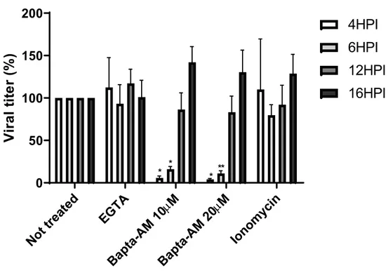
Calcium is required for HSV-1 egress
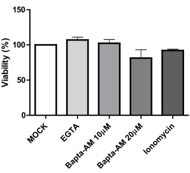
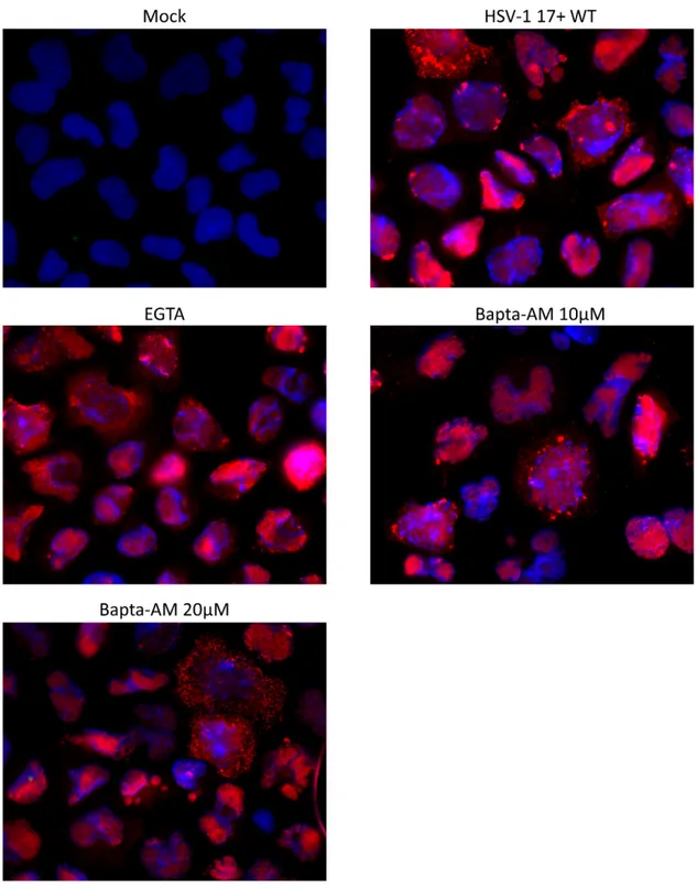
As mentioned previously, efficient viral entry is associated with transient calcium elevation inside the cells. To determine if calcium was also required during viral egress, Hela cells were infected with HSV-1 17+ WT virus at a MOI of 5 then subjected to the calcium chelator Bapta-AM (10 μM and 20 μM) or EGTA (0.5 mM) at 6 hpi. This time was selected to avoid measuring calcium impacts during the entry phase. Both chelators are very similar as they reversibly bind free calcium, but the acetoxymethyl (AM) ester derivative renders Bapta-AM cell-permeable. In fact, AM ester groups bind and neutralize the aminopolycarboxylic acid moieties of Bapta, which enable it to passively cross the PM [194, 195]. In the cytoplasm, the AM ester group is hydrolysed by cellular esterase, which enables the chelator to bind to two calcium molecules and prevent it from crossing the PM again [196]. Hence, EGTA chelates exclusively extracellular calcium in the media whereas Bapta-AM chelates cytosolic calcium. Those two calcium chelators were used at concentrations that were previously shown to significantly decrease the calcium concentration in the cytosol or the supernatant [197, 198]. Furthermore, a viability assay using Alamar blue was performed to ensure that those drug concentrations were not cytotoxic for Hela cells (figure 4). At 18hpi (i.e. 12 hours in the presence of drugs), the supernatant was harvested as described in the method section and viral titers were determined by titrating each sample on a monolayer of Vero cells. Data from figure 5 demonstrate that the presence of Bapta-AM reduced significantly the viral load in the supernatant whereas EGTA did not have much effect compared to mock-treated infected cells. Additionally, the effect of Bapta-AM was concentration dependent as the decrease of the viral load was greater with a higher concentration of Bapta-AM. Next, the location of viral particles was compared between each treatment by immunofluorescence (IF) to determine if those drugs affected the trafficking of newly synthesized viruses inside the cell. To observe de novo viral particles, an antibody against the capsid protein VP5 was employed. VP5 is detected at the nucleus at early stages during the infection as it is the site of capsid assembly [199]. During viral egress, VP5 is also detected in the cytoplasm as the newly synthesized viral particles circulate toward the PM [200,
201]. Thus, the immunodetection of VP5 concurs with the detection of viral particles as VP5 is not found as free protein in the cytoplasm. From figure 6, we can see that the viral localization was similar among each condition. Taken together, the data indicates that sequestering intracellular calcium affects viral egress without disturbing the localization of viral particles inside the cell.
MOC K EGTA µM Bapta -AM 1 0 µM Bapta -AM 2 0 Ionom ycin 0 50 100 150 Via bilit y ( % )
Fig 4. Hela cell viability is not affected by the different calcium chelators or ionomycin treatments
Hela cells were treated with 0.5 mM EGTA, 10 or 20 μM Bapta-AM or 2 μM ionomycin, as mentioned in the results section. Untreated cells (mock) were used to normalize the data (100%). Twelve hours post-treatment, Alamar blue was added to each condition and fluorescence was measured at 18 hours post-treatment. Each measurement was performed in triplicate and the average of three independent experiments is represented. Error bars represent the SEM and statistical analysis was performed by one-way ANOVA. No statistically significant differences were noted.
Not trea ted EGTA Bapt a-AM 10 Bapt a-AM 20 0 50 100 150 Vi ra l ti te r ( % )
**
***
Fig 5. Chelation of intracellular calcium inhibits viral egress
Hela cells were infected for 18 hours at a MOI of 5 with HSV-1 17+ WT virus. Drugs were added at 6 hpi and the supernatant of each condition was harvested at 18 hpi. Viral titers were measured by plaque assay (see Materials and Methods). EGTA= 0.5 mM, Bapta-AM= 10 or 20μM. n=3, error bars represent the SEM, statistical analysis by one-way ANOVA where p values are ≤ 0.05;*, ≤ 0.01; ** and ≤ 0.001; ***.
Fig 6. Viral localization inside the cell is not affected by post entry calcium level changes
Hela cells were infected for 18 hours at a MOI of 5 with HSV-1 17+ WT virus. Drugs were added at 6 hpi and infected cells were fixed and permeabilized at 18hpi. Viral particles were detected using the