Article accepted on 8/3/2006
parameters with a 50 lm resolution. A series of conditions have been
explored using skin capacitance imaging. This review summarizes
rel-evant findings about regional variability on the body, changes occurring
with ageing, effects of a hydrating formulation, reactivity kinetics of
corneocytes to surfactants, acne and skin pores characteristics, as well as
hyperkeratotic dermatoses and tumours.
Key words
:microrelief, hydration, capacitance, stratum corneum
S
ince the mid eighties, several imaging methods have been developed for non invasively studying the skin in health and disease. Some of them (ultrasound imaging, magnetic resonance imaging, confocal micros-copy, optical coherent tomography, two photons imaging) are dedicated to the structure and/or to the measurement of some of its properties.Beside those methods which allow the investigation of internal layers of the skin with resolutions varying from cell dimension to tissue dimension, some others are only dedi-cated to imaging the skin surface. Today, skin colour, tem-perature and microrelief can be routinely recorded. Quite recently, a new type of skin surface imaging was designed under the heading of Capacitance Imaging (CI). This new method allows us to picture the skin surface capacitance corresponding to the skin surface hydration.
This paper deals with the presentation of the functioning principle of skin CI, and with the description of its various domains of application.
Functioning principle
Skin CI is based on silicon image sensor (SIS) technology developed by electronic companies in order to record fin-gerprints for security reasons [1]. The sensor is composed of 92,160 microcapacitors located on a 1.8 × 1.28 cm plate measuring skin capacitance every 50 lm. These microca-pacitors are protected by a very thin silicon oxide layer. The dedicated device for skin recordings is called SkinChip® (L’Oréal, Paris) [1, 2]. It can be plugged directly to the USB port of any computer.
When the measuring plate is closely applied to the skin surface, images are produced corresponding to the hydra-tion map of the skin surface. Such images are coded in 256 gray levels with the darker pixels representing high capaci-tance and the clear ones, the lower capacicapaci-tance values. Beside the generic software of the sensor providing images, three other main softwares were developed for routinely
characterizing some specific skin parameters. The Mean Gray Level (MGL) of the image histograms allows measur-ing the mean skin surface hydration. The Corner Density (CD) parameter corresponds to the number per cm2 of crossings between the primary lines [4]. The main orienta-tions of the primary lines can also be detected.
Capacitance imaging of the skin surface
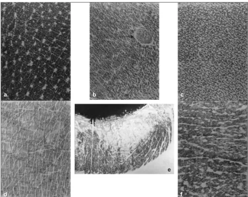
The CI method allows quite easy observation of the skin surface texture. Indeed, most of the features defining the skin microrelief (lines, pores, furrows wrinkles etc) appear in white because their bottoms are not in contact with the measuring plate [1, 3, 5-7]. The gray levels of the skin surface, which is in close contact with the measuring plate, is interpreted in terms of capacitance or water content of the stratum corneum (SC).Some typical aspects of skin CI found in adults are pre-sented in figure 1. Pilo-sebaceous openings at the skin surface of the lateral side of the neck appear as whitish objects (figure 1A). The skin of the abdomen is less studded by such pores (figure 1B). The inner side of the arm is quite protected from light and the microrelief appears very dense (figure 1C). The skin of the dorsum of the hand of an elderly person shows microrelief lines mostly oriented along one direction (figure 1D), and some whitish zones correspond to pigmented areas. The lower lip exhibits a distinct CI map (figure 1E). Fine furrows are visible. In addition, a whitish zone corresponding to a drier area is surprisingly located at the most internal part of the lip. SkinChip®was recently used for classifying lips according to their surface patterns [6]. Skin CI of forehead skin can reveal shallow frown lines (figure 1F).
As shown above the interest of skin CI is not only to routinely supply images of the skin surface patterns, but also to characterize them according to important skin sur-face properties, namely the hydration and the microrelief patterns.
doi:
Skin surface hydration
Quite a close correlation has been established between the MGL of the skin CI and the capacitance values given by a Corneometer®(C+K electronic, Cologne) [1, 3]. This is not surprising because the SkinChip®measuring plate “sees” exactly what a Corneometer® electrode captures. Both techniques establish an impaired contact with the skin surface because of its microrelief. The Corneometer®gives the average capacitance of the contact area, while the Skin-Chip® displays the repartition histogram of the values, MGL representing their mean value.
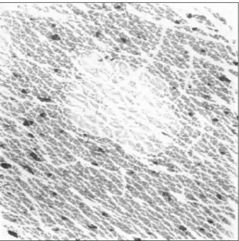
Treating dry skin with a high-performance moisturizer modifies its CI characteristics. Images become darker (more hydrated) with, in some cases, recovery of a more regular pattern of the primary lines of the microrelief (fig-ure 2). The meaning of such a phenomenon which may appear either after a single or repeated applications is presently under investigation.
Sweating is also easily observed by skin CI. At its onset, which remains clinically imperceptible, only black dots appear, marking the active sweat gland openings. This finding questions the interpretation to be given to the blind transepidermal water loss (TEWL) determinations which may indeed be influenced by imperceptible sweating. Pro-gressively, the CI black dots become larger and larger till merging to farm in a continuous black area (figure 3). Because sweat appears as black dots, it is quite easy to measure its contribution to the MGL of the skin CI by thresholding the histogram.
Another great advantage of skin CI is to supply a hydration map of the skin surface. On photo-aged skin, CI may be
heterogeneous. Some regions look quite dry, some others, just in the vicinity looking normal (figure 1D and figure 4). Such a patchy heterogeneity in hydration of the skin surface in the elderly could be related to focal variations in the epidermal differentiation of photoexposed skin.
Skin surface pattern
As shown above, the primary and secondary lines of the microrelief network can easily be viewed by CI. Of course,
a b c
f d
e
Figure 1.Capacitance imaging of the skin surface of six anatomical sites. A) Lateral side of the neck with numerous pore openings, B) Abdomen. The arrow points to a ruby angioma appearing as a circumscribed lesion with an altered pattern of skin line network. C) Inner aspect of the arm with a very dense network of microrelief lines. D) Dorsum of the hand with a parallel pattern of lines. E) Lower lip, the inner portion appears drier than the outer part. F) Skin of the forehead with many shallow frown lines.
T0 T21
Figure 2.Aspect of the skin of the dorsal side of the forearm of an elderly man before and after 21 days of applications of moisturizer. The texture of the skin is improved after treat-ment. CI values are 154 and 234/cm2at T0 and T21,
only the 2D network can be characterized through CI. On the forearm, CD varies from about 250 to 400/cm2 accord-ing to age. This findaccord-ing is in agreement with previous findings [8-10], showing a decrease in the microrelief line density with ageing.
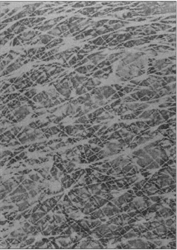
Another way to explore skin ageing using CI is to display the main orientations of the microrelief lines. CI can indeed routinely show the two main perpendicular orientations of the skin microrelief and their rotation when the skin is stressed (figure 5). This is in line with previous observa-tions made on replicas [8].
Other elements of the skin microrelief (pores, wrinkles) are also imaged and can be manually quantified [7].
Surfactant-induced skin reaction
The dynamics of stratum corneum reactivity to surfactants is complex. Surfactants present in hygiene and skin care products are in part adsorbed at the skin surface [11], and they also permeate the SC where they interact with proteins and lipids. A number of physicochemical interactions exist between corneocytes and surfactants [12]. One of the ear-liest events following surfactant-induced protein denatur-ation is perceived as corneocyte swelling [13]. This condi-tion leads to a paradoxical and transient SC hydracondi-tion following surfactant challenge in vivo [14]. The structure and physical properties of the SC can be altered profoundly following prolonged contact with anionic surfactants [13, 15, 16]. As a consequence, minimal to severe irritation may
develop with variable severity. Full-blown lesions show inflammatory erythema, increased TEWL, altered cutane-ous microrelief, increased SC roughness and erratic desquamation. Some of the changes can be assessed using non-invasive instrumental methods. In particular, the SC water content can be assessed in vivo using devices mea-suring changes in electrical properties of skin at different frequencies and at different depths inside the SC [17-19]. Skin CI has an added value to the conventional methods of assessment. Indeed, the sensitivity of skin CI allows the disclosure of focal and minute changes that are blurred by methods averaging data on a relatively large area corre-sponding to the size of the measuring probe. In addition, the CI method allows us to see the invisible sweating that interferes with any global electrometric assessment and supposedly TEWL measurement.
Two experimental studies have been performed to assess the discrete effects of mild surfactants on human SC [20, 21]. One study used the short-term patch-testing method [20]. The other one used an open method close to the in-use conditions [21]. Both procedures disclosed the early step of corneocyte swelling induced by surfactants (figure 6A). Delayed assessments after a couple of hours, as well as repeated surfactant insults showed a second event charac-terized by a skin surface drying effect (figure 6B). A corre-lation was found with data gained by the corneosurfametry bioassay [21].
Figure 3.Onset of the sweating process. Black spots corre-spond to very high capacitance zones due to the presence of
sweat. Figure 4
.Uneven CI aspect of the photoaged skin of the forearm.
Skin adnexal disorders
Skin pore is a dermocosmetic term which does not encom-pass one single feature. In the dermatological language, it is replaced to the best advantage by acroinfundibulum and acrosyringium to distinguish the openings of the folliculo-sebaceous ducts and the sweat gland apparatus, respec-tively. Skin CI is a rapid and sensitive method revealing the acrosyringia of discretely active glands. The same approach can reveal the open and the keratin-filled funnel-like
acro-infundibular structures [7, 22]. These structures are re-vealed as whitish low capacitance spots due to the absence of contact between the probe and the epithelial lining of each empty infundibulum, or to the dry nature of a micro-comedo.
Acne is a typical skin condition where skin capacitance imaging can highlight the heterogeneous patchwork of the electrical properties of the skin. Among the typical pin-point pattern of normal-looking pores, microcomedones and open comedones appear as larger low-capacitance ob-jects. When inflammation is present, the papules appear as
A: Young Skin: two equivalent maxima at about 90° B: SkinChip 195.94 190.00 185.00 180.00 Occurence 175.00 170.00 167.44 0 20 40 60 80 Angle (°) 100 120 140 160 180 138° 46°
Figure 5.Capacitance imaging of the skin microrelief. A) The two main orientations of the primary lines on the volar forearm of an adult. B) Automatic presentation of the capacitance values given by the SkinChip®softaware.
A
B Figure 6.Cornecoytes reactivity to surfactants. A) Immediate
corneocyte swelling characterized by a darker CI aspect. B) Delayed corneocyte drying characterized by a whitish CI aspect.
surrounded by a darker rim revealing a weakened skin barrier function and the presence of a discrete serosity exsudate [23].
Hyperkeratotic non-tumoral dermatoses
Epidermal hyperkeratosis is a typical feature of pityriasis versicolor. The condition is easily highlighted by skin CI because the skin surface is dryer than the surrounding skin (figure 8). Interestingly enough, the lesional skin appears anhidrotic, perhaps due to the occlusion of the acrosyringia [24]. The method allows the detection of small lesions of pityriasis versicolor almost invisible to the naked eye. Psoriasis is the paradigm of the inflammatory hyperkera-totic dermatoses. Skin CI reveals a map of heterogeneous electrical properties on lesional skin [25]. Whitish low capacitance is characteristic for stable hyperkeratotic plaques. More inflammatory and evolving plaques show darker high capacitance spots (figure 9). This aspect is most likely related to sites exhibiting increased TEWL [26]. Skin CI can thus provide clues of disease activity in the plaque stage of psoriasis.Benign keratotic or pigmented tumours
Viral warts are typically hyperkeratotic. They are easily identified using skin CI (figure 10) exhibiting the dry aspectFigure 9.Psoriatic lesion combining white hyperkeratotic areas and darker inflammatory sites.
these lesions (figures 11A and B). Low capacitance is commonly yielded, but increased capacitance is also pos-sible, particularly on minimally inflammed lesions [27].
Conclusion
Skin CI is a novel procedure allowing both visualization and quantification of the skin microrelief, SC hydration and imperceptible sweating. The method brings sound informa-tion in dermocosmetology. It also brings insights into phys-iopathological disorders revealing some unexpected fea-tures. j
References
1. Lévêque JL, Querleux B. SkinChip®, a new tool for investigating
the skin surface in vivo. Skin Res Technol 2003; 9: 343-7.
2. Piérard GE, Lévêque JL. What is SkinChip®? From silicon image
sensor technology to SkinChip®. Dermatology 2004; 208: 291-2.
3. Batisse D, Giron F, Lévêque JL. Capacitance imaging of the skin
surface. Skin Res Technol (in press).
4. Berardesca E, Primavera G, Zahouani H, Lévêque JL.
Capaci-tance imaging: new parameters for characterizing the skin surface texture, effect of hydration. Skin Res Technol 2005; 11: 293.
5. Piérard GE, Uhoda I, Piérard-Franchimont C. From skin
microre-lief to wrinkles: an area ripe for investigations. J Cosmet Dermatol 2003; 2: 21-8.
6. Lévêque JL, Goubanova E. Influence of age on lips and the
perioral skin. Dermatology 2004; 208: 307-13.
7. Uhoda E, Piérard-Franchimont C, Petit L, Piérard GE. The
conun-drum of skin pores in dermocosmetology. Dermatology 2005; 210: 3-7.
8. Corcuff P, de Rigal J, Makki S, Lévêque JL, Agache P. Skin relief
and ageing. J Soc Cosmet Chem 1983; 34: 177-90.
9. Piérard-Franchimont C, Piérard GE. Skin surface strippings in
diagnosing and monitoring inflammatory, xerotic and neoplastic diseases. Ped Dermatol 1985; 2: 180-4.
Figure 10.Viral wart of the foot.
A
B
Figure 11.Keratotic pigmented tumours. A) Seborrheic kera-tosis. B) Moderately inflamed melanocytic naevus.
ric assessments of the impact of surfactants on forearm skin. Exog
Dermatol 2003; 2: 64-9.
17. Berardesca E. EEMCO guidance for the assessment of stratum
corneum hydration: electrical methods. Skin Res Technol 1997; 3: 126-32.
18. Fluhr JW, Gloor V. Comparative study of five instruments
mea-suring stratum corneum hydration. Skin Res Technol 1999; 5: 171-8.
capacitance mapping of psoriasis. J Eur Acad Dermatol Venereol (in press).
26. Goon ATJ, Yosipovitch G, Chan YH, Goh CL. Barrier repair in
chronic plaque-type psoriasis. Skin Res Technol 2004; 10: 10-3.
27. Xhauflaire-Uhoda E, Piérard GE. Contrasted skin capacitance
imaging of seborrheic keratoses and melanocytic naevi.