A
NTIMICROBIALA
GENTS ANDC
HEMOTHERAPY,
0066-4804/98/$04.0010
Feb. 1998, p. 228–230
Vol. 42, No. 2
Copyright © 1998, American Society for Microbiology
Chromogenic Detection of Aminoglycoside Phosphotransferases
ANA M. AMOROSO
ANDGABRIEL O. GUTKIND*
Laboratorio de Resistencia Microbiana, Ca´tedra de Microbiologı´a, Facultad de Farmacia y Bioquı´mica,
Universidad de Buenos Aires, Buenos Aires, Argentina
Received 14 May 1997/Returned for modification 14 August 1997/Accepted 18 November 1997
A coupled chromogenic reaction (based on an agar overlay combining NADH, pyruvate kinase, lactate
dehydrogenase, phosphoenolpyruvate, ATP, and kanamycin sulfate with thiazolyl blue-phenazine methosulfate
for detection of NADH consumption) was optimized for the detection of aminoglycoside phosphotransferases
(APHs). When used after analytical isoelectrofocusing of bacterial extracts from APH-producing strains, this
method revealed one band in each of two strains with a genetically confirmed APH (3
*) I and two bands in
another strain with both APH (3
*) I and APH (3*) VI, whereas no bands were detected in susceptible control
strains or in aminoglycoside-resistant microorganisms without APH genes.
The most common mechanism of acquired resistance to
aminoglycosides in clinical isolates is that of
aminoglycoside-modifying enzymes (3). These enzymes are capable of three
general reactions: N acetylation, O nucleotidylation, and O
phosphorylation (1). The modifying enzymes present in
resis-tant bacteria may be studied by examining the resistance
phe-notype with substrate or nonsubstrate antibiotics, which merely
provides an indication of enzyme types (5). However, as
sev-eral different enzyme types (6) and isozymes may occur in an
individual isolate, attempts to recognize each component may
be difficult, since several resistance phenotypes may be
super-imposed.
Resistance phenotypes and hybridization with DNA probes
specific for known enzymes provide a reliable assessment of
resistance in an isolate (2). However, as the complete set of
probes is available to only a few laboratories around the world,
most laboratories are unable to test for them. Moreover, any
putative new enzyme would be detected only after failing to
hybridize to all known probes while carrying a new resistance
pattern or a resistance pattern resembling that corresponding
to a known gene.
In this study, an attempt was made to develop a coupled
chromogenic system to detect aminoglycoside
phosphotrans-ferase (APH) activity in cell extracts after separation of the
enzymes by analytical isoelectrofocusing, which is currently
used for
b-lactamase characterization.
MATERIALS AND METHODS
Resistant bacteria with phenotypically and genetically characterized enzymes were kindly provided by A. Rossi from the Institute C. G. Malbra´n, Buenos Aires, Argentina. The strains and the genes encoding the modifying enzymes are as follows: Proteus mirabilis INM 8628, aph (39)I 1 ant (29); P. mirabilis INM 900,
aph (39)I 1 aph (39)VI 1 ant (29); Escherichia coli INM 7251, aph (39)I 1 aac (69); E. coli ATCC 11105, none; and P. mirabilis CCMA-29 1157, none (INM, Instituto Nacional de Microbiologı´a Carlos Malbra´n; ATCC, American Type Culture Collection; CCMA-29, Coleccio´n de Cultivos Microbianos, [Facultad de Farmacia y Bioquı´mica, Universidad de Buenos Aires]).
Bacterial cells were grown to the late logarithmic phase in 500 ml of brain heart infusion (Merck Quimica Argentina) at 37°C in the presence of 50mg of kanamycin (Armstrong) per ml, harvested by centrifugation (6,0003 g, 30 min, 4°C), resuspended in 10 mM Tris HCl–10 mM magnesium acetate–25 mM ammonium chloride, pH 7.8 (all chemicals were purchased from Merck Quimica
* Corresponding author. Mailing address: Laboratorio de
Resisten-cia Microbiana, Ca´tedra de Microbiologı´a, Facultad de FarmaResisten-cia y
Bioquı´mica, Universidad de Buenos Aires, Junı´n 954, Piso 8, 1113
Buenos Aires, Argentina. Phone: 54 1 964 8285. Fax: 54 1 962 5341.
E-mail: ggutkind@huemul.ffyb.uba.ar.
FIG. 1. Enzymatic reactions used for APH activity detection.228
on February 29, 2016 by UNIV DE LIEGE
http://aac.asm.org/
Argentina), collected by centrifugation, and resuspended in the same buffer. The suspension was ice cooled and disrupted with six 30-s bursts in a Vibracell VC 500 sonicator. Cell extracts were then clarified by centrifugation (15,0003 g, 30 min, at 4°C), and the supernatants were concentrated to 1 ml by ultrafiltration through an Amicon 10 membrane.
Enzymatic activity was confirmed by incubating extracts with a small amount of kanamycin and ATP and monitoring the loss of drug activity by a standard disk method (2).
Biochemical detection of phosphotransferase activity in the concentrated su-pernatants was carried out by a coupled colorimetric reaction as follows. The melted reaction medium thermostated to 42°C contained 0.9% agar, 10 mM Tris HCl, 10 mM magnesium acetate, and 25 mM ammonium chloride, pH 7.8. The following freshly prepared solutions were added to obtain the indicated final concentrations: NADH, 16 mM; pyruvate kinase, 76 IU; lactate dehydrogenase, 640 IU; phosphoenolpyruvate (32 mM; ATP, 8 mM (all purchased from Sigma Chemical Company), as well as kanamycin sulfate, gentamicin, or neomycin (0.5 to 2.3 mM) (kindly supplied by Armstrong) by adding concentrated solutions (4). For qualitative detection, the mixture was poured onto glass plates. Once it had solidified, a disk containing 10ml of the concentrated supernatant or the same amount of an extract lacking phosphotransferase genes (negative control) was poured on and incubated for an hour in darkness at 37°C. A 10 mM thiazolyl blue (MTT) (Sigma)–1 mM phenazine methosulfate (PMS) (Sigma) water solution (7) was added and allowed to react at room temperature, until a blue color became evident (between 1 and 5 min). The enzymatic cascade is presented in Fig. 1. Analytical isoelectrofocusing of the concentrated supernatant was carried out with Pharmacia precast gel Inmobiline dry plates, pH 4.0 to 7.0 (Pharmacia Biotech, Uppsala, Sweden) according to the manufacturer’s instructions.
Local-ization of phosphotransferase activity was carried out by pouring the reaction mixture onto the gels, as described above.
RESULTS AND DISCUSSION
Figure 2 shows preliminary biochemical detection of
phos-photransferase activity in crude extracts, using neomycin 1 mM
as substrate. The same results were obtained when gentamicin
was used at the same or higher concentration (data not shown).
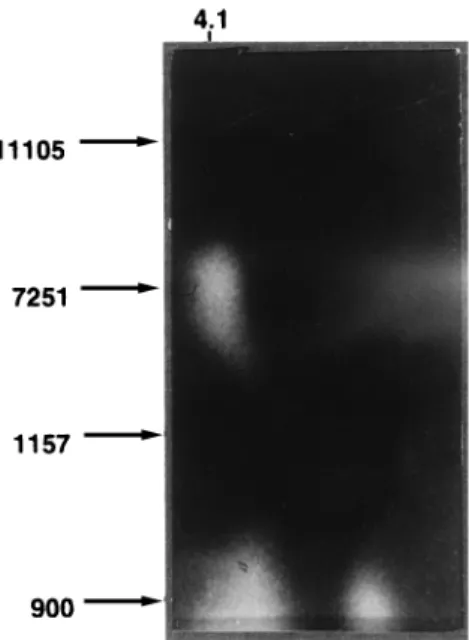
As shown in Fig. 3, at an apparent pI of roughly 4.1, one
active band appeared in two strains known to express a single
phosphotransferase. A second band was observed at pI 4.4 in a
strain harboring two such enzymes (Fig. 4). The band at 4.4 is
in good agreement with the predicted pI value (8).
As expected from a reaction with an overall consumption of
aminoglycoside, phosphoenolpyruvate, and MTT-PMS
(re-duced form) and production of aminoglycoside phosphate,
lac-tate, and MTT-PMS (oxidized form), in the presence of the
specific enzymatic system, only extracts with APHs were
posi-tive by this method. Although the enzymatic cascade
theoret-ically might be less specific due to the action of other NADH
or ATP-consuming or -producing systems, the absence of a
suitable substrate in the reaction mixture precluded these
re-actions. Moreover, control extracts from strains lacking APH
activity produced no decolorization. Furthermore, the
wide-spread aminoglycoside acetyltransferase-producing strains in
Argentina, most of which lack APH genes, also did not show
any nonspecific reaction (data not shown).
Although only three APH-producing strains have been
an-alyzed so far, the lack of nonspecific reactions and the
detec-tion of two active bands in the strain with two different APH
genes suggest that this will be a useful method especially for
the analysis of clinical isolates where phenotypic resistance
cannot be attributed to any particular enzyme or isozyme.
Hopefully, it may provide an easy and fast method for
semi-quantitative studies of APH activity in strains encoding the
same (or different) information at the DNA level or presenting
dissimilar expression. Lastly, our approach may also be used to
FIG. 2. Ten microliters of an extract of E. coli INM 7251 was poured onto disk A. The same amount of a nonresistant strain (E. coli ATCC 11105) was poured onto disk B. Phosphotransferase activity was detected as described in the text.
FIG. 3. Twenty microliters from each bacterial extract was electrofocused on Inmobiline dry plates (pH 4.0 to 7.0) at 2,000 V for 3.5 h. Phosphotransferase activity was detected as described in the text. Strains are indicated on the left.
FIG. 4. Twenty microliters from each bacterial extract was electrofocused on Inmobiline dry plates (pH 4.0 to 7.0) at 2,000 V for 3.5 h. Phosphotransferase activity was detected as described in the text. Strains are indicated on the left.
V
OL. 42, 1998
CHROMOGENIC DETECTION OF APHs
229
on February 29, 2016 by UNIV DE LIEGE
http://aac.asm.org/
detect presumptively novel enzymes, when specific probes are
not yet available.
ACKNOWLEDGMENTS
This work was supported, in part, by grants from UBACYT and
CONICET to G.O.G.
We thank A. Rossi for providing resistant strains previously
charac-terized by DNA hybridization to aminoglycoside-modifying enzyme
probes, which belonged to a group of local resistant strains analyzed by
Shaw et al. (8) at the Schering-Plough Research Institute (Bloomfield,
N.J.).
REFERENCES
1. Bryan, L. E. 1984. Aminoglycoside resistance, p. 241–277. In L. Bryan (ed.), Antimicrobial drug resistance. Academic Press, San Diego, Calif. 2. Davies, J. E. 1991. Aminoglycoside aminocyclitol antibiotics and their
modi-fying enzymes, p. 691–713. In V. Lorian (ed.), Antibiotics in laboratory med-icine. Williams and Wilkins, Baltimore, Md.
3. Foster, T. J. 1983. Plasmid determined resistance to antimicrobial drugs and toxic ions in bacteria. Microbiol. Rev. 47:361–409.
4. Goldman, P., and D. Northrop. 1976. Purification and spectrophotometric assay of neomycin phosphotransferase II. Biochem. Biophys. Res. Commun. 69:230–236.
5. Miller, G. H., F. J. Sabatelli, R. S. Hare, and J. A. Waitz. 1980. Survey of aminoglycoside resistant patterns. Dev. Ind. Microbiol. 21:91–104. 6. Rossi, M. A., G. O. Gutkind, M. Quinteros, M. Marino, E. Couto, M.
Toku-moto, M. Woloj, G. Miller, and A. Medeiros.1991. A Proteus mirabilis with a novel extended spectrum beta-lactamase and six different aminoglycoside resistant genes, abstr. 939. In Program and abstracts of the 31st Interscience Conference on Antimicrobial Agents and Chemotherapy. American Society for Microbiology, Washington, D.C.
7. Shaw, C. R., and R. Prasad. 1970. Starch gel electrophoresis of enzymes: a compilation of recipes. Biochem. Genet. 4:297–320.
8. Shaw, K. J., P. N. Rather, R. S. Hare, and G. H. Miller. 1993. Molecular genetics of aminoglycoside resistance genes and familial relationships of the aminoglycoside modifying enzymes. Microbiol. Rev. 57:138–163.