Université de Montréal
Étude de l’interaction entre le champignon mycorhizien Glomus irregulare et les bactéries du sol
Par Julie Lecomte
Département de sciences biologiques Faculté des arts et des lettres
Mémoire présenté à la Faculté des études supérieures en vue de l’obtention du grade de M. Sc.
en sciences biologiques
Juillet 2010
Université de Montréal Faculté des études supérieures
Ce mémoire intitulé :
Étude de l’interaction entre le champignon mycorhizien Glomus irregulare et les bactéries du sol
présenté par : Julie Lecomte
a été évaluée par un jury composé des personnes suivantes :
David Morse, président-rapporteur Mohamed Hijri, directeur de recherche
Marc St-Arnaud, co-directeur Anja Geitmann, membre du jury
R
ÉSUMÉ
Dans cette étude, nous avons isolé et cultivé des bactéries intimement liées aux spores du champignon mycorhizien Glomus irregulare prélevées dans la rhizosphère de plants d’Agrostis stolonifera L. récoltés dans un sol naturel. Le séquençage des 29 morphotypes isolés a révélé la présence de seulement sept taxons bactériens (Variovorax paradoxus, Microbacterium ginsengiosoli, Sphingomonas sp., Bacillus megaterium, B. simplex, B. cereus et Kocuria rhizophila). Des isolats de chacun de ces sept taxons ont ensuite été cultivés in vitro sur le mycélium de G. irregulare afin d’observer par microscopie leur capacité à croitre et à s’attacher au mycélium en absence d’éléments nutritifs autres que ceux fournis par le champignon. Tous les isolats, sauf B. cereus, ont été capables de bien croitre dans le système expérimental et de s’attacher au mycélium en formant des structures ressemblant à des biofilms sur la surface du champignon. Toutefois, B. simplex formait ces structures plus rapidement, soit en 15 jours, alors que les autres isolats les ont formés après 30 jours (K. rhizophila et B. megaterium) ou 45 jours (V. paradoxus, M. ginsengiosoli et Sphingomonas sp.). D’autre part, la technique PCR-DGGE a permis d’analyser la diversité bactérienne associée aux spores. La diversité des taxons associés aux spores de G. irregulare qu’il a été possible d’isoler et de cultiver in vitro a été nettement moindre que celle qui était présente sur la surface des spores,
alors que la biodiversité bactérienne totale du sol a été encore beaucoup plus élevée. Les bactéries associées aux champignons mycorhiziens jouent probablement un rôle important dans la capacité des plantes à résister aux stress biotiques et abiotiques auxquels elles sont soumises.
M
OTS-
CLÉS:
Champignons mycorhiziens à arbuscule (AMF), bactéries, biodiversité, biofilm, microscopie.
A
BSTRACT
In this study, we isolated and cultivated bacterial cells intimately associated with Glomus irregulare spores in a natural soil Agrostis stolonifera rhizosphere. Sequencing of the 29 morphotypes isolated revealed the presence of only seven bacterial taxa (Variovorax paradoxus, Microbacterium ginsengiosoli, Sphingomonas sp., Bacillus megaterium, B. simplex, B. cereus and Kocuria rhizophila). These seven isolates were cultivated in vitro on the mycelium of G. irregulare to allow microscopic observation of growth and attachment to the mycelium in absence of nutritive sources other than those derived from the fungal mycelium. All isolates but B. cereus were able to grow on the experimental system and to attach to the mycelium to form biofilm-like structures on their surface. However, B. simplex formed these structures more quickly, in 15 days, than the remaining isolates that have formed them only after 30 days (K. rhizophila and B. megaterium) or 45 days (V. paradoxus, M. ginsengiosoli and Sphingomonas sp.). In addition, PCR-DGGE was used to compare bacterial diversity. The bacterial biodiversity associated with spores of G. irregulare that were isolated and cultured in vitro was significantly lower than that present on the spore surface, while total soil bacterial diversity was much higher. The bacteria associated with mycorrhizal fungi probably have an
important role in the ability of plants to withstand biotic and abiotic stresses to which they are submitted.
KEYWORDS :
Arbuscular mycorhizal fungi (AMF), bacteria, biodiversity, biofilm, microscopy.
T
ABLE DES MATIÈRES
Résumé ... i
Mots-clés : ...ii
Abstract ... iii
Keywords : ... iv
Table des matières ... v
Liste des tableaux ... ix
Liste des figures ... x
Liste des abréviations...xiii
Remerciements ... xviii
Avant-propos ... xx
Chapitre 1. Introduction ... 1
Symbioses mycorhiziennes ... 1
Champignons mycorhiziens à arbuscules ... 6
Biofilms bactériens ... 10
Observations microscopiques ... 12
Analyse et comparaison de la biodiversité microbienne ... 14
Hypothèses ... 15
Objectifs ... 16
Chapitre 2. Spatio-temporal interactions between soil bacteria and arbuscular mycorrhizal fungi ... 17
Abstract ... 17
Introduction ... 19
Material & Method ... 22
Sampling ... 22
Assessment of root mycorrhizal colonisation ... 23
Isolation and identification of Glomus irregulare spores harvested from soil ... 23
Isolation of bacteria from spore surface ... 24
Identification of isolated bacteria ... 25
Bacterial biodiversity associated with G. irregulare spores ... 26
Assessment of interactions between G. irregulare and bacteria ... 27
Results ... 28
Assessment of bacterial biodiversity... 32
Interactions between bacteria and G. irregulare mycelium. ... 34
Discussion ... 38
Acknowledgments ... 44
Chapitre 3. Discussion et conclusion générale ... 45
Réferences ... 50
Annexe 1 ... 67
Observation des interactions entre bactéries et mycélium de champignon mycorhizien à arbuscules par microscopie confocale ... 67
Matériel et méthode ... 70
Transformation des bactéries avec des gènes codant pour des protéines fluorescentes ... 70
Mise en interaction des bactéries transformées avec le mycélium de Glomus irregulare ... 73
Observation des bactéries transformées au microscope confocal ... 74
Résultats ... 76
Discussion... 87
Annexe 2 ... 90
Matériel et méthode ... 91
Biodiversité et identification des bactéries ... 91
Résultats ... 93
Discussion... 100
Annexe 3 - Milieux de culture ... 102
Milieu M ... 102
Milieu Eau ... 106
Milieu de congélation des bactéries ... 107
Annexe 4 - Marqueur utilisé pour le DGGE ... 108
L
ISTE DES TABLEAUX
Tableau I Les différents types de symbioses mycorhiziennes, les champignons impliqués, leurs plantes hôtes, les structures formées et leurs impacts physiologiques…………...………..….4
Table II Bacterial taxa to which belong the 29 bacterial isolates recovered from Glomus irregulare spores isolated from natural soil and identified by sequencing of a 16S rRNA gene fragment...29
Table III Growth and attachment onto Glomus irregulare mycelium of bacteria isolated from field-harvested AM fungus spores, on a water media at 25 ºC after 15, 30 and 45 days of incubation...35
L
ISTE DES FIGURES
Figure 1 Classification phylogénétique du règne des Mycota (Fungi) basée sur l’analyse des séquences nucléotidiques du gène 18S de l’ARNr………...………..5
Figure 2 Classification phylogénétique du phylum Glomeromycota...7
Figure 3 A- Morphology of spores isolated from soil. B and C -
Patterns of mixed bacterial colonies growing from washed Glomus irregulare spores extracted from natural soil and incubated 1 month on G. irregulare. isolate DAOM 197198 hyphae growing in vitro on a gellan gum medium without nutrients...30
Figure 4 DGGE pattern of V9 portion of 16S rRNA gene...33
Figure 5 Bacterial growth patterns on Glomus irregulare hyphae cultivated in vitro observed on DIC microscope using 63X objective. Panel A, Bacillus simplex; B, Kocuria rhyzophila; C, Bacillus megaterium; D, Variovorax paradoxus; E, Sphingomonas sp; F, Microbacterium ginsengisoli; G, Pseudomonas sp.; and H, E. coli...35
Figure 6 Plasmides utilisés pour les transformations des bactéries par électroporation...72
Figure 7 Schéma du système expérimental utilisé pour la croissance des bactéries transformées avec les protéines fluorescentes sur les hyphes du champignon mycorhizien G. irregulare...74
Figure 8 A. Bactéries B. cereus exprimant la protéine «DsRed». B et C. Bactéries Sphingomonas sp. exprimant les protéines «enhance cyan» et «enhance green» respectivement. D. Bactérie Pseudomonas sp. exprimant la protéine «enhance yellow»...77
Figure 9 Photographie de B. simplex poussant sur G. irregulare…...79
Figure 10 Photographie de B. megaterium poussant sur G. irregulare..80
Figure 11 Photographie de Sphingomonas sp. poussant sur G. irregulare...81
Figure 12 Photographie de Microbacterium ginsengiosoli. poussant sur G. irregulare...82
Figure 13A Photographie de Variovorax paradoxus poussant sur G. irregulare...83
Figure 13B Photographie de Variovorax paradoxus poussant sur G. irregulare...84
Figure 14 Photographie de Kocuria rhizophila poussant sur G. irregulare...85
Figure 15 Photographie d’E. coli poussant sur G. irregulare…………86
Figure 16 Photographie d’un gel d’électrophorèse des produits de PCR du gène de l’ARNr 16S montrant une bande d’environ 1500 pb………94
Figure 17 Photographie d’un gel d’électrophorèse des produits de PCR correspondant à la portion V3 du gène de l’ARNr 16S montrant une bande d’environ 200 pb………...95
Figure 18 Photographie d’un gel DGGE des produits PCR correspondant à la portion V3 du gène de l’ARNr 16S montrant des bandes d’environ 200 pb..………...98
Figure 19 Photographie d’un gel DGGE des produits PCR correspondant à la portion V3 du gène de l’ARNr 16S montrant des bandes d’environ 200 pb. ...99
L
ISTE DES ABRÉVIATIONS
ADN Acide désoxyribonucléique
ADNr ADN ribosomique
AMF Champignon mycorhizien à arbuscule
BLASTn Nucleotide blast (Basic Local Alignment Search Tool)
BSA Bovine serum albumine
CFU Colony-forming unit
DGGE Denaturing gradient gel electrophoresis
DO Densité optique
DIC Differential interference contrast (microscopy)
DMSO Dimethyl sulfoxide
DNA Deoxyribonucleic acid
dNTP Deoxyribonucleotide triphosphate / désoxyribonucléotide tri-phosphate
FBSB Fonds de bourses en sciences biologiques, Université de Montréal
g Force de gravité
GI Glomus irregulare (intraradices)
GPS Global Positioning System
INVAM International Culture Collection of Arbuscular & Vesicular-Arbuscular Mycorrhizal Fungi
kb Kilobase
kV Kilovolts
LB Milieu Luria Bertani/ Luria Bertani Media
mL Mililiter / mililitre
N North
NCBI National Center for Biotechnology Information
ng Nanogramme
Pb Paire de base
PCR Polymerase chain reaction / Amplification en chaîne par polymérase
rDNA Ribosomal deoxyribonucleic acid
Ri Root inducing
RPM Rotation par minute
Taq Thermostable DNA polymerase named after the thermophilic bacterium Thermus aquaticus
TSA Tryptone soy Agar media / milieu tryptone soya agar
TSB Tryptone soy broth / bouillon tryptone soya
T-DNA Transferred DNA of the root-inducing (Ri) plasmid
U Unit
W West
w/v Weight/ volume
µl Microlitre
µg Microgramme
µm Micromètre
µM Micromolaire
Le microbe n'a pas le temps d'examiner le biologiste. [Henri Michaux]
La science, comme l'amour, est aveugle. Voilà pourquoi elle se plaît à procéder par tâtonnements.
[Jean O'Neil]
La science progresse en indiquant l'immensité de l'ignoré. [Louis Pauwels]
R
EMERCIEMENTS
Je veux d’abord remercier mes directeurs de recherche, Mohamed Hijri et Marc St-Arnaud, pour m’avoir confié cet intéressant projet ainsi que le support nécessaire pour le mener à bien. Ce travail a été possible grâce aux fonds CRSNG octroyés à M. Hijri et M. St-Arnaud. Un support financier a aussi été obtenu par la bourse du département de sciences biologiques de l’Université de Montréal (FBSB).
Je tiens aussi à remercier particulièrement Hugo Germain pour sa patience, ses explications en réponse à mes nombreuses questions et son aide avec la technique d’électroporation. Un merci tout spécial aussi à Marie-Soleil Beauregard pour l’aide avec la technique DGGE et le support moral. Merci à Marie-Pierre Gauthier et Sugir Selliah pour l’aide avec l’optimisation de la PCR, à Maureen Marie-Joseph, stagiaire française à l’été 2007, pour l’aide précieuse avec l’extraction des spores du sol et à Éric Chevalier pour ses conseils sur l’utilisation du microscope. Merci à Éric Claeyssen pour son écoute, ses conseils et son support moral. Je n’oublie pas non plus tous les autres membres de notre laboratoire et de l’IRBV sans qui mon séjour n’aurait pas été aussi agréable.
Pour terminer, je ne peux passer sous silence l’appui et les encouragements constants de mon amoureux Frédéric et la joie de vivre que nous procure notre merveilleuse petite Béatrice. Merci aussi à la famille Lecomte, particulièrement mon père Pierre, ma mère Jeannette et mes tantes Lorraine et Michèle, ainsi qu’à mes amis qui ont su me soutenir dans les moments les plus difficiles pour que je persévère dans ce projet.
A
VANT
-
PROPOS
Ce mémoire a été rédigé par articles. Les résultats présentés dans le deuxième chapitre ont donc été rédigés en anglais sous forme d’article scientifique. Le projet a été entamé à l’été 2006 avec l’échantillonnage de sol sur le terrain. Les manipulations de laboratoire et la recherche bibliographique ont aussitôt suivi. Mon directeur, Mohamed Hijri, et mon codirecteur, Marc St-Arnaud, ont su me guider à travers ce processus complexe, tout en me laissant libre dans mes choix. L’élaboration des dispositifs expérimentaux, toutes les manipulations de laboratoire et la rédaction de l'article ont été effectuées par moi en tant qu’auteure principale, avec les conseils avisés de messieurs St-Arnaud et Hijri en plus de leur soutient financier (CRSNG) au projet.
Le premier chapitre du mémoire inclut l’introduction avec la revue de littérature et la présentation des hypothèses de recherche. Le second chapitre est l’article comme tel. Il traite de la visualisation individuelle de chacune des bactéries du sol isolées lorsqu’associées à la surface du champignon mycorhizien G. irregulare par microscopie visible. Le troisième chapitre est une conclusion générale qui termine le mémoire.
Les annexes contiennent deux chapitres qui concernent les expériences qui ont été faites, mais qui n’ont pas donné de résultats publiables. L’annexe 1
traite donc de la tentative d’observation des interactions spaciotemporelles entre plusieurs de ces bactéries et le champignon mycorhizien G. irregulare par la microscopie confocale. L’annexe 2 tente de comparer la biodiversité microbienne totale du sol étudié, celle associée au champignon G. irregulare ainsi que les taxons bactériens qui ont été isolés à partir des spores de celui-ci, par biologie moléculaire. L’annexe 3 contient les recettes des milieux de croissance utilisés. L’annexe 4 donne l’identité des bandes d’ADN du marqueur utilisé pour le DGGE au chapitre 2. Finalement, l’annexe 4 termine le mémoire avec quelques photographies du lieu d’échantillonnage.
Veuillez noter que les références bibliographiques se trouvent toutes à la fin du présent mémoire juste avant les différentes annexes.
C
HAPITRE
1.
I
NTRODUCTION
S
YMBIOSES MYCORHIZIENNESIl existe une multitude d’interactions entre les plantes et leur environnement biotique et abiotique depuis l’émergence de celles-ci. Leur survie et leur évolution en dépendent grandement. Plus de 80 % des plantes forment des symbioses avec des champignons du sol, des associations appelées mycorhizes. Il s’agit de symbioses omniprésentes dans la presque totalité des écosystèmes terrestres, des forêts tropicales et tempérées jusqu’aux déserts en passant par l’Arctique jusqu’aux sommets montagneux. La plupart des mycorhizes sont des associations mutualistes et la symbiose mycorhizienne à arbuscules existerait depuis l’émergence des premières bryophytes au Dévonien, il y a 400 millions d’années (Simon, et al., 1993 , Brundrett, 2002).
L’impact bénéfique des associations mycorhiziennes sur la survie et la santé des plantes est considérable et largement documenté (Gianinazzi-Pearson, 1996, Brundrett, 2002, St-Arnaud & Vujanovic, 2007, Smith & Read, 2008). Celles-ci bénéficient de leur association avec les champignons mycorhiziens par une meilleure acquisition d’eau et des minéraux du sol (Tawaraya, et al., 2006). Les réseaux
d'hyphes extraradiculaires des champignons s'étendent bien au-delà de la zone du sol qui est explorée par les racines des plantes. Ces hyphes possèdent une grande capacité d'absorption des éléments nutritifs qui sont alors transportés jusqu’aux racines. En échange, la plante fournit au champignon des composés carbonés qu’elle synthétise par la photosynthèse, principalement des sucres (Gianinazzi-Pearson, 1996). Les plantes retirent d’autres bénéfices de telles associations, par exemple une résistance accrue aux stress abiotiques et biotiques (St-Arnaud & Vujanovic, 2007). Il a aussi été démontré que la présence de champignons mycorhiziens a une influence sur la structure physique (aération, formation d’agrégats, etc.) et biologique (populations microbiennes) du sol (Marschner & Baumann, 2003).
Il existe différentes sortes d’associations mycorhiziennes. Les principales sont classifiées dans le Tableau 1 (Fortin, et al., 2008). Ces associations mycorhiziennes varient dans leurs formes et leurs fonctions, mais plusieurs sont similaires (Brundrett, 2002). La symbiose ectomycorhizienne se forme principalement avec les racines de plantes ligneuses. Il s’agit de champignons macrophytes principalement des Basidiomycètes, mais aussi de quelques membres des Ascomycètes. La Figure 1 montre la classification phylogénétique du règne des Mycota (Fungi) basée sur l’analyse des séquences nucléotidiques 18S de l’ARNr (Schüler, et al., 2001). Les champignons ectomycorhiziens forment un manteau fongique dense entourant les racines, un réseau intercellulaire (réseau d’Hartig) entre les cellules corticales des racines et un réseau de mycélium externe qui explore le sol. Certaines racines, les racines courtes, sont aussi recouvertes d’un manchon de mycélium. Les plantes des
familles des Éricacées et des Orchidées forment, quant à elles, des associations mycorhiziennes qui leur sont typiques. Il existe aussi des formes d’ectendomycorhizes chez quelques conifères et arbres (Smith & Read, 2008). Contrairement à la plupart des plantes, certains taxa, par exemple les membres de la famille des Brassicaceae, ne forment pas de symbioses mycorhiziennes reconnaissables et ont même développé des mécanismes pour éviter d’être colonisées (Tester, et al., 1987).
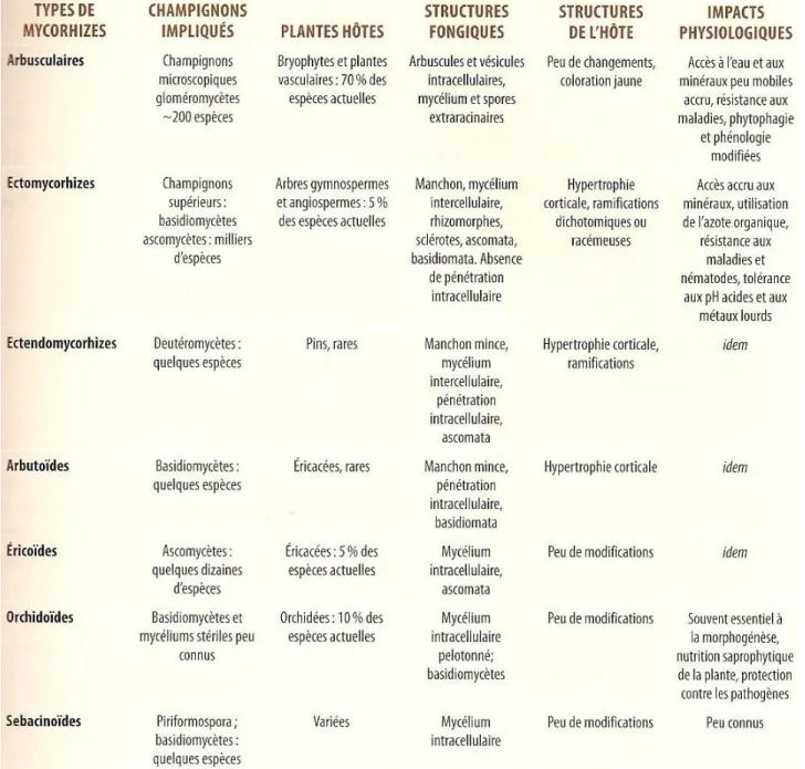
Tableau 1. Les différents types de symbioses mycorhiziennes, les champignons
impliqués, leurs plantes hôtes, les structures formées et les impacts physiologiques (Fortin, et al., 2008).
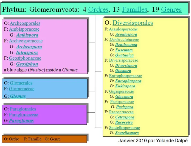
Figure 1. Classification phylogénétique du règne des Mycota (Fungi) basée sur l’analyse des séquences nucléotidiques du gène
C
HAMPIGNONS MYCORHIZIENS À ARBUSCULESLa symbiose mycorhizienne à arbuscules est la plus commune puisqu’elle concerne environ 80% des plantes terrestres. Ces champignons mycorhiziens sont microscopiques et biotrophes obligatoires. Ils sont classés dans le phylum Glomeromycota. La Figure 2, préparée et fournie gracieusement par Yolande Dalpé en janvier 2010, montre la classification phylogénétique récente de ce phylum. Les Glomeromycota forment des structures spécialisées intracellulaires appelées arbuscules dans les cellules corticales des racines. Les vésicules sont des structures fongiques de réserves intercellulaires formées dans le cortex racinaire, et qui contiennent des lipides et des noyaux. Elles n’existent pas chez toutes les espèces de champignons endomycorhiziens arbusculaires, par exemple, les genres Gigaspora et Scutellospora n’en forment pas. Les arbuscules sont des structures fugaces considérablement ramifiées qui augmentent la surface d’échange entre le champignon et la plante. Une fois la colonisation racinaire mise en place, un large réseau de mycélium extraracinaire se met en place pour explorer le sol environnant et infecter de nouvelles racines (Smith & Read, 2008). Les espèces de ce groupe forment les associations mycorhiziennes les plus répandues et fondamentaux dans la productivité des écosystèmes (Harrier, 2001). .
Le Glomus irregulare est une espèce modèle très bien étudiée. Il était auparavant nommé Glomus intraradices, mais des recherches récentes en phylogénie moléculaire ont mis au jour que l’isolat utilisé (DAOM197198) fait plutôt partie de l’espèce récemment décrite Glomus irregulare (Stockinger, et al., 2009). Sa croissance est rapide et il est facilement cultivable in vitro avec les racines de carotte transformées par Agrobacterium rhizogenes. Il est omniprésent et abondant dans toutes sortes d’écosystèmes. Cette espèce biotrophe obligatoire s’associe à la plupart des plantes. Pour ces raisons, elle a été choisie pour ce projet de recherche.
L’objectif principal de ce projet de recherche étant d’isoler et d’identifier les bactéries intimement associées au champignon mycorhizien Glomus irregulare afin de pouvoir les étudier dans leurs interactions avec le champignon. Donc, les bactéries qui ont été étudiées dans ce travail de recherche ont été isolées sur des spores de G. irregulare prélevées dans des échantillons de sol naturel. Ces bactéries ont ensuite été mises en croissance sur du mycélium in vitro de G. irregulare pour que nous puissions étudier, par microscopie et par des méthodes de biologie moléculaires, les interactions qu’elles entretiennent avec les hyphes du champignon.
B
ACTÉRIES ASSOCIÉES AUX CHAMPIGNONS MYCORHIZIENSDe nombreuses études ont montré que les mycorhizes jouent un rôle important dans la protection des plantes contre certains agents pathogènes, notamment des bactéries, des champignons, des protistes et des nématodes. L’article de synthèse de St-Arnaud et Vujanovic (2007) dans le manuel Mycorrhizae in Crop Production résume bien les différents effets des champignons mycorhiziens à arbuscules sur les maladies des plantes et les organismes pathogènes.
Dans le sol, les champignons mycorhiziens eux-mêmes sont associés avec des bactéries qui pourraient être impliquées dans des processus de biocontrôle (ou lutte biologique) et dans la mobilisation de nutriments. La biodiversité microbienne est très importante pour l’équilibre entre les végétaux et leur milieu. Un rôle central dans cet équilibre est joué par les symbioses mycorhiziennes qui permettent aux plantes de mieux s’adapter à leur milieu et d’améliorer leur croissance. De nombreuses études ont mis en évidence des associations entre les bactéries du sol et les champignons mycorhiziens associés aux racines des plantes (Bianciotto, et al., 1996, Andrade, et al., 1997, Andrade, et al., 1998, Filion, et al., 1999, Whipps, 2001, Barea, et al., 2005, Marschner & Timonen, 2005, Artursson, et al., 2006, Hildebrandt, et al., 2006, Toljander,
et al., 2006). La notion de symbiose dans les sols est beaucoup plus complexe qu’on le croyait jusqu’alors. Ces associations symbiotiques complexes très importantes dans l’écologie des sols sont encore très mal connues et encore moins bien comprises (Barea, et al., 2005).
B
IOFILMS BACTÉRIENSUn biofilm est formé de microorganismes établis en population sessile et emprisonnés dans une matrice de polysaccharides (Sutherland, 2001, Sutherland, 2001, Fujishige, et al., 2006). Les bactéries capables de s’organiser en biofilm adhèrent à un support solide et ont une meilleure capacité d’adaptation et de résistance aux conditions du milieu (Espinosa-Urgel, 2004). La plupart des bactéries du sol s’organisent en biofilm sur les racines et les particules du sol (Costerton, et al., 1987). Toutefois, l’étude de ces structures est compliquée par la difficulté à cultiver une grande partie des espèces présentes (Burmolle, et al., 2006). Des méthodes physiologiques, biochimique et moléculaires peuvent être utilisées pour contourner ces problèmes (Amann, et al., 1995, Aoi, 2002, Singh, et al., 2006). Les structures bactériennes formant des biofilms autour des hyphes des champignons mycorhiziens ont probablement un rôle important dans la capacité accrue que certaines plantes
ont à faire face aux agents pathogènes dans le sol (Whipps, 2001, Timmusk, et al., 2005 ).
La grande majorité des études faites sur les biofilms portent sur la plaque dentaire ou les biofilms qui se forment en surface des équipements de dentisterie, en industrie alimentaire, en médecine et dans les usines de traitement des eaux (Watnick & Kolter, 2000, Sutherland, 2001, Aoi, 2002, Rickard, et al., 2003, Beyenal, et al., 2004). La formation des biofilms sur les surfaces biotiques (ici les hyphes de G. irregulare) est relativement peu connue jusqu’à maintenant. Quelques équipes de recherche ont néanmoins étudié des biofilms se formant sur les racines de plantes (Timmusk, et al., 2005 , Fujishige, et al., 2006 , Rudrappa, et al., 2008). Des recherches récentes s’intéressent maintenant à la formation des biofilms bacterien associés aux surfaces de champignons du sol et leurs éventuelles applications technologiques (González-Chávez, et al., 2008, Seneviratne, et al., 2008). Certaines études sur les biofilms se font avec des bactéries qui proviennent de collections de cultures (Timmusk, et al., 2005, Li, et al., 2007, Paramonova, et al., 2007, Wijman, et al., 2007), d’autre se font avec des bactéries isolées directement d’échantillons environnementaux (Burmolle, et al., 2006, Burmolle, et al., 2007).
O
BSERVATIONS MICROSCOPIQUESPlusieurs techniques de microscopie peuvent être utilisées pour étudier les champignons mycorhiziens, la formation de structures ressemblant à des biofilms et les associations bactériennes sur différentes surfaces. Les techniques de microscopie optique (confocale) et électronique ont été utilisées avec succès à cette fin (Bianciotto, et al., 1996, Bloemberg, et al., 2000, Vierheilig, et al., 2005, Bloemberg, 2007, González-Chávez, et al., 2008). L’article de synthèse de l’équipe de Vierheilig (2005) donne un aperçu des différentes méthodes de visualisation des champignons mycorhiziens dans les racines des plantes. La microscopie confocale avec fluorescence a été utilisée pour observer l’attachement de souches bactériennes choisies cultivées en laboratoires sur la surface des champignons mycorhiziens à arbuscule (Bianciotto, et al., 1996). Des bactéries endosymbiotiques ont aussi pu être observées par microscopie électronique à transmission à l’intérieur des spores du champignon mycorhizien Gigaspora margarita (Bianciotto, et al., 1996). L’équipe de Bloomberg a, quant à elle, étudié la capacité du Pseudomonas fluorescent à exprimer différentes couleurs de fluorescence lorsqu’il colonisait des racines de tomate et la possibilité d’observer par microscopie confocale à balayage laser plusieurs couleurs de fluorescence simultanément (Bloemberg, et al., 2000). Plus récemment, l’équipe de González-Chávez (2008) a utilisé la microscopie à
fluorescence et la microscopie électronique à transmission pour observer la présence de bactéries sur la surface des hyphes du champignon mycorhizien Glomus claroideum dans du sol pollué avec des métaux lourds sans toutefois s’intéresser à l’identification de celles-ci (González-Chávez, et al., 2008).
Ce qui diffère dans notre projet de recherche, c’est que nous avons isolé, identifié et utilisé des bactéries présentes sur la surface des spores de champignons mycorhiziens prélevées dans un sol naturel non pollué, contrairement aux autres projets de recherche qui utilisaient généralement des souches bactériennes choisies et élevées en laboratoire. De plus, ces bactéries «sauvages» ont été utilisées pour tenter d’observer la formation de structures ressemblant à des biofilms sur la surface des hyphes de champignons mycorhiziens, des structures vivantes et relativement fugaces. La majorité des études de ce genre portent sur des supports inertes, ou du moins plus résistants, telles les racines des plantes. Très peu d’études relatent d’observations de bactéries poussant sur les hyphes de champignons mycorhiziens.
A
NALYSE ET COMPARAISON DE LA BIODIVERSITE MICROBIENNESeule une très petite proportion des bactéries du sol est cultivable in vitro (Amann, et al., 1995, Burmolle, et al., 2006). Il en est probablement de même avec les bactéries associées aux hyphes des champignons mycorhiziens. Cette situation est attribuable aux besoins nutritifs complexes et à la biotrophie obligatoire de plusieurs d’entre elles. Des techniques de biologie moléculaire permettent d’avoir une vision globale de cette diversité microbienne sans avoir à passer par les étapes d’isolation et de culture in vitro. L’amplification par PCR suivie d’électrophorèse en gradient de gel dénaturant (PCR-DGGE) de la région V3 du gène 16s de l’ARNr est une technique qui est déjà utilisée avec succès dans ce but (Roesti, et al., 2005).
H
YPOTHÈSESSelon les informations retrouvées dans la littérature sur le sujet, les hypothèses recherche suivantes ont été formulées :
1- Les bactéries intimement associées aux spores des champignons mycorhiziens à arbuscules peuvent être isolées et peuvent pousser in vitro sur les hyphes de ces champignons (mycosphère) même en l’absence d’éléments nutritifs autres que les exsudats libérés par les hyphes dans le milieu environnant.
2- Ces bactéries associées aux champignons mycorhiziens s’organisent en structures ressemblant à des biofilms sur la surface du mycélium fongique.
3- La biodiversité microbienne intimement associée aux champignons mycorhiziens est nettement moins grande que celle de la rhizosphère d’où celle-ci est isolée.
O
BJECTIFSL’objectif principal de ce projet de maîtrise était d’isoler, de cultiver et d’identifier les bactéries intimement associées au champignon mycorhizien Glomus irregulare. Il s’agissait alors de vérifier par microscopie leur capacité à former des structures ressemblant à des biofilms en interaction avec le mycélium de ce même champignon mycorhizien (chapitre 2 et annexe 1). Il était aussi prévu de faire une comparaison par biologie moléculaire de la biodiversité microbienne totale du sol étudié et de celle associée au champignon G. irregulare (annexe 2), ainsi que d’identifier les taxons bactériens qui ont été isolés à partir des spores de celui-ci (chapitre 2).
C
HAPITRE
2.
S
PATIO
-
TEMPORAL INTERACTIONS
BETWEEN SOIL BACTERIA AND ARBUSCULAR
MYCORRHIZAL FUNGI
1A
BSTRACTSoil-microbe symbioses are of fundamental importance for plant adaptation to their environment. Research in microbial ecology has revealed that some soil bacteria are associated with arbuscular mycorrhizal fungi (AMF). However, these interactions may be much more complex than originally thought. To assess the type of bacteria associated with AMF, we initially isolated spores of Glomus irregulare from an Agrostis stolonifera rhizosphere. The spores were washed with sterile water and plated onto G. irregulare mycelium growing in vitro in a root free compartment of bi-compartmented
1
Julie Lecomte, Marc St-Arnaud and Mohamed Hjiri, Institut de recherche en biologie végétale, Département de sciences biologiques, Université de Montréal, 4101 rue Sherbrooke Est, Montréal, Québec, H1X 2B2, Canada. Ce manuscrit sera
Petri dishes. We hypothesized that this system should select for bacteria closely associated with the fungus since the only nutrients available to the bacteria were those derived from the hyphae. Twenty-nine bacterial colonies growing on the AMF hyphae were subcultured, and identified using 16S rRNA gene sequences. All bacterial isolates showed high sequence identity to Bacillus cereus, B. megaterium, B. simplex, Kocuria rhizophila, Microbacterium ginsengisoli, Sphingomonas sp. and Variovorax paradoxus. We also assessed bacterial diversity on the surface of spores by PCR-DGGE. We used live cellular imaging to show that bacteria are able to grow on the surface of hyphae with different growing patterns and formed biofilm-like structures in contrast to E. coli as a control.
Keywords : Arbuscular Mycorrhizal Fungi, symbiosis, bacteria, soil, DGGE,
I
NTRODUCTIONSoil microorganisms constitute an important source of biodiversity in soil and are an integral part of terrestrial ecosystems. They contribute to major biological functions such as nutrient and gas cycling, biogeochemical processes and the decomposition and transformation of organic matter. Fungi are also very abundant in the soil and may represent up to 80% of soil microbial biomass (Kirk, et al., 2004). Arbuscular mycorrhizal fungi (AMF) are plant-root symbionts, and are the most abundant and widely distributed fungi in the soil (Smith & Read, 2008). They act as a root extension via their extensive mycelial network, which can explore a much larger soil volume than roots themselves, and can therefore increase the plant’s uptake of essential minerals and water. These fungi also play an important role in plant growth and protection against soil-borne pathogens (Hamel, 2004, St-Arnaud & Vujanovic, 2007).
Rhizosphere bacterial communities can be affected by mycorrhizal root colonization (Mansfeld-Giese, et al., 2002, Marschner & Baumann, 2003, Marschner & Timonen, 2005, Marschner & Timonen, 2006). Many researchers have reported that some soil bacteria are specifically associated with AMF; for example, Bianciotto et al. (1996) showed that soil microorganisms can directly
or indirectly interact with AMF via the exudates the latter released in soil. AMF exudates were also shown to influence the vitality of soil bacteria (Toljander, et al., 2007). Microbial interactions in rhizosphere are much more complex than was originally believed (St-Arnaud, et al., 1995, Filion, et al., 1999). Recently, AMF root colonization was shown to modify the bacterial community structure of tomato rhizosphere through mechanisms unrelated to root exudation by the host plant (Lioussanne, et al., 2010).
The dominant bacterial organization in nature is a biofilm, a population of bacteria embedded in an exopolysaccharide secreted matrix on a surface (Fujishige, et al., 2006). This organization has several advantages for bacteria since it promotes higher resistance to environmental and biological stresses than for planctonics cells (Burmolle, et al., 2006). In natural ecosystems, it has been shown that up to 99% of all bacterial activities are associated with biofilms attached to solid surfaces (Costerton, et al., 1987, Potera, 1996). However, biofilms are generally more complex than a single population, involving different bacterial taxa and other microorganisms living in a community (Watnick & Kolter, 2000). Standard microbiological techniques may allow the culture of as few as 1% of the soil bacterial taxa, and this 1% may not represent the bacterial community in a biofilm (Kirk, et al., 2004). However, other authors have suggested that the majority of bacterial strains present in some soil-biofilms are cultivable (Burmolle, et al., 2007). One could speculate that bacteria which are capable of biofilm formation on the surface of
AMF mycelia may play an important role in certain functions associated with AMF such as nutrient mobilization and protection against pathogens.
The objective of this study was to analyze the spatio-temporal interactions between soil bacteria and the mycelium of the AMF Glomus irregulare isolate DAOM 197198 previously known as Glomus intraradices (Stockinger, et al., 2009). Bacterial strains were isolated from G. irregulare spores harvested from the rhizosphere of Agrostis stolonifera growing in a natural stand. Bacteria were inoculated on mycelium of G. irregulare, grown in vitro on a water media without host roots, and were analyzed microscopically after 15, 30 and 45 days. We hypothesized that the bacteria closely associated with fungus spores would be able to grow on the surface of AMF hyphae, which constitute the sole source of energy in this system. E. coli (non-soil bacteria) and Pseudomonas sp. (soil bacteria not isolated from AMF spores) were used as controls to compare the bacterial growth and attachment on fungal mycelium.
M
ATERIALS&
M
ETHODSSampling
Sampling was conducted in the Mirabel-Lachute area (Quebec, Canada) on June 2006 in a natural stand colonized with herbaceous and deciduous shrubs growing on a sandy soil. The sampling site was chosen because a G. irregulare strain has been previously identified from AM fungal spores harvested from Agrostis stolonifera L. roots collected from the same site in 1991 (Y. Dalpé, personal communication). Six A. stolonifera plants with roots and approximately 1 L of the surrounding soil were collected. GPS coordinates of the six sampling site were between 45º41’36.35’’N 74º08’33.32’’W and 45º41’33.52’’N 74º08’31.50’’W. There was about 10 m between each plant collected. Samples were kept at 4º C and processed in a few days after sampling.
Assessment of root mycorrhizal colonisation
The percentage of root length bearing mycorrhizal colonisation was assessed prior selecting the samples for isolation of Glomus irregulare spores. Roots of Agrostis stolonifera were washed, cut into 1 cm sections and mixed in water. Ink and vinegar (Vierheilig, et al., 1998) were used to stain fungal structures in the roots. Assessment of the colonization extent was done using a dissection microscope using the gridline intercept method (Giovannetti & Mosse, 1980).
Isolation and identification of Glomus irregulare spores harvested from soil
To reduce the bias due to variation in local composition of the soil, a composite sample was prepared by mixing six 100 g rhizosphere soil samples from each sampling point. This composite soil sample was used for spore extraction. AM fungal spores were isolated by wet soil sieving (45 µm pore size) and sucrose density gradient centrifugation (Vilarino & Arines, 1990). Collected spores were kept in sterile water at 4°C until use. The identity of
collected spores was determined by PCR amplification and sequencing of 18S rRNA genes, as described in Kowalchuk et al. (2002) and modified by Yergeau et al. (2006).
Isolation of bacteria from spore surface
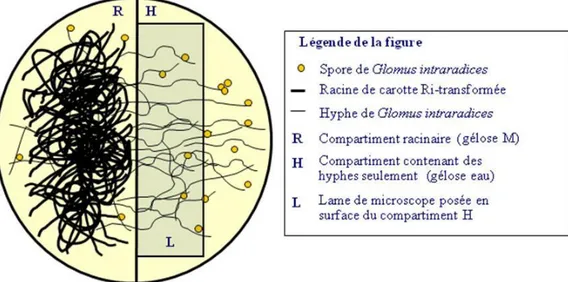
Glomus irregulare isolate DAOM 197198 (previously known as G. intraradices Schenck & Smith) was grown on root inducing (Ri T-DNA) transformed carrot (Daucus carota L.) roots in two-compartment 100 × 15 mm Petri dishes, as described in St-Arnaud et al. (1996). The first compartment containing the transformed carrot roots was filled with 20 mL M medium, while the second compartment received 20 ml sterile water solidified with 0.4 % (w/v) gellan gum (Sigma), containing 0.74 g L-1 MgSO4 but lacking nutrients. Plates were incubated for approximately 6 weeks at 25°C in the dark until hypha crossed the central wall and colonized the second compartment. Glomus irregulare spores harvested from the field were cleaned with autoclaved MilliQ water and placed directly in contact with hyphae growing in the second compartment. An additional incubation period of 4 weeks at 25°C in darkness was required to allow bacterial growth. Mixed bacterial colonies growing around hyphae were isolated and purified by successive inoculations on 10% Tryptic-Soy Agar medium (TSA, QueLab Laboratories, Canada).
Identification of isolated bacteria
The complete 16S rRNA gene was used to identify bacterial isolates. Bacterial cells (1 µL of 20-fold dilution of overnight bacterial cultures) were directly used as a template in PCR reactions. All reactions were conducted in 50 µL volume containing PCR buffer with 1.5 mM MgCl2, 0.2 mM dNTP, 0.5 µM each of primers pA (AGAGTTTGATCCTGGCTCAG) and pH (AAGGAGGTGATCCAGCCGCA) designed by Edwards et al. (1989), 0.6 µL of DMSO and 1.25 U of Taq polymerase (Qiagen TAQ PCR Core Kit). PCR was performed using a Mastercycler ep S gradient thermocycler (Eppendorf, Canada) with the following conditions: 5 min at 94C, followed by 29 cycles of 30 s at 94C, 30 s at 58C and 1 min at 72C, and finally one cycle of 7 min at 72C. PCR amplicons were sequenced at Genome Quebec Innovation Center (Montreal, Canada). E. coli cells and sterile water were respectively used as positive and negative controls. Sequences were identified by to BLASTn searches in the NCBI nucleotides database, and the seven different sequences obtained were deposited in the EMBL database under accession numbers: FN668006 to FN668012.
Bacterial biodiversity associated with G. irregulare spores
A single washed G. irregulare spore isolated from soil was directly put in a PCR tube and crushed with pipette tip, and the 16S rRNA gene was amplified using a nested-PCR protocol. The first round was performed with pA and pH primers (Edwards, et al., 1989) and the second round using primers 968-GC/1378 (Heuer, et al., 1997) to amplify approximately a 500 bp fragment corresponding to the hypervariable regions V6 to V9. Bacterial biodiversity was assessed by running the amplicons through denaturating gradient gel electrophoresis (DGGE), as described in Yergeau et al. (2006) modified from Heuer et al. (1997) with a 45-65% denaturant gradient using a DCode Universal Mutation Detection System (BioRad).
DGGE is a molecular fingerprinting electrophoresis-based method that separates PCR products. Its principal is based on separation of DNA fragment in a denaturing gradient in an acrylamide gel. A G-C clamp is added to one to the primer used in PCR. This pairing insures stability of double strand DNA allowing separation of fragment that differs in their sequence. This method is commonly used to assess microbial diversity.
Assessment of interactions between G. irregulare and bacteria
Two-compartment Petri dishes were prepared as described above (St-Arnaud, et al., 1995) except that after solidification of the gel, sterile 22 × 22 microscope cover glasses were placed along the central wall, and a further 3 mL of gellified sterile water was poured over the edge of cover glasses to form a bridge helping the fungus to cross (see Figure 7, annexe 1 p. 74). Plates received transformed carrot roots inoculated with Glomus irregulare in compartment filled with M medium and the roots were regularly trimmed to avoid any crossing into the second compartment where only the hyphae were allowed to grow. When hyphae grew over the coverslip, they were inoculated with various bacterial isolates from cultures grown in liquid TSB medium for 24 H. All bacterial cells were rinsed and the concentrations were adjusted to 106 CFU mL-1 with sterile 0.9% NaCl solution before use. A 150 µl aliquot of each bacterial suspension was deposited directly on the top of the coverslip where hyphae were growing. Each bacterial isolate was replicated five times and the controls included E. coli JM101 strain as a non-soil bacteria, Pseudomonas sp. strain B129 as a soil bacteria not isolated from AM fungi spores, and sterile water, with or without fungi, as a negative control. Pseudomonas sp. (B129) was previously isolated from the rhizosphere of black spruce [P. mariana (Mill.) B.S.P.] grown at the St-Modeste Forest Nursery (Québec, Canada)
(Filion, et al., 2004). Cultures were incubated in the dark at 25ºC for 15, 30 and 45 days prior to observations with a Axio Imager M1 microscope equipped with DIC (Zeiss).
R
ESULTSIsolation of bacteria from Glomus irregulare spores
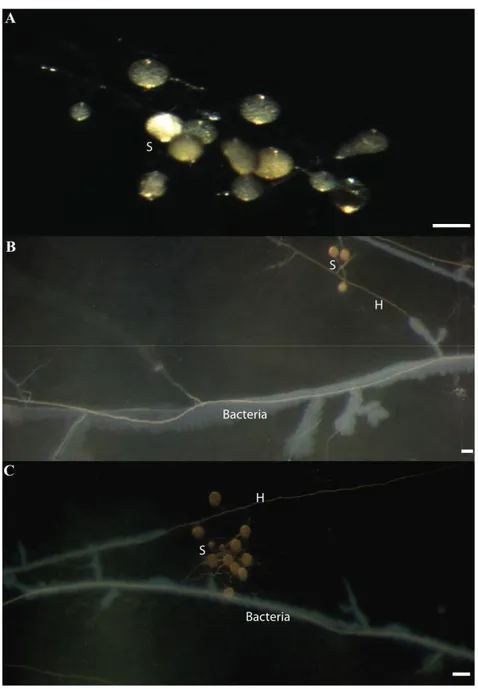
AM fungal spores were collected from the soil samples (Figure 3A). Morphological features such as color, size and shape were typical to Glomus irregulare. We confirmed the identity of these spores by sequencing of the 18S rRNA gene amplified by PCR from single spores. The obtained sequences showed 100% homology with G. irregulare isolate DAOM197198 (accession number AJ852526). After one month of incubation of these spores on the G. irregulare hyphae growing in vitro on water/gellan gum medium, bacterial growth was clearly visible around hyphae as shown in Figure 3B and C. These colonies were re-inoculated repeatedly until single morphotypes were obtained on TSA medium. In total, 29 morphotypes were recovered. PCR amplification
and sequencing of the 16S rRNA gene allowed grouping these 29 morphotypes into seven different bacterial taxa (Table II). BLASTn searches of 16S rRNA gene showed sequence homologies higher than 99% for all isolates except B. simplex (98.8%).
Figure 3. A- Morphology of spores (S) isolated from soil. B and C - Patterns of
mixed bacterial colonies growing from washed Glomus irregulare spores extracted from natural soil and incubated 1 month on G. irregulare. isolate DAOM 197198 hyphae (H) growing in vitro on a gellan gum medium without nutrients. Scale bars represent 200 µm in panel A; 100 µm in panels B and C.
Table II. Bacterial taxa to which belong the 29 bacterial isolates recovered
from Glomus irregulare spores isolated from natural soil and identified by sequencing of a 16S rRNA gene fragment.
Isolate
Number of morphotypes
Phylogenetic affiliation (strain, accession number)
% Identity (sequence length) SE712 1 Bacillus cereus (ATCC 14579, AE016877) 99.1 (1438 bp) SE342 2 B. megaterium (SB 3112, GU191918) 99.7 (1445 bp) SE713B 3 B. simplex (N25, GU086427) 98.8 (874 bp) SE713J 2 Kocuria rhizophila (TA68, NR026452) 99.9 (1424 bp) SE33c 5 Microbacterium ginsengisoli (unknown, AB271048) 99.9 (1412 bp) SE31c 3 Sphingomonas sp. (MUELAK1, EF628247) 99.8 (1386 bp) SE1342c 13 Variovorax paradoxus (rif200835, FJ527675) 99.8 (1430 bp)
Assessment of bacterial biodiversity
DGGE patterns of 16S bacterial gene fragments amplified from field-collected Glomus irregulare spores showed a total of 37 migration positions, with 17 to 24 bands per sample (Figure 4, lanes 1 to 3). The three individual spores showed fairly different banding patterns with only seven bands common to all spores and between five to nine bands unique to each spore, indicating that bacterial communities varied markedly among spores. The positive control E. coli showed one very bright band (Figure 4, lane 5) and few very faint band that are probably contamination of the lane 5 by marker of the lane 4 that occurred when charging the gel. However, the negative control did not show any band (Figure 4, lane 6).
1 2
3 M 5 6
M
Figure 4. DGGE pattern of V9 portion of 16S rRNA gene. Lanes 1 to 3 are
bacterial DNA from one individual G. irregulare spore taken in natural soil. Lane 5 is positive control (E. coli) and lane 6 is negative control (sterile water). Lanes M are markers (see annexe 4).
Interactions between bacteria and G. irregulare mycelium.
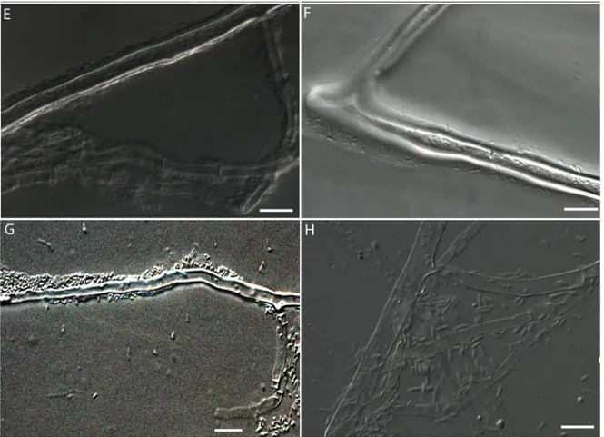
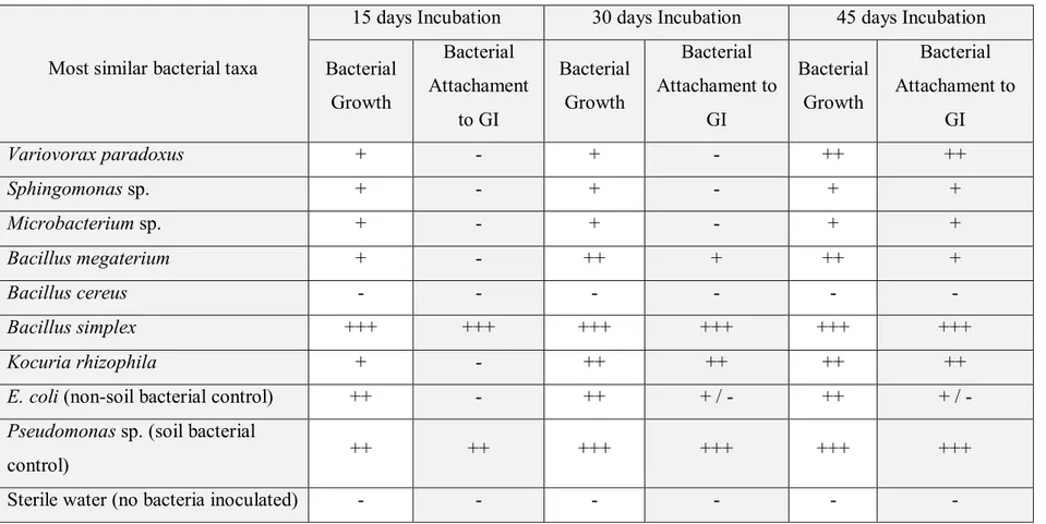
When inoculated on G. irregulare mycelium grown in vitro, bacterial isolates grew exclusively along hyphae and around spores and showed different growth speed and patterns. Some bacterial isolates, such as Bacillus simplex and Pseudomonas sp. (Figure 5A and G), showed profuse development around hyphae after 15-30 days of incubation. Other isolates, as K. rhizophila, V. paradoxus and Microbacterium ginsengisoli (Figure 5B, D and F) showed very little growth after 30 days but significant growth and attachment after 45 days of incubation. On the other hand, B. cereus showed little if any growth on the fungal surface (data not shown). Contrary to the spore-collected bacteria, the non-soil control bacteria E. coli showed no growth on the water media and little affinity to the fungal surface (Figure 5H). The negatives controls, sterile wated inoculated on the mycelium and sterile water inoculated on the media without mycelium, showed no growth (data not shown). The growth and attachment of the bacterial isolates on the hyphal surface are summarized in the Table III.
Figure 5. Bacterial growth patterns on G. irregulare hyphae cultivated in vitro observed with a DIC microscope using a 63X objective. Panel A, Bacillus simplex; B, Kocuria rhyzophila; C, Bacillus megaterium; D, Variovorax paradoxus; E, Sphingomonas sp; F, Microbacterium ginsengisoli; G, Pseudomonas sp.; and H, E. coli. A and G bacteria have 15 days of growth; B and C - 30 days of growth and D, F, E and H - 45 days of growth. Scale bars represent 10 µm.
Table III. Growth and attachment on G. irregulare (GI) mycelium of bacteria isolated from field-harvested AM fungus spores,
on a water media at 25 ºC after 15, 30 and 45 days of incubation.
Most similar bacterial taxa
15 days Incubation 30 days Incubation 45 days Incubation Bacterial Growth Bacterial Attachament to GI Bacterial Growth Bacterial Attachament to GI Bacterial Growth Bacterial Attachament to GI Variovorax paradoxus + - + - ++ ++ Sphingomonas sp. + - + - + + Microbacterium sp. + - + - + + Bacillus megaterium + - ++ + ++ + Bacillus cereus - - - - Bacillus simplex +++ +++ +++ +++ +++ +++ Kocuria rhizophila + - ++ ++ ++ ++
E. coli (non-soil bacterial control) ++ - ++ + / - ++ + / -
Pseudomonas sp. (soil bacterial
control) ++ ++ +++ +++ +++ +++
D
ISCUSSIONTwenty-nine bacterial morphotypes associated with G. irregulare spores were successfully recovered in the present work. 16S rRNA gene sequencing showed that they belong to only seven different bacterial taxa: Bacillus cereus, B. megaterium, B. simplex, Kocuria rhizophila, Microbacterium ginsengisoli, Sphingomonas sp. and Variovorax paradoxus (Table II), which was the most frequent bacterial taxa isolated (13/29 morphotypes). All these bacterial taxa are commonly found in soil. The results supported the hypothesis that bacteria adhering onto the spore surface and likely living on AMF mycelium surface, were able to grow in vitro with AMF hyphae as the sole energy source. It is likely that the bacterial taxa having grown around Glomus irregulare hyphae in vitro were mostly taxa adapted to metabolize the molecules released by the hypha since no other nutrients were available. However, it is also likely that this method would isolate only the most competitive and fast-growing taxa whose portion of the total bacterial biodiversity is unknown. Kirk et al. (2004) estimated that standard microbiological techniques may allow the growth of only about 1% of the soil bacterial taxa from environmental samples. Some bacterial taxa possibly present in the samples could have been obligatory biotrophs since such associations have been previously found in AMF spores (Bianciotto, et al., 2000). However, the bacteria used as a soil control
(Pseudomonas sp.) but which had not been detected in bacterial isolates from field collected spores was also able to grow on the experimental system and to form biofilm-like structures on mycelia surface (Figure 5G). This could be explained by the low number of field collected spores used for bacterial inoculation. A higher number of replicates could have allowed isolation of numerous other bacterial taxa as suggested by DGGE that explored biodiversity of bacterial communities present on AM field collected spores and in total soil bacterial DNA. Moreover, Lioussanne, et al. (2010) have found different bacteria (Pseudomonas spp., Herbaspirilium sp., Acidobacterium sp., Bacillus spp., Verrucomicrobium sp.) specifically associated with Glomus irregulare (intraradices) or G. mosseae (Lioussanne, et al., 2010). Of these bacteria, only B. simplex was found in our isolates thus supporting the hypothesis that using a higher number of samples would have permit to isolate more and probably very different bacterial taxa.
In addition, the non soil bacterial control (E. coli) grew on the experimental system without nutrient inputs other than AM exudates, but did not form biofilm-like structures. This suggests that the AM exudates contain, among other things, sugars (Hooker, et al., 2007, Toljander, et al., 2007) and organic acids such as citric acid (Tawaraya, et al., 2006) that are sufficient to support a large variety of bacterial taxa. It was also demonstrated that AM exudates changes vitality and bacterial soil community in in vitro conditions (Toljander, et al., 2006, Toljander, et al., 2007) and among the same bacterial
species, nutritive needs could change substantially as shown with different strains of Bacillus simplex and their carbon source utilisation (Sikorski, et al., 2008). However, we must not forget that the in vitro experimental conditions do not fully reflect the environmental soil conditions, where biotic and abiotic conditions are highly variable in space and time. AM spore-associated bacteria themselves have been found to change the mycorrhizal colonisation, plant growth and also pathogen relationship in potato plants (Bharadwaj, et al., 2008, Bharadwaj, et al., 2008).
Using DGGE, we assessed bacterial biodiversity of washed spores of G. irregulare isolated from soil and of total bacterial DNA extracted from soil samples (Figure 4). DGGE patterns from three field-collected spores were markedly different in the number of bands formed but mainly on their migration position, indicating a widely different community structure between spores. The number of bands ranged from 17 to 24, and although 29-41% of band positions were common to all spores, 28-38% were unique to each one. The markedly variable banding pattern seen on the DGGE clearly shows that a much higher number of bacterial taxa were associated with the spores than the number suggested by isolation. Soils may contain non-cultivable taxa or taxa with specific nutritional needs that were not met with the isolation protocol used in this study. Certain of the bacterial isolates recovered from the spores were also analyzed in DGGE. However, our DGGE analyses confirmed that we isolated only a very small proportion of bacterial taxa living on the surface of
AMF spores. In addition, bacterial community structure greatly varies among spores. It should be remembered that we cannot tell for certain if these spores originate from the same location because we mixed 6 samples taken from 6 sites to reduce the bias due to variation in local composition of the soil. Soil biotic and abiotic conditions where each AM spore used were taken could be very different and change bacterial pattern associated with AM fungal spores.
Spatio-temporal interactions between the isolated bacteria and the mycelium of Glomus irregulare isolate DAOM-197198 were assessed in vitro in absence of nutrients other than those derived from the mycelium. The seven bacterial taxa were inoculated at similar concentrations on the mycelium and incubated for 15, 30 or 45 days prior to observation. All bacterial taxa except for Bacillus cereus grew on the surface of hyphae and spores (Table III and Figure 5). Growth rates and patterns were however different between taxa. For example, B. cereus was rarely detected and very slow growing. In contrast, B. simplex was a fast grower on the hyphal surface and formed morphological structures analogous to a biofilm. Bacterial species of genus Bacillus are ubiquitous in soil. Most taxa are not known as pathogens for plants or fungi, but B. cereus can produce an enterotoxin and be a source of food-related illness for animals including humans (McKillip, 2000). This isolate might be opportunistic, contaminating the bacterial community growing on mycelium at the isolation step. In this study, B. megaterium shows important growth and little ability to form biofilm-like structure on the G. irregulare mycelium. B.
megaterium has been reported as improving plant growth when inoculated with Glomus irregulare (intraradices) (Marulanda-Aguirre, et al., 2008). Variovorax paradoxus wast fast growing, formed a dense colony around hyphae yet only after 45 days of incubation, and was the most frequently isolated species in the present study. This Proteobacteria was also previously reported as a frequently isolated species in the G. irregulare (intraradices) hyphosphere (Mansfeld-Giese, et al., 2002) and was also recovered from the hyphosphere of Glomus mosseae (Andrade, et al., 1997). The taxon was shown to promote plant growth (Belimov, et al., 2001, Schmalenberger, et al., 2008). Leadbetter and Greenberg (Leadbetter & Greenberg, 2000) showed that Variovorax spp. have the ability to survive with acyl-homoserine lactone as their only source of nitrogen and energy. These types of molecules are quorum sensing signals dedicated to gene-regulation in response to the population density (Parsek, et al., 1999). Interestingly, acyl-homoserine lactone has been more frequently reported to be synthesised by plant associated Pseudomonas spp. than from soilborn isolates (Elasri, et al., 2001). Members of genus Variovorax have a high ecological importance and biotechnological application potential because they are involved in many types of biodegradation (Abou-Shanab, et al., 2007, Schmalenberger, et al., 2008). The second most often isolated species was Microbacterium ginsengisoli. Two other species from the same genus, M. esteraromaticum and M. liquefaciens were previously isolated from G. irregulare (intraradices) hyphosphere (Mansfeld-Giese, et al., 2002). This
strain showed evidence of biofilm-like structure after 45 days of incubation. Kocuria rhizophila, a soil actinomycete (Kovacs, et al., 1999), also showed abundant growth and evidence of formation of biofilm-like structure after 30 days of incubation. On the other hand, the Sphingomonas sp. isolate showed a slow growth and only low evidence of biofilm-like structure formation. Microbacterium genus were shown to have a potential for bioremediation by degrading hydrocarbon (Harwati, et al., 2007) and Sphingomonas was also shown to have potential in bioremediation (Ni'matuzahroh, et al., 1999). The Pseudomonas isolate used here as a control soil bacteria was not isolated from AMF spores but was rather recovered from black spruce rhizosphere, an ectomycorrhizal tree species not forming association with AMF (Filion, et al., 2004). Pseudomonas is a widely distributed bacterial genus in various environments including soil, plant rhizospheres and fungal hyphospheres (Andrade, et al., 1997). In the present study, this isolate formed biofilm-like structure in association with the Glomus mycelium which indicates that this feature would not be specific to taxa showing specificity for the AMF mycelium, but would rather depend on the bacterial taxa and the surface of the mycelium being a favourable substrate for biofilm formation. The ability of certain Pseudomonas strains to form attachments with the fungal surface was reported previously (Bianciotto, et al., 1996). An E. coli strain was used as non-soil bacterial control. It showed abundant growth on the water media but no affinity to the fungal surface or any evidence of biofilm-like structure
formation, such as observed with most of the isolates collected from Glomus spores. The bacterial isolates growing in close to loose association with the AMF mycelium may play important roles in association with the mycorrhizal symbiosis. For example, certain bacterial strains could improve mineral availability for AMF and the plant or could be antagonistic to certain opportunistic pathogenic organisms and improve stability of the plant-AMF association (Xavier & Germida, 2003, Rillig, et al., 2005, Marulanda-Aguirre, et al., 2008).
Understanding interactions between AMF and bacteria and their biodiversity will advance our knowledge on microbial ecology in soil and therefore could have the potential to sustain modern agriculture systems by the use of AMF and associated bacterial as biofertilizers or in bioremediation.
A
CKNOWLEDGMENTSThis work was supported by the NSERC discovery grant to Marc St-Arnaud and Mohamed Hijri. We thank the Canada Foundation for Innovation (CFI) for microscopy facility support to MH. We also thank Maureen Marie-Joseph for technical assistance.
C
HAPITRE
3.
D
ISCUSSION ET CONCLUSION
GÉNÉRALE
À partir du champignon mycorhizien Glomus irregulare, les expériences mises de l’avant dans ce projet de recherche ont permis d’isoler, de cultiver et d’étudier certaines bactéries. Les hypothèses mises de l’avant pour lancer cette recherche ne sont toutefois que partiellement confirmées par les expériences effectuées.
Il avait été supposé que les bactéries intimement associées aux spores du champignon mycorhizien Glomus irregulare pourraient être isolées et pourraient pousser sur le mycélium de ce même champignon in vitro sans aucune autre source nutritive que les exsudats relâchés dans le milieu environnant par celui-ci et seraient isolables. En effet, il a été possible d’isoler sept taxons bactériens à partir des spores de G. irregulare prélevées dans un sol naturel et de les cultiver sur du milieu TSA. Toutefois, un de ces morphotypes cultivé s’est avéré être un isolat de Bacillus cereus. Les bactéries de type B. cereus sont omniprésentes dans l’environnement et cet isolat n’est donc probablement pas spécifiquement associé aux champignons mycorhiziens. Il s’agit plutôt d’une bactérie opportuniste qui colonise le sol et ce qui s’y trouve, y compris les hyphes de champignons mycorhiziens. Cette espèce n’a pas poussé sur les hyphes du champignon in vitro. Deux bactéries ont été utilisées