RESEARCH ARTICLE
In vitro screening of neuroprotective activity of Indian medicinal plant
Withania somnifera
Manjeet Singh
1* and Charles Ramassamy
1,21INRS– Institut Armand Frappier, 531, boul. des Prairies, Laval, Québec, Canada H7V 1B7 2Department of Medical Biology, Faculty of Medicine, Laval University, Québec, Canada G1K 7P4
(Received 25 December 2016– Final revision received 11 July 2017 – Accepted 14 July 2017)
Journal of Nutritional Science(2017), vol. 6, e54, page 1 of 5 doi:10.1017/jns.2017.48
Abstract
Canine cognitive dysfunction (CCD) is an age-dependent neurodegenerative condition characterised by changes in decline in learning and memory patterns. The neurodegenerative features of CCD in ageing dogs and cats are similar to human ageing and Alzheimer’s disease (AD). Discovering neuroprotective disease-modifying therapies against CCD and AD is a major challenge. Strong evidence supports the role of amyloidβ peptide deposition and oxidative stress in the pathophysiology of CCD and AD. In both the human and canine brain, oxidative damage progressively increases with age. Dietary antioxidants from natural sources hold a great promise in halting the progression of CCD and AD. Withania somnifera (WS), an Ayurvedic tonic medicine, also known as ‘Indian ginseng’ or ashwagandha has a long history of use in memory-enhancing therapy but there is a dearth of studies on its neuroprotective effects. The objective of this study was to investigate whether WS extract can protect against Aβ peptide- and acrolein-induced toxicity. We demonstrated that treatment with WS extract significantly protected the human neuroblastoma cell line SK-N-SH against Aβ peptide and acrolein in various cell survival assays. Furthermore, treatment with WS extract significantly reduced the generation of reactive oxygen species in SK-N-SH cells. Finally, our results showed that WS extract is also a potent inhibitor of acetylcholinesterase activity. Thus, our initialfindings indicate that WS extract may act as an antioxidant and cholinergic modulator and may have beneficial effects in CCD and AD therapy.
Key words:Withania somnifera: Amyloid β-peptide: Acrolein: Oxidative stress: Acetylcholinesterase
Canine cognitive dysfunction (CCD) and Alzheimer’s disease (AD) are the most common age-related neurodegenerative dis-orders affecting millions of pets and people worldwide. It is now well established that both oxidative stress and Aβ peptide have been implicated in the pathogenesis of CCD and AD(1). Aβ peptide is formed and released after the sequential cleavage of the amyloid precursor protein by β- and γ-secretases, respectively. Aβ peptide, a 40–42 amino acid-long peptide, is an important component of senile plaque formation in AD brain(2)and its toxicity is mediated through the generation of hydrogen peroxide (H2O2)(3,4). Reactive oxygen species
(ROS), on the other hand, generate highly electrophilic
α,β-unsaturated carbonyl derivatives including acrolein, 4-hydroxy-2-nonenal, and 4-oxononenal from the peroxidation of membrane lipids(5). In AD brain, levels of acrolein were found to be significantly higher in several brain regions such as in the hippocampus, amygdala, middle temporal gyrus and cerebellum(6,7). In primary neuronal cultures from the hippo-campus, the toxicity of acrolein was higher than 4-hydroxy-2-nonenal(8). We have recently established on a neuronal cell line SK-N-SH the details of the multiprocess step of the toxicity of acrolein(9). We showed that, in addition to being a byproduct of lipid peroxidation, acrolein could also potentiate oxidative damage and activate several redox-sensitive pathways.
Abbreviations:AChE, acetylcholinesterase; AD, Alzheimer’s disease; CCD, canine cognitive dysfunction; DCF-DA, 2′,7′-dichlorofluorescein diacetate; DMEM, Dulbecco’s minimum essential medium; DTNB, 5,5′-dithiobis-2-nitrobenzoic acid; LDH, lactate dehydrogenase; ROS, reactive oxygen species; WS, Withania somnifera.
* Corresponding author: Dr M. Singh, emailmsinghmore@gmail.com
© The Author(s) 2017. This is an Open Access article, distributed under the terms of the Creative Commons Attribution licence (http://creative-commons.org/licenses/by/4.0/), which permits unrestricted re-use, distribution, and reproduction in any medium, provided the original work is
JOURNAL OF NUTRITIONAL SCIENCE
Incidence rates of age-specific AD in rural India are at least three times lower than those of an age-matched American ref-erence population(10), which could be attributed to genetic, environmental or dietary causes. Withania somnifera (WS) is one of the most important medicinal plants used in the Indian system of traditional medicine as a nootropic agent and brain tonic to restore age-related decline in mental abil-ities(11). The major biochemical constituents of WS are steroidal alkaloids and steroidal lactones saponins, together called witha-nolides(12). Presently more than twelve alkaloids and forty with-anolides have been identified and characterised from the leaves, roots and berries of Withania species which either exist in free form, i.e. withanone, withaferin A, withanolide A, withanolides D-M, withanolides I–III, or in glycosidic form, i.e. withanosides I–VI(13)
. Recently WS extract has been shown to protect against Aβ peptide- and H2O2-induced toxicities(14–16). In spite of its
widespread use as a brain tonic, its neuroprotective activity against acrolein remains poorly investigated.
Since multi-factorial causes have been recognised in CCD and AD as well as in other neurodegenerative disorders, CCD and AD will require multiple drug therapy to address the varied pathological aspects. Even if the strategy of combin-ing drugs with different therapeutic targets is workable, the new pharmacological approach is to develop a multi-functional compound or extract to target multiple sites in the brain. Thus, the purpose of the present investigation was to demon-strate if a treatment with a standardised extract of WS can protect the human neuroblastoma cell line SK-N-SH against Aβ peptide- and acrolein-induced toxicity, decrease ROS levels and inhibit acetylcholinesterase (AChE) activity.
Materials and methods Drugs and chemicals
A standardised extract of WS was kindly provided by Natural Remedies Private Limited, Bangalore, India. WS extract was certified to contain 2·6 % withanolides including withanosides IV (0·87 %) and V (0·65 %), withaferin A (0·56 %), withano-lide A (0·20 %) and B (0·06 %), and 12-deoxy withastramono-lide (0·26 %) as determined by HPLC. Dulbecco’s minimum essential medium (DMEM), fetal bovine serum, penicillin/ streptomycin, acrolein, acetylthiocholine iodide, neostigmine bromide, 5,5′-dithiobis-2-nitrobenzoic acid (DTNB), H2O2
and TOX-2 (XTT based) were obtained from Sigma-Aldrich Inc. The cytotoxicity detection kit based on the lactate dehydrogenase (LDH) assay was from Roche Diagnostics and 2′,7′-dichlorofluorescein diacetate (DCF-DA) was from Invitrogen. β-Amyloid peptide(25–35) was purchased from
American Peptide.
Aβ25–35preparation
The synthetic peptide Aβ25–35 was dissolved in sterilised tap
water and incubated at 37°C for 72 h for fibrillisation before use as previously described(17). Although both soluble and fibrillar Aβ peptide can induce oxidative stress, the latter is more efficient in causing lipid peroxidation(18)
.
Cell culture and treatment
For neuroprotective effects of WS, SK-N-SH cells, a human neuroblastoma cell line from American Type Cell Culture (ATCC), were maintained in DMEM supplemented with 10 % (v/v) fetal bovine serum, 100 U/ml penicillin and 100 µg/ml streptomycin. SK-N-SH cells were plated at a dens-ity of 1·5 × 104
cells/well in ninety-six-well-plates (Corning) and incubated at 37°C. After 24 h of plating, the medium was completely removed and cells were kept in DMEM with antibiotics but without serum. Cells were then treated with toxic doses of Aβ (50 µg/ml) for 48 h or with acrolein (20 µM) for 24 h.
Cell viability assays
Cytotoxicity and cell survival were measured by LDH and XTT assays by using commercial kits as previously described(19). The XTT assay measures mitochondrial dehy-drogenases activity and may reflect the cellular metabolic state and serves as an indicator of cell survival. Because of the proliferative effects of the WS extract, cells may have higher dehydrogenases activity and the XTT test alone would not be sufficient to measure the neuroprotective effects. To further confirm the protective effects of the WS extract, we employed the LDH test; which measures cellular membrane integrity and is a mean of quantifying dead cells.
Intracellular reactive oxygen species level
The antioxidant activity of WS was confirmed by measuring its ability to scavenge ROS in SK-N-SH cells by using the fluor-escent dye DCF-DA as previously described(19). DCF-DA, a cell0permeable dye, is enzymically converted to a strongly fluorescent compound DCF in the presence of cellular ROS and the fluorescent intensity is directly proportional to the intracellular ROS level.
Acetylcholinesterase inhibition
The relative AChE- inhibitory activity of WS extract was deter-mined in rat brain homogenates by a spectrophotometric method(20,21). The reaction mixture containing 50 µl of DTNB (3 mM), 25 µl WS extract, 25 µl brain homogenate
from a pool of several Sprague–Dawley rats (100 µg total pro-tein) and 100 µl of phosphate buffer. The reaction was started with the addition of 50 µl acetylthiocholine iodide (15 mM).
Enzymic hydrolysis of acetylthiocholine iodide releases thio-choline, which forms a yellow 5-thio-2-nitrobenzoate anion as a result of the reaction with DTNB. The plate was read kinetically atλ 412 nm for 300 s and change in optical dens-ity/min was calculated. Neostigmine bromide (6·25 nM) was
taken as a positive control.
Data analysis
All results were confirmed in four or five separate independent experiments with at least three technical replicates each time
and expressed as means with their standard errors. The GraphPad InStat3 program (GraphPad Software) was used for data analysis. Statistical analysis was done by one-way ANOVA followed by Dunnett’s multiple means comparison test and significance was considered when P was <0·05.
Results
Withania somnifera extract protects SK-N-SH cells against Aβ peptide- and acrolein-induced toxicity
Results inTables 1 and 2 show that treatment with the WS extract protects SK-N-SH cells against Aβ peptide- and acrolein-induced toxicity. This protection was assessed 48 h after the addition of Aβ peptide and was significant from 1·0 and 5·0 µg/ml for the LDH and XTT assays, respectively (Table 1). Interestingly, treatment with the WS extract could also protect SK-N-SH cells against toxicity induced by acrolein from 12·5 µg/ml of WS and reached a maximum at 25 µg/ml (Table 2). To rule out any false-positive effects and interfer-ence with the XTT and LDH assays, absorbance was read and no interference was observed with the different doses of WStested.
Withania somnifera extract decreases intracellular reactive oxygen species level
Since ROS and oxidative damages are involved in the toxicity of Aβ peptide and acrolein, we investigated the antioxidant activity of WS. The WS extract was able to decrease the levels of ROS in SK-N-SH cells treated with H2O2(500 µM), with a
significant effect observed from 5·0 µg/ml (Supplementary Fig. S1).
Withania somnifera extract has potent acetylcholinesterase-inhibitory activity
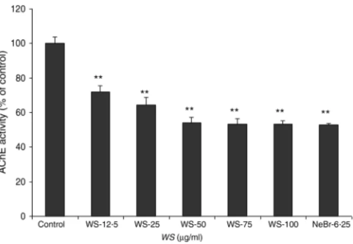
The so-called cholinergic hypothesis of AD proposed that degeneration of cholinergic neurons in the basal forebrain and the associated loss of cholinergic neurotransmission in the cerebral cortex and other areas contribute significantly to the deterioration in cognitive function seen in patients with AD(22). AChE is the key component of cholinergic synaptic transmission and plays a major role in the termination of impulse transmission by rapid hydrolysis of the neurotransmit-ter acetylcholine. This serves as a rationale for the use of three Food and Drug Administration-approved AChE inhibitors in the symptomatic treatment of AD(23). We thus investigated the effect of WS on the activity of AChE on brain homogenates. As shown inFig. 1, AChE activity was reduced by 30 % using 12·50 µg/ml of WS and reached about 50 % of inhibition at 50 µg/ml of WS.
Discussion
There is growing interest in naturally derived bioactive com-pounds for pharmacological application for the treatment of CCD and AD. Different strategies are being investigated such as the neuroprotective therapeutic approach based on protection against Aβ-induced neurotoxicity and oxidative damage or inhibition of AChE. In the present study, our results showed that the WS extract could significantly protect SK-N-SH cells against Aβ- and acrolein-induced toxicity, decrease ROS levels in SK-N-SH cells and inhibit the activity of AChE. The protect-ive effect against Aβ is consistent with other studies that have suggested that WS may be neuroprotective by mediating antioxi-dant effects. Similar to curcuminoids and ginkgolides,
Table 1. Effect of treatment with Withania somnifera (WS) on SK-N-SH cell survival after 24 h with Aβ peptide (50 µg/ml) using lactate dehydrogenase (LDH) and XTT assays†
(Mean values with their standard errors of four or five separate experiments performed in triplicate at least in each group)
Aβ (50 µg/ml) + WS (μg/ml)
Control Aβ (50 μg/ml) WS-0·5 WS-1·0 WS-5·0
Assay Mean SEM Mean SEM Mean SEM Mean SEM Mean SEM
LDH 100·0** 3·61 134·32 6·32 122·11 4·10 114·25** 3·79 109·28** 2·80
XTT 100·0** 4·98 42·31 6·08 62·17 7·32 69·03 13·75 71·87* 4·03
Mean value was significantly different from that of the Aβ group: * P < 0·05, ** P < 0·01. † Results are expressed as percentage of control (taken as 100 %).
Table 2. Effect of treatment with Withania somnifera (WS) on SK-N-SH cell survival after 24 h with acrolein (20·0 µM) using lactate dehydrogenase (LDH) and XTT assays†
(Mean values with their standard errors of four or five separate experiments performed in triplicate at least in each group)
Acrolein (20 µM) +WS (μg/ml)
Control Acrolein (20 µM) WS-12·5 WS-25·0
Assay Mean SEM Mean SEM Mean SEM Mean SEM
LDH 100·0** 4·42 215·24 7·37 139·73** 9·90 127·21** 5·96
XTT 100·0** 2·23 48·92 9·95 80·78** 6·37 81·67** 1·05
** Mean value was significantly different from that of the acrolein group (P < 0·01). † Results are expressed as percentage of control (taken as 100 %).
withanamides from WS fruit showed potent antioxidant activity as indicated by their ability to inhibit lipid oxidation(24). In our study, the neuroprotective effect against Aβ could not be attrib-uted to the effects of WS on ROS scavenging as its effect on ROS levels was significant from 5 µg/ml while cytoprotection was observed at lower concentrations (1 µg/ml). Other mechan-isms should be investigated such as the effects of WS on redox-sensitive pathways because it has been demonstrated that with-aferin A, the major constituents of the extract, could inhibit the activity of the transcription factors NF-κB, or AP-1(25). Also, the WS extract was more effective than withanamide A, one of the major components of WS, against Aβ because with-anamide A was inactive until 12·5 µg/ml(15). These results indi-cate that other components from the WS extract are impliindi-cated in the neuroprotective effect against Aβ such as sominone, the active metabolite of withanoside IV(14).
The present study demonstrated the protective effect of WS against acrolein. This result is of great interest as acrolein has been shown to be elevated in the brain from AD, from mild cognitive impairment and from preclinical AD(26) and could induce neuronal and glial toxicity(9). Different mechanisms could underlie this protection such as the antioxidant activity of WS because acrolein has been shown to increase the pro-duction of superoxide, activate NADPH oxidase activity and deplete reduced glutathione levels(27). Moreover, among the α,β-unsaturated aldehydes, acrolein reacts 110–150 times fas-ter with glutathione than 4-hydroxy-2-nonenal or crotonal(9). The effects of WS on the activity of redox-sensitive pathways could also be involved as we have recently shown that the tox-icity of acrolein modulates different signalling pathways(9).
The inhibition of AChE activity by WS from 12·5 µg/ml described here is in line with the effects of withanolides on AChE with an half maximal inhibitory concentration (IC50)
ranging between 20·5 and 49·2 µg/ml(28).
WScomponents have been shown to induce outgrowth of axons and dendrites, and memory enhancement. Tohda & Joyashiki(29) have demonstrated that sominone, an active
metabolite of withanoside IV, could induce the phosphorylation of RET (rearranged during transfection), a receptor for glial cell line induced neurotropic factor (GDNF). Pharmacokinetics studies in mice suggested rapid oral absorption of withanolides and revealed that withaferin A has one and half times more rela-tive bioavailability as compared with withanolide A(30). Recent studies have shown that WS extract reversed brain pathology in mouse models of Alzheimer’s and amyotropic lateral sclerosis, thereby indicating that certain components are bioavailable and cross the blood brain–barrier(31,32)
.
Thus, cholinesterase-inhibiting potential along with antioxi-dant ability and neurotrophic and neuroprotective activity against Aβ peptide and acrolein could make WS extract and its constituents possible therapeutic agents in CCD, AD and senile dementia. Our investigation further strengthens the trad-itional use of WS as a nootropic agent for restoring age-related decline in mental abilities. However, further exploratory animal studies are needed to optimise therapeutic doses, to find out the right therapeutic compounds that can cross the blood– brain barrier and the duration of WS treatments aiming to yield desired beneficial outcomes; this further represents chal-lenges as some of the components such as withaferin A are potentially cytotoxic(33).
Supplementary material
The supplementary material for this article can be found at
https://doi.org/10.1017/jns.2017.48
Acknowledgements
Financial support was obtained from the Natural Sciences and Engineering Research Council of Canada (NSERC) for C. R.; M. S. gratefully acknowledgesfinancial support from the Foundation Armand-Frappier.
There were no conflicts of interest.
References
1. Schütt T, Helboe L, Pedersen LØ, et al. (2016) Dogs with cognitive dysfunction as a spontaneous model for early Alzheimer’s disease: a translational study of neuropathological and inflammatory markers. J Alzheimers Dis 52, 433–449.
2. Mattson MP (2004) Pathways towards and away from Alzheimer’s disease. Nature 430, 631–639.
3. Behl C, Davis JB, Lesley R, et al. (1994) Hydrogen peroxide med-iates amyloidβ protein toxicity. Cell 77, 817–827.
4. Opazo C, Huang X, Cherny RA, et al. (2002) Metalloenzyme-like activity of Alzheimer’s disease β-amyloid. Cu-dependent catalytic conversion of dopamine, cholesterol, and biological reducing agents to neurotoxic H2O2. J Biol Chem 277, 40302–40308.
5. LoPachin RM, Gavin T, Petersen DR, et al. (2009) Molecular mechanisms of 4 hydroxy 2-nonenal and acrolein toxicity: nucleo-philic targets and adduct formation. Chem Res Toxicol 22, 1499– 1508.
6. Williams TI, Lynn BC, Markesbery WR, et al. (2006) Increased levels of 4-hydroxynonenal and acrolein, neurotoxic markers of lipid peroxidation, in the brain in mild cognitive impairment and early Alzheimer’s disease. Neurobiol Aging 27, 1094–1099. 7. Bradley MA, Markesbery WR & Lovell MA (2010) Increased levels
of 4-hydroxynonenal and acrolein in the brain in preclinical Alzheimer disease. Free Radic Biol Med 48, 1570–1576.
Fig. 1. Effect of Withania somnifera (WS) extract on rat brain acetylcholines-terase (AChE) activity. Results are expressed as percentage of control (taken as 100 %). Neostigmine bromide (NeBr; 6·25 nM) was taken as a positive con-trol. Values are means, with standard errors represented by vertical bars, of four or five separate experiments performed in triplicate at least in each group. ** Mean value was significantly different from that of the control group (P < 0·01).
8. Lovell MA, Xie C & Markesbery WR (2001) Acrolein is increased in Alzheimer’s disease brain and is toxic to primary hippocampal cultures. Neurobiol Aging 22, 187–194.
9. Thanh Nam D, Arseneault M, Zarkovic N, et al. (2010) Molecular regulations induced by acrolein in neuroblastoma SK-N-SH cell: relevance to Alzheimer’s disease. J Alzheimers Dis 21, 1196–1216. 10. Chandra V, Pandav R, Dodge HH, et al. (2001) Incidence of
Alzheimer’s disease in a rural community in India: the Indo-US study. Neurology 57, 985–989.
11. Ven Murthy MR, Ranjekar PK, Ramassamy C, et al. (2010) Scientific basis for the use of Indian Ayurvedic medicinal plants in the treatment of neurodegenerative disorders: ashwagandha. Cent Nerv Sys Agents Med Chem 10, 238–246.
12. Elsakka M, Grigorescu E, Stănescu U, et al. (1990) New data refer-ring to chemistry of Withania somnifera species. Rev Med Chir Soc Med Nat Iasi 94, 385–387.
13. Mirjalili MH, Moyano E, Bonfill M, et al. (2009) Steroidal lactones from Withania somnifera, an ancient plant for novel medicine. Molecules 14, 2373–2393.
14. Kuboyama T, Tohda C & Komatsu K (2006) Withanoside IV and its active metabolite, sominone, attenuate Aβ(25–35)-induced neu-rodegeneration. Eur J Neurosci 23, 1417–1426.
15. Jayaprakasam B, Padmanabhan K & Nair MG (2010) Withanamides in Withania somnifera fruit protect PC-12 cells fromβ-amyloid respon-sible for Alzheimer’s disease. Phytother Res 24, 859–863.
16. Kumar S, Seal CJ, Howes MJ, et al. (2010) In vitro protective effects of Withania somnifera (L.) dunal root extract against hydrogen perox-ide and β-amyloid(1–42)-induced cytotoxicity in differentiated PC12 cells. Phytother Res 24, 1567–1574.
17. Longpré F, Garneau P, Christen Y, et al. (2006) Protection by EGb 761 against β-amyloid-induced neurotoxicity: involvement of NF-κB, SIRT1, and MAPKs pathways and inhibition of amyloid fibril formation. Free Radic Biol Med 1, 81–94.
18. Melo JB, Sousa C, Garção P, et al. (2009) Galantamine protects against oxidative stress induced by amyloid-β peptide in cortical neurons. Eur J Neurosci 29, 455–464.
19. Singh M, Murthy V & Ramassamy C (2010) Modulation of hydro-gen peroxide and acrolein-induced oxidative stress, mitochondrial dysfunctions and redox regulated pathways by the Bacopa monniera extract: potential implication in Alzheimer’s disease. J Alzheimers Dis 21, 229–247.
20. Ellman GL, Courtney KD, Andres V Jr, et al. (1961) A new and rapid colorimetric determination of acetylcholinesterase activity. Biochem Pharmacol 7, 88–95.
21. Singh M & Rishi S (2005) Plasma acetylcholinesterase as a bio-marker of triazophos neurotoxicity in young and adult rats. Environ Toxicol Pharmacol 19, 471–476.
22. Francis PT, Palmer AM, Snape M, et al. (1999) The cholinergic hypothesis of Alzheimer’s disease: a review of progress. J Neurol Neurosurg Psychiatry 66, 137–147.
23. Cummings JL (2004) Alzheimer’s disease. New Engl J Med 351, 56–67.
24. Jayaprakasam B, Strasburg GA & Nair MG (2004) Potent lipid per-oxidation inhibitors from Withania somnifera. Tetrahedron 60, 3109– 3121.
25. Kaileh M, Vanden Berghe W, Heyerick A, et al. (2007) Withaferin A strongly elicits IκB kinase β hyperphosphorylation concomitant with potent inhibition of its kinase activity. J Biol Chem 282, 4253–4264.
26. Singh M, Nam DT, Arseneault M, et al. (2010) Role of by-products of lipid oxidation in Alzheimer’s disease brain: a focus on acrolein. J Alzheimers Dis 21, 741–756.
27. Ansari MA, Keller JN & Scheff SW (2008) Protective effect of Pycnogenol in human neuroblastoma SH-SY5Y cells follow-ing acrolein-induced cytotoxicity. Free Radic Biol Med 45, 1510– 1519.
28. Choudhary MI, Nawaz SA, ul-Haq Z, et al. (2005) Withanolides, a new class of natural cholinesterase inhibitors with calcium antagon-istic properties. Biochem Biophys Res Commun 334, 276–287. 29. Tohda C & Joyashiki E (2009) Sominone enhances neurite
out-growth and spatial memory mediated by the neurotrophic factor receptor, RET. Br J Pharmacol 157, 1427–1440.
30. Patil D, Gautam M, Mishra S, et al. (2013) Determination of with-aferin A and withanolide A in mice plasma using high-performance liquid chromatography-tandem mass spectrometry: application to pharmacokinetics after oral administration of Withania somnifera aqueous extract. J Pharm Biomed Anal 80, 203–212.
31. Sehgal N, Gupta A, Valli RK, et al. (2012) Withania somnifera reverses Alzheimer’s disease pathology by enhancing low-density lipoprotein receptor-related protein in liver. Proc Natl Acad Sci U S A 109, 3510–3515.
32. Dutta K, Patel P, Rahimian R, et al. (2017) Withania somnifera reverses transactive response DNA binding protein 43 proteinopa-thy in a mouse model of amyotrophic lateral sclerosis/frontotem-poral lobar degeneration. Neurotherapeutics 14, 447–462.
33. Wadhwa R, Konar A & Kaul SC (2016) Nootropic potential of ash-wagandhaleaves: beyond traditional root extracts. Neurochem Int 95, 109–118.