Open Archive Toulouse Archive Ouverte (OATAO)
OATAO is an open access repository that collects the work of some Toulouse
researchers and makes it freely available over the web where possible.
This is
an author'sversion published in:
https://oatao.univ-toulouse.fr/23080Official URL :
https://doi.org/10.1016/j.diii.2017.10.001
To cite this version :
Any correspondence concerning this service should be sent to the repository administrator: tech-oatao@listes-diff.inp-toulouse.fr
Nougarolis, Florence and Mokrane, Fatima-Zohra and Brouchet, Laurent and Telmon,
Norbert and Rousseau, Hervé and Sans, Nicolas and Dedouit, Fabrice An unusual
intracardiac foreign body following penetrating thoracic injury. (2017) Diagnostic and
Interventional Imaging, 98 (12). 901-902. ISSN 2211-5684
OATAO
An unusual intracardiac foreign
body following penetrating
thoracic injury
Keywords Cardiac foreign body; Thoracic trauma; Pericardial sac
Foreign bodies in the heart after thoracic trauma may result in fatal outcome [1]. We report a pendant inside
the pericardium after penetrating injury that did not cause major cardiac injury with a favorable outcome.
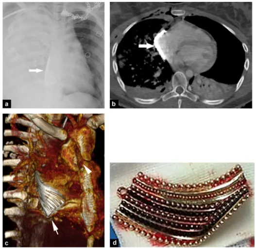
Figure 1. A 20-year-old woman with thoracic injury following motorcycle accident. a: chest X-ray shows a «white right lung» aspect associated with a dense foreign body in contact with the right cardiac border (arrow); b: computed tomography (CT) image of the thorax in the transverse plane shows a thin and regular metallic foreign body (arrow) adjacent to the right atrium accompanied by a left pneumothorax and right pulmonary contusion; c: three-dimensional CT image of the thorax reveals a linear and grooved foreign body (arrow), which is in contact with the heart, associated with a longitudinal sternal fracture (arrowhead); d: photograph shows the guilty pendant immediately after surgical removal.
A 20-year-old woman was admitted to the hospital fol-lowing a motorcycle traffic accident. She hit a metallic barrier at a speed of 30—50 km/h. Clinical examination revealed several traumatic injuries but her most severe injury was a transfixed thoracic wound. Chest X-ray revealed a right hemopneumothorax, that was subsequently drained (Fig. 1). An unidentified metallic body was detected in the projection of the thoracic spine and the right cardiac silhou-ette. Computed tomography (CT) showed an intrapericardial metallic foreign body, close to the right atrium. Other traumatic lesions included a longitudinal manubrium frac-ture, a right sternoclavicular dislocation, and a left drained
hemopneumothorax. Exploratory thoracotomy revealed a foreign body nestled against the right cardiac cavities, which was responsible of a slight dissection of the right auriculoventricular junction. The foreign body was a 5-cm pendant. No complications were observed immediately after surgery. The clinical follow-up was uneventful.
Cardiac foreign bodies are rarely seen because most patients with penetrating cardiac injuries die from hemor-rhagic shock or cardiac tamponade[1]. The most common foreign bodies that penetrate the heart are bullets and lawn-mower projectiles[1]. Their mechanisms are known[2]. In our patient, chest fractures and wounds were due to direct impact. The handlebars of the motorcycle and/or the metal-lic barrier are the two potential causes of chest injury. Of interest in our patient, the impact to the pendant produced enough strength to cut the clothes, dilacerate the skin, frac-ture the sternum, and push the object within the anterior mediastinum. This all happened despite the slow speed. The direct impact focused the force on a specific and restricted portion of the sternum, provoking a longitudinal sternum fracture and pushing the pendant inside the thorax. How-ever, there was no major cardiac injury. The foreign body was able to penetrate because of its position in front of an assumably weak anatomical mediastinal area. To our knowl-edge, only one case has been reported about a foreign body that came in contact with the auriculoventricular junction
[3]. In our patient, the foreign body opened the pericardial sac and made its way next to the right heart cavities despite the existence of a large mid-thoracic wound. Our case also highlights the major role of CT in the management of trauma patient[4].
Disclosure of interest
The authors declare that they have no competing interest.
References
[1]Leitman M, Vered Z. Foreign bodies in the heart.
Echocardiog-raphy 2015;32:365—71.
[2]Gunn ML, Clark RT, Sadro CT, Linnau KF, Sandstrom CK. Current
concepts in imaging evaluation of penetrating transmediastinal injury. Radiographics 2014;34:1824—41.
[3]Harrer J, Holubec T, Brtko M. A foreign body in the heart due to
an unusual injury. Ann Thorac Surg 2009;88:985—7.
[4]Daghfous A, Bouzaïdi K, Abdelkefi M, Rebai S, Zoghlemi A,
Mbarek M. Contribution of imaging in the initial management of ballistic trauma. Diagn Interv Imaging 2015;96:45—55.
F. Nougarolisa,∗, F.-Z. Mokraneb,
L. Brouchetc, N. Telmona, H. Rousseaub,
N. Sansd, F. Dedouita
aForensic Medicine Department, Rangueil University Hospital, 1, avenue du Pr-Jean-Poulhès, TSA 50032, 31059 Toulouse, France bRadiology Department, Rangueil University Hospital, 1, avenue du Pr-Jean-Poulhès, TSA 50032, 31059 Toulouse, France cThoracic Surgery Department, Rangueil-Larrey University Hospital, 31059 Toulouse, France dRadiology Department, Purpan University Hospital, place du Docteur-Baylac, TSA 40031, 31059 Toulouse, France
∗Corresponding author.
E-mail address:florence.nougarolis@gmail.com