Chorionic Gonadotropin Stimulation of Angiogenesis
and Pericyte Recruitment
Sarah Berndt, Silvia Blacher, Sophie Perrier d’Hauterive, Marc Thiry,
Marie Tsampalas, Andre´ Cruz, Christel Pe´queux, Sophie Lorquet, Carine Munaut, Agne`s Noe¨l,* and Jean-Michel Foidart*
Laboratory of Tumor and Development Biology (S.Be., S.Bl., A.C., C.P., S.L., C.M., A.N., J.-M.F.), Groupe Interdisciplinaire de Ge´noprote´omique Applique´-Cancer, Center of Immunology (S.P.H., M.Ts.), Laboratory of Cellular Biology (M.Th.), University of Lie`ge, and Department of Obstetrics and Gynecology (S.P.H., S.L., J.-M.F.), Centre Hospitalier Regional, B-4000 Lie`ge, Belgium
Context: During the periimplantation period, human chorionic gonadotropin (hCG) plays a key role by increasing the uterine blood flow through uterine vessel vasodilatation but also through angiogenesis. Indeed, we previously demonstrated that hCG contributes to endothelial cell re-cruitment and vessel formation.
Objective: In this study, hCG was proposed as an arteriogenic factor that could promote perivas-cular cell recruitment and vessel stabilization.
Design: The aortic ring assay, a three-dimensional ex vivo angiogenesis system mimicking all the steps of the angiogenesis process was used to study the impact of hCG on pericyte recruitment and vessel maturation.
Setting: The study was conducted at a university hospital laboratory.
Main Outcome Measures: Perivascular cell proliferation, migration, and apposition were quanti-fied by computerized image analysis.
Results: Physiological concentrations of hCG (10 – 400 IU/ml) significantly enhanced pericyte sprouting and migration and gave rise to the maturation and coverage of endothelial capil-laries. In a three-dimensional coculture model of endothelial and perivascular cells, hCG en-hanced vessel tube formation and endothelial/mural cell adhesion. In addition, hCG stimulated the proliferation of human umbilical vein endothelial cells and smooth muscle cells. The spec-ificity of these effects was determined by using an anti-hCG blocking antibody. Signaling pathways implicated on this hCG effect is protein kinase A and phospholipase C/protein kinase C dependent for the proliferative effect but only phospholipase C/protein kinase C for the migrative process.
Conclusions: Our findings highlight a novel paracrine role of this early embryonic signal in vessel maturation by stimulating perivascular cell recruitment, migration, and proliferation. (J Clin Endocrinol Metab 94: 4567– 4574, 2009)
P
eritrophoblastic angiogenesis at the implantation site is an important step for successful pregnancy. Among the wide range of contributing hormones, cytokines, and growthfactors, human chorionic gonadotropin (hCG) plays a key role and modulates: 1) the implantation process by control-ling leukemia inhibitory factor and macrophage colony
stim-ISSN Print 0021-972X ISSN Online 1945-7197 Printed in U.S.A.
Copyright © 2009 by The Endocrine Society
doi: 10.1210/jc.2009-0443 Received March 2, 2009. Accepted August 17, 2009. First Published Online October 16, 2009
* A.N. and J.-M.F. are both senior authors.
Abbreviations: AoSMC, Aortic SMC; BIM, bisindolylmaleimide I; CMFDA, 5-chlorometh-ylfluorescein diacetate; EC, endothelial cell; FBS, fetal bovine serum; Fmax, perivascular cells maximal migration distance from the explant; hCG, human chorionic gonadotropin; hCGR, hCG receptor; HUVEC, human umbilical vein endothelial cell; Nf, number of perivas-cular cells; PKA, protein kinase A; PKC, protein kinase C; PLC, phospholipase C; SMC, smooth muscle cell; VEGF, vascular endothelial growth factor.
E n d o c r i n e R e s e a r c h
ulating factor release, 2) tissue remodeling by increasing ma-trix metalloproteinase-9 production, and 3) angiogenesis by inducing vascular endothelial growth factor (VEGF) up-reg-ulation (1, 2). LH/hCG receptor (LH/hCGR) was first de-scribed on endometrial and myometrial vascular smooth muscle cells and thereafter in vascular endothelial cells (ECs) of the uterine vessels (3, 4). In vivo administration of hCG reduced vascular resistance in human uterus and decreased in
vitro the formation of vasoconstrictor eicosanoids of the
vas-cular wall (4, 5). Zygmunt et al (6). showed that hCG was able to promote angiogenesis by stimulating EC migration and capillary sprout formation. We previously showed that the angiogenic activity of hCG is mediated through a direct effect on ECs and through a paracrine dialogue between ECs and endometrial epithelial cells (1). Upon hCG binding to its LH/hCGR at the surface of endometrial ECs, VEGF synthe-sis and secretion is up-regulated, thereby activating the an-giogenic switch in ECs. The endometrial epithelium is thus sensitive to specific embryonic signals, leading to the pro-duction of VEGF, the main angiogenic factor (7). As classi-cally observed in pathological conditions (8), VEGF-driven angiogenesis results in the formation of highly abnormal ves-sels deficient in mural/pericyte coverage. The putative effect of hCG on perivascular cell recruitment and hence on vessel maturation is currently unknown. To address this issue, we used in this study the aortic ring assay, which mimics the different steps of the angiogenic process including EC pro-liferation, migration, and differentiation into capillaries (9). In addition, we evaluated the effect of hCG on perivascular cell outgrowth and EC coverage by mural cells. The effects of hCG was also investigated in monocultures and cocultures of
ECs and smooth muscle cells (SMCs). In this study, we pro-vide epro-vidence for a promoting effect of hCG on vessel mat-uration through the recruitment of perivascular cells. These novel findings underline a much broader spectrum of activ-ities for hCG than initially anticipated.
Materials and Methods Materials
Reagents tested were purified urinary hCG (Pregnyl, Organon, Roseland, NJ), recombinant hCG (Ovitrelle; Serono, Geneva, Swit-zerland), protein kinase A (PKA) inhibitor H89 dihydrochloride (Calbiochem, La Jolla, CA), phospholipase C (PLC) inhibitor U73122 (Calbiochem), protein kinase C (PKC) inhibitor bisindolyl-maleimide I (BIM; Calbiochem), anti-hCG neutralizing antibody (Sc-7821; Santa Cruz Biotechnology, Santa Cruz, CA), cell tracker Green 5-chloromethylfluorescein diacetate (CMFDA; Molecular Probes, Invitrogen, Carlsbad, CA), and CellTracker red CMRA (Molecular Probes, Invitrogen).
Animals
Wistar rats (⫾250 g) and C57/Bl 6 mice (10 –12 wk old) were provided by the Animal Center of the University of Lie`ge. Experiments were conducted with the approval of the local ethical committee for the care of experimental animals. Mice were maintained with a 12-h light, 12-h dark cycle, with food and water available ad libitum.
Aortic ring assay
Rat and mice aortic rings were cultured in three-dimensional type I collagen gels as previously described (10, 11). Effects of dif-ferent compounds were tested by addition to culture medium at d 0. For quantification, computerized image analysis was performed on a Sun SPARC30 workstation with the soft-ware Visilog 5.0 (Noesis, St. Aubin, France) according to Blacher et al. (10). The following parameters were determined: number of mi-crovessels; maximal microvessel length, total number of branching in microvessels, number of perivascular cells (Nf), and perivascular cells maximal migration distance from the ex-plant (Fmax).
Whole-mount immunostaining of aortic rings
At d 9 of culture, aortic fragments em-bedded in collagen gels were washed three times in PBS for 10 min followed by a fix-ation in 4% paraformaldehyde for 10 min. Samples were washed again in PBS three times for 10 min, and then nonspecific an-tibody binding was blocked with milk 1.5% for 20 min. Samples were incubated over-night at room temperature with a mix of a lectin and a primary antibody: Griffonia Simplifolia isolectin-B4/Alexa Fluor 488 at 5 g/ml (Molecular Probes; I21411) and rabbit anti-NG2 chondroitin sulfate
pro-FIG. 1. Effect of hCG on pericyte recruitment in the aortic ring assay. A, Photomicrographs of aortic rings cultured in the absence (0) or presence of increasing concentrations of recombinant hCG (rhCG; 0 –100 IU/ml). Endothelial capillaries are depicted by the arrow, and fibroblast like-cells are indicated by the dotted arrow. B, Quantification of the Nf and Fmax (C) in response to increasing rhCG concentrations (10 – 400 IU/ml) after 9 d of culture. Data are mean⫾SDof one representative experiment, each condition running at least four rings. *, P⬍ 0.05; ***, P ⬍ 0.001.
teoglycan antibody (Chemicon, Temecula, CA; AB5320) at 10 g/ml. After three washes in PBS for 10 min each, the tissues were incubated at room temperature with a secondary goat anti-rabbit biotin antibody (Dako, Glostrup, Denmark; E432) at 1.9g/ml. Finally, an incubation with streptavidin Cy3 (Sigma, St. Louis, MO; S6402) was performed for 1 h at room temperature at 1 g/ml. Samples were mounted with Vectashield-Dapi mounting medium (Vector Laboratories, Burlingame, CA; H-1200), after three washes in PBS for 10 min at room temperature.
Electron transmission microscopy
Aortic three-dimensional gels culture were stopped after 10 d, and gels were fixed 1 h at 4 C with 2.5% glutaraldehyde in a So¨rensen 0.1Mphosphate buffer (pH 7.4). Gels were postfixed for 30 min with 1% osmium tetroxide. After deshydratation, samples were embedded into Epon. Ultrathin sections, obtained with a Reichert Ultracut S ultramicrotome (Leica, Germany), were contrasted with uranyl acetate and lead citrate. Observa-tions were done with a Jeol 100 CX II transmission electron microscope at 60 kV (Peabody, MA).
Cell culture
Human umbilical vein endothelial cells (HUVECs) were grown on 0.2% gelatin-coated dishes in MCDB 131 medium (Life Technologies, Inc., Carlsbad, CA) supplemented with 10% fetal bovine serum (FBS), HEPES (25 mM), glutamine (2 mM),
endothelial cell growth supplement (12 ng/ml; BD Biosciences, Bedford, MA), heparin (2.5 mg/ml), and antibiotics (penicillin-streptomycin, 100 U/ml); for experiments, cells at passages 2–10 were used. Human aortic SMCs (AoSMC) purchased from Lonza (Basel, Switzerland; CC-2671) were grown in complete smooth muscle medium containing 10% FBS (SmBm; Lonza; CC-3181). Cells were at passages 6 –10 used for experiments. Mouse SMCs C3
H/10T1/2, clone 8 cell line (American Type Culture Collection, Manassas, VA; CCL-226) were grown in basal medium (Life Technologies, Inc.) supplemented with 10% fetal calf serum, glutamine (2 mM), penicillin/streptomycin (100 g/ml), HEPES (15 mM), and bicarbonate. All cell types were
cultured in 5% CO2at 37 C, and media were replaced every 2 d.
10 T1/2 cell migration
To evaluate SMC migration, the wound assay model was used as described by Savani et al. (12). 10 T1/2 cells (105
) were seeded on 24-well microplates and grown overnight in 10% FBS basal medium. A cross-shape scratch was made with a tip in cell mono-layers. After 4 and 9 h, pictures were acquired at the same mag-nification and location in the bottom of the dish. Quantification was done using ImageJ software (National Institutes of Health, Bethesda, MD) by measurements of wounded areas at time 0, 4, and 9 h.
Proliferation assays
Proliferation tests were performed on AoSMCs, 10 T1/2 cells, and HUVECs cultured for 24 h. Cells (5⫻ 103
) were seeded and allowed to adhere in 96-well microplates. For HUVEC prolifer-ation, 96-well microplates were coated with gelatin at 2 mg/ml. Cells were treated with or without hCG (10 –1000 U/ml), H89 (10M), U73122 (10M), or BIM (1g/ml) alone or combined
with hCG (100 IU/ml) in their respective culture media contain-ing 2% FBS. Cell proliferation was evaluated uscontain-ing 5-bromo-2⬘-deoxyuridine incorporation into cells followed by a cell
pro-liferation colorimetric assay (Roche Applied Science, Penzberg, Germany) as previously described (13).
Boyden chamber migration assay
AoSMC cell migration was assessed using Boyden chamber assay. Cells were seeded on top of polycarbonate filters (8m pore) of Transwell Permeable Support (Costar, Corning Inc., Lowell, MA). Cells in stock culture were trypsinized, suspended in serum-free medium containing 0.1% BSA, and placed on the upper compartment of the chamber (7.104
cells/filter). The lower compartment of the chamber was filled with serum-free medium containing 1% BSA, supplemented with or without hCG, anti-hCG, H89, U73122, and BIM. After an incubation period of 24 h at 37 C, filters were fixed in methanol. Cells on filters were stained with crystal violet solution 0.1% (Sigma). Nonmigrated
FIG. 2. Effect of hCG on pericyte outgrowth from the aortic rings. A and B, Photomicrographs of aortic rings cultured in the absence (control) (A, C, E, and G) or presence of uhCG 100 IU/ml (B, D, F, and H) (n⫽ 5 rings per condition). Whole-mount double immunostainings of the collagen gels at d 9 of culture with an anti-EC-specific isolectin (IB4) in green and a pericyte-specific antibody (NG2) in red; nuclei were counterstained with 4⬘,6⬘-diamino-2-phenylindole (blue) (C–F). Representative electron micrographs (G) showing a section of immature nude endothelial tubes composed of ECs (E) delimiting a lumen (L) and a mature endothelial tube (H) composed of endothelial cells (E) delimiting a larger lumen (L) and surrounded by a perivascular cell (P).
cells at the upper surface of the filters were wiped away with a cotton swab. Quantification of the migration assay was done by colorimetric measurement ( ⫽ 560 nm) of cells at the lower surface of the filter.
HUVEC and AoSMC coculture assays
AoSMC and HUVEC coculture assays were performed in a modified matrigel overlay assay as previously reported (14). Briefly, HUVEC cells (15⫻ 103
CMFDA green labeled cells) were seeded on top of a 300l matrigel layer (15) in 24-well microplates and allowed to form tubular structures. Six hours later, AoSMCs (15⫻ 103
CMRA red labeled) were overseeded on HUVECs, and 100 IU/ml hCG were added or not. After 24 h, cultures were stopped, rinsed with PBS, and fixed with 4% para-formaldehyde until microscopic examination. To analyze the AoSMC distribution around the EC tube network, an original computer-assisted quantification was developed by implementing an algorithm using the image analysis tool box of MATLAB7.1 software (The Mathworks, Natick, MA). This method calcu-lated the smallest distance separating green pixel corresponding to EC and red pixel corresponding to AoSMCs identified by cell tracker green CMFDA and CellTracker red CMRA, respectively.
Statistical analyses
Statistical analysis were conducted with GraphPad Prism software (GraphPad Inc., La Jolla, CA). Aortic rings experiments
including four explants per condition were repeated at least twice. Student’s t test was used to evaluate whether differences among groups were significant. For computerized image analysis, statistical analysis were performed with the statistics toolbox of MATLAB 7.1 (MathWorks) using Stu-dent’s t test or using Wilcoxon test, with regard to heteroscedasticity. In the other ex-periments, Mann-Whitney tests were used for statistical analysis. P⬍ 0.05 was con-sidered as statistically significant.
Results
hCG promotes perivascular cell recruitment in the aortic ring assay
According to our previous results (1), the incubation of rat aortic frag-ments embedded in a collagen gel with recombinant hCG induced an angio-genic response characterized by en-hanced capillary formation (Fig. 1. A). Interestingly, dispersed cells spread out, suggesting a possible mobilization of perivascular cells in addition to that of EC. These cells have been quantified through a computerized quantification (10) and were identified as mural cells by immunostaining. The addition of hCG (10 – 400 IU/ml) to the aortic ring culture media re-sulted in an increase of Nf (Fig. 1B) and distance of cell migration from the aortic explant (Fmax) (Fig. 1C). Sim-ilar results were obtained with urinary hCG. For next ex-periments, urinary hCG (uhCG) was used at its optimal concentration (100 IU/ml).
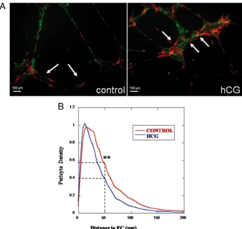
Whole-mount immunostainings of mice aortic ring cul-tures (d 9 of culture) were performed to identify the ECs through Isolectin B4 staining and pericytes through NG2 detection. They revealed the pericyte phenotype of perivascular cells that were growing out in the presence of stimulatory dose of hCG (100 IU/ml) (Fig. 2). In control conditions, in the absence of hCG, two types of capillary-like structures were detected, one composed of naked en-dothelial cells and the other one composed of pericytes (Fig. 2, C and E). In sharp contrast, on hCG treatment, pericyte proliferation and migration were drastically stim-ulated (Fig. 2D), and endothelial tubes displayed a mature phenotype with a pericyte coating (Fig. 2, F and H). Ob-servations by electron transmission microscopy confirmed the presence of perivascular cells upon treatment with hCG (Fig. 2H). In sharp contrast, in the absence of hCG, FIG. 3. Effect of hCG on EC-pericyte interactions in a three-dimensional matrigel coculture
model. A, Photomicrographs (scale bars, 100m) of HUVECs (green labeled) and AoSMCs (red-labeled) after 12 h of coculture without (ctrl) or with uhCG (100 IU/ml hCG). Arrows point area in which AoSMCs are close to endothelial tube-like structures. B, Computer-assisted quantification of AoSMC distribution around HUVEC tube network. Upon hCG treatment, pericytes were closer to ECs (blue curve). The Student t test was used for statistical analysis (n⫽ 20). **, P ⬍ 0.05.
capillaries were formed of a single layer of naked ECs delimiting a lumen (Fig. 2G).
hCG promotes EC and SMC interactions in a three-dimensional in vitro coculture model of HUVECs and human AoSMCs
The interactions between EC and human AoSMCs were then investigated in vitro in a coculture system on matrigel. When HUVECs were seeded on matrigel, they organized into tube-like structures. AoSMCs were then seeded on top of the HUVEC culture to study pericyte-endothelial cell interactions (Fig. 3). Under hCG treat-ment, pericytes became more closely apposed to the ECs (Fig. 3A). An original computer-assisted method of quan-tification was used to measure the distance separating pericytes from ECs (Fig. 3B). The density of mural cells apposed to ECs (distanceⱕ5M) was 20% higher under
hCG treatment (**, P⬍ 0.05) (Fig. 3B). In contrast, the density of mural cells present at a distance higher than 50m from ECs was 1.5-fold lower on hCG treatment (**, P⬍ 0.05).
hCG promotes SMC and EC proliferation through PKA/PKC signaling pathways
We previously reported that hCG can directly modulate the proliferation of ECs (1). To evaluate the putative di-rect effect of hCG on perivascular cells, we next determined the impact of hCG on the proliferation of human AoSMCs. Increasing concentrations of hCG (10 – 500 IU/ml) stimulated AoSMC prolifer-ation in a dose-dependent manner (Fig. 4A). This dose-response curve displayed a bell shape with maximal effect of hCG between 50 and 100 IU/ml. Similar re-sults were obtained by using murine smooth muscle cells (10 T1/2) (Fig. 4B). The specificity of this effect was assessed by the addition of an anti-hCG-blocking antibody together with hCG (Fig. 4C).
Cell signaling pathways involved in the proliferative effects exerted by hCG on ECs and SMCs have next been investigated by inhibiting differ-ent pathways putatively involved in LH/hCGR signaling (Fig. 4, D and E). Addition of a PKC inhibitor (1g/ml BIM) and a PLC inhibitor (10 M
U73122) abolished the effect of hCG on HUVEC proliferation (Fig. 4D). Addition of a PKA inhibitor (10 MH89) and a PLC
inhibitor (10MU73122) abolished the effect of hCG
on AoSMCs (Fig. 4E). hCG induces SMC migration
A wound healing in vitro assay was first applied to determine the effect of hCG on SMCs (10T1/2). Recolo-nization of wounded areas was evaluated after 4 and 9 h by measuring the width of the gap in the same areas. At each time point, the size of the gap was measured, nor-malized to each native wound surface (time 0) and data were averaged (n⫽ 24). Already after 4 h, hCG induced cell migration at a higher extent than in control condition (Fig. 5, A and B). After 9 h the recolonization was in-creased 1.4-fold (Fig. 5, A and B). A Boyden migration chamber assay was next used to further determine the promigratory effect of hCG on both smooth muscle cells FIG. 4. Effect of hCG on human and murine SMC proliferation. Proliferation of AoSMCs
treated for 24 h with 10 –500 IU/ml uhCG (A) and 10 T1/2 cell line (B) were evaluated by 5-bromo-2⬘-deoxyuridine colorimetric immunoassay. Data are expressed as means ⫾SD(n⫽ 10). **, P⬍ 0.01. C, Proliferation of 10 T1/2 cells treated or not with uhCG (50 IU/ml) or anti-hCG antibody (micrograms per milliliter) for 48 h. Data are expressed as means⫾SD(n⫽ 10). ***, P⬍ 0.001. D, Proliferation of HUVECs after 24 h of culture with or without uhCG 100 IU/ml, combined or not with BIM (1g/ml) or U73122 (10 M). E, Effect of hCG (100 IU/ml), H89 (10M), or U73122 (10M) alone or in combination on AoSMC cell proliferation rate. Data are expressed as means⫾SD(n⫽ 4). *, P ⬍ 0.05; **, P ⬍ 0.05; ***, P ⬍ 0.001. Results are expressed as percentage of control.
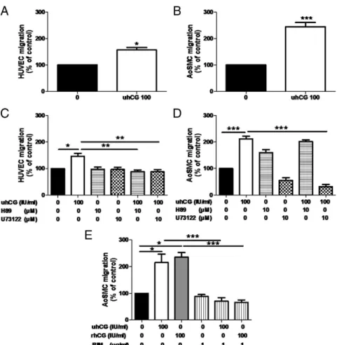
(AoSMCs) and ECs (HUVECs). Incubations of a stimu-latory dose of hCG (100 IU/ml) resulted in a significant enhancement of both HUVEC (Fig. 6 A) and AoSMC mi-gration (Fig. 6B). HUVECs and AoSMCs seeded on Boy-den chambers filters were incubated for 24 h with hCG (100 IU/ml) alone or in combination with signaling path-way inhibitors. Whereas hCG-induced migration of HUVECs was abolished by PKA (H89) or PLC (U73122) inhibitor (Fig. 6C), only the PLC and PKC inhibitor (BIM) was able to block the hCG-stimulated migration of AoSMCs (Fig. 6, D and E). All biological effects described above were observed in the presence of urinary and re-combinant hCG.
Discussion
hCG is a key actor of placental vascularization (16 –18), which is one of the crucial local adaptations to successful pregnancy. hCG stimulates directly angiogenesis through EC recruitment, enhanced proliferation, and migration (19). It also stimulates the release of VEGF by human epithelial endometrial cells that causes a paracrine stim-ulation of ECs. VEGF-mediated angiogenesis results in the formation of naked, dilated, and leaky capillaries resulting in tissue ischemia. Indeed, normal efficient blood circula-tion requires the coverage of newly formed endothelial tubes by vascular SMCs. In this study, through the use of
five different models (aortic ring, cell proliferation assay, wound healing assay, Boyden chamber assay, and tube formation on matrigel), we report a potent effect of re-combinant and urinary hCG on various SMC activities including proliferation, migration, and capacity to cover EC capillaries.
Because of the complexity of the mechanism underlying the angiogenic reaction and vessel maturation through pericyte recruitment, a panel of complementary models (9) has been used to investigate the mechanisms of action of hCG. The aortic ring assay comprises three different cell types (ECs, fibroblastic cells, and SMCs) and mimics four steps of the angiogenic process: 1) EC proliferation, 2) EC migration, 3) EC differentiation into capillaries, and 4) maturation by pericyte coverage (20). hCG was able to enhance the sprouting of both IB4-positive EC and NG2-positive pericytes. Interestingly, both their number and distance of migration were stimulated on hCG treatment. Of great interest is the finding that hCG promoted the coverage of endothelial capillaries by pericytes, whereas in its absence, EC and pericytes spread out in an independent manner. The impact of hCG on EC/mural cell interactions was then confirmed in a three-dimensional coculture model leading to tube-like structure formation. When EC and pericytes were cocultured on matrigel, hCG stimu-lated an affixing of close pericytes to ECs, which mimics the vascular maturation process. Our data are in accor-dance with clinical observations performed on hCG-res-cued human corpus luteum. Indeed, hCG treatment of women increased endothelial cell proliferation and peri-cyte recruitment indicating an increase rate of angiogen-esis together with vessel stabilization (21).
The presence of LH/hCGRs has been evidenced in vivo in the endothelium and smooth muscle of uterine blood vessels. Their expression levels are significantly increased in the intramyometrial segment (3, 4). In line with these findings, immunostaining of LH/hCGR revealed positiv-ity on AoSMCs (data not shown). Our previous work re-ported that hCG has a direct stimulating effect on ECs (1). Accordingly, vascular uterine endothelial cells expressing LH/hCGR responded to physiological doses of hCG with increased capillary formation in vitro (6). The LH/hCGR-dependent effect of hCG is further supported by the ap-plication of the aortic ring assay to LH/hCG-deficient mice (22). This genetic approach clearly demonstrated an ab-rogation of hCG angiostimulation in the absence of LH/hCGR.
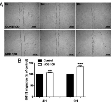
To further dissect the mechanism of hCG action, more simple models consisting of monocultures of EC or peri-cytes/SMCs were used to determine whether hCG can ex-ert an effect on cell proliferation and/or cell migration. hCG induced a strong proliferative effect on both ECs and FIG. 5. Effect of hCG on SMC migration in a wound-healing assay.
A, Photomicrographs of the wound area in 10 T1/2 cell monolayer in a kinetic assay (at time 4 and 9 h) without (CTRL) or with addition of uhCG 100 IU/ml to the culture medium. B,
Semiautomatic quantification of recolonized areas defined as the primitive wound surface divided by wound surfaces persisting after 4 and 9 h. Control condition was set to 100%. Data are expressed as means⫾SD(n⫽ 24). **, P ⬍ 0.05; ***, P ⬍ 0.001.
SMCs from murine or human origin, which is consistent with previous reports (23–25). Blocking antibody addi-tion and use of recombinant hCG further confirmed the specificity of this effect. This effect was dose dependent with an optimal effect at 50 –100 IU/ml corresponding to the physiological hCG concentration achieved in the first 2 wk of pregnancy. Such hCG concentration has proven a similar optimal angiogenic response in terms of endothe-lial cell proliferation and sprouting in different in vitro models (1, 19) and led to a stimulation of trophoblast migration in vitro (26).
We previously reported that hCG-mediated prolifera-tive effect was PKA dependent (1). In the present study, we provide additional data that the stimulation of HUVEC proliferation by hCG is also sensitive to PKC and PLC inhibition. AoSMC proliferation was affected by PKA and PLC pathway inhibitions under hCG treatment. This is in line with previous reports showing that in porcine myo-metrial cells, LH/hCGR activation is coupled with two different signaling systems: adenylate cyclase and PLC (27).
SMC-induced migration was further studied in a wound healing assay in which hCG enhanced 10T1/2 migra-tion already after 4 h of treatment and further after 9 h. This early promigra-tive effect signed a direct effect on mi-gration property and was not reflecting an enhancement of cell proliferation in the wounded area. hCG caused a 2.5-fold increase of AoSMC migration in a Boyden chamber assay and a 2-fold in-crease of HUVEC migration. The effect of hCG on EC migration was clearly more potent when uterine microvascu-lar endothelial cells were used (6). The present study demonstrates for the first time that hCG enhances pericyte/ SMC migration. To specify the signal-ing pathways involved in this process, PKA and PLC inhibitors were tested in combination or not with hCG in the Boyden chambers model. We demon-strated that HUVEC-hCG induced mi-gration signals through PKA and PLC pathways, whereas it involves only the PLC pathway in AoSMCs. Interest-ingly, this suggests that the signaling pathways involved in hCG’s promigra-tive property are cell type dependent.
In summary, our results support the complex and key role played by hCG in the angiogenic process occurring at the implantation site. They provide evidence for a novel func-tion of hCG in vessel maturafunc-tion through the stimulafunc-tion of pericyte recruitment.
Acknowledgments
The authors thank Benoît Brouwers for his skillful and excellent technical assistance.
Address all correspondence and requests for reprints to: Agne`s Noe¨l, Laboratory of Tumor and Developmental Biology, University of Lie`ge, Tour de Pathologie, Centre Hospitalier Uni-versitaire (B23), Sart Tilman, B-4000 Lie`ge, Belgium. E-mail: agnes.noel@ulg.ac.be.
This work was supported by grants from the Fonds National de la Recherche Scientifique (Belgium), European FP7 program (MI-CROENVIMET Grant 201279), IUAP (interuniversity attraction pole) program, Grant Te´le´vie, European Network of Excellence EMBryo Implantation Control, and the Fondation Le´on Fred-ericq (University of Lie`ge). S. B. is a recipient of a Te´le´vie grant. Disclosure Summary: The authors have nothing to disclose.
FIG. 6. Effect of hCG on HUVEC and AoSMC migration in a Boyden chamber assay. Migration of HUVECs (A) or AoSMCs (B) in the absence or presence of uhCG 100 IU/ml. HUVECs (C) and AoSMCs (D) were treated or not with uhCG 100 IU/ml, H89 (10M), or U73122 (10M) alone or in combination. Inhibition of hCG-induced migration by PKC inhibitor (BIM) (E). Data are (means⫾SD) expressed as percentage of control condition (n⫽ 4). *, P⬍ 0.05; **, P ⬍ 0.05; ***, P ⬍ 0.001.
References
1. Berndt S, Perrier d’Hauterive S, Blacher S, Pe´queux C, Lorquet S, Munaut C, Applanat M, Herve´ MA, Lamande´ N, Corvol P, van den Bruˆle F, Frankenne F, Poutanen M, Huhtaniemi I, Geenen V, Noe¨l A, Foidart JM 2006 Angiogenic activity of human chorionic gonad-otropin through LH receptor activation on endothelial and epithelial cells of the endometrium. FASEB J 20:2630 –2632
2. Licht P, Russu V, Lehmeyer S, Mo¨ll J, Siebzehnru¨bl E, Wildt L 2002 Intrauterine microdialysis reveals cycle-dependent regulation of en-dometrial insulin-like growth factor binding protein-1 secretion by human chorionic gonadotropin. Fertil Steril 78:252–258
3. Lei ZM, Reshef E, Rao V 1992 The expression of human chorionic gonadotropin/luteinizing hormone receptors in human endometrial and myometrial blood vessels. J Clin Endocrinol Metab 75:651– 659 4. Toth P, Li X, Rao CV, Lincoln SR, Sanfilippo JS, Spinnato 2nd JA, Yussman MA 1994 Expression of functional human chorionic go-nadotropin/human luteinizing hormone receptor gene in human uterine arteries. J Clin Endocrinol Metab 79:307–315
5. Toth P, Lukacs H, Gimes G, Sebestyen A, Pasztor N, Paulin F, Rao CV 2001 Clinical importance of vascular LH/hCG receptors—a re-view. Reprod Biol 1:5–11
6. Zygmunt M, Herr F, Keller-Schoenwetter S, Kunzi-Rapp K, Mu¨nstedt K, Rao CV, Lang U, Preissner KT 2002 Characterization of human chorionic gonadotropin as a novel angiogenic factor. J Clin Endocrinol Metab 87:5290 –5296
7. Ferrara N, Gerber HP, LeCouter J 2003 The biology of VEGF and its receptors. Nat Med 9:669 – 676
8. Carmeliet P 2003 Angiogenesis in health and disease. Nat Med 9:653– 660
9. Berndt S, Bruye`re F, Jost M, Noel A 2008 In vitro and in vivo models of angiogenesis to dissect MMP functions. Edwards D, Hoyer-Hansen G, Blasi F, Sloane BF, eds. The cancer degradome. Pro-teases and cancer biology. New York: Springer; 303–323 10. Blacher S, Devy L, Burbridge MF, Roland G, Tucker G, Noe¨l A,
Foidart JM 2001 Improved quantification of angiogenesis in the rat aortic ring assay. Angiogenesis 4:133–142
11. Masson V, Devy L, Grignet-Debrus C, Bernt S, Bajou K, Blacher S, Roland G, Chang Y, Fong T, Carmeliet P, Foidart JM, Noe¨l A 2002 Mouse Aortic Ring Assay: A New Approach of the Molecular Ge-netics of Angiogenesis. Biol Proced Online 4:24 –31
12. Savani RC, Wang C, Yang B, Zhang S, Kinsella MG, Wight TN, Stern R, Nance DM, Turley EA 1995 Migration of bovine aortic smooth muscle cells after wounding injury. The role of hyaluronan and RHAMM. J Clin Invest 95:1158 –1168
13. Perrier d’Hauterive S, Charlet-Renard C, Berndt S, Dubois M, Munaut C, Goffin F, Hagelstein MT, Noe¨l A, Hazout A, Foidart JM, Geenen V 2004 Human chorionic gonadotropin and growth factors at the em-bryonic-endometrial interface control leukemia inhibitory factor (LIF)
and interleukin 6 (IL-6) secretion by human endometrial epithelium. Hum Reprod 19:2633–2643
14. Saunders WB, Bohnsack BL, Faske JB, Anthis NJ, Bayless KJ, Hirschi KK, Davis GE 2006 Coregulation of vascular tube stabilization by endothelial cell TIMP-2 and pericyte TIMP-3. J Cell Biol 175:179 –191 15. Timpl R, Rohde H, Robey PG, Rennard SI, Foidart JM, Martin GR 1979 Laminin—a glycoprotein from basement membranes. J Biol Chem 254:9933–9937
16. Zygmunt M, Herr F, Munstedt K, Lang U, Liang OD 2003 Angio-genesis and vasculoAngio-genesis in pregnancy. Eur J Obstet Gynecol Re-prod Biol 110(Suppl 1):S10 –S18
17. Licht P, Russu V, Wildt L 2001 On the role of human chorionic gonadotropin (hCG) in the embryo-endometrial microenvironment: implications for differentiation and implantation. Semin Reprod Med 19:37– 47
18. Licht P, Fluhr H, Neuwinger J, Wallwiener D, Wildt L 2007 Is human chorionic gonadotropin directly involved in the regulation of human implantation? Mol Cell Endocrinol 269:85–92
19. Herr F, Baal N, Reisinger K, Lorenz A, McKinnon T, Preissner KT, Zygmunt M 2007 HCG in the regulation of placental angiogenesis. Results of an in vitro study. Placenta 28(Suppl A):S85–S93 20. Nicosia RF, Ottinetti A 1990 Growth of microvessels in serum-free
matrix culture of rat aorta. A quantitative assay of angiogenesis in
vitro. Lab Invest 63:115–122
21. Wulff C, Dickson SE, Duncan WC, Fraser HM 2001 Angiogenesis in the human corpus luteum: simulated early pregnancy by HCG treatment is associated with both angiogenesis and vessel stabiliza-tion. Hum Reprod 16:2515–2524
22. Zhang FP, Poutanen M, Wilbertz J, Huhtaniemi I 2001 Normal prenatal but arrested postnatal sexual development of luteinizing hormone receptor knockout (LuRKO) mice. Mol Endocrinol 15: 172–183
23. Ko¨rnyei JL, Lei ZM, Rao CV 1993 Human myometrial smooth muscle cells are novel targets of direct regulation by human chori-onic gonadotropin. Biol Reprod 49:1149 –1157
24. Lee TK, Kim DI, Song YL, Lee YC, Kim HM, Kim CH 2004 Dif-ferential inhibition of Scutellaria barbata D. Don (Lamiaceae) on HCG-promoted proliferation of cultured uterine leiomyomal and myometrial smooth muscle cells. Immunopharmacol Immunotoxi-col 26:329 –342
25. Horiuchi A, Nikaido T, Yoshizawa T, Itoh K, Kobayashi Y, Toki T, Konishi I, Fujii S 2000 HCG promotes proliferation of uterine leiomyomal cells more strongly than that of myometrial smooth muscle cells in vitro. Mol Hum Reprod 6:523–528
26. Zygmunt M, McKinnon T, Herr F, Lala PK, Han VK 2005 HCG increases trophoblast migration in vitro via the insulin-like growth fac-tor-II/mannose-6 phosphate receptor. Mol Hum Reprod 11:261–267 27. Kisielewska J, Flint AP, Ziecik AJ 1996 Phospholipase C and ade-nylate cyclase signalling systems in the action of hCG on porcine myometrial smooth muscle cells. J Endocrinol 148:175–180