Université de Montréal
Assessing locomotion in cats trained on a flat treadmill and
on a ladder treadmill before and after spinal cord injury (SCI)
par
Manuel Jose Escalona Castillo
Département de neurosciences. Université de Montréal Faculté de Médecine
Mémoire présenté à la Faculté des études supérieures et postdoctorales en vue de l’obtention du grade de Maître ès sciences (M.Sc.) en
Neurosciences
Septembre, 2015
Université de Montréal
Faculté des études supérieures et postdoctorales
Ce mémoire intitulé:
Assessing locomotion in cats trained on a flat treadmill and on a
ladder treadmill before and after spinal cord injury (SCI)
Présenté par :
Manuel Jose Escalona Castillo
A été évalué par un jury composé des personnes suivantes :
Elaine Chapman, président-rapporteur Serge Rossignol, directeur de recherche Jean-Pierre Gossard, codirecteur de recherche
i
Résumé
Les effets des lésions de la moelle épinière sur la locomotion sont souvent évalués sur un tapis roulant avec une surface plane, ce qui demande peu d’implication active des structures supraspinales. L’objectif du présent travail est d’évaluer si un type d’entraînement nécessitant une plus grande part de contrôle volontaire (c.-à-d. supraspinal) pourrait améliorer la récupération de la marche chez le chat après une hémilésion unilatérale spinale au niveau thoracique (T10). Pour ce faire, pendant 6 semaines les chats ont été entrainés sur un tapis roulant conventionnel ou sur un tapis-échelle roulante, tâche requérant un placement des pattes plus précis. Les paramètres de la marche ont été évalués par cinématique et électromyographie (EMG) avant et une fois par semaine pendant 6 semaines après lésion.
Nos résultats comparant la marche sur tapis conventionnel à celle sur échelle roulante montrent des différences dans les excursions angulaires et les couplages entre les membres. On observe aussi des différences dans l’amplitude des EMG notamment une augmentation de la deuxième bouffée du muscle Semitendineux (St) sur l’échelle roulante. Après l’hémilésion spinale cette bouffée disparait du côté de la lésion tandis qu’elle est maintenue du côté intact. Après l’entrainement sur échelle roulante, on observe des changements de trajectoire de la patte et une disparition du pied tombant (foot drag) qui suggèrent une amélioration du contrôle de la musculature distale.
Nos résultats montrent que le patron locomoteur observé sur tapis conventionnel est influencé par le type d’entraînement procuré. De plus, certains paramètres de la locomotion suggèrent que l’entraînement sur échelle roulante, qui requiert plus de contrôle supraspinal, favorise une meilleure récupération de la marche après lésion spinale.
Mots-clés : Entraînement locomoteur, inputs supraspinal, hémilésion spinale, échelle roulante,
ii
Abstract
The effects of spinal cord lesions on locomotion in animals is often assessed on a flat surface which requires minimal supraspinal demands. Here we investigated whether locomotor training requiring more voluntary commands could improve recovery in cats after a unilateral hemisection on the left side (Thoracic 10). Cats were trained for 6 weeks on a conventional flat treadmill (FTM) or on a ‘ladder treadmill’ (LTM) requiring precise foot positioning. Locomotor parameters such as kinematics and electromyographic activity (EMG) were evaluated before and once a week for 6 weeks after spinal hemisection.
There were differences in angular excursions and limb couplings when comparing walking on FTM and LTM and important changes occurred in the amplitude of EMGs such as an increased second burst in Semitendinosus (St) during LTM stepping. After the left hemisection, this burst disappeared on the left side while it was maintained on the right (non lesioned) side. With LTM training, we observed changes in limb trajectories and disappearance of foot drag of the left foot during FTM stepping suggesting a better control of distal musculature.
Our results show that locomotor patterns observed during FTM stepping are influenced by different training modalities and also that training on the LTM after a spinal hemisection, demanding more supraspinal control, leads to a better stepping pattern.
Keywords: locomotor training, supraspinal inputs, spinal hemisection, ladder treadmill, flat
iii
Table of contents
RÉSUMÉ ... i
ABSTRACT ... ii
LIST OF TABLES ... vi
LIST OF FIGURES ... vii
LIST OF ABBREVIATIONS ... ix
ACKNOWLEDGEMENTS ... xi
CHAPTER 1: INTRODUCTION ... 12
1.1 Background ... 12
1.2 Animal models to study locomotion ... 13
1.2.1 The Central pattern Generator ... 13
1.2.2 Organization of the CPG ... 14
1.2.3 Localization of the CPG ... 16
1.3 Locomotion and evaluation methods ... 17
1.3.1 Treadmill ... 18
1.3.2 Locomotor cycle and kinematics ... 18
1.3.3 Interlimb coordination ... 20
1.3.4 General muscle activity in cats ... 21
1.3.5 Activity of flexor and extensor muscles. ... 23
Extensors ... 23
Flexors ... 23
Flexor-Extensor coordination ... 24
1.3.6 Individual muscular activity of relevance to this study ... 24
Flexors ... 24
Semitendinosus (St) ... 24
Extensor digitorum brevis (EDB) ... 25
Tibialis anterior (TA) ... 25
Biceps Brachii (Bra) ... 26
Extensors ... 26
Vastus lateralis (VL) ... 26
iv
Triceps Brachii (Tri) ... 27
1.4 Control and modulation of Locomotion ... 27
1.4.1 Sensory inputs ... 29
1.4.1.1 Proprioceptive inputs ... 29
1.4.1.2 Cutaneous inputs ... 30
1.4.2 Propriospinal system ... 31
1.4.3 Supraspinal inputs and pathways ... 32
1.4.3.1 Initiation of locomotion ... 32
1.4.3.2 Posture and corrections of steps during locomotion ... 32
1.5 Skilled locomotion ... 35
1.6 Animal models of SCI and the importance of locomotor training. ... 37
1.6.1 Complete lesions ... 37 1.6.2 Incomplete lesions ... 38 1.7 Problem statement ... 40 CHAPTER 2: ARTICLE ... 43 Abstract ... 44 Introduction ... 45 Methods ... 47 Results ... 51
Step cycle characteristics ... 51
Angular excursions and limb trajectory ... 51
Interlimb coupling and walking gaits ... 52
Changes of EMG activity ... 53
Hindlimb flexor muscles ... 54
Hindlimb extensor muscles ... 54
Forelimb flexor and extensor muscles ... 55
EMG phase shifts ... 55
Discussion ... 56
Kinematics of walking on the FTM and LTM ... 56
EMG changes in LTM and FTM walking ... 59
Acknowledgements ... 60
v
CHAPTER 3: SPINAL HEMISECTION AND TRAINING PARADIGMS ... 74
3.1 Methods ... 74 3.1.1 Training paradigm ... 74 3.1.2 Spinal lesions ... 75 3.1.3 Histology ... 75 3.1.4 Kinematics analysis ... 76 3.1.5 Statistical analysis ... 76 3.2 Results ... 77 CHAPTER 4: DISCUSSION ... 87 4.1 Kinematics ... 88 4.2 EMG adaptations ... 90 4.3 Foot drag ... 92
4.4 Training and human SCI ... 93
4.5 Limitations ... 94
4.5 Concluding remarks and future work ... 95
vi
List of tables
Chapter 2: Article
Table 1. Values of kinematic parameters of averaged step length and averaged cycle duration
on the FTM and on the LTM for each cat.
vii
List of figures
Chapter 1: Introduction
Figure 1: Schematic representation of the two-level hierarchical architecture of the CPG Figure 2: Representation of limb joint kinematics (stick diagrams) and angular excursions. Figure 3: Schematic representation of chronically implanted muscles used in this study showing
origins and point of insertion and their respectives raw EMG signals.
Figure 4: Schematic representation of the tripartite system controlling locomotion.
Chapter 2: Article
Figure 1: Schematic overview of the experimental protocol and ladder treadmill setup. Figure 2: Comparisons of length and step cycle duration on the FTM and on the LTM.
Figure 3: Averaged angular excursion and stick figures of the left hindlimb synchronized on
the left paw contact for three cats on the FTM and on the LTM.
Figure 4: Graphical 2d representations of left toe trajectories during FTM and LTM stepping. Figure 5: Polar representation of interlimb coupling during FTM locomotion and ladder LTM
locomotion.
Figure 6: Changes in interlimb coordination during FTM and LTM stepping.
Figure 7: Averaged implanted muscles electromyogram (EMG) profile on the FTM
superimposed on the LTM from cat 1 and cat 3.
Figure 8: EMG amplitude and duration on the LTM task for implanted flexor and extensor
viii
Chapter 3: Spinal hemisection and training paradigms
Figure H1: Schematic overview of the experimental protocol.
Figure H2: Schematic drawings of the hemisections at T10 in individual cats.
Figure H3: Averaged angular excursion and stick figures of the left hindlimb of cat 3 during
FTM stepping 42 days after spinal hemisection and trained on the LTM compared to the same cat before spinal lesion.
Figure H4: Graphical 2d representations of left toe trajectories before and after spinal
hemisection.
Figure H5: Averaged rectified EMG synchronized on the left paw contact in cat 1 and cat 3
comparing stepping on the LTM before and 42 days after a left lateral hemisection.
Figure H6: Comparisons of the frequency and duration of foot drag between a hemisected cat
ix
List of abbreviations
CPG: central pattern generator. EMG: electromyography CNS: central nervous system FTM: flat treadmill
LTM: ladder treadmill SCI: spinal cord injury MC: motor cortex F: flexion phase
E1: first extension phase E2: second extension phase E3: third extension phase
MTP: metatarso-phalangeal articulation Anatomy St: semitendinosus VL: vastus lateralis Srt: sartorious GM: gastrocnemius medialis TA: tibialis anterior
EDB: extensor digitorum brevis T: thoracique segment
L: lumbar segment S: sacral segment
x
To my parents, Manuel and Maritza, and my sisters July, Mary and Anto who always encouraged me to go on every adventure, including this one.
xi
Acknowledgements
Firstly, I would like to express my sincere gratitude to my supervisor Dr Serge Rossignol. I have been amazingly fortunate to have a supervisor who gave me the opportunity and the motivation to explore such an amazing domain as the spinal cord. His vast knowledge, patience and his passion for the research on spinal cord inspired me to continue my career on this domain. I could not have imagined having a better advisor and mentor for my master’s studies.
My sincere thanks also go to Dr Jean-Pierre Gossard who provided me direction, teaching on surgical skills and shared a little part of his vast knowledge not only about locomotor circuits and fictive locomotion but also about life in general.
I would like to thank the rest of my thesis committee: Dr Elaine Chapman and Dr Johanne Higgins for taking the time to evaluate my thesis.
A very special thanks to those with whom I share this marvelous 2 years of my life and became as a family to me. First to Hugo Delivet-Mongrain whose persistence, understanding and his incredible patience and help allowed me to achieve this work. Thanks to Aritra Kundu whose unconditional friendship kept me going in the hardest moments. To Philippe Drapeau for all his technical support and his disposition to always be there to help and answer the endless technical questions. To Maxime Delcour whose passion and knowledge made me always go further in my research. Thanks to Marina Martinez, Olivier Alluin, Melvin Dea and Lea Ahmed for their teachings.
Finally, I would also like to thank my family for the support they provided me through my entire life. Even in the distance they are always present to help me overcome myself.
12
Chapter 1: Introduction
1.1 Background
Locomotor behaviors such as walking or swimming are fundamental motor acts giving animals and humans the ability to move in order to satisfy their needs and to survive.
There are in the spinal cord of all vertebrates, including humans, neural networks capable of generating much of the timing and pattern of complex, coordinated muscle activities such as walking. For locomotion one usually refers to the term “central pattern generator” or CPG to indicate a set of neurons whose properties and connectivity may give rise to rhythmic motor patterns. However, locomotor commands originating from the brain stem and cortex as well as sensory afferents can influence or modulate the CPG to meet environmental demands.
A better knowledge of the CPG and its interactions would allow developing strategies to reactivate or maintain locomotion when an insult has been produced in the Central Nervous System (CNS). For instance, after a complete spinal cord injury, locomotor training on a treadmill makes use of movement-related sensory information to reactivate and modulate the CPG activity. However, following a partial spinal cord injury, there is substantial evidence that residual supraspinal tracts also contribute to the recovery of locomotion. It is believed that activity in residual supraspinal pathways from cortical, subcortical, and brainstem motor areas as well as other chemically-defined nuclei (such Locus Coeruleus for norepinephrine and Raphe Nuclei for serotonin) induce plasticity at all levels of the central nervous system. Therefore, rehabilitative techniques, to be successful in re-establishing goal-directed locomotion, must activate supraspinal tracts to optimize plastic changes in spinal locomotor circuits.
The main objective of the present study was to investigate whether a locomotor training method that demands more supraspinal contribution will result in a better recovery of
13 locomotion in hemisected cats. The need for this is that often, after CNS lesions, locomotion in cats is assessed on an ordinary treadmill with little voluntary demands. We developed a training method based on a voluntary locomotor task that requires continuous adaptation of the steps through various feedbacks including vision. We modified a conventional flat treadmill by adding regularly spaced quadrangular rungs requiring the animal to voluntarily position the paws on the top of rungs. Thus, cats could walk and be trained on either a conventional treadmill with a flat belt (FTM) or on a moving horizontal ladder treadmill (LTM) fixed to the treadmill within the same treadmill enclosure.
Before presenting our results, the introduction will first summarize how animal models helped our comprehension of the CPG for locomotion and its organization, how locomotion is assessed and how certain locomotor tasks allow evaluating the adaptations necessary to voluntarily walk in highly demanding environments. Secondly, the interactions of the CPG with supraspinal and sensory inputs to regulate locomotion will be summarized. Finally, there will be a brief section on how animal models of complete and incomplete SCI have contributed to establish the importance of locomotor training.
1.2 Animal models to study locomotion
1.2.1 The Central pattern Generator
More than 100 years ago, in 1874, experiments in dogs with a spinal cord transection in the lower thoracic region reported locomotor rhythms that appeared in response to various non-rhythmic stimuli and also spontaneously if the animal was held in the air with the limbs extended (Eichhorst and Naunyn, 1874;Goltz and Freusberg, 1874). Years later, Sherrington attributed this rhythm (alternations between flexor- extensors) to reflex inputs of peripheral origin and their integration with the posture of the animal. He also brought forward the idea that these aspects were produced by cells centrally i.e. within the spinal cord, but that the peripheral input from sensory afferents must be provided (Sherrington, 1910a).
14 Brown later demonstrated that the cat spinal cord can generate a locomotor rhythm in the absence of such inputs from higher centers and afferent feedback. He proved that cats with a transected spinal cord and dorsal rhizotomy still showed rhythmic alternating contractions in ankle flexors and extensors. This was the basis of the concept of a spinal locomotor center which Brown termed the ‘half-center’ model in which one group of spinal interneurons induced activity in flexor motoneurons, and another group of interneurons in extensor motoneurons for each limb and individual signature discharge patterns of individuals muscles were molded by the afferent inputs (Brown, 1911;Brown, 1914).
Alternating activities between flexors and extensors can also be recorded in muscle nerves after immobilization of the limbs with curarization, therefore removing all movement-related sensory feedback. This preparation is called ‘fictive locomotion’ because there is no movement (Grillner, 1981). Fictive locomotion preparations not only provide evidence that the spinal cord, isolated from sensory feedback and descending inputs, can produce the same regular alternation in the activity of flexor and extensor at all joints in the cat hindlimb as observed during normal treadmill or overground locomotion, but also provide further insight into the organization of these circuits.
These and later investigations on animal models led to the concept of central pattern generators (CPGs), defined as a functional network which resides within the spinal cord of invertebrates and vertebrates that generates the rhythm and shapes the pattern of various rhythmic movements (Grillner, 1981). The CPG represents a central concept over which we construct and assess models of plasticity, for example after a spinal lesion.
1.2.2 Organization of the CPG
In mammals, the spinal CPGs are composed of populations of spinal interneurons (Grillner, 1981). The spinal locomotor network is regulated by intrinsic excitatory and inhibitory connections. During locomotion, motoneurons receive rhythmic alternating discharge patterns of glutamatergic excitation and glycinergic inhibition during the active and inactive phases, respectively (Cazalets, Borde et al., 1996;Grillner, 2003;Shefchyk and Jordan, 1985). Other synchronous gaits, such as galloping and hopping probably involve
15 reconfiguring inhibitory and excitatory connections within the spinal locomotor CPG (Cowley and Schmidt, 1995). The production of different complex activity patterns led Grillner to conclude that the locomotor CPG “... does not simply generate an alternate activation of flexors and extensors but a more delicate pattern that will sequentially start and terminate the activity in the appropriate muscles at the correct instance” (Grillner and Zangger, 1975). In other words, according to Grillner, the CPG produces both the rhythmic commands and the pattern of individual muscle activities (‘signature’ discharges).
Brown's theory of half centers assumes that there is one CPG for every limb. This assumption could explain the coordination of the limbs in various gaits. But walking backwards changes the coupling between hip and knee, while that between knee and ankle remains unchanged (Edgerton, Grillner et al., 1976). Rhythmic alternating activity can also occur in one group of flexors (for example in the knee) with a discharge in other flexors (for example those of the ankle) (Grillner and Zangger, 1979). Because of these results, Grillner proposed in 1981 that the mammalian locomotor CPG is composed of interconnected modules that coordinate activity around specific joints (Grillner, 1981), the so called ‘unit’ CPG.
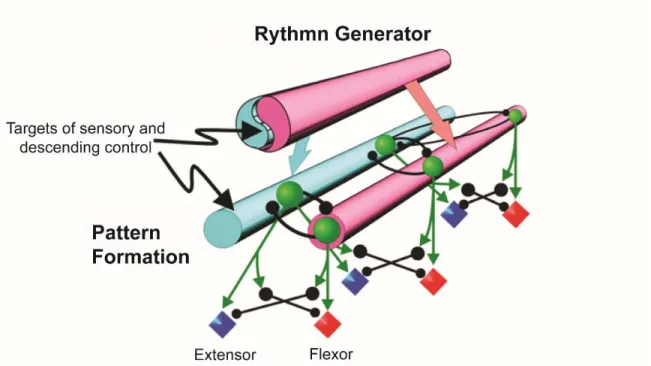
More recent studies have shown that this unit of modules may or may not be dissociated from the rhythm-generating circuitry, especially during fictive locomotion. Therefore, a multilayered spinal locomotor CPG, in which rhythm-generation and pattern formation are functionally separated has been proposed (McCrea and Rybak, 2008). In this two-level CPG, the rhythm generator controls features of the rhythm (i.e., cycle period, phase durations/transitions) and projects to the pattern-formation level, which coordinates and distributes activity to individual motor pools (Fig. 1). Inputs from peripheral mechanoreceptors or supraspinal structures can regulate activity at each of two levels, including spinal motoneurons (Rossignol and Frigon, 2011).
16
1.2.3 Localization of the CPG
Transversal sections of the cat spinal cord have been used to determine the rostro-caudal localization of the CPG. By isolating the L6-S1 segment of the spinal cord it was found that they could generate alternating activity between flexors and extensors in the cat (Grillner and Zangger, 1979). There are also different gradients of rhythmogenesis throughout the spinal cord. For instance, the rostral and caudal parts of the spinal cord present different gradients of excitation (Kiehn, 2006). In the cat the L3-L4 segments have greater rhythmogenic capacity than other spinal segments. The importance of the L3-L4 segments have been shown by localized topical application of clonidine (an α-2 noradrenergic agonist) into these specific regions which was sufficient to induce walking movements. On the contrary, injection of the α-2 NA antagonist yohimbine into these
Figure 1. Schematic representation of the two-level hierarchical architecture of the CPG. In this model a single rhythm generator and multiple unit pattern formation modules are separated and would be replicated in all four limbs. Both levels could be influenced by supraspinal and sensory information. Figure modified from McCrea and Rybak, 2008.
17 segments could block locomotion (Marcoux and Rossignol, 2000). Moreover, after a chronic spinal lesion at T13 on cats having recovered treadmill locomotion, a spinal transection performed on the L3-L4 segment abolished all locomotor activity demonstrating that the integrity of these segments is crucial for the expression of locomotion in spinal cats (Langlet, Leblond et al., 2005).
Several others experiments on the hen (Ho and O'Donovan, 1993), turtles (Mortin and Stein, 1989), rats (Cazalets, Borde et al., 1995) and in the cat (Deliagina, Orlovsky et al., 1983) have shown these specificity on the localization of this rhythmogenic areas in the spinal cord.
The transversal localization of these circuits have been revealed by several activity-labeling studies (Cina and Hochman, 2000;Dai, Noga et al., 2005;Kjaerulff, Barajon et al., 1994) and electrophysiological evidence (Tresch and Kiehn, 1999) showing that locomotor-related neurons are concentrated in a ventral location (laminae VII, VIII, and X). This finding have been confirmed in microlesion studies in the rodent (Bracci, Ballerini et al., 1996).
1.3 Locomotion and evaluation methods
Interactions between spinal locomotor CPG along with sensory afferents and descending inputs form a tripartite organization which allows gait modifications and adaptations to external conditions by generating various patterns of locomotion. This organization is central to our current understanding of how rhythmic patterns are continuously adapted to internal and external demands.
This chapter will first describe the kinematics and electromyography (EMG) activity recorded in intact animals during locomotion. This information is needed to specify the normal parameters generated by the nervous system for locomotion. Knowledge of limb kinematics has led to insights about the requirements for modifiable motor patterns (Stein and Smith, 1997).
18
1.3.1 Treadmill
The analysis of locomotion is usually done on a flat treadmill because the speed can be adjusted to different walking conditions of the cats while the sensory information from auditory, vestibular and visual systems remains approximately constant. Therefore, one of the main advantages of using treadmill over ground walking to study locomotion is that the velocity of locomotion can be controlled better and a large number of steps can be recorded and averaged allowing a statistical analysis of the different gait parameters. Also, the speed of the treadmill belt can be changed to cover the range used by the animal. This procedure allows collection of reliable data of continuous step cycles and the analysis of movement (Kinematics) and EMG activity.
However, in more natural surroundings the locomotor pattern is constantly adapted to the terrain and to the goals of the animal and the visual information varies continually (Halbertsma, 1983). This could introduce a variable in which adaptation through sensory feedback and voluntary corrections are more important for over ground walking than on an ordinary treadmill.
Variables such as speed or confinement to a constant environment are key to assess the efficacy of training methods and allows comparisons between them. Such variables could then be maintained on a treadmill and represent an important advantage of the treadmill compared to over ground locomotion especially in experimental locomotor training in which that same task (or steps) have to be repeated several times as will be used in this study.
1.3.2 Locomotor cycle and kinematics
A step cycle is defined as two successive contacts of the same foot on the treadmill. When any limb is in contact with the ground, it extends, and thus serves to propel the animal forward (stance phase). At the end of this phase, the limb is lifted from the ground by a movement of flexion, carried forward (swing phase), and finally is again placed upon the ground to repeat the cycle.
19 Other subdivisions of the swing and stance phases of the step cycle are commonly used. These patterns
were refined by describing the movements of different joints (hip, knee, ankle and metatarsophalangeal or MTP joints) in each phase (Phillipson, 1905). According to this subdivision, swing starts by a flexion (F) of all joints; while the hip continues its flexion, the ankle and knee start extending (El) until the paw touches the ground. At paw contact, the knee and ankle are passively flexed during weight
acceptance (E2 or yield phase), the MTP joint continues the extension initiated in El. During the third extension phase (E3 or push-off), all joints extend to propel the body forward. This phase ends when the foot is lifted off the ground and a new cycle starts (Fig. 2). Thus the swing phase is subdivided in F and E1 while stance is subdivided in E2 and E3.
Figure 2. Representation of limb joint measurements used to extract limb kinematics (stick diagrams) displaying separately the swing and stance phases and angular excursions. For angular excursions, flexion is always represented by a downward deflection of the traces. Phillipson (1905) subdivisions are shown: F and E1 constitute swing while E2 and E3 constitute stance. Figure modified from (Rossignol and Bouyer, 2004).
20
1.3.3 Interlimb coordination
Accurate coordination between the limbs is essential in locomotion. Appropriate coordination ensures the dynamic stability of head and trunk, prevents stepping of limbs on one another, allows a precise and smooth contact against ground for each limb, and reduces oscillatory movements of head and trunk, thus decreasing the energy needed for locomotion.
Interlimb stepping is linked by both rigid neural programs and modifiable programs that give the animals the flexibility necessary to meet changing postural demands imposed by a dynamic environment (English, 1979). This suggests that limb coordination somehow results from an interaction between independent neuronal centres (CPGs) controlling each extremity (see (Halbertsma, 1983).
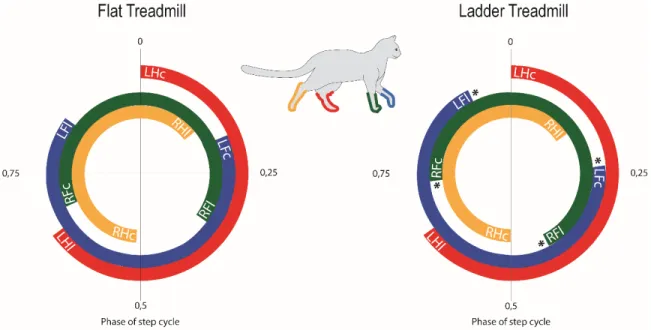
For walking gaits, the principal criterion for gait selection is stability. The body is always supported by two, three, or four paws at a time, so stability is increased by maximizing support by four feet, minimizing support by two feet, and, by selecting the combinations of two and three feet that are favorable to balance (Hildebrand, 1980). There seems to be a tendency to use more frequently a parallel coupling (same side of the body) between the forelimb and the hindlimb on the treadmill (Blaszczyk and Loeb, 1993) whereas during trot the forelimb and hindlimb are coupled to the contralateral hindlimb (diagonal coupling) (Hildebrand, 1980;Rossignol, 1996).
In symmetrical gaits, both hindlimbs and both forelimbs are coupled out of phase. During walking, the right hindpaw contacts the ground at 50% of the left cycle. Thus, a typical sequence of normal walking starting from the left hindlimb contact with the ground will pursue as follows: Left hindlimb – left forelimb – right hindlimb – right forelimb. Many neural mechanisms may participate in the control of interlimb coupling through different crossed pathways (Jankowska and Edgley, 1993;Jankowska and Noga, 1990) and other interlimb reflexes (Schomburg, 1990), and also through descending inputs to adapt to various external conditions (Blaszczyk and Loeb, 1993).
21 Interneurons may be responsible for the coordination between the CPGs, and they have been identified in invertebrate motor systems, such as the one controlling walking in the cockroach (Pearson and Iles, 1973). These interneurons appear to provide the next ganglion with an efferent copy, which might be used to produce intersegmental coordination. In cats a part of the ascending neuronal activity to the cerebellum provides efferent copy signals (Arshavsky, Berkinblit et al., 1972). Such signals could also be utilized to coordinate the CPGs (Grillner, 1981). Also, propriospinal system (see below) could also play an important role in relaying locomotor information between cervical and lumbar enlargements and therefore, the coupling of motor rhythms between the forelimbs and the hindlimbs (Cowley and Schmidt, 2000).
We studied limb coordination because its preservation or changes during the task we evaluate could be an expression of similar or different neural programs controlling locomotion within two different tasks (see chapter 2) presented in this project.
1.3.4 General muscle activity in cats
Functional classification on flexor and extensor muscles was first stablished based on experiments on decerebrate cats and dogs where flexion responses where produce in response to cutaneous stimulation on the limb or foot (Sherrington, 1910a). Muscles excited during the flexion reflex were generally classified as flexors and those muscles which responded by inhibition were classified as extensors. Flexor muscles generally close the joint angle whereas extensor do the opposite. For small toe muscles such as Extensor Digitorum Brevis, the dorsiflexion closes the metatarsophalangeal joint although the name suggests rather an extension.
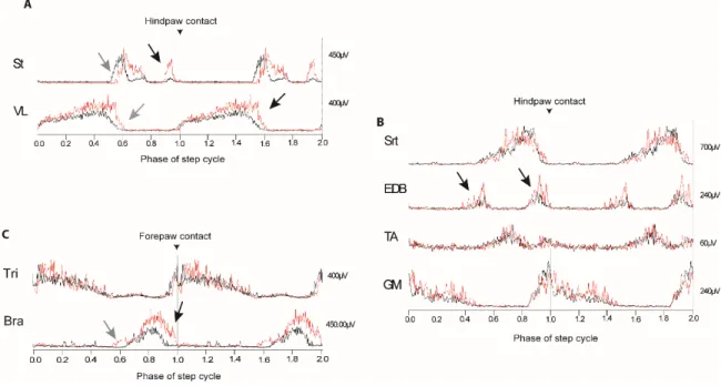
Describing in detail the normal pattern of flexors and extensors EMGs reveals the complexity of the mechanisms generating locomotion. Groups of agonist and antagonist muscles, in both the forelimbs and the hindlimb, are activated sequentially during locomotion. The sequential activation of muscle gives the flexibility of movements by which descending systems can regulate and modify limb activity (Krouchev, Kalaska et al., 2006) as seen in Fig.3.
22 Cats with chronically implanted EMG electrodes allow to assess how various muscles are utilized in such behaviors and how the nervous system specifically selects various anatomical patterns and combinations in appropriate phases.
Differences in EMG descriptions found in the literature might depend on the animal’s inherent variability, strategies, anatomical variability, on what part of the muscle was recorded or what kind of electrodes was used (Loeb, 1993). However, some muscles have a more or less consistent pattern under various experimental conditions and are described below.
The knowledge of muscular dynamics, its specific EMG shapes or “signatures” and their normal activation during the different phases of the step cycle helps us identify changes when altering external conditions or intrinsic variables within the animal. Thus, it allows Figure 3. Schematic representation of chronically implated muscles used in this study showing origins and point of insertion and their respective raw EMG signals. Flexors and extensor are activated in an alternate and rythmic manner. Each muscle presents particular EMG “signatures” or shapes. St: Semitendinosus, VL: Vastus lateralis, EDB: Extensor digitorum brevis, GM: gastrocnemius medialis, TA: Tibialis anterior, Bra: biceps brachii, Tri: Triceps brachii.
23 us to understand how the locomotor system works, how it is modified and, depending on the study approach, hypothesize about the structures responsible for these changes.
1.3.5 Activity of flexor and extensor muscles
Extensors
During forward walking, extensor muscles across the hindlimb are co-active during most of the stance phase to counteract ground-reaction forces that tend to flex the joints (Perell, Gregor et al., 1993;Smith, Carlson-Kuhta et al., 1998;Smith, Edgerton et al., 1977). Extensor muscles have very similar patterns of activity. They are activated in cats some 20-80 ms before the paw contact so that their discharge is not normally triggered by sensory events associated with contact. Except for Vastus Lateralis muscle (see below), most of the extensor muscles present a large main burst beginning just before ground contact and terminating just before the end of the stance phase. Although the activity in these muscles generally shows an on-off behavior, characteristic differences exist between extensor muscles (Grillner, 1981).
Flexors
Muscles related to the swing phase may have a more complex and versatile discharge pattern. These muscles usually have either one or two bursts of activity during swing. During all forms of walking, the cat's hip and ankle flexor muscles exhibit one burst of activity, and the burst is associated with a flexor muscle torque at each joint (Stein and Smith, 1997).
Flexor muscles are not all activated simultaneously. Instead, they show variations in the initiation, duration and termination of their activity during the step cycle. For instance, the initial period of activation in the most distal muscles, occurs just before the cat lifts the paw from the treadmill. Subsequently there is activation of the knee flexor muscles which serves to raise the paw from the support surface. Flexion of the ankle and transport of the
24 limb is followed by the braking action of bi-functional muscles (Wisleder, Zernicke et al., 1990) such as Semitendinosus (see below) and by activation of the distal flexors in preparation for landing (Krouchev, Kalaska et al., 2006).
Flexor-Extensor coordination
The main flexors (hip, knee, ankle and foot) reciprocate with the extensors in a complex way. The coordination between flexors and extensors can be illustrated on the dynamics of a few main muscles of the hindlimb (Fig.3). For example, one of the periods of activity in the semitendinous (St) occurred at the same time as a brief period of activity in an extensor muscle, the gastrocnemius lateralis (GL), whereas the other period of activity in the St occurred at the same time as a period of activity in the extensor digitorum brevis (EDB). The major periods of activity in the lateral (GL) and medial (GM) heads of the gastrocnemius also occurred at the same time during stance (Krouchev, Kalaska et al., 2006).
1.3.6 Individual muscular activity of relevance to this study
Flexors:
Semitendinosus (St)
Semitendinosus (St) has its origin on the tuberosity of the ischium and passes to the medial side of the tibia to insert into the crest of the tibia about 1 cm from its proximal end. St is a bifunctional muscle: it acts as a knee flexor and a hip extensor. This muscle has a particular discharge pattern: it has two EMG bursts during the swing phase of forward walking. One larger burst (first) with a sudden onset occurs around paw lift-off and a shorter burst (second), which is not always present at very low speeds of locomotion, precedes paw contact. Changes in the duration and amplitude of both bursts are closely tied to changes in the magnitude and duration of the flexor muscle torque at the knee joint (Smith, Chung et al., 1993).
25 The first St burst involved in the earliest part of swing when the paw is lifted off the ground is active before and during the F phase of the knee, indicating a knee flexor and not a hip extensor action (Halbertsma, 1983). The second St burst coincides with the first part of F phase of the hip and the first part of the E1 phase of the hip. This could be a combined hip extensor action. This second burst is more consistent at higher speeds and may be related to the need for a greater torque in flexion to decelerate both the hip and knee at the end of swing in preparation for foot contact (Smith, Chung et al., 1993;Wisleder, Zernicke et al., 1990).
Extensor digitorum brevis (EDB)
Ankle and digits extensor muscles are interesting in many aspects since they have an important role in positioning and stabilizing the foot, as well as flexing (plantar-flexion) the digits.
The extensor digitorum brevis (EDB) has its origin from the distal border of the calcaneal ligament and from the dorsal surfaces of the proximal ends of the three lateral metatarsals. It ends in three flat tendons which run in the interspaces between the four tendons of the extensor digitorum longus. This muscle prepares the paw for placement on the support surface. It has a large burst of activity at the beginning of the E1 phase, which declines when the ankle and foot extensors start their main activity and exhibits some residual activity throughout the support phase. The main extensor activity, evident by a second burst in the E2 phase, often has a rapid onset in connexion with the placing of the foot (Engberg, 1964).This muscle in particular is maximally active during the placing of the foot and differs from all other flexors in being relaxed in the flexion phase and the third extension phase (Engberg and Lundberg, 1969).
Tibialis anterior (TA)
Tibialis anterior muscle functions as an ankle dorsiflexor following the regular flexor pattern activity. It originates is from the proximal lateral side of the tibia and it ends distally in a strong tendon that insets into the outer surface of the first metatarsal. TA is strongly activated for a short period at the end of the third extension phase but this is followed by
26 another strong activation in the flexion phase and the muscle is only relaxing for a short period before a very strong burst of activity at the end of the first extension phase. The very striking increase in flexor activity late in the first extension phase may be needed for a deceleration of the more rapid forward swing of the leg in the trot (Engberg and Lundberg, 1969).
Biceps Brachii (Bra)
Biceps Brachii is an elbow flexor of the forelimb. It originates from the supracondyloid foramen and the humerus and it ends by a flat tendon which joins the tendon of the cleidobrachialis on the lateral surface of the ulna. This muscle starts its discharge before the F phase (paw-off), maintaining its activity during the whole swing phase and terminates before or at the paw contact.
Extensors: Vastus lateralis (VL)
M. vastus lateralis is a part of the large quadriceps femoris muscle which covers the cranial, lateral and medial part of the femur or thigh. It is a knee extensor whose activity gradually starts before the onset of the stance phase, and usually is most active in the later part of the E3 phase. The VL activity terminates abruptly and almost always before the onset of the swing phase. (Engberg and Lundberg, 1969).
VL is thus active just before and during the major part of the support phase. This corroborates its supporting function and it could propel the animal during the last part of its activity.
Gastrocnemius medialis (GM)
Gastrocnemius medialis is an ankle extensor and knee flexor. It originates from the medial sesamoid bones of the femur and it inserts along with the gastrocnemius lateralis by the calcaneal tendon into the proximal end of the calcaneus. It has an intense burst of activity at the onset of the E1 phase of the ankle and 60 to 80 ms before the onset of the
27 stance phase. The activity drops gradually and terminates before the VL activity around 200 ms before the onset of the swing phase. This large activity occurring before foot contact has played an important role in defining the preprograming of muscle activity by a central generator in advance of the actual foot contact (Gorassini, Prochazka et al., 1994).
GM is active during the first part of the stance phase, which suggests a support function of the body after touch-down. GM is less active during the last part of the stance phase, the propulsive phase, where the knee extensors are still active. The delay between the EMG activity and the force development in the muscle could be responsible for the substantial ankle torque, which is needed during this phase (Manter, 1938).
Triceps Brachii (Tri)
Triceps Brachii is an elbow extensor of the forelimb. This muscle has three heads (lateral, long and medial) which originate from various areas on the scapula or on the humerus and ends at various points of the ulna. The three heads start their discharge prior to paw contact. This precedence increases with speed, from practically null during walking into several milliseconds during trotting and galloping in dogs. The long head of triceps that attaches to the scapula often has a short distinct burst just prior to swing (Drew and Rossignol, 1987;Rossignol, 1996).
1.4 Control and modulation of Locomotion
Locomotion results from intricate dynamic interactions between a central program and feedback mechanisms. The central program relies fundamentally on a genetically determined spinal circuitry (CPG) capable of generating the basic locomotor pattern and on various descending pathways that can trigger, stop, and steer locomotion. The feedback originates from muscles and skin afferents as well as from special senses (vision, audition, vestibular) and adapts the locomotor pattern to the requirements of the environment.
28 Afferent inputs
from muscles or the skin reach the spinal cord, project to motoneurons directly
or through interneurons, or through the CPG itself. Afferents also send collaterals through ascending pathways (directly or via relay interneurons) reaching supraspinal structures (telencephalon, brain stem, cerebellum), which in turn via cerebral cortex or brain stem, project down to the spinal cord on neurons that may or may not also
be contacted by the same primary afferent. At the spinal cord level, sets of interneurons will be selected to modulate transmission during a given task or during a particular phase of a task. In this way it is ensured that reflex activations of given muscles occur only at the appropriate times in the step cycle (phase-dependent modulation) and generate the right locomotor pattern (Rossignol, Dubuc et al., 2006). See Fig. 4.
The study of the tripartite system reveals at the same time the autonomy and the interdependence of the various control parts as it will be described in the next sections.
Figure 4. The control of locomotion is tripartite. Spinal CPG is at the core of the system and it can be altered or modulated by sensory inputs (propiospinal and/or cutaneous) and descending signals from supraspinal structures (cortical or subcortical). These 3 strucutres are in constant interaction to drive the appropiate commands to specific muscles to adapt locomotion to external situations. Interneurons in the spinal cord could participate in this modulation before reaching their respective motoneurons.Figure modified from Rossignol et al,. 2006.
29
1.4.1 Sensory inputs
Sensory inputs include proprioceptive and cutaneous receptors. The implication of the sensory inputs to locomotion has been studied with various approaches such as recording the activity of afferents, inactivating the afferents through neurectomy or by stimulating the afferents throughout locomotion or in discrete phases of the cycle (as reviewed in (Rossignol, 1996). Results from fictive locomotion have shown that sensory inputs are not necessary to produce the basic locomotor pattern. What are then the roles of sensory information during locomotion?
The hindlimbs of a spinal cat, when walking on split-belt treadmill, can separately adapt their speed to that of their corresponding belt. This is the result of the sensory activity generated by foot contact with the belt (Forssberg, Grillner et al., 1980b). Obstruction of the hip movement (Grillner and Rossignol, 1978) or loading of extensor muscles (Duysens and Pearson, 1980) can completely suppress rhythmicity in a leg while the other legs continue to be rhythmically active.
This and many other experiments have proven that sensory inputs are crucial in adapting and modulating the operation of the CPG in the real environment. Sensory inputs can have global influences in allowing, preventing, or selecting motor patterns. Through dynamic interactions, sensory inputs can participate in the correct positioning of the paw in uneven terrain and they can also modify the frequency of the pattern, its intrinsic structure, or the amplitude of muscle discharges within locomotor phases (Rossignol, Dubuc et al., 2006). Afferent inputs most relevant to walking primarily arise from stretch- and load-sensitive mechanoreceptors located in muscles and skin.
1.4.1.1 Proprioceptive inputs
Proprioceptors (muscle, joint and deep fascia receptors) as well as cutaneous receptors have general roles such as providing signals acting as on-off switch to set the range of joint angular excursion within which locomotion can take place. However, an important role of muscle afferent feedback appears to be in setting the overall timing of the step cycle by
30 adjusting the duration of the various phases of the locomotor cycle and facilitating the switch between phases. Another important role is to regulate the output amplitude of muscles in various phases (as reviewed in Rossignol, Dubuc et al., 2006).
Experiments suggest that hip proprioceptors exert a powerful control over the initiation of the locomotor rhythm because lifting a spinal cat or dog by the thorax, thus extending passively the hindlimbs, triggers air stepping. This is largely due to stimulation of proprioceptors (especially at the hip), since removal of the skin does not abolish the response (Sherrington, 1910b). Contrarily, flexing the hip in the same preparation can prevent air stepping. Similarly, in chronic spinal cats, flexion of the hip joint on one side can abolish treadmill stepping on that side, while the other side continues to walk (Grillner and Rossignol, 1978).
As mentioned above, spinal cats can modify the structure of the step cycle of individual limbs when walking on a split treadmill moving at different speeds. Although cutaneous receptors of the foot pads cannot be discarded as an important source of inputs for such speed adaptation, the experimental evidence suggests that proprioceptive inputs from muscles and/or joints are the main contributors (Forssberg, Grillner et al., 1980b).
1.4.1.2 Cutaneous inputs
Early work of Sherrington showed that removing cutaneous inputs from the hindlimbs did not prevent locomotion even after spinalization (Sherrington, 1910a). This was largely supported by others who reported little deficits when cutting cutaneous nerves in intact cats (Duysens and Stein, 1978) or infiltrating the central foot pad with a local anesthetic (Engberg, 1964). In these experiments on cats with an intact spinal cord and with extensive denervation on the hindlimb, stepping was almost normal on a regular treadmill after compensation of the deficits through locomotor training.
Stimulation of the perineal region (scrotum, vulva and base of the tail, inguinal fold) (Krawitz, Fedirchuk et al., 2001), is most effective in triggering locomotion. The activation of unspecific afferents from the perineal region presumably underlies some important survival function (such as escape from a predator), but the mechanisms of interactions of these perineal afferents with the CPG are not known. Other types of tonic stimuli can
31 completely inhibit these locomotor movements in both normal and fictive locomotion. For example, pressure of the skin of the lumbar region (Viala and Buser, 1974) or controlled electrical stimulation (Viala, Orsal et al., 1978) will abolish locomotion in the rabbit.
Although cutaneous inputs may have some general roles to play in locomotion such as triggering walking (perineal stimulation) or inhibiting walking (inhibitory inputs from the skin of the back), skin afferents participate predominantly in the correction of limb, foot placement during stance on uneven terrain and also on the expression of locomotion after spinal lesions. For example, after a complete spinal lesion denervated cats never recovered locomotion even when submitted to training showing the importance of cutaneous inputs on the compensatory mechanisms to regain stepping (Bouyer and Rossignol, 2001).
1.4.2 Propriospinal system
Spinal segments are interconnected by short and long intraspinal pathways (i.e., propriospinal) that run close to the gray matter bilaterally.
It was demonstrated that after corticospinal tract lesions in rats, new connections could be established with the lumbosacral cord through cervical propriospinal pathways (Bareyre, Kerschensteiner et al., 2004). Propriospinal pathways appear to be of considerable importance for volitional aspects of locomotor recovery after an incomplete SCI in adult mice (Courtine, Song et al., 2008) and cats (Kato, Murakami et al., 1984) through the formation of new functional circuits.
Moreover, high spinal preparations are capable of developing coordinated activity involving both the fore- and the hindlimbs (Miller and Van der Meche, 1976). This is apparently achieved via propriospinal pathways which reciprocally interconnect the cervical and lumbar enlargements of the cord (Halbertsma, Miller et al., 1976;Miller, Van der Burg et al., 1975). Indeed, in experiments where synaptic transmission in the thoracic region was supressed in neonatal rats, a disruption in the motor rhythms between cervical and lumbar regions resulted (Cowley and Schmidt, 2000).
32
1.4.3 Supraspinal inputs and pathways
Supraspinal control of locomotion can be viewed from many aspects: initiation of locomotion, control of posture and propulsion, and corrections to adapt one or several steps to the environment. Several structures can be implicated in the initiation and can play a role in posture and correction and they will be described in this section.
1.4.3.1 Initiation of locomotion
Experiments applying transections and electrical stimulation at different levels of the neural axis have shown that the regions for initiation of locomotion are located in the brain stem, at supraspinal level (Shik, Severin et al., 1966). Electrical stimulation of a region of the midbrain can evoke locomotion in decerebrate cats and other mammals (Gelfand, Orlovsky et al., 1988;Shik and Orlovsky, 1976). The structure implicated on this initiation is the mesencephalic locomotor region (MLR).
The MLR region receives projections from several brain structures including the basal ganglia, the sensorimotor cortex and the limbic system and projects toward the middle part
of the reticular formation (Garcia-Rill, Skinner et al., 1983;Shik, 1983;Steeves and Jordan,
1984). This last projection to the reticular formation would be one important connection
implicated in the initiation of locomotion (Steeves and Jordan, 1980). The MLR does not
project directly to the spinal cord. Instead it initiates and controls locomotion through monosynaptic connections to the reticular formation of the brain stem which, through the ventrolateral funiculi, activate the spinal locomotor networks to initiate locomotion (Garcia-Rill and Skinner, 1987;Shik, 1983).
1.4.3.2 Posture and corrections of steps during locomotion
In addition to a role in the initiation and termination of locomotion, the brain stem contains centers which are important for the modulation of locomotor activity. Reticulospinal, rubrospinal and vestibulospinal pathways are capable of influencing locomotor neural circuits in the spinal cord (Drew, Jiang et al., 2002a).
33 Descending pathways that influence motor activity, including locomotion, can be divided into two principal systems (Lawrence and Kuypers, 1968):
A medial system that includes the reticulospinal and vestibulospinal pathways.
The reticulospinal pathway arise from regions of the reticular formation in the brainstem projecting through the anterior funiculus of the spinal cord to the motoneurons of the axial musculature (Kuypers, 1981). Commands that initiate and modulate locomotor circuits in the spinal cord are transmitted through the reticulospinal tract. Moreover, the reticulospinal pathways are involved in the modifications of posture that anticipate and accompany voluntary movements and which ensure stability of the body during locomotion (Drew, Dubuc et al., 1986;Rossignol and Frigon, 2011).
The vestibulospinal pathway can be divided in a lateral vestibulospinal tract and medial vestibulospinal tract, both originate from 2 of the 4 vestibular nuclei in the brainstem. They course through the anterior funiculus of the spinal cord on the ipsilateral side. Because of its origin from the vestibular nucleus (lateral or medial), this pathway is implicated in the control over postural changes necessary to compensate for sloping ground by acting specially over the extensor muscles during the stance phase (Matsuyama and Drew, 2000). The destruction of the lateral vestibular nucleus decreases the activity of the extensor muscles in a decerebrate cat (Yu and Eidelberg, 1981).
Overall, the medial system has a relatively diffuse action on flexor and extensor muscles of the more proximal limb and the axial musculature and serves to adjust the posture of the animal. Results from lesion on the ventrolateral quadrant (Brustein and Rossignol, 1998;Gorska, Bem et al., 1990;Kuypers, 1963) of the spinal cord (which contains these pathways) and also microstimulation (Degtyarenko, Zavadskaya et al., 1993;Drew, 1991a;Drew and Rossignol, 1984) suggest that this system is responsible for producing the requisite muscle tonus necessary to support the body, for ensuring the lateral stability of the animal, and for producing step by step regulation of the level of muscle activity during locomotion.
Another important structure related to postural adaptations to external conditions is the cerebellum. Cerebellar contributions to locomotion has been hypothesised to help provide
34 adaptability to motor patterns but its specific role is still not clear. One theory suggest that the cerebellum might act as “real-time sensory processing device” (Bower, 1997), by processing sensory inputs and making alterations of the ongoing movement patterns. However other theory suggest that the cerebellum might alter ongoing movement patterns in a predictive manner based on a stored internal representation of a specific movement (Maschke et al., 2004; Smith and Shadmehr, 2005).
A lateral system, includes the corticospinal and rubrospinal pathways.
The rubrospinal pathway originates in the red nucleus of the midbrain. The axons immediately cross to the contralateral side of the midbrain, and course through the lateral funiculus reaching motoneurons in the spinal cord. The motor cortex and premotor cortex both project to the red nucleus from which the rubrospinal tracts arise (in the cat) resulting in an alternative way by witch voluntary commands can be sent to the spinal cord (Schieber, 2007).
The corticospinal pathway provides the most direct pathway over which the cerebral cortex controls movement. It originates mainly in the motor cortex from which its axons courses through the internal capsule and then to the cerebral peduncle on the brainstem. The axons then form the medullary pyramids on the ventral surface of the brainstem splitting in two from there. Most of the axons (90%) cross over to the contralateral side at the pyramidal decussation forming the lateral corticospinal tract that courses through the lateral funiculus of the spinal cord. The 10% of remaining axons form the anterior corticospinal tract that courses ipsilaterally through the anterior funiculus (Kuypers, 1981). Once they reach the spinal segment in which they end the axons cross to the contralateral side to innervate motoneurons of the ventral horn. Even though the corticospinal tract is not indispensable for the production of the basic locomotor rhythm in cats, it contributes to the regulation of locomotion, especially when, in order to adapt to external conditions, a requirement for precise control over paw placement is needed (Drew, Jiang et al., 2002b). Altogether, the lateral system is responsible for voluntary and goal-directed aspects of locomotion, as well as fine control of the distal musculature. It also plays a critical role in more difficult locomotor tasks, such as obstacle avoidance and ladder walking (Drew, Prentice et al., 2004;Liddell and Phillips, 1944).
35 Lesions of the dorsolateral funiculi at the low thoracic level that completely interrupted both the corticospinal and rubrospinal pathways produced long-term deficits in locomotion on a level surface. These deficits included a foot drag that was probably caused by both a loss of cortico- and rubrospinal input to motoneurones controlling distal muscles as well as by a change in the relative timing of muscles acting around the hip and knee. Smaller lesions produced similar deficits from which the cats recovered relatively quickly. Cats with the largest lesions of the dorsolateral funiculi were unable to modify their gait sufficiently to step over obstacles attached to the treadmill belt even 3–5 months after the lesion suggesting that the corticospinal tract provides an important contribution to the modification of the basic locomotor rhythm to take into account the variations of the walking environment (Drew, Jiang et al., 2002a). These results also imply that the medial pathways, the reticulo- and vestibulospinal pathways, are unable to fully compensate for damage to the lateral pathways.
The knowledge of supraspinal pathways and of their functions in normal locomotion allows us to infer which supraspinal pathway is affected based on observed motor deficits. Also, based on the reduction of specific deficits, for example after locomotor training, we can infer which pathway is being activated and then compare advantages of a specific locomotor training method over another.
1.5 Skilled locomotion
Even though decerebrate cat preparations allow studying the organization of the CPG and its interaction with sensory information, these preparations do not throw any light on the roles of more rostral structures, such as, for example, the motor cortex (MC). For this, other neurophysiological investigations in conditions that require dynamic voluntary adaptations to the environment such as skilled locomotion are necessary (Armstrong, 1986a).
It has been hypothesized that the single most important event in the step cycle is the placement of the foot at touch-down. A precise control over this event is important to avoid
36 obstacles and holes in the terrain and to ensure a stable support (Halbertsma, 1983). So it is logical to think that many of the voluntary modifications are done to improve this aspect of skilled locomotion.
The contribution of the MC to the control of voluntary movement has been demonstrated by a large number of studies using single neuron recording techniques to probe the relationship between neuronal activity and behavior (Drew, 1988). Many authors have shown for example that, in cats, the discharge of identified projection neurons from motor cortex (pyramidal tract neuros, PTNs) are increased in locomotor tasks in which animals are required to modify their gait or to accurately control the position at which the foot is placed. These tasks include stepping over moving obstacles (Drew, 1988), peg walking (Donelan, McVea et al., 2009) and walking on a horizontal fixed ladder (Amos, Armstrong et al., 1990;Armstrong, 1986a;Beloozerova and Sirota, 1993).
These experiments suggested that the motor cortex is involved in the transformation of a visual signal (obstacle, barrier, or ladder rung) into a motor act (the modification of the basic locomotor rhythm) (Kalaska and Drew, 1993).
Experiments involving destruction of the MC or interruptions of the pyramidal tract have demonstrated the inability of lesioned cats to walk on wire mesh or on elevated horizontal bars (Chambers and Liu, 1957;Trendelenburg W, 1911). Without the MC, dogs standing stationary lost the ability to place the paw in the appropriate space zone (Grillner, 1981). The pattern of impulse activity in motor cortex cells changes during unexpected perturbations of the stepping of the cat, as well as during voluntary modification during walking (Amos, Armstrong et al., 1989;Drew, 1988). Patterns of activity on the MC can also be seen at different times of the step cycle. For example, recordings have shown that the mean activity of motor cortical neurons was elevated in late stance and early swing during accurate stepping, suggesting that there is an important cortical regulation at the moment of the paw contact with the ground (Beloozerova, Farrell et al., 2010).
Descending drives certainly contribute to the production of locomotion in normal conditions by refining the basic synergy and improving the level of inter-limb coordination which are otherwise limited at the spinal level only (Armstrong, 1986a).
37 The importance of these skilled locomotor tasks is that they challenge the animal to walk in more demanding conditions. This allows us to investigate deficits after lesions of the nervous system that otherwise might be undetected during level treadmill walking. For example the inactivation of the motor cortex by TTX produces several deficits on locomotion over a horizontal fixed ladder while it does not affect locomotion over a conventional treadmill (Beloozerova, Farrell et al., 2010). Also, after extensive cutaneous denervation of the hindlimb on cats with an intact spinal cord almost normal locomotion was achievable on a regular treadmill but locomotion on a horizontal ladder was not possible (Bouyer and Rossignol, 2001).
1.6 Animal models of SCI and the importance of
locomotor training.
Spinal cord injury (SCI) in animal models provides an opportunity to study not only the functions of various CNS structures but how they adapt to different external conditions to maintain or recover locomotion.
1.6.1 Complete lesions
After complete section of the spinal cord, descending inputs to the spinal locomotor circuitry are altered because of the severance of pathways controlling the circuits for initiation and modification of locomotion. Remaining peripheral sensory afferents to spinal circuits as well as intrinsic changes of spinal circuits are then responsible of the plastic changes that allow locomotor recovery.
Even though it was first showed that kittens could recover locomotion after a complete spinal lesion (Forssberg, Grillner et al., 1980a;Forssberg, Grillner et al., 1980b;Grillner, 1973) subsequent studies showed that in adult cats recovery was also possible (Barbeau and Rossignol, 1987;Rossignol, Bélanger et al., 2000;Rossignol, Chau et al., 2002). Few days after spinalization, perineal stimulation can elicit rhythmic, alternating stepping in the hindlimbs, although plantar foot placement is absent and an important foot drag is seen.
38 Locomotor training on a treadmill during 3 weeks to 3 months (depending on the animal) allows such cats to recover alternate hindlimb movements, hindquarter support and plantar foot contact (Barbeau and Rossignol, 1987). These findings showed that locomotor recovery after a complete spinal lesion is achieved by interactions between the CPG with sensory feedback from the hindlimbs.
The importance of regular daily training (starting even after 1 month after transection) has been proved by many authors (Edgerton, de Guzman et al., 1991;Lovely, Gregor et al., 1986;Lovely, Gregor et al., 1990;Rossignol, Martinez et al., 2015). This is of great interest in the clinical situation, where training may be started late after the spinal injury (Barbeau, Dannakas et al., 1992;Wernig and Muller, 1992).
1.6.2 Incomplete lesions
Although models of complete SCI are important in determining intrinsic spinal mechanisms involved in locomotor recovery, most SCIs in humans are incomplete, and spared descending pathways can still access the spinal circuitry.
Lateral hemisection models, where several pathways are severed simultaneously on one side only, are relevant because they mimic the clinical situation in which spinal lesions due to accidents or falls damages several pathways producing characteristic deficits and directly influencing locomotor recovery (Rossignol and Frigon, 2011).
Although substantial recovery of hindlimb locomotion is observed following a lateral hemisection of the spinal cord, some deficits, mostly observed on the side of the lesion, can persist. Deficits observed after a lateral hemisection resemble those associated with a ventrolateral (transiently impaired body equilibrium, interlimb coupling) together with a dorsolateral (impaired skilled locomotion) lesions (Brustein and Rossignol, 1998;Gorska, Bem et al., 1990;Kuypers, 1963).
After incomplete SCI, spared pathways originating from supraspinal and propriospinal structures can play an active role in the recovery process, and also in restoring some voluntary control (Rossignol, Dubuc et al., 2006). However, intrinsic spinal circuits and peripheral afferents still remain to initiate and organize hindlimb locomotion. The
39 compensation by descending pathways may take different forms. Damaged pathways may regenerate while undamaged pathways may sprout or may change the efficacy of their transmission. In doing so, new circuits could result from new anatomical connections or from enhanced connectivity (enhancing existing circuits) (Rossignol and Frigon, 2011).
How important is the plasticity of the spinal CPG for the recovery of hindlimb locomotion after partial spinal lesions, considering that spared descending pathways still have access to the spinal cord? The essential role of the intrinsic spinal circuitry was clearly demonstrated by a dual spinal lesion paradigm, in which cats with a lateral hemisection at T10-T11 and then trained on a treadmill was followed several weeks later by a complete spinalization at T13 (Barrière, Cohen-Adad et al., 2008;Barrière, Frigon et al., 2010). Hindlimb walking was observed in all trained cats within hours following spinalization, a process that usually takes 2 to 3 weeks of treadmill training to appear in spinalized cats (Barbeau and Rossignol, 1987). This is a demonstration that important intrinsic changes had already occurred within the locomotor spinal circuitry following the partial spinal lesion. The first major difference between untrained cats compared to trained cats was an important asymmetry in stepping capacity between both hindlimbs observed during the first 10 days after complete spinalization. For instance, kinematic analysis in these cats showed that the right hindlimb remained most of the time in a flexed position while rhythmic activity was seen in the left lesioned side. However, in cats that were trained after the initial hemisection, the walking pattern after a complete spinalization was similar to what is observed in cats with an intact spinal cord, corroborating earlier evidence that the intrinsic spinal network can be improved by training (Barrière, Cohen-Adad et al., 2008;Barrière, Frigon et al., 2010;Martinez, Delivet-Mongrain et al., 2011). Plastic changes during the hemisected period might be the result of compensation by remnant descending inputs that imprint into the spinal cord new modes of functioning (Chen and Wolpaw, 2002).
Further experiments using the dual-lesion paradigm showed that minimizing the period of recovery between a spinal hemisection and the second complete lesion to 3 weeks and without locomotor training, asymmetric changes seen after the hemisection could be maintained after spinalization (Martinez, Mongrain et al., 2011;Martinez,
Delivet-40 Mongrain et al., 2012b). Results from these experiments proved that even though locomotor recovery is possible without locomotor training, the way the spinal circuits could be reorganized depends on the conditions during the hemisected period. For example, cats that re-expressed the best bilateral hindlimb locomotion after spinalization were the ones with the most prominent locomotor deficits and had the larger initial hemisection. This asymmetrical reorganization of the spinal circuits in absence of locomotor training could only lead to the conclusion that “treadmill training leads to a more symmetrical spinal reorganization and a bilateral stepping pattern after spinalization by forcing the two hindlimbs to walk after hemisection” (Martinez, Delivet-Mongrain et al., 2011).
If locomotor training after partial lesions can induce such changes in the spinal circuitry for locomotion, could a training requiring more supraspinal commands improve locomotor recovery differently than a regular training on FTM after an incomplete spinal lesion? This question is the main issue investigated in this project. We try to answer this question by using a new method that increases the influence from supraspinal structures to the locomotor training.
1.7 Problem statement
As we have seen in this introduction, locomotion is controlled at multiple levels of the CNS and more complex locomotor tasks such as skilled locomotion require complex interactions in order to take place. After spinal lesions, the recovery of function involves optimizing these interactions between remaining structures resulting in the development of compensatory mechanism to optimize the basic locomotor function.
Plasticity within intrinsic spinal circuits is a critical component of hindlimb locomotor recovery. Locomotor control, seen as a tripartite system where CPG, sensory feedback and supraspinal structures are in constant interaction, is changed or interrupted somehow after a spinal lesion. Thus, after SCI, targeting intrinsic spinal circuits by stimulating or
engaging remaining pathways and sensory afferents should be a focus for rehabilitative strategies (Barbeau and Rossignol, 1994;Harkema, 2008).