Université de Montréal
Dissecting the dynamic of Noc2p and its partners in
pre-60S particles maturation
par Katherine Cléroux
Département de biochimie Faculté de Médecine
Mémoire présenté à la Faculté des études supérieures en vue de l’obtention du grade de Maître ès Sciences
en biochimie
option génétique moléculaire
Avril, 2014
Résumé
Plusieurs études ont permis la caractérisation de la structure et de la fonction du ribosome. En ce qui attrait à la biogénèse du ribosome, nombreux aspects restent à être découverts et compris de façon plus dynamique. En effet, cette biogénèse englobe une variété de voies de modifications et d’assemblages requises pour la maturation des ARNr et pour leurs liaisons avec les protéines ribosomales. De ce fait, les protéines Noc ont été caractérisées comme des facteurs d’assemblages et ont permis la découverte d’une des premières indications sur l’ordre spatio-temporel de la maturation du ribosome. Ainsi, en utilisant la levure comme modèle, notre objectif est d’étudier d’avantage l’échange des complexes composés des protéines Noc ainsi que leur localisation intranucléaire. Ainsi, la nature des interactions de Noc2p avec Noc1p et Noc3p et l’influence de l’arrêt du transport intranucléaire ont été étudiés en utilisant des promoteurs inductibles, la microscopie à fluorescence, des immunobuvardages, qRT-PCR et des purifications par affinité.
Mots-clés :
Biogénèse du ribosome, maturation du ribosome, voie d’assemblage, ordre spatio-temporel, Noc1p, Noc2p, Noc3p, Rix7p
Abstract
Several studies have been performed to characterize the ribosome as far as to understand its structure and its function. However, major aspects of ribosome biogenesis remain elusive or gave only a static picture of the process. In fact, ribosome biogenesis involves dynamic processing and assembly pathways that are required for rRNA modification and folding, in addition to rRNA binding with some ribosomal proteins. One set of assembly factors, the Noc proteins, allowed one of the first indications about the spatio-temporal ordering of ribosome maturation. By using yeast as model, our objective is to provide a dynamic picture of the Noc proteins complexes exchange and nuclear localization by determining the nature of Noc2p interactions with Noc1p and Noc3p and by studying the influence of reversibly arrested intranuclear transport on these proteins and on Rix7p, an AAA-ATPase. In order to achieve these aims, inducible promoter, fluorescent microscopy, western blot, qRT-PCR and affinity purification analyses were used.
Keywords:
Ribosome biogenesis, ribosome maturation, assembly factors, spatio-temporal ordering, Noc1p, Noc2p, Noc3p, Rix7p.
Table of contents
1 Introduction ... 17
1.1 The ribosome ... 18
1.2 Ribosomal DNA transcription ... 20
1.3 Ribosomal processing pathway ... 24
1.3.1 90S processiong ... 24 1.3.2 Pre-40S processing... 26 1.3.3 Pre-60S processing... 26 1.4 Assembly pathway ... 27 1.4.1 Pre-40S assembly ... 29 1.4.2 Pre-60S assembly ... 29
1.5 Coordination and quality control system ... 31
1.5.1 AAA-ATPase Rix7p ... 33
1.6 The Noc proteins ... 36
1.6.1 Noc1p/Mak21p in S.cerevisiae ... 36
1.6.1.1 Homologs of Noc1p in higher eukaryotes... 38
1.6.2 Noc2p in S.cerevisiae... 40
1.6.2.1 Homologs of Noc2p in higher eukaryotes... 41
1.6.3 Noc3p in S.cerevisiae... 43
1.6.3.1 Homologs of Noc3p in other organisms ... 45
1.6.4 Noc4p in S.cerevisiae... 46
1.6.4.1 Homologs of Noc4p ... 47
1.6.5 The dynamics of Noc protein exchange... 47
1.7 The Project and its objectives... 55
1.7.1 Rationale and previous work ... 55
1.7.2 The use of ts mutants and ribosome biogenesis ... 56
1.7.3 Aims and objectives ... 58
2 Matériels et méthodes ... 61
2.1 Chemicals ... 62
2.2.1 Bacteral culture media ... 62
2.2.2 Yeast culture media... 62
2.3 Plasmids ... 64
2.4 Bacterial techniques ... 64
2.4.1 Expression and purification of recombinant anti-GFP nanobody... 64
2.4.1.1 Transformation of PET31-peIB-VHH plasmid into ArticExpress (DE3) cells ... 64
2.4.1.2 Induction and expression of anti-GFP nanobody ... 65
2.4.2 Transformation of competent E.coli DH5-α cells ... 66
2.5 Yeast techniques ... 67
2.5.1 Yeast strains ... 67
2.5.2 Yeast transformation and selection of positive clones ... 68
2.5.3 Serial dilution spot test... 68
2.5.4 Growth curves of genes under the control of a MET25 promoter ... 69
2.5.5 Nuclear transport arrest by metabolic poisoning ... 69
2.5.6 Harvesting of yeast cells and cryogenic cell lysis ... 70
2.6 DNA techniques ... 71
2.6.1 Plasmid purification ... 71
2.6.2 Chromosomal DNA extraction from yeast cells ... 71
2.6.3 Polymerase chain reaction ... 72
2.6.4 Phenol/chloroform/IAA DNA wash ... 75
2.6.5 Agarose gel ... 75 2.6.6 Psoralen experiment ... 75 2.7 RNA techniques ... 77 2.7.1 RNA extraction ... 77 2.7.2 RNA purification ... 77 2.7.3 Reverse transcription ... 77 2.7.4 qRT-PCR... 78 2.8 Protein techniques ... 82
2.8.1 Preparation of yeast extracts for protein gel electrophoresis ... 82
2.8.3 Singel-step affinity purification ... 83
2.8.4 Silver staining of polyacrylamide gels ... 85
2.8.5 Western blotting ... 86
2.9 Microscopy ... 87
2.9.1 Cell fixation ... 87
2.9.2 Fluorescent microscopy ... 88
3 Résultats ... 89
Part 1: The effect of Noc2p depletion on early steps of ribosome biogenesis ... 90
3.1 Studying the dunamic influence of Noc2p ... 91
3.1.1 Strain construction ... 92
3.1.2 Growth curves experiment ... 95
3.1.2.1 Noc protein localization affected by Noc2p depletion ... 98
3.1.2.2 Effect of Noc2p depletion on total protein levels ... 108
3.1.2.3 Effect of Noc2p depletion on gene expression... 112
3.1.3 The effect of Noc2p depletion on pre-60S subunit export ... 115
Part 2: The Noc proteins during nulcear translocation of pre-60S ribosomes ... 118
3.2 The effect of reversible nuclear transport arrest on Noc protein exchange ... 119
3.2.1 Effects of metabolic poison on protein localization ... 120
3.2.2 Influence of nuclear transport on proteins level ... 125
3.2.3 Influence of metabolic poison on mRNA levels ... 127
Part 3: Determining composotional changes of selected pre-ribosomes ... 131
3.3 Studying Noc2p containing RNP complexes under different conditions ... 132
3.3.1 Determining the composition of Noc2p-associated complexes under wild-type conditions ... 133
Part 4: Noc2p and its human homolog NIR ... 137
3.4 Determining links between Noc2p and its human homolog NIR ... 138
3.4.1 Chromatin state influenced by Noc2p... 139
4 Discussion et perspectives ... 140
4.1 Looking at the dynamic influence of Noc2p ... 142
4.1.1 The influence of Noc2p on nucleaolar integrity ... 144
4.1.3 Noc2p depletion influences Rix7p intra-nuclear localization ... 153
4.1.4 Noc2p depletion related phenotypes are reversible ... 154
4.1.5 Noc2p depletion infleunces levels of other nuclear proteins ... 155
4.2 Influence of reversibly depletion of free cellular energy ... 159
5 Bibliography ... 164
List of tables
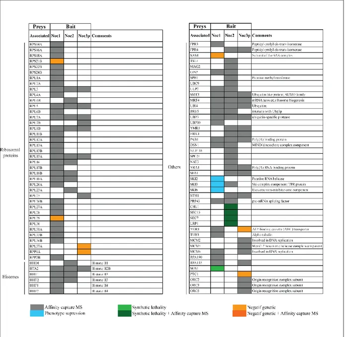
Table I: Summary of non-ribosomal proteins physically and genetically associated
with Noc1p, Noc2p and Noc3p pre-ribosomal particles ... 52
Table II: Summary proteins physically and genetically associated with Noc1p, Noc2p and Noc3p pre-ribosomal particles ... 53
Table III: List of the antibiotics for appropriate bacterial culture media ... 62
Table IV: List of the antibiotics for appropriate yeast culture media ... 63
Table V: Plasmids used in this study ... 64
Table VI: ArcticExpress (DE3) cell genotype ... 64
Table VII : Yeast strains used in this study ... 67
Table VIII : Primers used in this study for strains construction ... 73
Table IX : Primers for preparation of radio-labeled probes ... 77
Table X: Primers used in this study for qRT-PCR ... 79
List of figures
Figure 1.1 : X-ray structure of the S.cerevisiae 80S ribosome ... 19
Figure 1.2: Compositional analysis by comparative MS and classification of proteins co-purifying with distinct rDNA chromatin domains ... 22
Figure 1.3: Schematic Saccharomyces cerevisiae processing pathway ... 25
Figure 1.4: Assembly of non-ribosomal proteins with the different pre-ribosomal particles ... 28
Figure 1.5: Rix7p-GFP localization under growth conditions monotored by fluorescent microscopy ... 34
Figure 1.6: Potential model for Rix7p remodeling of pre-60S particle ... 35
Figure 1.7: Representation of homologous regions of Noc1p/Mak21p with its differnet homolog ... 39
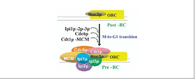
Figure 1.8: Hierarchical of replication-initiation proteins ... 44
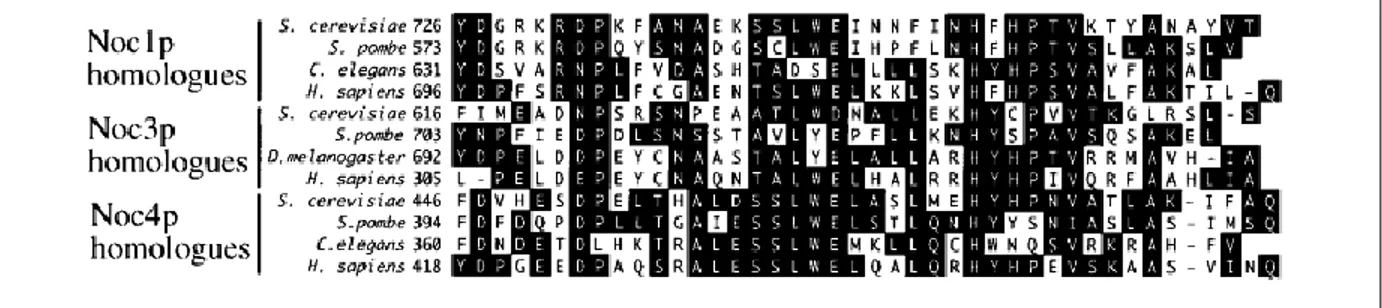
Figure 1.9: Sequence comparison of the conserved Noc-domain in Noc1p, Noc3p and Noc4p ... 48
Figure 1.10: Intranuclear localization of Noc1p, Noc2p and Noc3p ... 50
Figure 1.11: Changes in cellular expression networks after shift to restrictive temperature ... 57
Figure 2.1: Testing different IPTG concentration in order to induce anti-GFP nanobody ... 66
Figure 2.2: Schematic outline of harvesting cells and cryo-lysis protocol ... 70
Figure 2.3: NOC2 qRT-PCR melting and standard curves using to test primers sets ... 81
Figure 2.4: Schematic outline of the single-step purification technique ... 85
Figure 3.1: Testing MET25 promoter sensitivity and efficiency by spot test analysis ... 95
Figure 3.2: Noc2p depletion using repressible MET25 promoter affects cell growth rate ... 97
Figure 3.3: Noc1p and Gar1p are redistributed in cells in the absence of Noc2p... 102
Figure 3.4: Noc3p and Gar1p are redistributed in cells in the absence of Noc2p... 104
Figure 3.6: Noc2p depletion specifically influences proteins levels of the nucleolar
factors Noc1p and Rix7p ... 110
Figure 3.7: NOC2 level is influencing the genes expression of ribosomal factors and Histones ... 115
Figure 3.8: Rpl11p localization is not affected by Noc2p depletion ... 117
Figure 3.9: Noc protein localization is energy-dependent ... 122
Figure 3.10: Rix7p localization may be influenced by lack of ATP and GTP ... 124
Figure 3.11: Total levels of the Noc proteins and Rix7p are influenced by metabolic poison ... 126
Figure 3.12: Metabolic poison affects gene expression levels ... 129
Figure 3.13: Affinity-purified Noc2p complexes using a GFP antibody and different buffer conditions ... 134
Abbreviations
Amp ampicillinATP adenosine triphosphate
bp base pair
d deoxy-
DEPC diethylpyrocarbonate DNA deoxyribonucleic acid dNTP desoxyribonucleo DTT dithiothreitol
EDTA ethylendiaminetetra acetic acid
g gram
GAL galactose
GDP guanosine diphosphate GFP green fluorescent protein GTP guanosine triphosphate HIS histidine
H2O double destilled water, sterile IPTG isopropyl-D-thiogalactopyranoside
Kan kanamycyn
kb kilobase pair
kDa kilo Dalton
L liter
LSU large subunit
M molar MET methionine
pMET MET25 promotor
µ micro (10-6)
m mili (10-3)
min minute
mRNA messenger ribonucleic acid
MS mass spectrometry
OD optical density
o/n over night
ORF open reading frame
PAGE polyacrylamide gelelectrophoresis PCR polymerase chain reaction
qRT-PCR quantitative real-time polymerase chain reaction RNA ribonucleic acid
rRNA ribosomal ribonucleic acid rpm rotations per minute r-protein ribosomal protein
RT room temperature
SD synthetic dropout
SDS Sodium dodecyl sulphate
sec second
snoRNA small nucleolar RNA
snoRNP small nucleolar ribonucleoprotein SSU small subunit
TEMED N, N, N’, N’ –tetramethylethylendiamine Tris Tris-(hydroxymethyl)
tRNA transfer RNA
Acknowledgments
First, I want to thank the person who makes this possible, Marlene Oeffinger. Thank you for giving me this huge opportunity and believing in me during my entire master. It was an honor to work with and for you. I will always remember you as the good and understanding boss, the special Yoga teacher and the cat lover. Thank you for your understanding about my possible switch into the world of teaching… but I will always have the science passion!
I would like also to say a big thank you to the member of the lab. Karen Wei, thank you to showed me quite everything! Pierre Zindy, thank you for being with me every early morning and to helped me with the qRT-PCR and the microscopy stuff. Your help saved me many times. I would like to thank Christian Trahan and Mathew Karmar for sharing your knowledge in general. You were very helpful. Finally, a special thank to Carolina Lisbeth Agular, who shared coffee and lunch time with me and especially for the 24 hours-long experiments. It was a pleasure to work with all of you!
In addition, I would like to thank my friends. Mes amies de toujours, merci d’être tout simplement là… friendship never end! Mes amies de biochimie, merci d’avoir toujours été à l’écoute... étant donné que vous êtes les seules qui peuvent vraiment me comprendre!
Finally, I want to thank my family who was always so much supportive. Maman, même si tu ne comprenais pas toujours se que je faisais, merci d’avoir été présente, de m’avoir soutenue et d’avoir toujours accepté mes choix. Bruno, mon amour, merci de m’avoir supporté, d’avoir su me guider dans mes nombreuses remises en question et de m’avoir fait autant rire dans nos moments fous. Une chance que tu es là pour moi ! Finalement, un petit merci tout spécial à Gino et Maurice, nouveaux dans ma vie, mais qui m’apportent beaucoup de bonheur.
1.1 The Ribosome
The ribosome was discovered in 1955 by the Nobel laureate George Emil Palade who described it as small particles in the cytoplasm, associated with the endoplasmic reticulum membrane [1]. Through his work, ribosomes were found to be the biological protein synthesis machinery in cells. Since many years, a lot of structural and biological studies have allowed to understand the structure of the mature ribosome and its components. It is known that all prokaryotic and eukaryotic ribosomes are composed of ribosomal RNA (rRNA) and ribosomal proteins (r-proteins), in addition to having the same molecular function [2]. Due to additional regulatory steps during translation in eukaryote, these two ribosomes differ in structure and complexity [2]. In Escherichia coli, ribosome is composed of two unequal subunits namely the small 30S subunit (which contains the 16S rRNA) and the large 50S subunit (which contains the 5S, 23S rRNAs) [2]. In comparison , the eukaryotic ribosome of the budding yeast
Saccharomyces cerevisiae is made of two unequal subunits, the small 40S subunit (which
contains the 18S rRNA) and the large 60S subunit (which contains the 5S, 5.8S and 25S rRNA) [3]. Furthermore, these two subunits contain about 79 r-proteins that form the mature 80S ribosome, such as 33 in the small subunit and more than 46 in the large subunit (Figure 1.1) [4]. Synthesis of these proteins as well as rRNAs are made in equimolar amounts [5]. Thereby, mature 80S eukaryotic ribosome is much larger and complex, with more than 5500 nucleotides of rRNA and 79 r-protein, compared to the mature 70S prokaryotic ribosome with about 4500 nucleotides of rRNA and 54 r-proteins [6]. These differences in complexity are due to the large number of proteins that interact with ribosomes and to the diversity of cellular processes where ribosomes play a role [7]. This aspect will be elaborated later in the manuscript.
Since 2000, when the first structural model of eukaryotic ribosome was constructed using cryo-electron microscopy, our knowledge of ribosomal function and structure has increased significantly [8]. In fact, 33 r-proteins have been shown to exist in all domains of life, including 15 proteins of the small subunit (Rps2p-5p, Rps7p-15p, Rps17p and Rsp19p) and 18 of the large subunit (Rpl1p-6p, Rpl10p-15p, Rpl18p, Rpl22-24 and Rpl29p-30p) [9]. It has been shown that r-proteins contain a high amount of positively charged lysines and
arginines in their loops and tails [9]. Indeed, the specificity of interaction between r-proteins and rRNA was first shown for the Rpl11p-rRNA complex and revealed that the interaction is mainly determined by complementarities of the charges and shapes of the surface, but not by amino acid composition or combination [9]. Moreover, it has been shown that r-proteins are involved in compactization of rRNA into correct structures which determine the shape and features of ribosomal particles [9]. Crystal structures of yeast ribosomes allowed to show that RNA are present in the core of each subunit, with r-proteins enshrined on the surface (Figure 1.1) [4]. Sites for ribosome functions such as peptidyltransferase center, polypeptide exit tunnel, GTPase binding site in large subunit (LSU) and tRNA-binding site have been identified in bacterial, archeaeal and euckaryotic ribosome structures [4]. Indeed, r-proteins were found to be responsible for specific functions of the ribosome through their participation in more than half of the known inter-subunit bridges[9].
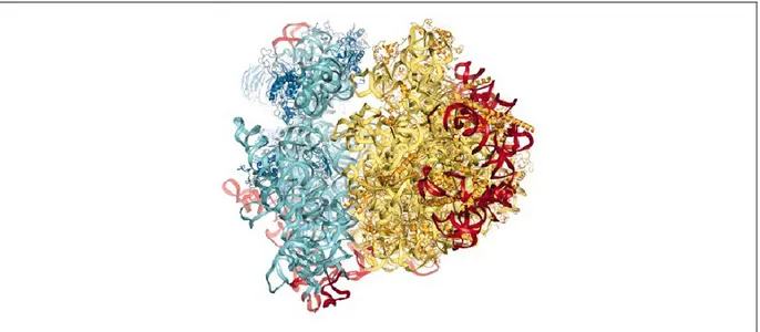
Figure 1.1. X-ray structure of the S.cerevisiae 80S ribosome
Proteins and rRNAs of the 40S subunit are colored in dark and light blue respectively. Proteins and rRNAs of the 60S subunit are colored in dark and pale yellow respectively. [7]
Although our understanding of the ribosomal composition and function has much significantly evolved, ribosomal biogenesis pathway remains elusive owing to a complex maturation and a tight regulation. Assembly of r-proteins and rRNA in ribosomal particles has been proposed to result in two principles: the incorporation of r-proteins is made step-by-step and the formation of ribosomal particles is accompanied by conformational changes in rRNA
structure [9]. However, eukaryotic ribosome formation is known to include the assembly of 5500nt of rRNA and about 79 r-proteins, in addition to requiring about 76 small nucleolar RNAs (snoRNA) and 200 different assembly factors [4]. This formation has been shown to be an extremely dynamic and expensive process for a cell and to required stringent inspection by specific control mechanisms coupled to nuclear export of pre-rRNPs [4]. Therefore, ribosome biogenesis will be under the microscope in this manuscript, especially the dynamic aspect of the assembly pathway.
1.2 Ribosomal DNA transcription
Synthesis of the eukaryotic ribosome has been extensively studied in the yeast
Saccharomyces cerevisiae because of its easy experimental accessibility by biochemical and
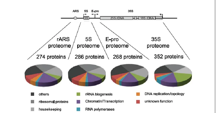
genetic methods [2]. Ribosome biogenesis is a dynamic process and is known to start in a specialized compartment of the nucleus called the nucleolus, which is composed of three substructures: fibrillar center (FC), dense fibrillar component (DFC) and granular component (GC) [10]. Ribosomal DNA (rDNA) is ~9.1 kb long and is present in 100 to 200 tandem copies on chromosome XII, located in the FC substructure [3, 11]. rRNA has been shown to elongate into the cortex of this substructure and to enter into the surrounding DFC to be processed, modified, folded and assembled with r-proteins by specific dynamic pathways from the nucleolus to the cytoplasm [11, 12]. As shown in Figure 1.2, a single rDNA unit is composed of an autonomously replicating sequence (ARS), the bidirectional promoter (E-pro), the 5S rRNA gene, which is transcribed by RNA polymerase III (Pol III) and flanked by two nontranscribed spacers (NTS1 and NTS2), and finally the 35S pre-rRNA operon, which is transcribed by RNA polymerase I (Pol I) [2, 3, 5, 13]. The 35S pre-rRNA contains the mature sequence of the 5.8S, 18S and 25S rRNAs separated by two internal transcribed spacers (ITS1 and ITS2) and flanked by two external transcribed spacers (5’-ETS and 3’-ETS) [3]. While the sole function of RNA polymerase I is to transcribe the 35S rRNA gene precursor, it has been shown that its function can be replaced by RNA polymerase II following select mutations [14].
In exponentially growing cells, only ~50% of rDNA is transcriptionally active [15]. By using a psoralen photo-crosslinking assay, in which cells are treated with a DNA intercalating
agent and UV-cross-linked (discussed in results and method section), rDNA genes were shown to adopt two distinct chromatin states that coexist throughout the entire cell cycle [15, 16]. Thus, rRNA gene can be psoralen-accessible or psoralen-inaccessible, which corresponds to transcriptionally active or inactive rDNA genes, respectively [15]. This transcriptionally inactive fraction has been shown to be important for the integrity of the rDNA locus since actively transcribed rDNA is sensitive to mutagen-induced DNA damage [15]. Moreover, experiments investigating chromatin dynamic have shown that replication is required to convert rDNA to a transcriptionally inactive state, and that Pol I transcription is required to establish an conformation, transcribed rDNA chromatin state [15]. This open-conformation rDNA was shown to be stabilized outside of S-phase by the HMG box protein Hmo1, which is believed to interfere with replication-independent nucleosome assembly [15]. It has been shown that when cells enter the diauxic shift, which is a transition triggered by nutrient depletion and characterized by massive reprogramming in metabolism that prepares cells for long-term survival during stationary phase, Pol I-mediated transcription of rDNA is repressed [16]. rDNA that becomes transcriptionally repressed has been found to return to a closed chromatin state until cells are provided with fresh nutrients [16].
Recent studies have taken this further by providing evidence that active rDNA genes are devoid of nucleosomes [16, 17]. ChIP experiments demonstrated that log and post-log cells have a severe difference in histones occupancy, including a 10-fold increase in H2B and H4 across the transcribed 35S region [16]. In this study, H2B and H4 were used and considered as indicators of the H3/H4 tetramer and H2A/H2B dimer respectively, which strongly implies that the entire nucleosome is likely removed during Pol I transcription [16]. In fact, it has been already shown that nucleosomes are acetylated and disrupted ahead of the polymerase during Pol II transcription elongation [16]. This disruption/reassembly cycle is known to require several proteins such as the histone deacetylase Rpd3p and histone chaperon/elongation factors Spt6p, Asf1p and FACT [16]. While Rpd3p has also been shown to associate with both the rDNA promoter (IGS2) and transcribed 35S regions, its depletion was found to cause a decrease in both H2B and H4 acetylation and in the loading of these histones onto rDNA genes [16]. Indeed, this factor was suggested to be important for maintaining the entire nucleosome structure [16]. Meanwhile the depletion of the FACT
component Spt16p was shown to be defective for loading of H2B only, suggesting that FACT is mainly responsible for assembling of the H2A/H2B dimer onto rDNA [16]. Due to the fact that the H3/H4 tetramer is known to assemble before H2A/H2B, a model has been proposed where Rpd3 could be responsible for either H3/H4 deposition or for maintaining stability of the nucleosome, while FACT could be required for H2A/H2B deposition [16]. Another recent study has developed a new purification technique enabling compositional and structural analyses of the multicopy rDNA gene cluster. Indeed, the autonomously replicating sequence (ARS), the 5S rRNA gene, the bidirectional promoter (E-pro) region and the 35S rRNA gene were found to exhibit distinct histone modification patterns [13]. As an example, histone H3 K4 methylation was enriched across the ARS region, whereas histone H3 K36 tri-methylation was preferentially found across the E-pro and 35S rDNA [13]. Analyses of proteins co-purifying with rDNA domains allowed for the identification and classification of 250 to 350 proteins associated with each of the four rDNA subdomains (Figure 1.2). In fact, yeast strains were constructed to constitutively express a LexA-TAP fusion protein and specific rDNA domains or a complete rRNA repeat were flanked by RS element and include LexA-binding sites [13].
Figure 1.2. Compositional analysis by comparative MS and classification of proteins co-purifying with distinct rDNA chromatin domains
Proteins co-purifying with LexA-TAP were subjected to iTRAQ analyses in direct comparison with control purifications [13]. Identified proteins were categorized according to their biological function. The pie charts depict the relative fraction for each protein class in the purification. The data represent the summary of three independent biological replicates.
In addition to transcription, ribosomal synthesis involves several post-transcriptional processing and assembly steps to ensure 40S and 60S subunit maturation. With the aim of providing a large number of 35S pre-rRNA transcripts, Pol I needs to efficiently elongate and maintain a high synthesis rate through the rDNA region [18]. It has been shown that Pol I transcription elongation rate is closely linked to the downstream events of the pre-rRNA processing pathway [18]. Indeed, mutation of the Pol I subunit Rpa135p was shown to have a 90% reduced elongation rate and to result in a multitude of rRNA processing permutations, in addition to a reduced 60S subunit level [18]. Thus, these results provided evidences for the link between Pol I transcription and pre-rRNA processing [18]. Further evidence came via electron microscopy analysis. Due to the tandem arrangement of rDNA, pre-rRNAs have been shown to adopt a characteristic Christmas tree-like structure by EM, whereas rDNA and Pol I molecules form the stem and rRNAs are the branches extending out from transcribing Pol I molecules along the rDNA [19]. In addition, identification of co-transcriptional assemblies of r-proteins and ribosomal factors has been possible by visualization of ball- or knot-like structures, which are believed to be compacted pre-rRNA transcripts [19]. Moreover, a new purification technique allowing for compositional and structural analyses of the multicopy rDNA gene cluster, revealed several different r-proteins and pre-rRNA biogenesis factors in each actively transcribed rDNA region, supporting once again the co-transcriptional aspect of the pre-rRNA processing (Figure 1.2) [13]. Finally, by using the temperature-sensitive (ts) allele rpa190-2 of RPA190, which is an essential gene encoding for the largest subunit of Pol I, in situ electron microscopy was used to study the effect of an rDNA transcription arrest [10]. Interestingly, this arrest has been shown to result in a dramatic nuclear reorganization, where the nucleolus was spatially rearranged and the pre-rRNA processing machinery dispersed [10]. In fact, some amount of Nop1p and Gar1p, core components of the box C/D and box H/ACA small nucleolar ribonucleoprotein complexes (snoRNP), respectively, were found re-localized to the nucleoplasm, while some still remained in the intermediate-electron-dense domain of
the segregated nucleoli [10]. Under normal conditions, both proteins are known to be involved in early pre-rRNA processing steps and are localized exclusively within the nucleolus. Indeed, rDNA transcription is now known to be required in order to maintain the structural integrity of the nucleolus as well as the early pre-rRNA processing machinery [10].
1.3 Ribosomal processing pathway
As it is being transcribed by Pol I, the nascent pre-rRNA is modified by about 75 different snoRNPs that target specific sites for pseudouridylation and 2’-O-ribose methylation, and assembled with trans-acting factors [2, 5]. The 35S pre-rRNA is co-transcriptionally assembled with a number of processing and assembly factors in addition to r-proteins devoted to the 40S biogenesis pathway and form the 90S particle [3, 12]. Following these early modifications and assembly steps, there are at least 11 endonucleolytic and exonucleolytic cleavages that process the precursor to remove external (ETS1 and 2) and internal spacer (ITS1 and 2) regions and generate mature 5’ and 3’ ends of the 5.8S, 18S and 25S rRNAs [20]. Indeed, pre-rRNA processing is carried out by a dynamic pathway, which involves more than 200 proteins, and which occurs in a hierarchical manner in the context of pre-ribosome particles.
1.3.1 90S processing
It has been showed that about 30% of the length of Pol I transcripts are completed before pre-rRNA processing is initiated [21]. The modular subcomplexes UTP-A, UTP-B and UTP-C, consisting of processing and assembly factors, some small subunit r-proteins and more than 20 other assembly factors, progressively assemble onto the primary transcript co-transcriptionally and form the first ribosomal particle called the 90S particle [2]. As shown in Figure 1.3 B, the 3’ external spacer (ETS) is immediately cleaved at the B0 site by the endoribonulcease Rnt1p, and the pre-rRNA simultaneously undergoes a large number of covalent modifications carried out by the box C/D and box H/ACA snoRNPs [2, 22]. SnoRNP complexes are composed of small nucleolar RNA (snoRNAs), which select the site to be modified by base pairing with the pre-rRNA target site, and RNA binding proteins, which are known to catalyze the modification reaction on the pre-rRNA [2, 23]. Based on their
conserved sequence and structural elements and their associations with specific proteins, snoRNPs can be divided in two classes named box H/ACA and C/D, which catalyze site-specific pseudouridylation and 2’-O-methylation of rRNA, respectively [23]. In fact, box H/ACA snoRNPs are composed of common core proteins (Cbf5, Nop10, Nhp2 and Gar1p) that base pair with the rRNA by a pseudouridylation loop where the unpaired uridine is converted to pseudouridine by the the pseudouridine synthase Cbf5p [23]. Next, the C/D box snoRNPs are known to be composed of a common core proteins (Snu13, Nop56, Nop58 and Nop1) [23]. It is known that the rRNA nucleotide five base pairs from the box D is 2’-O-methylated by Nop1p, the yeast homologue of Fibrillarin [23]. 35S pre-rRNA processing has been shown to require a subset of snoRNPs, comprising the box H/ACA snoRNPs snR30/U17 and snR10 and the boxC/D snoRNPs U3, U14, U8 and U22 [23].
Thereafter, the 35S pre-rRNA is cleaved at sites A0, A1 and A2, which are dependent on the small-subunit (SSU) processosome, a 2.2 MDa RNP complex composed of the U3 snoRNA and more than 30 proteins [24]. The cleavage at site A0 produces the 33S precursor, and, afterwards, a cleavage at site A1 yields the 32S pre-rRNA allowing for the precise removal of 5’ ETS [2, 25]. Cleavage at site A2 in the internal transcribed spacer 1 (ITS1) leads to the separation of the 32S pre-rRNA into 20S and 27SA2 pre-RNAs, which are part of the pre-43S and pre-66S particles, respectively [2]. These are the earliest pre-40S and pre-60S particles, and from here on out the separate pathways are marked by dramatic differences in particles compositions [2, 12]. It should also be noted that these early processing events are irreversible, which suggest a point of regulatory control [2, 25].
Figure 1.3. Schematic Saccharomyces cerevisiae processing pathway.
A) Location of the ETS and ITS sequences, rRNA regions and processing site. B) Pre-rRNA processing pathway showing steps which are performed in the 90S particle (gray), in the 66S particle (light blue) and in the 43S particle (light green). The 40S subunit contains 18S rRNA (dark green) and the 60S subunit contains 5.8S, 25S and 5S rRNAs (dark blue).Modified from [24, 26]
1.3.2 Pre-40S processing
In S.cerevisiae, the 20S rRNA within the pre-43S particle was shown to be directly exported from the nucleolus to the cytoplasm after cleavage at site A2, where 3’end maturation of 18S rRNA occurs, whereas its 5’end has been already formed through the removal of the 5’ETS in the nucleus [24]. Thereafter, particle composition is known to change when further modifications are carried out. The methyltransferase Dm1p has been found to methylate conserved adenosines near the 3’end of the 18S rRNA, and endonucleolytic cleavage at site D has been shown to require the nuclease Nob1p as well as factors Rio1p, Rio2p, Tsr1p and Fap7p [2, 27]. The Bud23p methyltransferase is also part of this complex; Bud23 catalyzes N7-methylation of guanosine 1575 and Nep1p, which is responsible for the N1-methylation of the hyper modified m1acp3Ψ 1191 base [28].
1.3.3 Pre-60S processing
Pre-60S rRNA maturation is much more complex than that of the 18S rRNA, since it comprises the maturation of two rRNAs, 5.8S and 25S, as well as two distinct processing pathways (major and minor), and requires several non-ribosomal processing factors, including exo- and endonucleases. As for the major pathway, it is known that the MRP RNase induces the cleavage at site A3 resulting in 85-90% of 27SA2 pre-rRNA, which is then endonucleolytically cleaved to form the 27SA3 precursor [4, 29]. The timing of this cleavage was found to occur posttranscriptionally, and to be linked to events at the 3’ end of the 35S pre-rRNA [29]. Moreover, A3 cleavage requires the presence of the non-ribosomal protein Rrp5p, which binds adjacent to sites A2 and A3 and which is also required for A0, A1 and A2 cleavage [29]. Indeed, the C-terminal domain of Rrp5p appears to be required for the A0-A2 cleavages on the pathway of 18S rRNA processing, whereas the N-terminal domain was demonstrated to be required for A3 cleavage along the 5.8S/25S rRNA processing pathway. This clearly illustrates the complexity and dynamic of the ribosomal biogenesis pathway [29]. Thereafter, the 5’ to 3’ exonucleases Rat1p, Xrn1p, Rrp17p and the cofactor of Rat1p, Rei1p, are required to generate the 27SBs pre-rRNA and thus the 5’ end of the major form of the 5.8S rRNA, 5.8SSHORT [4, 30, 31]. In contrast, the minor pathway is known to start with
endonucleolytic processing at the B1L site. In fact, 10-15% of 27SA2 pre-rRNA is not processed by the MNP RNAse, but directly cleaved at the B1L site by an unknown endonuclease, which generates the 27SBL pre-rRNA [4].
Simultaneous with formation of the 5’end of 5.8S rRNA into both major and minor pathways, the 3’ to 5’ exonuclease Rex1p generates the 3’end of 25S rRNA at the B2 site [32]. Subsequently, cleavage at the C2 site, again by an as yet unknown endonuclease, allows separations of 7S and 26S pre-rRNAs, the two large subunits rRNAs precursors [4]. The mature 3’ end of the 5.8S rRNA is generated by a multistep process, involving the exosome component Rrp6 and the exonucleases Rex1p and Rex2 [4]. The 5’ end of 25S rRNA is generated by Rat1p and Rrp17, which carry out exonuclease digestion from the C2 to C1 site [4].
1.4 Assembly pathway
Ribosomes were shown to contain ~79 r-proteins. In addition, research groups have identified a large number of non-ribosomal proteins involved in ribosome biogenesis. There are more than 200 non-ribosomal proteins, such as GTPases, DExD/H-box ATPases, AAA-type ATPases, nucleases, rRNA-binding proteins and RNA helicases that participate in rRNA processing, folding and assembling of the rRNA with ribosomal proteins at different stages of maturation [20, 33]. These proteins are believed to assemble with the pre-ribosomes in a transient manner [34]. Maturation of the pre-40S particle occurs mainly in the cytoplasm, while the pre-60S particle matures mostly in the nucleus and continues after its exportation into the cytoplasm [5]. In fact, pre-ribosomes start off in the nucleolus, traverse into or through the nucleoplasm to the nuclear periphery, and are finally exported to the cytoplasm [5, 33, 35]. It is believed that this spatial movement of maturing pre-ribosomes is linked to specific temporal maturation events, which underscores the importance of coordinating the processing and assembly steps. To date, little is known about the integration of processing steps with rRNA folding and assembly. By using affinity purification methods, several studies isolated and characterized different complexes associated with assembly factors, in addition to identifying their approximate positioning along the maturation pathway [2, 33, 34]. However,
ribosome biogenesis is a highly dynamic process, and so far most of these studies provided only a static image of the process and limited information on the pathway’s dynamics [34].
Figure 1.4. Assembly of non-ribosomal proteins with the different pre-ribosomal particles.
The upper and lower panels indicate 40S and 60S non-ribosomal proteins, respectively. The central panel illustrates the different pre-ribosomal particles in their sub-cellular locations. Pre-ribosomes are classified as early (E), middle (M), late (L) and cytoplasmic (C). Nuclear pore complexes (NPC) allow pre-ribosomes passage from the nucleoplam to the cytoplasm [33].
1.4.1 Pre-40S assembly
As mentioned above, pre-40S maturation occurs mainly in the cytoplasm, and the particle is exported directly following the A0, A1 and A2 cleavages. Thereafter, composition of the pre-40S drastically changes, where a group of non-ribosomal factors are dissociated whereas a novel set are associated and further r-proteins are recruited [2]. Rrp12p, a protein that was found in 90S complexes but also in pre-40S particles, is essential for maturation of the small subunit, and is believed to facilitate the export of pre-ribosome particles to the cytoplasm. [5]. This protein was also found in association with some pre-60S proteins (Nop4p, Nop15p and Noc1p), being one of the very few ribosome biogenesis factors involved in the maturation of both subunits [5].
The pre-40S particle was shown to already display typical morphological ‘landmarks’ of mature 40S subunits, while still lacking the typical “beak” structure [2]. At this point, the Enp1p-Ltv1p-Rps3p subcomplex and Hrr25p are known to promote phosphorylation/dephosphorylation events that finally allow the stable association of Rps3p, and the formation of the mature 40S subunit structure, including the ‘beak’ [2].
1.4.2 Pre-60S assembly
Pre-60S subunit maturation is known to be much more complex and to require many more non-ribosomal proteins that associated with the large number of different pre-ribosomal particles. In fact, distinct pre-60S particles have been characterized by affinity purification of definite non-ribosomal proteins. However, it is still unclear whether these particles are all along the same, or parallel, pathways.
In the nucleolus, the earliest pre-60S particles that have been identified are associated with Npa1p and Ssf1p. The Npa1p particle contains 27SA2 pre-rRNA and ~40 non-ribosomal proteins, such as early pre-60S factors (Noc1p and Nop4), snoRNP components, RNA helicases and some 90S-associated factors [2]. In contrast, the Ssf1p particle is composed of 27SA2 and 27SB pre-rRNA, and ~30 non-ribosomal proteins, such as early factors Noc1p and Rrp5p, but no snoRNP components [2]. There is also Rpf1p that has been found in the Ssf1p particles but only associated with 27SB rRNA intermediates [5]. Thereafter, another pre-60S particle has been isolated with Nsa1p, where the exchange of the Noc1p-Noc2p to Noc2p-Noc3p complex has already been made (see section 1.6) [2]. This intermediate particle contains the 27SB pre-rRNA only, the AAA-ATPase Rix7p and the Ytm1p-Erb1p-Nop7p subcomplex in addition to the 5’-3’exonucleases Rat1p, Rrp17p and Xrn1p, which participate in the 5’ trimming of the 27SA3 pre-rRNA [2, 31, 36]. The proteins Rpf2p and Rrs1p are both part of this particle, and have been shown to mediate the incorporation of the 5S rRNA:Rpl5p complex as well as Rpl11p into the pre-ribosome [2, 37]. Moreover, 5S rRNA is found in Nug1p and Nog2p associated complexes, but not with Ssf1p associated complex [5]. As mentioned previously, there are two distinct processing pathways (major and minor) that can lead to the formation of the 27SB pre-rRNA. Mostly, these two pathways were shown to require the same non-ribosomal factors, except for the so far unknown endonuclease that cleaves at the B1L site [34].
In the nucleoplasm, the pre-60S particle has been isolated using Rix1p as bait, which allowed visualization of drastic compositional changes at this point. Several intermediate factor such as Erb1p, Ytm1p, Nop2p, several DExD/H-ATPases and the Noc2p-Noc3p complex were found to have been dissociated and replaced by different factors such as Rea1p, Rsa4 and Nug2 [2]. This simplification of protein composition is consistent with the 5S rRNA association, and with the formation of the 7S pre-rRNA that occurs after the C2 cleavage [5]. In fact, the Rix1p particle contains the nearly mature 25S and 7S/5.8S rRNAs, which indicates that processing of the 27SB pre-RNA has been almost completed [2]. On top of this major compositional change, ATP-dependent structural rearrangements have been found to occur through the dynein related AAA-ATPase Rea1p/Mdn1p and the Rix1p-Ipi1p-Ipi3p complex [33].
Later pre-60S particles contain Arx1p, and can be found in both the nucleo- and cytoplasm, indicating the transitioning aspect of this particle; moreover, Arx1p has also been found associated with export factors Nmd3p and Mtr2p [5]. This particle also contains more mature 5.8S rRNA than 7S pre-rRNA [2]. It has been shown that the AAA-ATPase Drg1p binds the Arx1p-containing particle only once it has reached the cytoplasm [2]. This binding triggers Nog1p and Rlp24p release [2]. The later, solely cytoplasmic pre-ribosomal particle is characterized by the presence of Lsg1p/Kre35p [2]. This particle contains some cytoplasmic factors and a diminished amount of Arx1p, whose release depends on Rei1p, Ssa1/Ssa2p and Jjj1p [2, 4]. Another protein of the Lsg1p/Kre35p particles is Tif6p, the ortholog of the mammalian protein eIF6, which is functionally linked to the cytoplasmic GTPase Efl1p [2, 5]. Interestingly, Tif6p is an essential nucleolar protein that relocates to the cytoplasm when Efl1p is depleted in cells, and dissociates from the cytoplasmic pre-60S particle through the GTPase activity of Efl1p and Sdo1p [2, 5].
Some assembly factors are only transiently associated with distinct pre-60S particles, while others are found across several maturation steps. This is the case with Nsa3p/Cic1p, which is present in both late 90S particles and early pre-60S particles [5]. Another example is Rlp24p, which is found in the nucleolar, nucleoplasm and cytoplasm pre-60S complexes [5].
The enormous number of non-ribosomal proteins involved in the ribosome biogenesis has been a puzzle for some time, however, is most likely due to the coordination of the ribosomal synthesis with other cellular process such as growth rate and stress response [2]. Interestingly, although, the majority of these proteins are essential for cell viability, it seems that mutation or depletion of some of them does not affect the viability of the cell but more the growth rate [5, 31].Indeed, there is a concept of overlapping and parallel pathways to ensure the production of ribosome in the cell, that would not only explain this particular phenomenon, but also the existence of more than 200 ribosome biogenesis factors in yeast [38].
1.5 Coordination and quality control system
Ribosomes are essential to a cell’s viability, and ribosome biogenesis needs to be regulated in conjugation with other cellular pathways. In yeast, it is known that at least 60% of
the RNA polymerase II transcription and 90% of mRNA splicing are dedicated to mRNAs encoding r-proteins and non-ribosomal protein factors [4, 39]. All these mRNAs are processed and exported from the nucleus, translated, and the proteins then imported into the nucleus, where they associate with nascent pre-rRNAs [4]. Moreover, pre-rRNAs also need to be processed, modified and assembled as two separate and distinct subunits. Overall, 2000 ribosomes are produced per minute that correspond to 200,000 ribosomes per yeast cell, and about 4000 ribosomal subunits are exported from the nucleus to the cytoplasm every minute [39]. Therefore, cells need to expend a lot of energy to ensure the production of ribosomes.
It also demonstrates the incredible coordination that is required to ensure that correct ribosomes are made, as malfunctioning ribosome could cause mistakes during translation that may have deleterious effects for any cell or organism, as it has been recently shown in the cases of ribosomopathies and even cell cycle arrest in yeast [4, 33]. Indeed, there may be some ribosomal factors that have a role in quality control steps during ribosome biogenesis, or even in overlapping and alternative pathways, that allow pre-ribosomes to still be matured correctly by a minor factor if major ones are absent or not available at the necessary levels [40]. However, how pre-ribosomes are quality controlled, and whether any checkpoint exists, is poorly understood to date. It is known that ribosome, containing mal-processed pre-rRNAs, can be degraded by the exosome, following targeting and polyadenylation by Trf4/5p-Air1/2p-Mtr4p polyadenylation (TRAMP) complex [41, 42]. Moreover, it has been shown that release of Tif6p during late steps of pre-60S maturation is coupled to verification of P-site integrity [41]. This formation of sub-complexes is one possibility of quality-control system because it can physically block the binding-site of ribosomal proteins or other assembly factors [40]. Other proteins, such as the Noc proteins, are known to function together or, like Ssf1p and Nop7p, interact directly with the pre-rRNA [39]. Based on our knowledge about the translocation of pre-ribosomes, it is certainly conceivable that some of these proteins, or sub-complexes, are involved in the coordination of the controlled spatio-temporal movement of pre-ribosome, from nucleolar compartment to the next, through the nucleoplasm to the nuclear pore [5, 31, 33]. However, these kinds of control mechanisms remain, to this date, largely unexplored.
It is known that about 20% of ribosome assembly factors are nucleoside triphosphate hydrolyzing enzymes, such as kinases, ATPases and GTPases [40]. These enzymes are believed to provide the energy needed to confer directionality to the maturation pathway [2]. Ribosomal assembly requires one GTPase during pre-40S maturation (Bms1p), and five during pre-60S maturation (Nog1p, Nug1p, Nug2p/Nog2p, Ria1p and Lsg1p) [2, 20, 40]. Interestingly, these proteins contain an RNA-binding domain in addition to their GTPase domain, suggesting that they may be able to bind to the pre-rRNA directly, in the absence of bridging interactions via other factors [20]. From previous biochemical and genetic work, some possible functions for these proteins have been speculated on; one is that the energy from the GTP hydrolysis can be used to remove proteins or to promote a conformational rearrangement within the nascent ribosome [20]. But these studies have not been able to confer the exact function of these proteins [20].
In addition, seven DExD/H-box ATPases were found to be involved in pre-40S maturation (Dbp4-8p, Dhr1-2p, Fal1p, Rok1p, Rrp3p), ten in pre-60S maturation (Dbp2-3-6-7-9-10p, Drs1p, Mak5p, Mtr4p/Dob1p, Sbp4p), and two in the maturation of both subunits (Has1p, Prp43p) [2]. Although their precise roles remain unclear, these proteins are known to act as ATPases and can be viewed as chaperon/modulators of RNA or RNP structures [2].
There are also three kinases implicated in pre-40S maturation (Hrr25p, Rio1-2p), where recently Hrr25p has further been related to phosphorylation of Tif6p during pre-60S maturation [2, 40]. Moreover, both pre-40S and pre-60S maturation pathways involve two ATP-binding cassette (ABC) proteins, Arb1p, Rli1p [2]. This superfamily of proteins is known to use ATP hydrolysis to transport proteins against a concentration gradient [2].
Finally, there are three AAA-ATPases required during pre-60S maturation, Drg1p/Afg2p, Rea1p/Mdn1p and Rix7p [2]. These proteins are known to use ATP hydrolysis to potentially stimulate structural rearrangement, or protein dissociations from the particles through transmission of the tension of a nucleotide-dependent conformational switch to bound substrate proteins [2, 40].
1.5.1 AAA-ATPase Rix7p
Rix7p was the first AAA-ATPase characterized during pre-60S maturation. It is also the earliest acting in the 60S subunit biogenesis pathway [43]. Rix7p contains two AAA ATPase domains (D1 and D2) and one putative bipartite nuclear localization signal (NLS) [44]. Cdc48p has been identified as a yeast homologue of Rix7p; Cdc48p is known to recognize ubiquitinated proteins and dissociate them from unmodified binding partners [45]. However, sequence conservation of Rix7p is restricted to the two AAA ATPase domains, but not to the N-terminal domain [43]. Indeed, the highly homologous domain D1 is known to promote hexamer formation and to be a relatively stiff structure, whereas domain D2 is known to harbor the ATPase activity and perform conformational changes following ATP hydrolysis [45]. By mutating conserved residues implicated in ATP binding and ATP hydrolysis, the D1 domain was shown to require ATP or ADP binding, but not ATP hydrolysis to perform its function [41]. On the other hand, the D2 domain was shown to required both ATP and ADP binding as well as ATP hydrolysis [41].
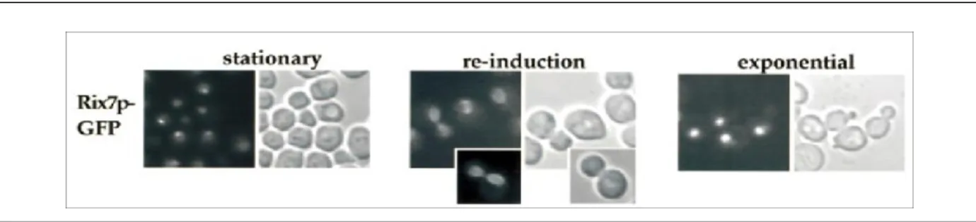
Due to its N-terminal domain, Rix7p shows a nuclear localization in normal condition [44]. Nevertheless, its intranuclear localization has been shown to change depending on the growth condition and nutrient availability (See Figure 5) [44]. Using fluorescent microscopy and growth density analysis, cells containing GFP-tagged Rix7p were grown to stationary phase and Rix7p-GFP was shown to concentrate in the nucleolus [44]. However, after entering stationary phase, cells were transferred to fresh medium and Rix7p-GFP was then shown to follow a transient perinuclear location [44]. Finally, when cells were grown until they reached an exponential phase, Rix7p-GFP was shown to have an overall nuclear distribution [44]. These localization changes have been suggested to correlate with some adjustments and alterations of ribosome biogenesis in response to nutrient availability [44].
Figure 1.5. Rix7p-GFP localization under different growth conditions monitored by fluorescent microscopy.
Yeast cells were grown in YPD medium at 23°C (A) Stationary phase (B) Re-induction in fresh medium for 3 h (C) Exponential phase [44].
Using the temperature-sensitive (ts) mutant strain rix7-1 and the large subunit reporter Rpl25p-eGFP, Rix7p has been predicted to have a role in pre-60S ribosomal export [44]. Indeed, at restrictive temperature (37°C), Rlp25p-eGFP was retained in the nucleolus and nucleoplasm, suggesting a defect in pre-60S export from the nucleus to the cytoplasm [44]. Moreover, sucrose density gradient profiling of polysomes in rix7-1 under restrictive conditions revealed a significant decrease of free 60S subunits, indicating a role in 60S biogenesis [44]. Northern blot, primer extension and pulse-chase analysis have indicated that Rix7p is not essential for pre-rRNA processing itself, but rather for structural rearrangement and stability of the pre-60S particles [44].
More recently, Rix7p has been shown to be genetically linked to Nsa1p, an essential pre-60S factor and conserved WD repeat protein that is known to bind pre-60S particles containing 27SA pre-rRNA [40, 45]. By mutation of the N-terminus of Rix7p in a GFP-tagged Nsa1p strain, fluorescent microscopy analysis showed that Nsa1p cannot be efficiently removed from late nucleolar pre-60S particles, and stays associated with aberrant cytoplasmic pre-60S particle [45]. Thus, Rix7p was suggested to be essential for removal of Nsa1p from pre-60S particle during pre-ribosomal maturation [45]. Moreover, Nsa1p removal by Rix7p coincides with compositional changes on the pre-60S particle; Nop7-TAP associated particles isolated in a rix7 mutant were found to be more complex than wild-type complexes, and to be composed of a set of very early factors such as Rrp5p, Noc1p, and Nop4p [45]. In addition, the later nucleoplasmic Rix1p particle was found to contain reduced quantities of the usual factors, such as Nug1p, Nog2p and Rsa4p in a rix7 mutant background [45]. Overall, Rix7p
has been proposed to detect the structural integrity of pre-60S particles and lead them into a clearance or check point, which, if the particles are correct, leads to Nsa1 removal and change in particle composition, including structural rearrangements [40, 41, 45].
Figure 1.6. Potential model for Rix7p remodeling of pre-60S particle.
Nsa1p and other early 60S assembly factors such as Rrp5p, Noc1p and Nop4p are bound to pre-60S subunit containing 27SA rRNA. Rix7p interacts with Nsa1p to remove it from the particle and this coincides with loss of the early and binding of later 60S-assembly factors. Orange: Energy-consuming factors. Yellow: Assembly factors. [40]
1.6 The Noc proteins
The Noc proteins (Noc1p, Noc2p, Noc3p and Noc4p) were heavily studied at the beginning of the last decade, and provided one of the initial clues about the spatio-temporal dynamic of ribosome maturation. In fact, mass spectrometric analysis of a large nucleolar substructure co-purified with RNA Polymerase I (Pol I) and Pol I-dependent transcription factors in a single chromatographic fraction, allowed for the identification of several nucleolar proteins implicated in pre-rRNA processing. Among these were the Noc proteins (NucleOlar Complex-associated proteins) [46, 47]. Later on, it was shown that, indeed, Noc1p, Noc2p and Noc3p are involved in intranuclear transport as well as export of pre-60S particles, and that Noc4p is required for the nuclear export of the pre-40S particles [47, 48]. Using the 3D-PSSM server (http://www.sbg.bio.ic.ac.uk/~3dpssm/), the Noc proteins were determined to be constituted of α-helical repeats, similar to HEAT/Armadillo repeats, with a confidence range of 70%-90% [49]. About 0.2% of eukaryotic proteins contain HEAT or Armadillo repeats,
including Rrp12p, Utp10p, Utp20p and Sda1p, all of which are essential for ribosome biogenesis, however, there is no detectable sequence similarity between these and the Noc proteins [49, 50]. Because some HEAT-repeats proteins, such as Rrp12p, play role in the export of both ribosomal subunit export, α-helical repeats of the Noc proteins could provide a possible explanation about their function [49, 50].
Interestingly, pre-60S particles associate with either Noc1p and Noc2p, or Noc2 and Noc3p, but never Noc1p and Noc3p, in a seemingly strict temporal order during ribosome biogenesis [47]. Finally, essential roles for the Noc proteins could potentially be inferred from their conserved homologues in human cells. Their dynamic common functions and distinct differences will be discussed in more detail in the following paragraphs. Noc1p/Mak21p
1.6.1 Noc1p/Mak21p in S.cerevisiae
Noc1p, also called Mak21p, presents in ~1500 molecules per cell, was first identified in studies on the “killer character” of the yeast Saccharomyces cerevisiae [51, 52]. Indeed, by testing the propagation of the M1 dsRNA, which encodes the secreted polypeptide “killer toxin” and immunity to this toxin, ~30 MAK genes (MAintenance of Killer) were identified [53]. Mutation of MAK genes resulted in the loss of M1 propagation [52]. Of these 30 genes, 20 were also found to affect 60S ribosomal subunit biogenesis, amongst them MAK21, but none were found to affect 40S subunit synthesis [52]. MAK21, or NOC1, is located on chromosome IV and is essential for cell viability in yeast [52]. Mutations in MAK21/NOC1 induce a decrease of free 60S ribosomal subunit on a polysome gradient [52]. Depletion of Mak21p/Noc1p, using a inducible/repressible GAL1 promoter, increases the doubling time by ~4.8-fold after 40 hours of growth in repressive glucose medium [52]. However, it is possible to delete the first 228 amino acids at the N-terminus as well as the last 127 amino acids from the C-terminus of Mak21p/Noc1p without affecting its function in ribosome subunit formation [52].
Using fluorescence microscopy, GFP-tagged Noc1p was found to be present in crescent-like nucleolus, and co-localized with the well-known nucleolar marker Nop1p [47, 54]. In addition, immunoelectron microscopy using Protein A-tagged Noc1p showed that 80%
of Noc1p was concentrated in the nucleolus, compared to 85% of Pol I [47]. In fact, polysome sucrose gradients have shown that Noc1p co-sediments with 90S complexes, and remains associated with 66S particles, which corresponds to early steps during ribosome biogenesis [47]. Moreover, an increase in temperature to 37°C caused accumulation of Noc1p into 90S particle due to a slowdown in ribosome biogenesis [47]. Affinity purification of Tap-tagged Noc1p coupled with Northern blot and primer extension analyses, revealed the co-purification of 27SA2 and 35S pre-rRNAs and, albeit less efficient, of 27SBS and 27SBL pre-rRNAs [55]. Moreover, Chromatin Immunoprecipitation (ChIP) analysis has shown that Noc1p associates specifically with the 25S rRNA coding region on 35S primary transcripts [55]. This suggests that Noc1p is recruited to the earliest particles, and not present in intermediate and late particles [55]. In addition, peptide count analysis of RNA polymerase subunits from mass spectrometry (MS) analysis of proteins co-purifying with Noc1p-TAP and Pol I affinity purification from chromatin fractions of formaldehyde-treated yeast cells, demonstrated that Noc1p is part of Pol I-associated rDNA chromatin, and is co-transcriptionally recruited to nascent pre-rRNA [55]. Finally, down-regulation of pre-rRNA synthesis using a ts mutant expressing defective Pol-I transcription factor Rrn3p under restrictive temperature has shown a 40% reduction of Noc1-TAP protein levels [54]. In contrast, expression levels of other TAP-tagged proteins such as Nop7p, Rix1p, Arx1p, Enp1p and Rio2p, all of which are either intermediate to late pre-60S complex components or 40S synthesis factors, was comparable in the wild-type and mutant backgrounds. Redistribution of Noc1p localization was also observed in this rnr3 mutant, showing an intra-nuclear redistribution, probably due to changes in nucleolar structure [54]. Indeed, Noc1p still co-localized with the nulceolar marker Nop1p, and both overlapped significantly with the DAPI stained nucleoplasmic region when pre-rRNA synthesis was down-regulated [54].
As previously mentioned, Noc1p was found to play a role in 60S ribosomal subunit biogenesis, when growth of the temperature sensitive noc1-1 strain under restrictive temperature conditions for 6 hours induced a reduction of 60S/40S ratio of polysome [47]. In addition, Northern blot analysis showed impairment in the rRNA processing pathway, leading to a reduction of the mature 25S and 5.8S rRNAs; indeed, a strong reduction of 27SB, 7S and 6S pre-rRNA was observed [47]. However, synthesis of the 27SA2 and 20S prRNA precursors
continued at a reduced rate, suggesting a delay in the three early cleavages sites A0, A1 and A2, while primer extension analysis showed the continuation of A3, B1L and B1S cleavages, demonstrating that the noc1-1 strain is not primarily defective in pre-rRNA processing, suggesting a potential role in assembly or transport of the pre-60S subunit [47]. In addition, a 60S subunit export assay in noc1-1 cells under restrictive temperature revealed a retention of Rpl25p-eGFP in the nucleolus, suggesting again that Noc1p is either involved in subunit transport, and possibly in the correct assembly of 60S ribosomal subunits [47].
1.6.1.1 Homologs of Noc1p in higher eukaryotes
Noc1p shares significant amino acid similarity throughout its 1025 residues with some open reading frames (ORFs) of unknown function in mouse, Caenorhabditis elegans, and
Schizosaccharomyces pombe, but especially with a human CCAAT-box-binding transcription
factor called CBF or CEBPZ for CCAAT/enhancer-binding protein zeta (See Figure 7) [52]. A NCBI Blast showed that Noc1p has 94% sequence similarity and 29% identity to human CBF (hCBF) [56]. hCBF is a transcription factor that specifically binds to the HSP70 CCAAT element and stimulates transcription from the HSP70 promoter in a CCAAT-element-dependent manner [57, 58]. Moreover, the human HSP70 promoter is transcriptionally activated by adenovirus E1a, which needs to interact with residues 1 to 192 of hCBF to induce transcription [58]. To determine if Noc1p shares a similar function to hCBF, expression from the SSA3 promoter was tested, since Ssa3p is the yeast homolog of human Hsp70 [52]. No decrease in the expression from the SSA3 promoter was detected in ts noc1-1 strain, which suggested that Noc1p is not required for Ssa3p expression [52]. hCBF also interacts with E1a and p53 via its N-terminal region, which is not homologous to Noc1p and also absent in the S.
pombe homolog [52]. Furthermore, subcloning of hCBF to test complementation of noc1-1
showed that hCBF was not able to rescue the noc1-1 mutant phenotype [52]. Finally, no specific role for hCBF in ribosome biogenesis was found in human cells [52]. Thus, while Noc1p shares high amino acid similarities with hCBF, no functional overlap seems to exist.
Figure 1.7: Representation of homologous regions of Noc1p/Mak21p with its different homolog.
Homologous regions of Noc1p/Mak21p with the mouse hCBF and ORFs form S. pombe and C. elegans were determined by using Multiple Alignment Construction and Analysis Workbench (MACAW) [52, 59].
Another Noc1p homolog is the protein Swa2p, with 31% sequence identity and 53% similarity at the amino acid level [60]. SWA2 is expressed ubiquitously in the Arabidopsis
thaliana in actively dividing tissues and female gametophytes [60]. The protein contains a
C-terminal nuclear localization signal and an N-C-terminal nucleic acid-binding domain and was suggested to be involved in ribosome biogenesis, transcription and RNA metabolism, based on the presence of motifs such as CCAAT-BOX binding factor (CBF) and DNA Topoisomerase I in eukaryote (TOPEUc) [60]. Analyses demonstrated nucleolar localization of Swa2p, and a physical interaction with At2g18220, one of the two yeast Noc2p homologues in Arabidopsis [60]. Although, similar to Noc1p, swa2 mutations impaired cell growth and division, SWA2 failed to functionally complement the yeast noc1-1 mutant [60].
1.6.2 Noc2p in S. cerevisiae
The second member of the Noc protein family that is part of a large nucleolar substructure is Noc2p. It is an essential and highly abundant protein (~ 29,000 molecules per cell) composed of 710 amino acids [51]. Its gene locus is located on the chromosome XV [47, 51]. Initially, Noc2p was identified by a screen from a bank of 900 randomly generated temperature sensitive mutants to test for factors required for ribosomal export (rix) [61]. A library of mutants was analyzed for nuclear accumulation of Rpl25p-eGFP at restrictive temperature [61]. Out of these, 20 mutants, such as rix3-1, were shown to exhibit a
Rpl25p-eGFP nuclear accumulation phenotype [61]. Indeed, rix3-1 cells showed an accumulation of Rpl25p-eGFP in two ways: between the nucleolus and nucleoplasm, and between the nucleoplasm and the cytoplasm, suggesting that the protein exists in two subsets [47]. Later, this mutant was complemented by the ORF YOR206W, which was already known to correspond to the nucleolar complex associated protein, Noc2p [46, 47].
Using fluorescent microscopy, N-terminally GFP-tagged Noc2p was shown to exhibit a predominantely nucleolar localization, with a weaker distribution throughout the nucleoplasm [47]. In fact, immuno-EM indicated an intermediate distribution for Noc2p with 60% nucleolar and 40% nucleoplasmic [47]. In addition, sucrose gradient analysis showed that Noc2p associated with 90S pre-ribosomes, and after cleavage at site A2 remained associated with the 66S pre-ribosome [47]. Unlike Noc1p, however, Noc2p was not detected associated with Pol I-containing particles, suggesting that Noc2p is not part of primary transcript containing, Pol I-associated complexes [55]. To date, it is not well understand if this result is due to limitations in sensitivity, or if Noc2p is truly not recruited co-transcriptionally to nascent pre-rRNA [55]. Indeed, affinity purification using Noc2p-TAP predominantly co-purified 27SA and 27SB pre-rRNAs, which correspond to late 90S and early to intermediate steps in pre-60S maturation [62]. Consistent with that, Noc2p-TAP co-purification also showed an underrepresentation of Rpl2p, Rpl43p and Rpl39p, all of which are described to play roles in 7S pre-rRNA processing at later pre-60S maturation steps [62]. Interestingly, increasing levels of Noc2p were detected in complexes co-purified with Nog1p-TAP from individual mutants that lacked expression of either RPL25, RPL43 or RPL2, while 7S pre-rRNA levels co-purified with Noc2-TAP increased after depletion of either rpL2 or rpL43 [62]. In addition, depletion of rpL25 induced a strong delay in ITS2 cleavage at site C2, causing a reduction in 7S and 25.5S pre-rRNA production from 27SB pre-rRNA [62]. In contrast, RpL2 and RpL43 are both required for conversion of 7S into 5.8S rRNA, and absence of these proteins does not block 7S pre-rRNA production [62]. Therefore, it has been suggested that release of Noc2p from the intermediate pre-60S particle is affected by lack or absence of rpL2, rpL25 and rpL43 [62].
As mentioned, in ts NOC2 mutants, Rpl25p-eGFP is accumulated in the nucleus at two distinct stages [47]. In addition, absence of functional Noc2p causes a reduction in the ratio of