Distribution and evolution of ferripyoverdine receptors
in Pseudomonas aeruginosa
Josselin Bodilis,1Bart Ghysels,2Julie Osayande,2 Sandra Matthijs,2Jean-Paul Pirnay,3Sarah Denayer,2 Daniel De Vos3and Pierre Cornelis2*
1Université de Rouen, Laboratoire M2C, UMR CNRS
6143, groupe microbiologie, Bâtiment IRESE B, UFR des Sciences, 76821 Mont Saint Aignan, France.
2Flanders Institute of Biotechnology (VIB), Laboratory of
Microbial Interactions, Department of Molecular and Cellular Interactions, Vrije Universiteit Brussel, Building E, room 6.6, Pleinlaan 2, B-1050 Brussels, Belgium.
3Laboratory for Molecular and Cellular Technology, Burn
Wound Center, Queen Astrid Military Hospital, Bruynstraat 1, B-1120 Brussels, Belgium.
Summaryemi_19322123..2135
Pseudomonas aeruginosa is a ubiquitous Gram-negative bacterium, which is also able to cause severe opportunistic infections in humans. The colonization of the host is importantly affected by the production of the high-affinity iron (III) scavenging peptidic siderophore pyoverdine. The species P. aeruginosa can be divided into three subgroups (‘siderovars’), each characterized by the production of a specific pyoverdine and receptor (FpvA). We used a multiplex PCR to determine the FpvA siderovar on 345 P. aeruginosa strains from environmental or clinical origin. We found about the same proportion of each type in clinical strains, while FpvA type I was slightly over-represented (49%) in environmental strains. Our multiplex PCR also detected the presence or absence of an addi-tional receptor for type I pyoverdine (FpvB). The fpvB gene was in fact present in the vast majority of P. aeruginosa strains (93%), regardless of their sid-erovar or their origin. Finally, molecular analyses of fpvA and fpvB genes highlighted a complex evolu-tionary history, probably linked to the central role of iron acquisition in the ecology and virulence of P. aeruginosa.
Introduction
Like other ubiquitous aerobic microorganisms, the differ-ent Pseudomonas species produce siderophores in order to satisfy their need for iron (Braun and Killmann, 1999). Pseudomonas aeruginosa, the type species of the genus, is able to thrive in very diverse environments, including water, soil, roots, plant and animal hosts where it is known as an opportunistic pathogen able to cause life-threatening infections (Goldberg, 2000). The common characteristic trait of fluorescent pseudomonads is their capacity to produce, under conditions of iron limitation, the yellow-green fluorescent pigment and siderophore pyoverdine (Meyer, 2000; Ravel and Cornelis, 2003; Cor-nelis et al., 2007; 2009; Visca et al., 2007). Pyoverdines are composed of a conserved dihydroxyquinoline chro-mophore, a variable peptide chain, comprising 6–12 amino acids, specific to a producing strain, and a side-chain, generally a dicarboxylic acid or an amide (Ravel and Cornelis, 2003; Visca et al., 2007). Both chromophore (Mossialos et al., 2002) and peptide chain of pyoverdines (Ravel and Cornelis, 2003) are synthesized by non-ribosomal peptide synthetases (NRPSs). A specific TonB-dependent outer membrane receptor recognizes and binds the cognate pyoverdine (Smith et al., 2005). The genes coding for the receptor and the NRPSs responsible for the synthesis of the peptide moiety of pyoverdine are part of the so-called ‘variable’ locus of pyoverdine genes (Ravel and Cornelis, 2003; Smith et al., 2005; Cornelis et al., 2007; Visca et al., 2007). Three siderovars of P. aeruginosa can be distinguished, producing three structurally different types of pyoverdine (type I, II, III) (Cornelis et al., 1989; Meyer et al., 1997; De Vos et al., 2001; Ernst et al., 2003; Spencer et al., 2003; Smith et al., 2005), each being recognized at the level of the outer membrane by a specific receptor (Cornelis et al., 1989; De Chial et al., 2003; Spencer et al., 2003). It has also been shown that the type II ferripyoverdine receptors are more diverse and it has been suggested that the type II receptor gene is under positive selection (Smith et al., 2005; Tümmler and Cornelis, 2005). This selection pres-sure could be due to the pyocin S3 bacteriocin which uses type II ferripyoverdine receptors in order to enter the cell and kill it (Baysse et al., 1999; De Chial et al., 2003). However, another pyocin, S2, was recently found to kill strains having the type I FpvA receptor, which does not Received 10 March, 2009; accepted 10 March, 2009. *For
correspon-dence. E-mail [email protected]; Tel. (+32) 2 6291906; Fax (+32) 2 6291902.
show such variability, contradicting this hypothesis (Denayer et al., 2007). A second receptor specific for type I pyoverdine, called FpvB, the gene of which is not part of the pyoverdine locus, has also been identified (Ghysels et al., 2004). The fpvB gene was also detected in other P. aeruginosa strains, including some that produce type II and type III pyoverdines, where it was found to confer the capacity to utilize type I pyoverdine as a source of iron (Ghysels et al., 2004). Here, using a multiplex PCR (MPCR) approach, we found a slightly different proportion of each pyoverdine receptor type between clinical and environmental strains and report that the fpvB gene is almost ubiquitous among P. aeruginosa strains. More-over, sequencing and molecular analyses of fpvA and fpvB genes from each P. aeruginosa siderotype high-lighted a complex evolutionary history.
Results
Existence of fpvA type II variants
With the previously developed MPCR method for identifi-cation of fpvAI, II and III receptor genes in P. aeruginosa (De Chial et al., 2003) we failed to amplify an fpvA frag-ment in some isolates known to produce type II PVD (as evidenced by IEF typing of pyoverdines), including the type II reference strain ATCC 27853. Spencer and col-leagues (2003) described a new FpvA receptor sequence (Accession No. AAO1728) and in silico analysis indicated that this receptor is a variant of the FpvAII receptor that we previously described (Accession No. AAN62913) (De Chial et al., 2003). At the nucleotide level, both genes share 89% of the residues in an overlap of more than 90% of their sequence. We therefore designed a primer set for the specific amplification of a fragment of this fpvAII gene variant and detected its presence (PCR detection) in P. aeruginosa ATCC 27853 and other type II P. aerugi-nosa strains that failed to give amplification with the pre-viously designed MPCR primer set (De Chial et al., 2003). We therefore called this second type II receptor ‘fpvAIIb’ and the original type II receptor from 7NSK2 ‘fpvAIIa’.
Multiplex PCR for the simultaneous detection of five P. aeruginosa ferripyoverdine receptor genes
Previously, we reported the presence of a second type I ferripyoverdine transport mediating receptor in P. aerugi-nosa PAO1, encoded by fpvB (PA4168) (Ghysels et al., 2004). We also demonstrated the presence of functional fpvB homologues in type II and type III P. aeruginosa strains (Ghysels et al., 2004). A primer set for fpvB detec-tion and one for detecdetec-tion of fpvAIIb were therefore added to the original MPCR primer set for detection of fpvA, fpvAIIa and fpvAIII (De Chial et al., 2003). With this
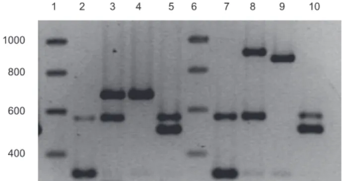
five-primer-pair MPCR set, we were able to detect simulta-neously five different ferripyoverdine receptor genes in different P. aeruginosa strains, two in the PVD type refer-ence strain PAO1 (fpvAI and fpvB), two in the type IIa reference strain 7NSK2 (fpvAIIa and fpvB), one in the type IIb reference strain ATCC 27853 (fpvAIIb) and two in the type III reference strain 59.20 (fpvAIII and fpvB) (Fig. 1). We also used an MPCR primer set in which the primers for detecting fpvAIIa and fpvAIIb are replaced by a single primer pair which detects both genes without discrimina-tion (Fig. 1). It is important to note that even with the bacterial cells directly inoculated as template in the PCR-mix (without prior boiling), clear amplifications were obtained.
Distribution of ferripyoverdine receptor genes in a P. aeruginosa population
The MPCR described above was applied to study the distribution of the currently identified ferripyoverdine receptor genes in a P. aeruginosa population comprising 345 clinical and environmental isolates from different locations throughout the world. The results are summa-rized in Table 1 and the complete list of strains with their origin is given in Table S1 in Supporting information. From only four isolates (1.2%) no amplification signal could be detected, while all the other strains were positive for at least one receptor gene (Table 1).
From these 341 MPCR-positive isolates, 122 (35.8%) had fpvAI, 48 had fpvAIIa (14.1%), 80 (23.5%) had fpvAIIb and 83 (24.3%) had the fpvAIII gene, while in eight strains (2.3%) only fpvB could be amplified. It is important to note that the distribution is slightly different according to the 1000
800 600
400
1 2 3 4 5 6 7 8 9 10
Fig. 1. Multiplex PCR-amplified fragments of four reference strains (PAO1: lanes 2 and 7, 7NSK2: lanes 3 and 8, ATCC 27853: lanes 4 and 9, and 59.20: lanes 5 and 10) with two different primer sets, electrophoretically separated. The first and sixth lanes contain the molecular weight markers (sizes in bp indicated on the left). The bands corresponding to the different PVD receptors have the following sizes in reverse order of size for set 1 (lanes 2–5): fpvAII, for both variants (682 bp), fpvB (562 bp), fpvAIII (505 bp) and fpvAI (324 bp), for set 2 (lanes 7–10): fpvAIIa (908 bp), fpvAIIb (863),
origin of the strains (Table S1). The clinical strains (220 strains) showed about the same proportion of each type, while FpvA type I was over-represented (49%) in environ-mental strains (79 strains).
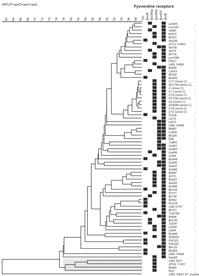
Altogether fpvB was amplified from 317 strains (93%) either alone or together with fpvAI, fpvAIIa, fpvAIIb or fpvAIII. The fpvB gene could not be amplified in 4.1%, 4.2%, 6.3% and 9.6% of the strains that were positive for fpvAI, fpvAIIa, fpvAIIb or fpvAIII respectively. Figure 2 shows a similarity tree based on AFLP patterns, sequences of oprI, oprL and oprD, and serotypes. Results of the MPCR are also shown for each strain. All 75 strains in the tree are mentioned in Table 1 and Table S1, except LMG 10643, which is not a P. aeruginosa, but a Pseudomonas oryzihabitans.
Comparison between IEF pyoverdine determination and receptor typing
Isoelectrofocalization of pyoverdines from the spent medium is a technique allowing fast and accurate deter-mination of the pyoverdine type in P. aeruginosa (Meyer et al., 1997; De Vos et al., 2001). However, some strains had lost the ability to produce pyoverdine, as evidenced in some cystic fibrosis isolates, but were still able to take up ferripyoverdine (De Vos et al., 2001; Ernst et al., 2003). For these pyoverdine-negative mutants, growth stimula-tion experiments with purified pyoverdines did not provide clear-cut answers because of the ability of some strains to utilize more than one type of ferripyoverdine as a source of iron (De Vos et al., 2001; Ghysels et al., 2004). This is due to the presence of FpvB, the alternative receptor for type I ferripyoverdine and also because the type III fer-ripyoverdine receptor also allows some level of utilization of the type II ferripyoverdine (Ghysels et al., 2004). All pyoverdine-positive strains, which were tested by IEF, showed the same fpvA receptor type as the correspond-ing pyoverdine, in addition to the presence or absence of fpvB gene (results not shown). Four of the fpvA-negative strains (So122, Lo059, Pr332 and Br700 strains) were found to produce type II pyoverdine, suggesting the
exist-ence of further type II receptor variants while the others were pyoverdine-negative (Table S1).
Functionality of the fpvB gene
In some pyoverdine-negative strains fpvB was amplified, either singly or together with an fpvA gene. The results presented in Fig. 3 for strains Mi159 and Mi162 show that fpvB is expressed and functional as judged by the growth stimulation assay using the three purified pyoverdines. Both isolates are pyoverdine-negative, but in Mi159 both fpvAIII and fpvB were amplified by PCR and only fpvB in Mi162. The growth of Mi159 was stimulated by the three pyoverdines, showing a good correlation with the pres-ence of FpvAIII and of FpvB. As already mentioned, FpvAIII allows the uptake not only of type III, but also, to some extent, of type II ferripyoverdine, and FpvB is responsible for the uptake of type I ferripyoverdine (Ghysels et al., 2004). In Mi162 only FpvB seems to be functional.
In strains SG17M, C2 and C19, which can be typed by IEF as type II pyoverdine producers (although their pyoverdine production is low), both fpvAIIb and fpvB can be amplified, although only in the case of SG17M could the growth be stimulated by type I pyoverdine, indicating either that in C2 and C19 the fpvB gene is not expressed or that its product is not functional (results not shown).
Nature of a supplementary 450 bp PCR fragment detected in some P. aeruginosa strains
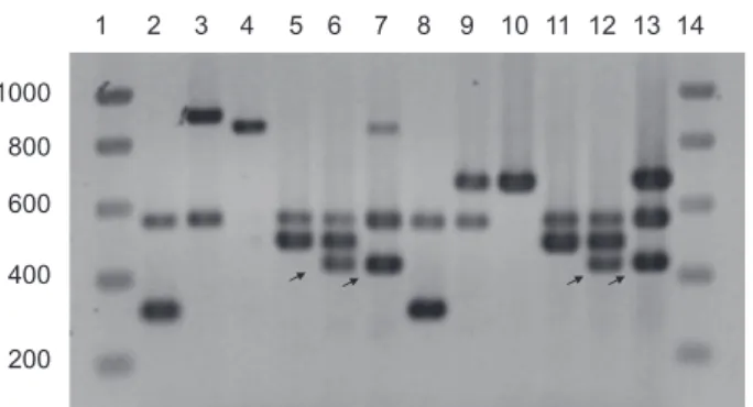
In a minority of the strains (8.9%) we obtained, in addition to the expected fragment associated with the different fpvA receptors, an additional amplicon of around 450 bp (Fig. 4). Closer analysis revealed that this fragment was the PCR product of the primer pair fpvAIf and fpvBf. A BLASTXsearch of Pseudomonas genomes revealed that the translated product had 94% identity with the products of two genes from PA7, PSPA7_0713 and PSPA7_5043 which are annotated as coding putative phage proteins. The fragment also appeared to be more frequently ampli-fied in type III strains (18%) than in type II (9%) and type I strains (2.5%).
Phylogeny of PVD receptors
In order to investigate the evolutionary history of the fer-ripyoverdine receptor genes in P. aeruginosa, we carried out a phylogenetic analysis with 8 fpvAI, 10 fpvAII (4 IIa and 6 IIb), 8 fpvAIII and 15 fpvB genes from 22 strains (Fig. 5A). While the dendrogram shows a great variability between fpvA and fpvB clusters, the variability within each fpvA and fpvB cluster is much lower, as highlighted by the scales on the dendrograms and the overall mean
variabil-Table 1. Results of the multiplex PCR of 345 P. aeruginosa strains. Positive strains fpvAI fpvAIIa fpvAIIb fpvAIII fpvB
5 + 117 + + 2 + 46 + + 5 + 75 + + 8 + 75 + + 8 + 4
fAFLP+oprD+oprI+oprL 100 98 96 94 92 90 88 86 84 82 80 78 76 74 72 70 68 66 64 62 Pyoverdine receptors fpvA I fpvA IIa fpvA IIb fpv AIII fpvB . . . . . . . . . . . . . . . . . . . . . . . . . . . . . . . . . . . . . . . . . . . . . . . . . . . . . . . . . . . . . . . . . . . . . . . . . . . . . . . . . . . . . . . . . . . . . . . . . . . . . . . . . . . . . . . . . . . . . . . . . . . . . . . . . . . . . . . . . . . . . . . . . . . . . . . . . . . . . . . . . . . . . . . . . . . . . . . . . . . . . . . . . . . . . . . . . . . . . . . . . . . . . . . . . . . . . . . . . . . . . . . . . Us448 Lw1047 Is580 Br670 Br257 Aa249 ATCC 27853 Mi159 Is579 Br776 Lw1048 PAO1 LMG 14083 Br908 Lo053 Mi162 Bu004 C17 (clone C) SG17M (clone C) C (clone C) C1 (clone C) C18 (clone C) PT31M (clone C) C2 (clone C) SG50M (clone C) C19 (clone C) C13 (clone C) Pr335 Li012 Li010 LMG 14085 Br667 Lo049 Br229 Pa6 Tu863 Us451 Us449 Us450 Li009 Bo548 So095 Us447 So099 Br692 Is573 Bu007 Aa245 So092 Be128 Pr317 Br735 Br642 Ro124 LMG 2107 Mi151 TuD199 Br906 Be136 TuD47 Lo050 Li004 Bo546 PhDW6 PAO23 PAO29 Be133 Bo559 LMG 14084 Aa246 LMG 5031 CPHL 11451 Br680 PA7 LMG 10643 (P. oryzihabitans) . . . . . . . . . . . . . . . . . . . . . . . . . . . . . . . . . . . . . . . . . . . . . . . . . . . . . . . . . . . . . . . . . . . . . . . . . . . . . . . . . . . . . . . . . . . . . . . . . . . . . . . . . . . . . . . . . . . . . . . . . . . . . . . . . . . . . . . . . . . . . . . . . . . . . . . . . . . . . . . . . . . . . . . . . . . . . . . . . . . . . . . . . . . . . . . . . . . . . . . . . . . . . . . . . . . . . . . . . . . . . . . . . . . . . . . . . . . . . . . . . . . . . . . . . . . . . . . . . . . . . . . . . . . . . . .
Fig. 2. Dendrogram (UPGMA, BioNumerics v5.2) based on the comparison of the composite data set consisting of the AFLP pattern, the oprI,
oprL and oprD nucleotide sequences and the serotype of 75 diverse P. aeruginosa strains isolated from different clinical and environmental
sites across the world. Black squares represent the type of receptor identified by MPCR. Strain name, geographical origin, isolation site and year and pyoverdine receptor profiles are shown in Table S1. The PA7 clade is highlighted in grey.
ity (Pi) in Table 2. Among these clusters, the fpvAII cluster is the most discriminatory (Pi is about twofold higher), with two robust subclusters (Bayesian posterior probabili-tiesⱖ 98%). The other fpv clusters contain more similar sequences revealing ambiguous topologies with statisti-cal supports frequently lower than 50%.
To explain the presence of these three very different types at the fpvA locus, it seems likely that some lateral transfers have occurred during the evolutionary history of the ferripyoverdine receptor genes of P. aeruginosa. Accordingly, several articles already suggested that several lateral transfers occurred in P. aeruginosa, espe-cially at pyoverdine locus (Pirnay et al., 2005; Smith et al., 2005; Wiehlmann et al., 2007).
In order to get insight into the evolutionary history of pyoverdine genes, we used several approaches. First, we carried out a comparative analysis between the ferripy-overdine receptor gene and organism phylogenies. Second, we looked at the synonymous codon usage [codon adaptation index (CAI index)] and the GC content.
Comparison between PVD receptor and organism phylogenies
We investigated the evolutionary history of organisms in a fine resolution by using a dendrogram (UPGMA, BioNumer-ics v5.2) based on the comparison of the composite data set consisting of AFLP patterns, oprI, oprL and oprD nucleotide sequences and serotypes (Fig. 2). In general, we can see that the closely related strains (i.e. with a similarity superior to 85%) presented the same fpvA/fpvB distribution, especially when the strains were epidemio-logically related (e.g. clone C) but also when they were not (e.g. Br692 versus Is573 strains). In contrast, some
closely related strains (e.g. PAO1 versus LMG14083 strains or Lo053 versus Mi162 strains) showed different fpvA/fpvB distribution, corresponding likely to some lateral transfers. Because the profiles become probably too dif-ferent when the strains are not closely related, almost all the dendrograms constructed from molecular fingerprints lose resolution in the deeper nodes.
In order to compensate for this putative limitation, we estimated an organism phylogeny at a larger resolution by using a set of 34 ribosomal concatenated genes from seven sequenced P. aeruginosa genomes (Fig. 5B). Inter-estingly, while the topology between six closely related strains (less than 0.3% of difference) has not been fully resolved (some weak statistical supports), the PA7 strain is well separated from the other strains in the organism phylogeny (maximum Bayesian posterior probabilities). We cannot formally exclude a faster evolution of the PA7 strain, as happens with a mutator strain. However, the position of the root as highlighted by out-grouping with Pseudomonas mendocina and Azotobacter vinelandii shows clearly an early divergence of this strain in the P. aeruginosa species (Fig. 5B). Moreover, from the last P. aeruginosa common ancestor, about the same evolu-tionary distance is observed to each strain.
Because both phylogenies (of ferripyoverdine receptor genes and concatenated ribosomal genes) have not been fully resolved, it is difficult to compare them. However, two observations can be made. (i) The presence of the same
Mi159
Mi162
II
III
III
II
I
I
fpvAIII
fpvB
fpvB
PCR
PCR
Fig. 3. Result of growth stimulation assays using disks impregnated with 2 mM purified pyoverdines (types I–III as indicated on the picture) for strains Mi159 and Mi162. The insert below shows the result of the multiplex PCR amplification.
1000 800 600 400 200 1 2 3 4 5 6 7 8 9 10 11 12 13 14
Fig. 4. Multiplex PCR-amplified fragments of six strains with two different primer sets, and separated by electrophoresis on 1% agarose. The first and 14th lanes contain the molecular weight markers as in Fig. 1. The bands corresponding to the different PVD receptors have the following sizes in reverse order of molecular weight for set 1 (lanes 2–7): fpvAIIa (908 bp), fpvAIIb (863 bp),
fpvB (562 bp), fpvAIII (505 bp) and fpvAI (324 bp), for set 2 (lanes
8–13): fpvAII, for both variants (682 bp), fpvB (562 bp), fpvAIII (505 bp) and fpvAI (324 bp). The four pyoverdine type reference strains are: PAO1 (lanes 2 and 8), 7NSK2 (lanes 3 and 9), ATCC 27853 (lanes 4 and 10) and 59.20 (lanes 5 and 11). In strains Br667 (lanes 6 and 12) and Is573 (lanes 7 and 13) we amplified, in addition to the bands corresponding to their receptor types (which is fpvAIII and fpvB for Br667 and fpvAIIb and fpvB for Is573), another band of around 450 bp (indicated by arrows) which appeared to be the result of amplification of a genomic fragment, probably of phage origin that is present in a small fraction of the
0.0005
I W15Dec6 fpvB(EU348660)
III W15Aug16 fpvB(EU348657) I PA14 fpvB
I 2192 fpvB I W15Dec1 fpvB(EU348659)
III W15Dec10 fpvB(EU348662)
III W15Dec9 fpvB(EU348661) IIb C3719 fpvB
III 59.20 fpvB(EU348655) I W15Aug21 fpvB(EU348658)
I PAO1 fpvB
IIb W15Dec11 fpvB(EU348663)
IIa W15Aug15 fpvB(EU348656)
IIa PACS2 fpvB III LES fpvB 54 58 71 87 99 75 99 94 0.0005
III W15Dec9 fpvA(EU348652)
III 206-12 fpvA(AY765261)
III W15Dec10 fpvA(EU348653)
III LES fpvA, III W15Aug16 fpvA(EU348648)
III ATCCO13 fpvA(AY765262)
IV Rprime fpvA(AY765260)
III 59.20 fpvA(AF537094)
89
88
0.0005
I PAO1 fpvA
I 2192 fpvA, I W15Dec1 fpvA(EU348650) I PAO29p fpvA(EU348646) I 10-15 fpvA(AY765259)
I W15Dec6 fpvA(EU348651) I W15Aug21 fpvA(EU348649)
I PA14 fpvA
57 79
91
0.005
IIa 7NSK2 fpvA(AF537095) IIa PACS2 fpvA
IIa W15Aug15 fpvA(EU348647) IIa MSH fpvA(AY765263) IIb PA7 fpvA
IIb C3719 fpvA
IIb W15Dec11 fpvA (EU348654) IIb 1-60 fpvA(AF540992),
IIb 2-164 fpvA(AF540993), IIb ATCC27853 fpvA (EU348645)
98 100 100 100 100 91
A
fpvB
fpvAI
fpvAIII
fpvAII
100
100
100
100
100
0.1
fpvAIIa subcluster fpvAIIb subcluster 0.001 PA7 PAO1 LES C3719 2192 PACS2 PA14 100 99 100 78 79B
Fig. 5. Phylogenetic relationships among ferripyoverdine receptor genes (A) and 34 concatenated ribosomal genes (B) from P. aeruginosa isolates. Strains for which the genome is sequenced are in bold. For the phylogenetic tree of ferripyoverdine receptor genes (A), each cluster (fpvAI, fpvAII, fpvAIII, fpvB) is highlighted separately, because of a great difference between intra- and inter-cluster variabilities. The names of the sequences on the subtrees correspond, respectively, to the fpvA type (I, IIa, IIb or III) of the corresponding strain, the strain name, the gene name (fpvA or fpvB), and the GenBank accession number (expect for genes from sequenced genome). For the phylogenetic tree of 34 concatenated ribosomal genes (B), the position of the root was determined by using P. mendocina and Azotobacter vinelandii (ymp and AvOP strains respectively) as out-group. All the dendrograms were generated using Bayesian inference under a GTR+g model of evolution. Numbers on tree branches report Bayesian posterior probabilities (expressed in percentage). Only statistical supportⱖ 50% are reported (majority rule consensus tree). The horizontal length of branches is proportional to the estimated number of substitutions. For each tree, the scale corresponds to the number of substitutions per site.
fpvA type (IIb) in two evolutionary-distant strains (PA7 and C3719 strains) added to the presence of all the possible fpvA types in closely related strains (PAO1, C3719, LES, 2192 and PACS2) confirmed that some lateral transfers have likely occurred at the fpvA locus. (ii) The second observation concerns the presence or absence of the fpvB gene in the seven sequenced genomes. Since the fpvB gene was detected in about 93% of our set of 345 P. aeruginosa strains and not in other Pseudomonas species (even in the close species P. mendocina), it might be useful to know whether the insertion of the fpvB gene was correlated with the P. aeruginosa speciation event, followed by some deletion events, or whether the insertion of the fpvB gene occurred after the speciation event, highlighted by an ancestral state of some P. aeruginosa strains without the fpvB gene. Interestingly, the peculiar PA7 strain is the only strain with a sequenced genome without the fpvB gene. Moreover, fpvB was not detected in three other strains forming a cluster with the PA7 strain (denominated ‘PA7 clade’ as highlighted in Fig. 2) in the composite dendrogram analysis. Since these three strains were not temporally and spatially related, we won-dered whether the fpvB insertion event occurred after the divergence of the PA7 clade, which could have inherited the ancestral state without the fpvB gene. An alternative
hypothesis would be a lateral transfer of fpvB gene before the divergence of the PA7 clade, followed by a deletion after this divergence. In both scenarios, the fpvB gene could have been introduced just before, during or just after the speciation event. It is worth to mention again that all strains in PA7 clade had the fpvAIIb gene. By studying the genomic context of the fpvB gene (http:// v2.pseudomonas.com), it can be observed that the regions upstream and downstream of fpvB are conserved in PAO1, LES and PA14. These genomic regions are also conserved in the genome of PA7 strain (Fig. 6). Interest-ingly, in PA7, the two genes flanking fpvB are conserved, and in place of fpvB a fragment of about 100 nucleotides showing 93% of identity with the end of the gene can be detected, highlighting an ancient deletion of the fpvB gene in the PA7 clade.
Study of the synonymous codon usage and the GC content
Finally, we studied the synonymous codon usage (CAI index) and the GC content to investigate the occurrence of lateral transfers during the evolutionary history of the ferripyoverdine receptor genes in P. aeruginosa. As expected, for each gene or set of genes, the CAI index
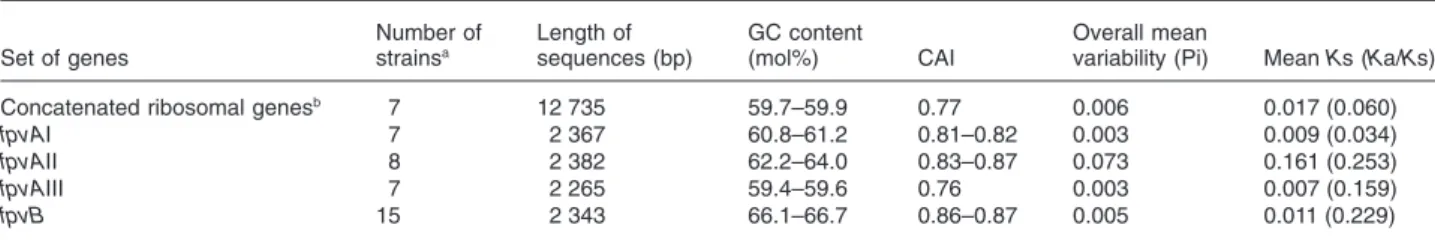
Table 2. Properties of the genes analysed in this study.
Set of genes Number of strainsa Length of sequences (bp) GC content (mol%) CAI Overall mean
variability (Pi) Mean Ks (Ka/Ks)
Concatenated ribosomal genesb 7 12 735 59.7–59.9 0.77 0.006 0.017 (0.060)
fpvAI 7 2 367 60.8–61.2 0.81–0.82 0.003 0.009 (0.034)
fpvAII 8 2 382 62.2–64.0 0.83–0.87 0.073 0.161 (0.253)
fpvAIII 7 2 265 59.4–59.6 0.76 0.003 0.007 (0.159)
fpvB 15 2 343 66.1–66.7 0.86–0.87 0.005 0.011 (0.229)
a. See the name of strains in Fig. 5A. Identical sequences were removed for this analysis. b. Corresponding to 34 concatenated ribosomal genes (see Experimental procedures).
prpL PA14_09910 PA14_09920 PA14_09930 PA14_09940 PA14_09950 PA14_09960 fpvB PA14_09980 PA14_09990 PA14_10010 PA14_10020 PA14_10040 PA14_10050
‘fpvB
PA7_0919 PA7_0920
xth2
PA7_0922 PA7_0923 PA7_0924 PA7_0925 PA7_0926 PA7_0927 PA7_0929 PA7_0928
Fig. 6. Schematic representation of the genomic region around the (complete or partial) fpvB gene in the PA14 (above) and PA7 (bellow) strains. ORFs oriented in the right are in the leading strand. Orthologous genes (according to the Pseudomonas Genome Project) are in grey and linked by double arrows.
and GC content were conserved between the sequenced P. aeruginosa strains for a given gene, while these features varied between the genes for a given strain (Table 2). However, it should be noted that, again, the PA7 strain had the highest value of GC content and CAI index for ribosomal genes and the fpvAIIb gene. When compar-ing the GC content of fpvA genes we can see a decrease in % GC from fpvAII to fpvAIII (Table 2). Since many driving forces are responsible for variations in GC content (e.g. position in the genome) or in synonymous codon usage (e.g. level of gene expression), it is usually difficult to compare these features between different genes. However, as expected, the concatenated ribosomal genes showed a GC content of about 60%, a classically lower value than the GC content calculated from the core genome of PAO1 strain (67.1%) (Wolfgang et al., 2003; Bodilis and Barray, 2006). Interestingly, the fpvB gene showed the same GC content as for the core genome and high CAI values, typical for a gene present in the lineage for a long time. These features are in agreement with both the evolutionary scenario described above, suggesting that the fpvB gene was introduced early in the P. aerugi-nosa lineage and subsequently lost in some strains such as those representing the PA7 clade. Concerning the fpvA genes, since the different alleles are roughly at the same locus, code a similar function and present a priori the same level of expression, we could expect the same CAI index and GC content values between the three fpvA types. Because this was not the case, we deduced that some inter-species lateral transfers occurred at different times and/or from different organisms, lateral transfer of the fpvAII gene being the more ancient event and/or from a closely related organism, followed by the fpvAI gene and finally the fpvAIII gene.
The fpvAIII gene of LES strain is triplicated
Analysis of the recently annotated genome of the Liverpool epidemic strain (LES) revealed that three identical copies of fpvAIII are present (http://www. pseudomonas.com). Also the pvdE gene is triplicated and there are two incomplete pvdF genes before each fpvAIII. The sequences of pvdE and fpvAIII are all identical to each other.
Discussion
Because most of the studies about the population struc-ture of P. aeruginosa had an epidemiological goal and focused on recent clonal expansions, geographic localiza-tions and links with virulence factors and pathogenicity, little was known about the early evolutionary history of the P. aeruginosa species. In this article, we have approached this aspect in order to study the distribution of
ferripyover-dine receptor genes from an evolutionary point of view. We therefore estimated an organism phylogeny in the scale of the P. aeruginosa species from parts of the core genome of the seven P. aeruginosa sequenced genomes. The phylogenetic tree obtained from ribosomal genes showed an early divergence of PA7 strain that was strongly distant from the six other closely related P. aeruginosa strains (Figs 2 and 5B). These six strains were not clearly evolutionarily distinct from each other, with a not fully supported topology, probably because of a very limited variability. In contrast, a composite dendro-gram (including AFLP pattern, oprI, oprL and oprD gene sequences, and serotype) was useful to discriminate between those more closely related strains but may have some limitations on a larger scale. Altogether, the use of these two phylogenic approaches permitted us to study the evolutionary history in the whole P. aeruginosa species.
The ribosomal genes have already been shown to be useful for constructing a robust phylogeny among Pseudomonas (Bodilis and Barray, 2006). It is important to note that there are some discussions about methods for estimating phylogeny from a set of genes (Gadagkar et al., 2005). Phylogeny could be estimated either from concatenated genes (as we did), or by carrying out a consensus from individual trees. The principal argument against phylogeny from concatenated genes is the varia-tion of the evoluvaria-tionary rate between funcvaria-tionally distinct genes. However, because the ribosomal genes code for functionally linked proteins and have likely evolved slowly at the same evolutionary rate (independent of environ-mental changes), we argue that this argument against phylogeny from concatenated genes is not valuable here. Second, from the 34 (generally not well supported) trees constructed from individual ribosomal genes, we arrived to the same conclusions, i.e. a strong separation of the PA7 strain and variable topologies for the six other closely related strains (data not shown).
Pseudomonas aeruginosa is a ubiquitous microorgan-ism, which is endowed with a high capacity for adapta-tion to different niches (Goldberg, 2000). This is reflected in its capacity to take up different siderophores next to the uptake of its own siderophores, pyoverdine and pyochelin (Cornelis and Matthijs, 2002; Cornelis et al., 2007; 2009). Pseudomonas aeruginosa strains can be subdivided into three groups based on the type of pyoverdine they produce (Cornelis et al., 1989; Meyer et al., 1997; De Vos et al., 2001). The receptors corre-sponding to these three ferripyoverdines have now been identified by different teams (Poole et al., 1993; De Chial et al., 2003; Spencer et al., 2003; Smith et al., 2005). Here, by using an MPCR, we typed 345 clinical and environmental isolates from different locations through-out the world and found a similar distribution of each
receptor type, type I being slightly over-represented in environmental strains. Interestingly, in a recent work on 240 P. aeruginosa strains (only a few strains were common with our study), Wiehlmann and colleagues (2007) found about the same proportion for each ferripy-overdine receptor type. Since competition for iron plays an important role for the fitness of Pseudomonas (Griffin et al., 2004) a link between the distribution of the FpvA types and the ecological niches could be expected. The slightly different proportion of each type between envi-ronmental and clinical strains would be interesting to investigate further by studying the coexistence of strains with different FpvA types, in terms of cooperation (which would tend to limit the number of different PVD type) and competition (which would tend to increase the number of different PVD type).
Another important observation concerns the conserva-tion of the fpvB gene among P. aeruginosa strains, sug-gesting that the ability to utilize type I ferripyoverdine as a source of iron is a common trait of the vast majority of P. aeruginosa strains (Ghysels et al., 2004). Although we did not investigate the functionality of FpvB in a large number of strains, it is evident that there are some instances where the gene is present (or at least the part we amplified with the primers used in this study), but the ability to utilize the heterologous type I pyoverdine could not be observed, perhaps because fpvB is not expressed in these strains. In the study of Wiehlmann and col-leagues (2007), the authors found that 10% of the P. aeruginosa tested do not have the fpvB gene, which is close to the 7% we found. Moreover, it could be deduced from both the study of Wiehlmann and colleagues (2007) and ours (Fig. 2) that at least a few deletions of fpvB genes have occurred as evidenced in PA7 (Fig. 6). Because fpvB was only found in P. aeruginosa and was absent in other Pseudomonas spp., we formulate the hypothesis of an ancestral state of some P. aeruginosa strains before the insertion of the fpvB gene. So, the fpvB gene was likely introduced early in the P. aeruginosa species (or just before the speciation event), and lost in the PA7 clade. The deletion of fpvB would therefore have occurred in the PA7 clade soon after its insertion. This observation refutes thus the most parsimonious hypoth-esis of an ancestral state without fpvB inherited by the PA7 clade.
Finally, the fact that the great majority (more than 90%) of P. aeruginosa have fpvB could highlight a fundamental role of this gene in the ecology of this species. Neverthe-less, it cannot be excluded that introduction of fpvB in the P. aeruginosa species would be concomitant with a trans-fer of a more important gene and so, would result from a genetic hitchhiking.
In their interesting study on the evolution of pyover-dine biosynthesis and uptake genes, Smith and
col-leagues (2005) propose that the pyoverdine region has been acquired by horizontal transfer, since the codon usage of the corresponding genes is unusual. Within the P. aeruginosa pyoverdine region, some genes show high divergence between types. These genes include the NRPS genes involved in the biosynthesis of the pyover-dine peptide chain, the pvdE gene coding for an ABC transporter, and the fpvA gene encoding the receptor (Ravel and Cornelis, 2003; Smith et al., 2005; Visca et al., 2007). Based on large strain collections, this study and two previous studies (Pirnay et al., 2005; Wiehl-mann et al., 2007) have arrived at the same conclusion of frequent intra-species lateral transfers of fpvA genes, correlated with the important role of the FpvA type in the fitness of P. aeruginosa. It is interesting to mention that in other fluorescent pseudomonads the genes involved in the biosynthesis and uptake of pyoverdine are also clustered, suggesting that horizontal gene transfers have also occurred in these species (Ravel and Cornelis, 2003). According to the study of Smith and colleagues (2005), from GC content and synonymous codon usage it seems that the type III ferripyoverdine receptor gene was transferred more recently or from a more distant organism than the other two types, in agreement with the low GC content of this gene (59%), the lowest of all other TonB-dependent receptor genes, which have an average value of 67% (P. Cornelis and J. Bodilis, in preparation). In contrast, the type IIb ferripyoverdine receptor gene was probably transferred before the other two types or from a more closely related organism. Inter-estingly, since FpvAIIb is the receptor of the peculiar PA7 clade, it may be the first fpvA type of the P. aerugi-nosa species.
Intra-type variability and tests for positive selection have highlighted a diversifying selection of the fpvAII gene (Smith et al., 2005; Tümmler and Cornelis, 2005). Smith and colleagues (2005) made the suggestion that the more rapid evolution of this gene might be driven by the need to resist killing by pyocin S3, for which FpvAII is the receptor (Baysse et al., 1999; De Chial et al., 2003). Although we also think that a Darwinian selection most likely occurred for the fpvA gene, we do not totally agree with this hypoth-esis of driving force proposed by Smith and colleagues (2005). First, we have recently shown that another soluble pyocin, S2, kills strains having the type I ferripyoverdine receptor, but sequences of different fpvAI alleles from S2-sensitive and S2-resistant strains did not reveal such a diversifying selection (Denayer et al., 2007). The second argument is the sensitivity to pyocin S3 of strains with both FpvAII receptor subtypes (IIa and IIb), highlighting that this positive selection gives no particular advantage for resis-tance to pyocin S3 (data not shown). So, the driving force may be unknown yet, e.g. the use of FpvAII as a phage receptor or the need to escape to the immune system. To
explain this observed positive selection and more gener-ally to explain the great diversity of the PVD/FpvA pairs, we suggest an alternative scenario where the evolution of the receptor is driven essentially by changes in pyoverdine structure. In the competition for iron, new pyoverdine struc-tures could offer a selective advantage. In this context, we hypothesize that the changes occur first in just one or only a few modules of the NRPS for the biosynthesis of a given pyoverdine. Since a receptor can sometimes recognize heterologous pyoverdines (Ghysels et al., 2004), a new pyoverdine variant could still be recognized by the recep-tor, although with lower efficiency. This could now drive the evolution of the receptor towards a finer specificity, by a positive selection. In this scenario, the type II pyoverdine would result from relatively recent modifications in its struc-ture (in fact, perhaps concomitant with the speciation event) and the recognition of the pyoverdine by the recep-tor would not yet be optimized. In this regard, it is important to mention that type II FpvA is the receptor showing the highest specificity, since it does not allow the transport of the other two P. aeruginosa pyoverdines (Ghysels et al., 2004). In order to check this hypothesis, it would be inter-esting to study the competition between bacteria with type IIa and those with type IIb FpvA in conditions of iron limitation, with or without pyocin S3. Since evolution of receptors could also be facilitated by gene duplications, it is of interest to notice that three copies of pvdE and fpvAIII exist in the LES strain. However, the three copies are identical, suggesting that this is a recent event. In Pseudomonas syringae genomes there are two copies of fpvA in tandem, but the two proteins are only 73% identical (P. Cornelis and J. Bodilis, in preparation).
Finally, in addition to changing or diversifying their pyoverdine and their associated FpvA receptor, acquisi-tion of alternative receptors (without the PVD genes), like FpvB but also like the 35 other putative TonB-dependent receptors identified in the PAO1 genome (Cornelis et al., 2007), can be considered as a cheap (and cheat) strategy to increase the fitness.
The MPCR described in this study allows a more rapid and accurate identification of the pyoverdine type
com-pared with the IEF-based method for siderotyping (Meyer et al., 1997) and should also be useful for the typing of pyoverdine-negative strains that are often isolated from Cystic Fibrosis (CF) lungs (De Vos et al., 2001). Since nine patterns are possible (fpvAI, fpvAIIa, fpvAIIb, fpvAIII, fpvAI-fpvB, fpvAIIa-fpvB, fpvAIIb-fpvB, fpvAIII-fpvB, fpvB), this MPCR could be useful as a complementary technique for typing P. aeruginosa isolates. Since it appears that several typing methods, with different degrees of resolu-tion, are necessary for the study of P. aeruginosa, similar MPCR assays could be designed by including other receptor genes, such as fptA for pyochelin (Ankenbauer and Quan, 1994) or pfeA and pirA for ferrienterobactin (Ghysels et al., 2005).
Experimental procedures Bacterial strains used in this study
The P. aeruginosa strains used for reference in this MPCR are PAO1, a type I pyoverdine producer (Stover et al., 2000), 7NSK2 and ATCC27853, both type II pyoverdine producers (De Chial et al., 2003), and 59.20 as an example of a type III pyoverdine producer (De Chial et al., 2003). Some (75 strains) of the 345 strains used in this study are reported in Fig. 2. A list of all the strains used for this study as well as their origin is available in Table S1.
Primers and PCR conditions
The primers used for this MPCR are listed in Table 3. The PCR was performed using ™Ex-Taq polymerase (Takara), supplied with buffer and dNTPs, according to the following cycling parameters: 94°C (5 min) followed by 30 cycles [94°C (30 s)-52°C (30 s)-72°C (2 min)] and a final extension [72°C (10 min step)]. All the primers were manufactured by Euro-gentec (Seraing, Belgium). The template for the PCR-mix was either a pipette tip of bacterial cells (without prior boiling), or 2ml of a chromosomal DNA preparation. Double-stranded DNA sequencing of some fpvA and fpvB genes was carried out by the VIB sequence facility. The nucleotide sequences determined in this study have been deposited in the GenBank database.
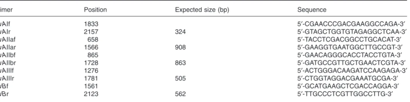
Table 3. Primers used in this study.
Primer Position Expected size (bp) Sequence
fpvAIf 1833 5′-CGAACCCGACGAAGGCCAGA-3′ fpvAIr 2157 324 5′-GTAGCTGGTGTAGAGGCTCAA-3′ fpvAIIaf 658 5′-TACCTCGACGGCCTGCACAT-3′ fpvAIIar 1566 908 5′-GAAGGTGAATGGCTTGCCGT-3′ fpvAIIbf 865 5′-GAACAGGGCACCTACCTGTA-3′ fpvAIIbr 1728 863 5′-GATGCCGTTGCTGAACTCGTA-3′ fpvAIIIf 1276 5′-ACTGGGACAAGATCCAAGAGA-3′ fpvAIIIr 1781 505 5′-CTGGTAGGACGAAATGCGA-3′ fpvBf 1561 5′-GCATGAAGCTCGACCAGGA-3′ fpvBr 2123 562 5′-TTGCCCTCGTTGGCCTTG-3′
Phylogenetic analyses
From 22 strains (including the seven strains for which the genomes were sequenced), nearly complete FpvA and/or FpvB sequences (41 sequences in total) were aligned using
CLUSTALXversion 1.81, with default parameters (Thompson
et al., 1997), and optimized visually. The nucleic acid
align-ment was deduced from the corrected protein alignalign-ment, leading to about 2300 aligned nucleotide positions.
A set of 34 ubiquitous ribosomal genes were retrieved from the seven (fully or partially) sequenced Pseudomonas
aerugi-nosa genomes (PAO1, LES, 2192, PACS2, C3719, PA7 and
PA14 strains). All the genes were aligned individually and concatenated, leading to 12 735 unambiguously aligned nucleotide positions.
From nucleic alignments, Bayesian analysis was per-formed using MrBayes 3.1 (Ronquist and Huelsenbeck, 2003). The Modeltest software (Posada and Crandall, 1998) was used to choose the evolutionary model. For both phy-logenies (PVD receptor and ribosomal genes), the model used is the complex GTR with an among-site rate hetero-geneity (GTR+ g). In addition, we also used a model that takes into account rate heterogeneity among positions in codon. Since the resulting topologies were identical for the two models, except for two weak-supported nodes in the
fpvB cluster, only the phylogenetic analyses from the first
model were presented in Fig. 5. All analyses were carried out with random starting trees. Four Metropolis coupled Markov chain Monte Carlo (MCMC) chains were run, stop-ping after 1 or 2 million generations (for ribosomal and PVD receptor genes respectively), when the standard deviation of split frequencies was less than 0.01. Trees were sampled every 100 generations and the first 25% burn-in cycles (i.e. 2500 or 5000 trees) were discarded prior to consensus trees construction. Analyses were repeated twice to ensure the correct topology. Consensus trees were visualized with TreeView 1.6.6 (Page, 1996) and posterior probabilities were employed to test the statistical support of clades. Addi-tionally, a data set consisting of the AFLP pattern, oprI, oprL and oprD gene sequences, and serotype of 75 P.
aerugi-nosa isolates was analysed using biological data analysis
software. AFLP band patterns were imported into BioNu-merics v5.2 software (Applied Maths, Belgium) for further normalization (background subtraction, filtering: arithmetic average, and band search: minimum profiling 0.5% relative to maximum value) and cluster analysis (similarity coeffi-cient: Pearson correlation, dendrogram type:UPGMA, optimi-zation: 0%, position tolerance: 1%, uncertain bands were ignored). Sequences were clustered (Pairwise alignment, open gap penalty: 100%, unit gap penalty 0%, minimum match sequence: 2, maximum number of gaps: 9, fast algo-rithm), aligned (multiple alignment, open gap penalty: 100%, unit gap penalty: 0%, minimum match sequence: 2, maximum number of gaps: 98) and clustered a second time (using the same parameters) using BioNumerics v5.2 soft-ware. The serotypes were compared using the Pearson cor-relation. These individual comparisons resulted in individual similarity matrices. These similarity matrices were averaged into the similarity matrix of the composite data set. No cor-rection for internal weights was applied. A dendrogram (UPGMA, BioNumerics v5.2) based on the comparison of the composite data set was built.
Sequence analyses
The synonymous and non-synonymous rates were deter-mined using the modified Nei-Gojobori method implemented in the MEGA v2.0 software (Kumar et al., 2001). The transition to transversion ratio was fixed at 2 and the Jukes–Cantor correction was used to account for multiple substitutions at the same site. Codon adaptation index (CAI) was calculated with the new method implemented in
DAMBE software which deals with several computational problems (Xia and Xie, 2001; Xia, 2007). All the measure-ments were also carried out with the classical method as implemented in EMBOSS.cai program (Rice et al., 2000) and, although the values were always lower than the ones presented here, the trends were the same (data not shown). As CAI is a measure of the relative codon usage bias of a gene towards the average codon usage of an organism, a reference codon usage table of the given organism is required. Because only the reference codon usage table of the PAO1 strain is available in EMBOSSand
DAMBE data (Epae), we wondered whether differences in codon usage between P. aeruginosa strains would prevent us using the same reference table for all P. aeruginosa strains. To deal with this problem, we estimated seven ref-erence codon usage tables from concatenated ribosomal genes of the seven P. aeruginosa sequenced genomes, by using the cusp program ofEMBOSS(Rice et al., 2000). Next, we used these reference codon usage tables to calculate CAI (with classical and new methods) for several genes and found almost identical results, whatever the strains used to construct the reference codon usage tables (data not shown), highlighting almost identical optimal codon usage between the different P. aeruginosa strains tested. Therefore only the results obtained with the reference codon usage table of the PAO1 strain (Epae) are presented here.
Pyoverdine typing by IEF
For IEF typing, pyoverdines were partially purified by chro-mabond C18-affinity chromatography from 10 ml supernatant of cell culture in casamino acid medium (CAA). Pyoverdine was eluted from this matrix with a 1:1 water/methanol mixture. Pyoverdine-IEF was carried out on Ampholine PAG plates (pH 3.5–9.5; Pharmacia) as described previously (Meyer et al., 1997). For growth stimulation assays, pyover-dines from the different reference strains (PAO1, 7NSK2 or 59.20) were semi-purified on a preparative scale on an XAD-4 amberlite column as described earlier (Budzikiewicz, 1993; Ghysels et al., 2004).
Acknowledgements
This work received the support of the OZR fund from the VUB, of the Belgian Federal Research Policy (contract No. C3/00/13), and of the Association Française de Lutte contre la Mucoviscidose. We thank Dr Paul De Vos (University of Gent) for his interesting comments.
References
Ankenbauer, R.G., and Quan, H.N. (1994) FptA, the Fe(III)-pyochelin receptor of Pseudomonas aeruginosa: a pheno-late siderophore receptor homologous to hydroxamate siderophore receptors. J Bacteriol 176: 307–319. Baysse, C., Meyer, J.M., Plesiat, P., Geoffroy, V.,
Michel-Briand, Y., and Cornelis, P. (1999) Uptake of pyocin S3 occurs through the outer membrane ferripyoverdine type II receptor of Pseudomonas aeruginosa. J Bacteriol 181: 3849–3851.
Bodilis, J., and Barray, S. (2006) Molecular evolution of the major outer-membrane protein gene (oprF) of
Pseudomo-nas. Microbiology 152: 1075–1088.
Braun, V., and Killmann, H. (1999) Bacterial solutions to the iron-supply problem. Trends Biochem Sci 24: 104–109. Budzikiewicz, H. (1993) Secondary metabolites from
fluores-cent pseudomonads. FEMS Microbiol Rev 10: 209–228. Cornelis, P., and Matthijs, S. (2002) Diversity of
siderophore-mediated iron uptake systems in fluorescent pseudo-monads: not only pyoverdines. Environ Microbiol 4: 787– 798.
Cornelis, P., Hohnadel, D., and Meyer, J.M. (1989) Evidence for different pyoverdine-mediated iron uptake systems among Pseudomonas aeruginosa strains. Infect Immun
57: 3491–3497.
Cornelis, P., Baysse, C., and Matthijs, S. (2007) Iron uptake in Pseudomonas. In Pseudomonas. Genomics and Molecular Biology. Cornelis, P. (ed.). Linton, UK: Caister
Academic Press, pp. 213–235.
Cornelis, P., Matthijs, S., and Van Oeffelen, L. (2009) Iron uptake regulation in Pseudomonas aeruginosa. Biometals
22: 15–22.
De Chial, M., Ghysels, B., Beatson, S.A., Geoffroy, V., Meyer, J.M., Pattery, T., et al. (2003) Identification of type II and type III pyoverdine receptors from Pseudomonas
aeruginosa. Microbiology 149: 821–831.
De Vos, D., De Chial, M., Cochez, C., Jansen, S., Tümmler, B., Meyer, J.M., and Cornelis, P. (2001) Study of pyover-dine type and production by Pseudomonas aeruginosa isolated from cystic fibrosis patients: prevalence of type II pyoverdine isolates and accumulation of pyoverdine-negative mutations. Arch Microbiol 175: 384–388. Denayer, S., Matthijs, S., and Cornelis, P. (2007) Pyocin S2
(Sa) kills Pseudomonas aeruginosa strains via the FpvA type I ferripyoverdine receptor. J Bacteriol 189: 7663– 7668.
Ernst, R.K., d’Argenio, D.A., Ichikawa, J.K., Bangera, M.G., Selgrade, S., Burns, J.L., et al. (2003) Genome mosaicism is conserved but not unique in Pseudomonas aeruginosa isolates from the airways of young children with cystic fibrosis. Environ Microbiol 5: 1341–1349.
Gadagkar, S.R., Rosenberg, M.S., and Kumar, S. (2005) Inferring species phylogenies from multiple genes – con-catenated sequence tree versus consensus gene tree.
J Exp Zool 304: 64–74.
Ghysels, B., Dieu, B.T., Beatson, S.A., Pirnay, J.P., Ochsner, U.A., Vasil, M.L., and Cornelis, P. (2004) FpvB, an alterna-tive type I ferripyoverdine receptor of Pseudomonas
aeruginosa. Microbiology 150: 1671–1680.
Ghysels, B., Ochsner, U., Mollman, U., Heinisch, L., Vasil, M.,
Cornelis, P., and Matthijs, S. (2005) The Pseudomonas
aeruginosa pirA gene encodes a second receptor for
ferri-enterobactin and synthetic catecholate analogues. FEMS
Microbiol Lett 246: 167–174.
Goldberg, J.B. (2000) Pseudomonas: global bacteria. Trends
Microbiol 8: 55–57.
Griffin, A.S., West, S.A., and Buckling, A. (2004) Cooperation and competition in pathogenic bacteria. Nature 430: 1024– 1027.
Kumar, S., Tamura, K., Jakobsen, I.B., and Nei, M. (2001) MEGA2: molecular evolutionary genetics analysis soft-ware. Bioinformatics 17: 1244–1245.
Meyer, J.M. (2000) Pyoverdines: pigments, siderophores and potential taxonomic markers of fluorescent Pseudomonas species. Arch Microbiol 174: 135–142.
Meyer, J.M., Stintzi, A., De Vos, D., Cornelis, P., Tappe, R., Taraz, K., and Budzikiewicz, H. (1997) Use of siderophores to type pseudomonads: the three Pseudomonas
aerugi-nosa pyoverdine systems. Microbiology 143: 35–43.
Mossialos, D., Ochsner, U., Baysse, C., Chablain, P., Pirnay, J.P., Koedam, N., et al. (2002) Identification of new, con-served, non-ribosomal peptide synthetases from fluores-cent pseudomonads involved in the biosynthesis of the siderophore pyoverdine. Mol Microbiol 45: 1673–1685. Page, R.D. (1996) TreeView: an application to display
phylo-genetic trees on personal computers. Comput Appl Biosci
12: 357–358.
Pirnay, J.-P., Matthijs, S., Colak, H., Chablain, P., Bilocq, F., Van Eldere, J., et al. (2005) Global Pseudomonas
aerugi-nosa biodiversity as reflected in a Belgian river. Environ Microbiol 7: 969–980.
Poole, K., Neshat, S., Krebes, K., and Heinrichs, D.E. (1993) Cloning and nucleotide sequence analysis of the ferripy-overdine receptor gene fpvA of Pseudomonas aeruginosa.
J Bacteriol 175: 4597–4604.
Posada, D., and Crandall, K.A. (1998) MODELTEST: testing the model of DNA substitution. Bioinformatics 14: 817–818. Ravel, J., and Cornelis, P. (2003) Genomics of pyoverdine-mediated iron uptake in pseudomonads. Trends Microbiol
11: 195–200.
Rice, P., Longden, I., and Bleasby, A. (2000)EMBOSS: the European Molecular Biology Open Software Suite. Trends
Genet 16: 276–277.
Ronquist, F., and Huelsenbeck, J.P. (2003) MrBayes 3: Bayesian phylogenetic inference under mixed models.
Bioinformatics 19: 1572–1574.
Smith, E.E., Sims, E.H., Spencer, D.H., Kaul, R., and Olson, M.V. (2005) Evidence for diversifying selection at the pyoverdine locus of Pseudomonas aeruginosa. J Bacteriol
187: 2138–2147.
Spencer, D.H., Kas, A., Smith, E.E., Raymond, C.K., Sims, E.H., Hastings, M., et al. (2003) Whole-genome sequence variation among multiple isolates of Pseudomonas
aerugi-nosa. J Bacteriol 185: 1316–1325.
Stover, C.K., Pham, X.Q., Erwin, A.L., Mizoguchi, S.D., War-rener, P., Hickey, M.J., et al. (2000) Complete genome sequence of Pseudomonas aeruginosa PA01, an opportu-nistic pathogen. Nature 406: 959–964.
Thompson, J.D., Gibson, T.J., Plewniak, F., Jeanmougin, F., and Higgins, D.G. (1997) TheCLUSTAL_Xwindows inter-face: flexible strategies for multiple sequence alignment
aided by quality analysis tools. Nucleic Acids Res 25: 4876–4882.
Tümmler, B., and Cornelis, P. (2005) Pyoverdine receptor: a case of positive Darwinian selection in Pseudomonas
aeruginosa. J Bacteriol 187: 3289–3292.
Visca, P., Imperi, F., and Lamont, I.L. (2007) Pyoverdine siderophores: from biogenesis to biosignificance. Trends
Microbiol 15: 22–30.
Wiehlmann, L., Wagner, G., Cramer, N., Siebert, B., Gudow-ius, P., Morales, G., et al. (2007) Population structure of
Pseudomonas aeruginosa. Proc Natl Acad Sci USA 104:
8101–8106.
Wolfgang, M.C., Kulasekara, B.R., Liang, X., Boyd, D., Wu, K., Yang, Q., et al. (2003) Conservation of genome content and virulence determinants among clinical and environ-mental isolates of Pseudomonas aeruginosa. Proc Natl
Acad Sci USA 100: 8484–8489.
Xia, X. (2007) An improved implementation of Codon Adap-tation Index. Evol Bioinformatics 3: 53–58.
Xia, X., and Xie, Z. (2001)DAMBE: software package for data analysis in molecular biology and evolution. J Hered 92: 371–373.
Supporting information
Additional Supporting Information may be found in the online version of this article:
Table S1. Strains used in this study, indicating their MPCR
results, origin, year, and source (environmental strains in yellow).
Please note: Wiley-Blackwell are not responsible for the content or functionality of any supporting materials supplied by the authors. Any queries (other than missing material) should be directed to the corresponding author for the article.