Université de Montréal
Alternative Strategies for Deciphering the Genetic Architecture of Childhood Pre-B Acute Lymphoblastic Leukemia
par
Jasmine Healy
Programmes de biologie moléculaire Faculté de médecine
Thèse présentée à la Faculté de médecine en vue de l‘obtention du grade de doctorat
en biologie moléculaire
Juin, 2010
Université de Montréal Faculté de médecine
Cette thèse intitulée :
Alternative Strategies for Deciphering the Genetic Architecture of Childhood Pre-B Acute Lymphoblastic Leukemia
présentée par:
Jasmine Healy
a été évaluée par un jury composé des personnes suivantes:
Dr. Bernard Brais ……….. président-rapporteur Dr. Daniel Sinnett ……….. directeur de recherche Dr. Guillaume Lettre ……….. membre du jury Dr. David Goldgar ……….. examinateur externe Dr. Patrick Cossette ……….. représentant du doyen
F
RENCHS
UMMARYLa leucémie lymphoblastique aigüe (LLA) est une maladie génétique complexe. Malgré que cette maladie hématologique soit le cancer pédiatrique le plus fréquent, ses causes demeurent inconnues. Des études antérieures ont
démontrées que le risque à la LLA chez l‘enfant pourrait être influencé par des gènes agissant dans le métabolisme des xénobiotiques, dans le maintient de l‘intégrité génomique et dans la réponse au stress oxydatif, ainsi que par des facteurs environnementaux. Au cours de mes études doctorales, j‘ai tenté de disséquer davantage les bases génétiques de la LLA de l‘enfant en postulant que la susceptibilité à cette maladie serait modulée, au moins en partie, par des variants génétiques agissant dans deux voies biologiques fondamentales : le point de contrôle G1/S du cycle cellulaire et la réparation des cassures double-brin de l‘ADN. En utilisant une approche unique reposant sur l‘analyse d‘une cohorte cas-contrôles jumelée à une cohorte de trios enfants-parents, j‘ai effectué une étude d‘association de type gènes/voies biologiques candidats. Ainsi, j‘ai évaluer le rôle de variants provenant de la séquence promotrice de 12 gènes du cycle cellulaire et de 7 gènes de la voie de réparation de l‘ADN, dans la susceptibilité à la LLA. De tels polymorphismes dans la région promotrice (pSNPs) pourraient perturber la liaison de facteurs de transcription et mener à des différences dans les niveaux d‘expression des gènes pouvant influencer le risque à la maladie.
En combinant différentes méthodes analytiques, j‘ai évalué le rôle de différents mécanismes génétiques dans le développement de la LLA chez l‘enfant. J‘ai tout d‘abord étudié les associations avec gènes/variants indépendants, et des essaies fonctionnels ont été effectués afin d‘évaluer l‘impact des pSNPs sur la liaison de facteurs de transcription et l‘activité promotrice allèle-spécifique. Ces analyses ont mené à quatre publications. Il est peu probable que ces gènes de
susceptibilité agissent seuls; j‘ai donc utilisé une approche intégrative afin d‘explorer la possibilité que plusieurs variants d‘une même voie biologique ou de voies connexes puissent moduler le risque de la maladie; ces travaux ont été soumis pour publication. En outre, le développement précoce de la LLA, voir même in utero, suggère que les parents, et plus particulièrement la mère,
pourraient jouer un rôle important dans le développement de cette maladie chez l‘enfant. Dans une étude par simulations, j‘ai évalué la performance des
méthodes d‘analyse existantes de détecter des effets fœto-maternels sous un design hybride trios/cas-contrôles. J‘ai également investigué l‘impact des effets génétiques agissant via la mère sur la susceptibilité à la LLA. Cette étude, récemment publiée, fût la première à démontrer que le risque de la leucémie chez l‘enfant peut être modulé par le génotype de sa mère.
En conclusions, mes études doctorales ont permis d‘identifier des nouveaux gènes de susceptibilité pour la LLA pédiatrique et de mettre en évidence le rôle du cycle cellulaire et de la voie de la réparation de l‘ADN dans la
leucémogenèse. À terme, ces travaux permettront de mieux comprendre les bases génétiques de la LLA, et conduiront au développement d‘outils cliniques qui amélioreront la détection, le diagnostique et le traitement de la leucémie chez l‘enfant.
Mots clés : leucémie lymphoblastique aigüe de l‘enfant, épidémiologie génétique, susceptibilité génétique, polymorphisme régulateur, expression génique, cycle cellulaire, réparation de l‘ADN, voie biologique, interaction gène-gène, association génétique fœto-maternelle
E
NGLISHS
UMMARYChildhood acute lymphoblastic leukemia (ALL) is a complex and heterogeneous genetic disease. Although it is the most common pediatric cancer, its etiology remains poorly understood. Previous studies provided evidence that childhood ALL might originate through the collective contribution of different genes
controlling the efficiency of carcinogen metabolism, the capacity of maintaining DNA integrity and the response to oxidative stress, as well as environmental factors. In my doctoral research project I attempted to further dissect the genetic intricacies underlying childhood ALL. I postulated that a child‘s susceptibility to ALL may be influenced, in part, by functional sequence variation in genes
encoding components of two core biologic pathways: G1/S cell cycle control and DNA double-strand break repair. Using a unique two-tiered study design
consisting of both unrelated ALL cases and healthy controls, as well as case-parent trios, I performed a pathway-based candidate-gene association study to investigate the role of sequence variants in the promoter regions of 12
candidate cell cycle genes and 7 DNA repair genes, in modulating ALL risk among children. Polymorphisms in promoter regions (pSNPs) could perturb transcription factor binding and lead to differences in gene expression levels that in turn could modify the risk of disease.
To better depict the complex genetic architecture of childhood ALL, I used multiple analytical approaches. First, individual genes/variants were tested for association with disease, while functional in vitro validation was performed to evaluate the impact of the pSNPs on differential transcription factor binding and allele-specific promoter activity. These analyses led to four published articles. Given that these genes are not likely to act alone to confer disease risk I used an integrative approach to explore the possibility that combinations of
interconnected pathways, could contribute to modified childhood ALL risk either through pathway-specific or epistatic effects; this work was recently submitted for publication. Finally, childhood ALL is thought to arise in utero suggesting that the parents, and in particular the mother, may play an important role in shaping disease susceptibility in their offspring. Using simulations, I investigated the performance of existing methods to test for maternal genotype associations using a case-parent trio/case-control hybrid design, and then assessed the impact of maternally-mediated genetic effects on ALL susceptibility among children. This published work was the first to show that the mother‘s genotype can indeed influence the risk of leukemia in children, further corroborating the importance of considering parentally-mediated effects in the study of early-onset diseases.
In conclusion, my doctoral work lead to the identification of novel genetic
susceptibility loci for childhood ALL and provided evidence for the implication of the cell cycle control and DNA repair pathways in leukemogenesis. Better elucidation of the genetic mechanisms underlying the pathogenesis of ALL in children could be of great diagnostic value and provide data to help guide risk-directed therapy and improve disease management and outcome. Ultimately, this study brings us one step closer to unraveling the genetic architecture of childhood ALL and provides a stepping-stone towards disease prevention.
Key words: childhood acute lymphoblastic leukemia, genetic epidemiology, genetic susceptibility, promoter SNP, gene expression, cell cycle, DNA repair, pathway, gene-gene interaction, maternally-mediated genotype effect
T
ABLE OFC
ONTENTSLIST OF FIGURES ... xi
LIST OF TABLES ... xiii
LIST OF ABBREVIATIONS... xvii
DEDICATION ... xix
PREFACE – Childhood Acute Lymphoblastic Leukemia in the Age of Genomics ... xx
CHAPTER ONE - Introduction ... 1
Childhood Cancer ... 2
Leukemia ... 3
The Natural History of Childhood Acute Lymphoblastic Leukemia ... 7
Incidence, Survival and Trends ... 7
Pathobiology of Childhood Acute Lymphoblastic Leukemia ... 8
Molecular Genetic Alterations ... 9
Prenatal Origin ... 12
Cooperating Oncogenic Lesions ... 12
Risk Factors ... 14
Environmental Exposures ... 16
Genetic Susceptibility Factors ... 23
Dissecting the Genetic Basis of Complex Diseases ... 27
The Landscape of Genetic Variation ... 28
Genetic Association Studies ... 35
Case-control Association Studies ... 37
Linkage Disequilibrium ... 38
Family-Based Association Studies ... 42
Hypothesis-Driven versus Hypothesis-Free Approaches ... 43
Finding the Missing Heritability ... 45
Epistasis ... 46
Maternal Genetic Effects ... 47
Candidate Cancer Pathways ... 48
Objectives of The Project ... 57
Expected Impact of the Project ... 58
References ... 59
CHAPTER TWO – Individual Main Effects of Candidate Genes on the Susceptibility ot Childhood Acute Lymphoblastic Leukemia ... 77
Candidate Gene Association Studies ... 78
ARTICLE I - Promoter SNPs in G1/S Checkpoint Regulators and their Impact
on the Susceptibility to Childhood Leukemia ... 92
Author Contributions ... 93
Abstract ... 94
Introduction ... 95
Patients, Materials, and Methods ... 96
Results ... 99 Discussion ... 103 Figures ... 107 Tables... 110 References ... 118 Reprint Permissions ... 124
ARTICLE II - No Evidence for Association Between TGFB1 Promoter SNPs and the Risk of Childhood Pre-B Acute Lymphoblastic Leukemia Among French Canadians ... 125
Author Contributions ... 126
Letter to the Editor ... 127
Tables... 130
References ... 132
Reprint Permissions ... 134
ARTICLE III - Functional Impact of Sequence Variation in the Promoter Region of TGFB1 ... 135
Author Contributions ... 136
Abstract ... 137
Introduction ... 138
Material and Methods ... 139
Results ... 142 Discussion ... 146 Figures ... 151 Tables... 155 References ... 157 Reprint Permissions ... 160
ARTICLE IV - Replication Analysis Confirms the Association of ARID5B with Childhood B-Cell Acute Lymphoblastic Leukemia ... 163
Author Contributions ... 164
Abstract ... 165
Introduction ... 166
Design and Methods ... 167
Results and Discussion ... 169
Tables... 174
References ... 182
CHAPTER THREE – Combined Pathway Effects and Gene-Gene Interactions in the Susceptibility to Childhood Acute Lymphoblastic
Leukemia ... 185
ARTICLE V - Promoter Variants in Genes Involved in the Cell Cycle and DNA Repair Pathways and the Susceptibility to Childhood Acute Lymphoblastic Leukemia ... 188
Author Contributions ... 189
Abstract ... 190
Introduction ... 191
Material and Methods ... 192
Results ... 201
Discussion ... 203
Tables... 209
References ... 226
Reprint Permissions ... 230
CHAPTER FOUR – The Role of Maternally-Mediated Genetic Effects in the Susceptibility to Childhood Acute Lymphoblastic Leukemia ... 231
ARTICLE VI - Detection of Fetomaternal Genotype Associations in Early-Onset Disorders: Evaluation of Different Methods and their Application to Childhood Leukemia ... 234
Author Contributions ... 235
Abstract ... 236
Introduction ... 237
Materials and Methods ... 240
Results and Discussion ... 245
Conclusions ... 254
Figures ... 256
Tables... 265
References ... 275
Reprint Permissions ... 279
CHAPTER FIVE – Conclusions and Future Perspectives ... 280
Summary and Discussion of Main Findings ... 281
A Candidate Pathway Strategy ... 291
Parental Genetics in Early-Onset Disorders ... 295
Limitations of the Study ... 298
Future Perspectives ... 300
Integrative Genomics ... 300
Gene-environment interactions ... 301
Candidate Gene Versus Genome-Wide Association Studies ... 302
Common Versus Rare Variants ... 303
Conclusion ... 306
APPENDIX I – Measures of Association ... xxiv APPENDIX II – Power of the Association Studies and Complete
Results of the Case-Control and Family-Based Association Tests ... xxxii APPENDIX III Results of the Fetomaternal Association Tests for
the DNA Double-Strand Break Repair Genes ... lxv APPENDIX IV Follow-Up Analysis of a Genome-Wide Association
Study Identifies CDKN2A as a Susceptibility Locus for Childhood
Acute Lymphoblastic Leukemia ... lxxi CURRICULUM VITAE ... lxxviii
L
IST OFF
IGURESCHAPTER ONE
Figure 1. Hematopoietic differentiation ... 4
Figure 2. Chromosomal translocations in childhood ALL ... 11
Figure 3. Greaves‘ multi-stage model for the development of childhood ALL ... 13
Figure 4. Critical windows of exposure for childhood ALL ... 17
Figure 5. Single nucleotide polymorphism ... 29
Figure 6. Gene-based single nucleotide polymorphisms ... 30
Figure 7. The impact of a SNP in a transcription factor binding site (TFBS) ... 32
Figure 8. Rationale for rSNP discovery ... 33
Figure 9. Disease mapping strategies ... 36
Figure 10. Rationale for association studies ... 40
Figure 11. Cellular responses to environmental exposures and cancer susceptibility... 49
Figure 12. Illustration of the G1/S cell cycle checkpoint ... 51
Figure 13. DNA repair mechanisms ... 55
CHAPTER TWO Figure 1. Design of the candidate gene association study ... 78
Figure 2. Candidate genes selected based on their function in two cancer-related pathways ... 84
Figure 3. Power calculations for main effects using a case-control design ... 88
Figure 4. Power calculations for main effects using a family-based case-parent trio design ... 89
ARTICLE I Figure 1. Polymorphisms detected in the promoter regions of CDKN2A, CDKN2B, CDKN1A, and CDKN1B ... 107
Figure 2. EMSA illustrating allelic DNA-protein interactions in the promoter region of CDKN2B ... 108
ARTICLE III Figure 1. TGFB1 haplotype network ... 151
Figure 2. Gene reporter assays evaluating promoter activity of 4 promoter haplotypes of TGFB1 ... 152
Figure 3. Representative EMSA analyses showing allelic DNA-protein interactions for pSNPs −1886G>A, −1571A>G, −I550DEL/AGG and −509C>T in Jeg-3 ... 153
CHAPTER FOUR ARTICLE VI
Figure 1. Cohort data used for simulations ... 256 Figure 2. Type I error rates and power for the maternal association test under mating asymmetry ... 257 Figure 3. Power of the forward stepwise procedure to detect joint fetomaternal associations ... 258 Figure 4. Type I error rate for the HD and HD-NPC approaches for the maternal association test assuming either mating symmetry or asymmetry ... 259 Figure 5. Power of HD and HD-NPC to detect mating asymmetry ... 260 Figure 6. Log-linear, likelihood-ratio association analysis between 29 regulatory SNPs from 12 cell cycle genes and childhood pre-B acute lymphoblastic
leukemia (ALL) ... 262 Supplementary Figure 1. Relative distribution of reciprocal mating types as a function of mating asymmetry ... 263 Supplementary Figure 2. Type I error rates for fetomaternal association testing under mating asymmetry ... 264
CHAPTER FIVE
Figure 1. Details of the linkage disequilibrium region at 9p21.3……….285 Figure 2. The V(D)J recombination mechanism………...….289 Figure 3. Cellular responses to environmental exposures and childhood ALL susceptibility... 294 Figure 4. A paradigm shift in genetic association testing – from common to rare variants ... 305
APPENDIX II
Figure I. Allelic associations of cell cycle checkpoint gene variants with
childhood ALL ...xl Figure II. Allelic associations of DNA double-strand break repair gene variants with childhood ALL ... xlviii
APPENDIX III
Figure I. Log-linear, likelihood-ratio association analysis between 20 regulatory SNPs from 7 DNA double-strand break repair genes and childhood pre-B acute lymphoblastic leukemia ... lxvi
L
IST OFT
ABLESCHAPTER ONE
Table 1. Childhood leukemia – a heterogeneous group of disorders categorized by cell type and level of differentiation ... 5 Table 2. Main chromosomal changes in childhood acute lymphoblastic leukemia ... 9 Table 3. Exposure-dependent risk factors reported to be significantly associated with childhood ALL ... 19 Table 4. Genetic risk factors reported to be significantly associated with
childhood ALL ... 25
CHAPTER TWO
Table 1. Characteristics of the pre-B ALL patients from the Quebec Childhood ALL cohort ... 80 Table 2. Characteristics of genes and corresponding DNA variants genotyped in the association studies ... 85 ARTICLE I
Table 1. Characteristics of the PCR primers and ASO probes used to genotype the selected pSNPs ... 110 Table 2. Allele frequencies of promoter SNPs in CDKN2A, CDKN2B, CDKN1A, and CDKN1B in childhood pre-B ALL patients and controls ... 111 Table 3. Distribution of CDKN2A, CDKN2B, CDKN1A, and CDKN1B promoter-based genotypes among childhood patients with pre-B ALL and controls ... 112 Table 4. Impact of promoter SNPs on predicted transcription factor binding sites ... 113 Table 5. Distribution of CDKN2B, CDKN1A, and CDKN1B promoter haplotypes in patients with pre-B ALL and controls ... 114 Table 6. Family-based association analysis of promoter SNPs in CDKN2A, CDKN2B, CDKN1A, and CDKN1B ... 116 Table 7. FBAT analysis of promoter haplotypes in CDKN2B, CDKN1A, and CDKN1B ... 117 ARTICLE II
Table 1. Allele and genotype frequencies of pSNPs in TGFB1 in childhood pre-B ALL patients and controls. ... 130 Table 2. Distribution of TGFB1 promoter haplotypes in pre-B ALL patients and controls. ... 131 ARTICLE III
Table 1. TGFB1 promoter haplotype distributions among five continental
Table 2. Impact of pSNPs on predicted transcription factor binding sites and summary of EMSA results ... 156 ARTICLE IV
Table 1. Characteristics of the B-cell ALL patients from the Quebec Childhood ALL cohort ... 174 Table 2. Replication analysis in the Quebec Childhood ALL cohort of germline SNPs whose allele frequencies differed between children with ALL and control groups in two genome-wide association studies ... 175 Table 3. Distribution of ARID5B genotypes among B-cell ALL cases and
controls from the Quebec Childhood ALL cohort and gender-specific genotype risks estimates ... 177 Supplementary Table 1. Summary of primers used in the PCR and
allele-specific primer extension (ASPE) assays for SNP genotyping ... 179 Supplementary Table 2. Distribution of ARID5B haplotypes among B-cell ALL cases and controls from the Quebec Childhood ALL cohort and gender-specific haplotype risks estimates ... 180
CHAPTER THREE ARTICLE V
Table 1. Gens and DNA variants genotyped in B-cell ALL patients, their parents, and healthy controls ... 209 Table 2. Prior matrix used in the hierarchical model ... 211 Table 3. Characteristics of the French-Canadian pre-B ALL patients from the Quebec Childhood ALL cohort ... 213 Table 4. Main effects of pSNPs on pre-B ALL risk among children, as estimated by conventional maximum-likelihood analysis and hierarchical modeling ... 214 Supplementary Table 1. Distribution of DNA repair and cell cycle genotypes among B-cell ALL cases and controls from the Quebec Childhood ALL cohort and their effect on ALL risk, as estimated by logistic regression ... 216 Supplementary Table 2. Main effects of genetic polymorphisms on ALL risk among children, as estimated by hierarchical modeling with a range of second-stage residual variance values ... 220 Supplementary Table 3. Gene-gene interaction effects of selected
polymorphisms on ALL risk among children, as estimated by hierarchical
modeling with a range of second-stage residual variance ... 222 Supplementary Table 4. Gene-gene interaction effects on pre-B ALL risk among children, as estimated by conventional maximum-likelihood analysis and
hierarchical modeling ... 224
CHAPTER FOUR ARTICLE VI
Table 1. Forward stepwise likelihood-ratio testing procedure used to dissect child and maternal genotype associations ... 265
Table 2. The eight simulation models used for evaluation of the fetomaternal association tests ... 266 Table 3. Parental genotype distributions under mating symmetry and mating asymmetry ... 267 Table 4. Genes and DNA variants genotyped in the pre-B ALL association study ... 268 Table 5. Distribution of CDKN2A rs36228834 and CDKN2B rs36229158
genotypes and associated risk estimates for pre-B ALL susceptibility among children ... 270 Supplementary Table 1. Log-linear, likelihood-ratio association analysis
between 29 regulatory SNPs from 12 cell cycle genes and childhood pre-B ALL ... 272
APPENDIX I
Table I. Presentation of data from a cohort study or a case-control study of a binary risk factor ... xxv Table II. Presentation of data from a family-based case-trio study of a binary risk factor ... xxix
APPENDIX II
Table I. Power calculations for main effects using a case-control design ... xxxiii Table II. Power calculations for main effects in the family-based study design ... xxxv Table III. Power calculations for gene-gene interaction effects in the
case-control study design ... xxxviii Table IV. Allele frequencies of promoter SNPs in G1/S cell cycle checkpoint genes among B-cell ALL cases and controls from the Quebec childhood ALL cohort and their effect on ALL risk ... xli Table V. Distribution of G1/S cell cycle genotypes among B-cell ALL cases and controls from the Quebec childhood ALL cohort and their effect on ALL risk, as estimated by logistic regression ... xliii Table VI. Distribution of G1/S cell cycle haplotypes among B-cell ALL cases and controls from the Quebec childhood ALL cohort and their effect on ALL risk ... xlvi Table VII. Allele frequencies of promoter SNPs in DNA double-strand break repair genes among B-cell ALL cases and controls from the Quebec childhood ALL cohort and their effect on ALL risk ... xlix Table VIII. Distribution of DNA double-strand break repair genotypes among B-cell ALL cases and controls from the Quebec childhood ALL cohort and their effect on ALL risk, as estimated by logistic regression ...li Table IX. Distribution of DNA double-strand break repair haplotypes among B-cell ALL cases and controls from the Quebec childhood ALL cohort and their effect on ALL risk ... liv Table X. Family-based association analysis of promoter SNPs in G1/S cell cycle checkpoint genes ... lvii
Table XI. Family-based association analysis of promoter haplotypes in G1/S cell cycle checkpoint genes ... lix Table XII. Family-based association analysis of promoter SNPs in DNA double-strand break repair genes ... lxi Table XIII. Family-based association analysis of promoter haplotypes in DNA double-strand break repair genes ... lxii
APPENDIX III
Table I. Log-linear, likelihood-ratio association analysis between 20 regulatory SNPs from 7 double-strand break repair genes and childhood pre-B ALL ... lxvii
L
IST OFA
BBREVIATIONSALG, automatic luminex genotyping ALL, acute lymphoblastic leukemia AML, acute myelogenous leukemia
ASO, allele-specific oligonucleotide hybridization ASPE, allele-specific primer extension
BER, base excision repair bp, basepair
CDCV, common disease common variant hypothesis CDKI, cyclin-dependent kinase inhibitor
CEPG, conditioning on exchangeable parental genotypes CI, confidence interval
CLP, common lymphoid progenitor CLR, conditional logistic regression CML, chronic myelogenous leukemia CMP, common myeloid progenitor CNV, copy-number variant
CRM, cis-regulatory module
cSNP, coding single nucleotide polymorphism df, degree of freedom
dHPLC, denaturing high-performance liquid chromatography DNA, deoxyribonucleic acid
DSBR, double-strand break repair EM, expectation-maximization
EMSA, electrophoretic mobility shift assay
FAB, French-American-British classification system FBAT, family-based association test
FDR, false discovery rate GxG, gene-gene interaction
GMP, granulocytic myelomonocytic progenitor GRR, genotype relative risk
GWAS, genome-wide association study HD, hybrid design
HM, hierarchical modeling HR, homologous recombination HSC, hematopoietic stem cell HWE, Hardy-Weinberg equilibrium
IBD, identity by descent
ICCC, International Classification of Childhood Cancer indel, insertion/deletion
iSNP, intronic single nucleotide polymorphism kb, kilobase
LD, linkage disequilibrium
LEM, Log-linear and event history analysis with missing data using the EM algorithm
LT-HSC, long-term hematopoietic stem cell LR, likelihood ratio
MA, mating asymmetry MAF, minor allele frequency
MEP, megakaryocytic/erythroid progenitor MH, Mantel-Haenszel
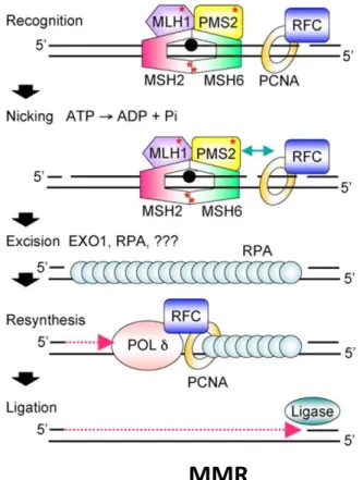
MMR, mismatch repair
MPP, multipotent progenitor cell MRV, multiple rare variant hypothesis MS, mating symmetry
NER, nucleotide excision repair NHEJ, non-homologous end-joining NK, natural killer
OR, odds ratio
PCR, polymerase chain reaction PGL, pathway genetic load pHap, promoter haplotype
pSNP, promoter single nucleotide polymorhphism
QcALL, Quebec Childhood Acute Lymphoblastic Leukemia Cohort RAF, risk allele frequency
RNA, ribonucleic acid RR, relative risk
rSNP, regulatory single nucleotide polymorphism SD, standard deviation
SNP, single nucleotide polymorphisms ST-HSC, short-term hematopoietic stem cell TF, transcription factor
TFBS, transcription factor binding site UTR, untranslated region
I dedicate this thesis to my parents, Deborah and Patrick, whose love and support nurtured and guided me along this journey; and to my two best friends, my sister Rose and my brother Zachary, for holding my hand every step of the way.
PREFACE
C
HILDHOOD
A
CUTE
L
YMPHOBLASTIC
“Cancer research driven by the allure of miracle cures is impoverished if it does not pay equal attention to possible causal mechanisms and prospects for prevention.”
––– M. Greaves, 1999.
Over the past decade, a key focus of cancer research has been geared toward dissecting variation in cancer predisposition through the identification of
inherited genetic changes that influence cancer risk, with the ultimate goal of decreasing mortality by reducing risk and improving diagnosis and treatment. Following the sequencing of the human genome, we moved rapidly into the age of genomics leading us from linkage analysis that were successful for identifying high-risk gene mutations involved in familial cancers to ever-growing association studies that now allow us to identify low to moderate risk alleles involved in sporadic cancers. From genotyping a handful of variants in a few candidate genes, to genotyping millions of variants genome-wide, to sequencing entire genomes, rapid technological advances have allowed us to peer ever so deeper into the genomes of cancer cells and into the genomes of individuals genetically predisposed to cancer, shedding light on the complex underpinnings of this multifaceted disease.
Childhood acute lymphoblastic leukemia (ALL) is one of the great success stories of modern medicine; thirty years ago a child that was diagnosed with ALL had little chances of survival, today through the application of intensive multiagent chemotherapeutic regimens, the cure rate for childhood ALL exceeds an impressive 80% in the developed world. This achievement stems mainly from advances in our knowledge of the pathobiology of the disease. Several clinical and biological features that correlate with outcome are now used to assign
risk-based treatment regimes to ALL sufferers. However a significant proportion of patients still fail therapy for unknown reasons and the long-term effects of the intense chemotherapeutic cocktails that are administered to patients can in certain instances be as debilitating as the disease itself. And on the downside of this success story is the shadowed fact that, in developed countries, ALL is still the leading cause of death by disease among children. More striking still is the fact that despite success in treating the disease, very little is known of the
etiology of childhood ALL; and to better treat the disease is to better understand it.
ALL results from a series of mutational events within an immature blood cell that halt cell maturation and eventually lead to malignant proliferation and disruption of normal blood production. Over 200 genetic alterations have been identified so far in ALL tumors, with a handful of recurrent chromosomal rearrangements and mutations characterizing most of the cases. While there is well-established evidence that these mutational events play an important role in driving the leukemic process, the events leading up to leukemogenesis are not known. Initiation of the disease occurs during fetal life or in early infancy and, as with most other cancers, is likely caused by a combination of environmental and genetic factors. The assertion that ALL has a genetic basis has long been pursued through association studies based on candidate genes. These studies were recently complemented by genome-wide studies that vindicated the role of common inherited genetic variation in childhood ALL susceptibility.
There is no doubt that the genomics era will have a profound impact on the diagnosis and therapy of childhood ALL. But with progress comes new challenges; already the issue of missing heritability is daunting researchers interested in dissected the genetic architecture of complex diseases. In the research presented here I explore the effects of genetic variation on childhood
ALL susceptibility by addressing the following questions. Can we deviate from traditional analytical approaches to further illuminate the genetic basis of
childhood ALL? Can deregulation of core cellular functions such as cell division and maintenance of genomic integrity influence a child‘s susceptibility to ALL? How important are the mother‘s genes in shaping her offspring‘s susceptibility to disease? And in light of the growing popularity of agnostic genome-wide
searches, can candidate gene approaches still help explain some of the interindividual variability in complex disease susceptibility? Using a unique design that involves collecting DNA from childhood ALL cases as well as their parents and unrelated control individuals, I investigate the role of genes involved in two cancer-related pathways, the cell cycle control and DNA repair
mechanisms, in childhood ALL. This pathway-based candidate gene association study provides a unique opportunity to investigate some of the genetic subtleties involved in this pediatric disorder. It is my hope that this research will provide greater insight into the etiologic intricacies of childhood ALL and help pave the way toward new opportunities for prevention. Unraveling the genetic
architecture of childhood ALL will bring us one step closer to that ultimate goal of decreasing mortality by reducing risk and improving diagnosis and treatment.
CHAPTER ONE
C
HILDHOODC
ANCERCancer is thought of as a disease of ageing, one in which the DNA of a normal cell accumulates sufficient mutations that it acquires a selective advantage and becomes capable of uncontrolled, unlimited proliferation. Yet cancer is also the leading cause of death by disease among
children (1). It is estimated that each year, approximately 150 children out of every million children younger than 20 years of age will be diagnosed with cancer (2). And while incidence rates have been
increasing steadily since the mid-1970‘s, the etiology of many childhood cancers remains elusive.
Cancer in children differs markedly from its adult counterpart with regard to cancer type, site of occurrence, as well as clinical behavior. Beyond the shorter latency period observed in pediatric cancers, they are often more aggressive and more invasive. The majority of tumors diagnosed in children stem from immature ―embryonic-like‖ cell types, whereas adult cancers are mainly
carcinomas that arise in epithelial tissue (3). Consequently, a more appropriate classification system was developed for childhood cancers based on cell
morphology and tissue of origin rather than on the primary anatomical site of appearance as in adult cancers (4). The most common pediatric neoplasms are leukemias (cancer of the blood) representing 25% of all cancer cases among children younger than 20 years of age, followed by brain and central nervous system cancers (17%) and lymphomas (cancer of the lymphatic system) (16%) (5). The relative contribution of leukemia to the total childhood cancer burden rises as high as 46% among children aged 2-3 years, making this disease the leading cause of cancer-related deaths in children.
L
EUKEMIAHematopoiesis is the highly regulated and hierarchical process during which blood cells are formed. Self-renewing progenitors in the bone marrow, the hematopoietic stem cells (HSCs), give rise to multipotent progenitors that in turn produce lineage-committed progenitors that give rise to the mature blood cells of either the myeloid or lymphoid lineages (Figure 1). In leukemia, normal hematopoiesis is disrupted. Development of normal hematopoietic cells is arrested in an early stage of differentiation in the bone marrow, and malignant proliferation of the immature lymphoid or myeloid cells depletes the pool of functionally mature blood cells and eventually invades the blood, lymph nodes, central nervous system and other vital organs (6). Leukemia is a clonal disease arising from the neoplastic transformation of a single cell and in many respects the initial leukemia cell resembles a stem cell with unlimited proliferation
potential and self renewal capabilities and the ability to give rise to a new, albeit abnormal, hematopoietic tissue (7).
Leukemia is a heterogeneous group of neoplasms classified on the basis of cell type of origin. Leukemias can arise during any step of the hematopoietic
process and are either acute, aggressive diseases affecting mostly immature, undifferentiated cells, or chronic, less rapidly progressing diseases affecting more mature and differentiated hematopoietic cells. The three major
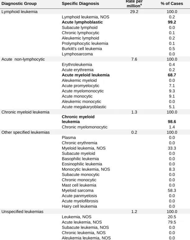
classifications of childhood leukemia are acute lymphoblastic leukemia (ALL), accounting for 75% to 80% of childhood leukemia cases; acute myelogenous leukemia (AML), accounting for 20% to 25% of cases; and chronic myelogenous leukemia (CML), accounting for ~3% of cases (6) (Table 1).
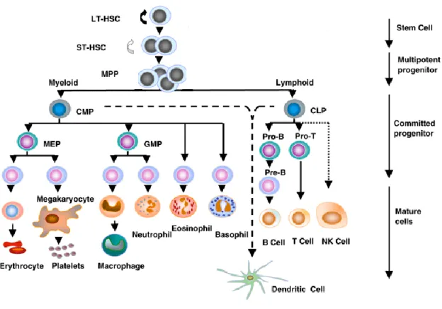
Figure 1. Hematopoietic differentiation
As HSCs divide, they can either self-renew, commit to differentiation or die by apoptosis in order to maintain a steady-state level of HSCs in the bone marrow and constantly provide progenitors for the various haematological lineages. The HSC population is made up of long-term HSCs (LT-HSCs) capable of lifetime self-renewal and short-term HSCs (ST-HSCs) that only briefly self-renew and give rise to multipotent progenitor cells (MPPs) which differentiate into the mutually exclusive myeloid and lymphoid lineages. The common myeloid progenitors (CMPs) give rise to the myelomonocytic progenitors (GMPs) which produce macrophages and granulocytes, and to the megakaryocytic/erythroid progenitors (MEPs) which produce megakaryocytes, platelets and erythrocytes. The common lymphoid progenitors (CLPs) give rise to B and T lymphocytes and natural killer (NK) cells. Both CMPs and CLPs can give rise to dendritic cells. Cell surface markers can be used to discriminate between all stem and progenitor hematopoietic cell populations. (7)
Table 1. Childhood leukemia – a heterogeneous group of disorders categorized by cell type and level of differentiation
Diagnostic Group Specific Diagnosis Rate per
milliona % of Cases
Lymphoid leukemia 29.2 100.0
Lymphoid leukemia, NOS 0.2
Acute lymphoblastic 99.2
Subacute lymphoid 0.0
Chronic lymphocytic 0.1
Aleukemic lymphoid 0.2
Prolymphocytic leukemia 0.1
Burkitt's cell leukemia 0.5
Lymphosarcoma 0.0
Acute non-lymphocytic 7.6 100.0
Erythroleukemia 0.4
Acute erythremia 0.2
Acute myeloid leukemia 68.7
Aleukemic myeloid 0.0 Acute promyelocytic 7.1 Acute myelomonocytic 9.3 Acute monocytic 9.1 Aleukemic monocytic 0.0 Acute megakaryoblastic 5.1
Chronic myeloid leukemia 1.3 100.0
Chronic myeloid
leukemia 98.6
Chronic myelomonocytic 1.4
Other specified leukemias 0.2 100.0
Plasma 0.0
Chronic erythremia 0.0
Myeloid leukemia, NOS 33.3
Subacute myeloid 0.0
Basophilic leukemia 0.0
Eosinophilic leukemia 0.0
Monocytic leukemia, NOS 8.3
Subacute monocytic 0.0
Chronic monocytic 0.0
Mast cell leukemia 0.0
Myeloid sarcoma 58.3
Acute panmyelosis 0.0
Acute myelofibrosis 0.0
Hairy cell leukemia 0.0
Unspecified leukemias 1.2 100.0
Leukemia, NOS 20.5
Acute leukemia, NOS 79.5
Subacute leukemia, NOS 0.0
Chronic leukemia, NOS 0.0
Aleukemia leukemia, NOS 0.0
Diagnostic groups and subcategories are based on the International
to the 1970 US standard population. In bold are the three major leukemia subtypes diagnosed in children. NOS, not otherwise specified.
a
Age-adjusted incidence rates and percent distribution for specific diagnostic subcategories of leukemia based on data from the Surveillance Epidemiology and End Results, 1975-95 for patients age <20.
T
HEN
ATURALH
ISTORY OFC
HILDHOODA
CUTEL
YMPHOBLASTICL
EUKEMIAAcute lymphoblastic leukemia (ALL) is the most frequent pediatric cancer and is itself a diverse group of diseases distinct both biologically and clinically. ALL can arise in either the B or T lineage of lymphocyte cells (Figure 1). Classification of B- and T-cell ALL is done based on cell morphology, using cytological features distinguished by the French-American-British (FAB) classification system (9) – including cell size, nuclear chromatin, nuclear shape, nucleoli, amount of cytoplasm, basophilia of cytoplasm, and cytoplasmic vacuolation – and using immunophenotyping of lineage- and maturation-specific cell surface antigens via flow cytometry (10). B-lineage ALL can be further subdivided into pro-B cell ALL (also known as early pre-B and pre-pre-B ALL) and pre-B ALL (also known as cALL for common ALL or simply as B-cell ALL) (11). Pro-B ALL is one of the most immature ALL subtypes and occurs mainly in infants aged birth to 1 year; it is rare and accounts for only ~5% of childhood ALL cases (12). B-cell ALL is the most common subtype, accounting for 80%, while T-cell ALL represents
approximately 15% of all newly diagnosed pediatric ALL cases (13).
Incidence, Survival and Trends
It is estimated that approximately 1,100 children are diagnosed with ALL each year in Canada (14). While ALL occurs in children worldwide, its incidence varies between nations with Costa Rica, Finland and Canada experiencing the highest rates, China and India the lowest (15). As for most childhood cancers, ALL is slightly more prevalent in boys compared to girls with a 1.2:1 male to female ratio, except for pro-B ALL which exhibits a slight predominance in females (12). Incidence peaks between 2-5 years of age and tends to be higher in socioeconomically developed populations compared to developing countries
that tend to have lower rates with no obvious age-specific incidence peak (16). A two- to three-fold higher incidence rate is observed in white children
compared to black children and rates among Hispanics are highest of all, suggesting differences in disease frequency associated with race and/or socioeconomic status (17).
Childhood ALL is one of the great cancer success stories of the 20th century. While less than 20% of diagnosed childhood ALL patients survived their disease 40 years ago, modern treatment protocols have managed to completely reverse survival rates. Among patients receiving contemporary chemotherapy treatment, the five-year survival rate is now 80% (18). However, 20% of the cases remain resilient to current treatment protocols and ultimately succumb to their disease. These numbers also conceal that, of the patients that become five-year
survivors of childhood ALL, a substantial number develop long-term treatment-related complications including death (19, 20). Therefore, while the marked improvement in the overall cure rate for ALL is undoubtedly very impressive, treatment is far from being optimal. And importantly, despite decreasing mortality rates, the incidence of ALL among children younger than 20 years of age has been increasing with improved socio-economic conditions (5).
Pathobiology of Childhood Acute Lymphoblastic Leukemia Success in treatment is due in large part to our increased understanding of the pathobiology of ALL. Leukemia is a disease of the genome characterized by gross genomic and chromosomal alterations which ultimately provide the leukemic cell with a selective and proliferative advantage.
Molecular Genetic Alterations
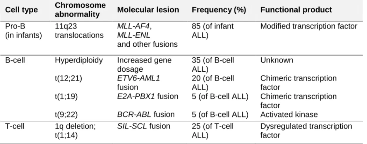
Although the primary causes of childhood ALL remain unknown, the
mechanisms through which the disease arises can be postulated based on specific genetic alterations found in ALL. Over 200 somatic genetic alterations have been identified in ALL but for the most part they are characterized by gross chromosomal changes such as changes in DNA ploidy, chromosomal
translocations and deletions. The acquired genetic lesions lead to precise stage-specific developmental arrest and allow unlimited self-renewal and clonal
expansion of the B or T progenitor cell. Table 2 summarizes the main molecular alterations observed in ALL. The most frequent are hyperdiploidy (>50
chromosomes) and the t(12;21)/ETV6-AML1 gene fusion; together they
constitute 50% of all childhood ALL and almost 80% of ALL occurring between the ages of 2-5 years (21-23).
Table 2. Main chromosomal changes in childhood acute lymphoblastic leukemia
Cell type Chromosome
abnormality Molecular lesion Frequency (%) Functional product Pro-B (in infants) 11q23 translocations MLL-AF4, MLL-ENL
and other fusions
85 (of infant ALL)
Modified transcription factor
B-cell Hyperdiploidy Increased gene
dosage 35 (of B-cell ALL) Unknown t(12;21) ETV6-AML1 fusion 20 (of B-cell ALL) Chimeric transcription factor
t(1;19) E2A-PBX1 fusion 5 (of B-cell ALL) Chimeric transcription
factor
t(9;22) BCR-ABL fusion 5 (of B-cell ALL) Activated kinase
T-cell 1q deletion;
t(1;14)
SIL-SCL fusion 25 (of T-cell ALL)
Dysregulated transcription factor
Hyperdiploid ALL is characterized by the nonrandom gain of chromosomes X, 4, 6, 10, 14, 17, 18, and 21. Though hyperdiploidy is suspected to incur a selective advantage to leukemic cells through gene dosage effects, the precise
mechanism through which it occurs and its impact on leukemogenesis are unknown (23).
Chromosomal translocations are initiated by DNA double-strand breaks that occur simultaneously in a single cell. Through the process of repair, fusion gene products are formed resulting in functionally viable chimeric proteins with altered or dysregulated function (24). Several recurrent translocations are found in childhood ALL patients (Figure 2). The most prevalent is the t(12;21) translocation which creates a fusion gene that involves the transcriptional repressor ETV6 and the hematopoietic-specific transcription factor AML1. The resulting ETV6-AML1 chimera leads to impaired hematopoietic differentiation, however the precise mechanisms by which it causes leukemia remain elusive.
Figure 2. Chromosomal translocations in childhood ALL
The t(12;21) translocation which gives rise to the TEL/AML1 (also known as the ETV6/AML1) fusion gene is the most prevalent among pre-B ALL cases. Note however that while chromosomal translocations are frequent in leukemia, roughly 30% of childhood ALL cases harbor no apparent alterations. Figure adapted from Pui et al., 2004 (25).
Prenatal Origin
There is well-established evidence for prenatal initiation of the leukemogenesis process in children (26-28). The early age of onset in childhood ALL (peak incidence in patients aged 2 to 5 years) is highly suggestive of a latency period that begins before the birth of the child. Moreover, molecular studies have shown that several of the common chromosomal translocations, mainly
t(4;11)MLL/AF4 in infant ALL and t(12;21)ETV6/AML1 in childhood B-cell ALL, occur in utero. First, retrospective DNA screening of archived neonatal blood spots revealed the presence of the fusion genes in patients that developed ALL later on between the ages of 5 months to 2 years (26, 29). And concordance of ALL in identical, monozygotic twins – ranging from 50% in twin pairs diagnosed before the age of 1 to about 5% for older ages – has been shown to result from intraplacental metastasis of leukemic clones that initiate in one identical twin and are passed to the other in utero through their shared blood system (30). This was shown by mapping the unique genetic breakpoints in translocations which were shown to be shared among affected twin pairs (24). Moreover, screening of newborn cord blood revealed that 1% of babies carry the
ETV6/AML1 fusion (31), a frequency much higher than the actual prevalence of leukemia (about 1 in 10 000), providing a proof-of-principle that chromosomal translocations occur prenatally and are likely insufficient to cause leukemia. Other subtypes of ALL are expected to be initiated prenatally as well, but lack of genetic markers such as gene fusions precludes their identification.
Cooperating Oncogenic Lesions
While chromosomal alterations play an important role in driving the leukemic process by affecting molecular pathways that halt lymphoid progenitor cell differentiation and promote cell proliferation and survival, they are incapable, on their own, of causing full leukemic transformation (32). The observation that these abnormalities are often detected years before the onset of leukemia and
the use of experimental models to show that they do not alone result in
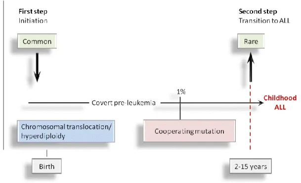
leukemia (33) suggest the need for additional cooperating genetic mutations for full leukemic transformation. Greaves suggested that a minimum of two steps are required for leukemia development; the first genetic insult is expected to occur in utero during pregnancy, in perinatality or at the very least in early infancy, followed by a second postnatal genetic insult, that is required to induce overt leukemia (Figure 3).
Figure 3. Greaves’ multi-stage model for the development of childhood ALL
The most frequent genetic lesions in ALL are generated during normal fetal development and are fairly common events (31). Transition between covert pre-leukemia and covert clinical pre-leukemia occurs in only 1% of the cases and involves at least a second hit involving cooperating mutations that occur postnatally.
Recent effort has gone into identifying the full complement of somatically
acquired genetic alterations that contribute to acute leukemogenesis in children (35, 36). In addition to known recurrent chromosomal translocations, high frequency DNA copy number alterations, loss-of-heterozygosity events, deletions, amplifications and point mutations in genes involved in lymphoid differentiation, as well as in other critical cancer-related pathways such as cell cycle regulation, apoptosis, tumor suppression and xenobiotic metabolism have been identified in both B- and T-lineage ALL (37-39). Epigenetic analysis
revealed that recurrent inactivation of tumor suppressor genes through DNA hypermethylation also contributes to oncogenesis in ALL (40, 41).
The recurrent chromosomal abnormalities observed in childhood ALL define unique subtypes of the disease and together with cooperating genetic and epigenetic alterations shed light on the pathogenesis of ALL in children (35, 42). Many of these alterations are associated with disease outcome and have helped guide risk-stratified treatment regimes contributing to the success of modern day therapy in curing childhood ALL. But while it is likely that these distinct
prognostic subgroups also reflect divergent disease etiologies, very little is known still of the underlying causes of childhood ALL.
Risk Factors
Acquired genetic changes (somatic mutations) are involved in the molecular pathogenesis of childhood ALL and its progression but do not explain the origin of the disease. It is the more elusive inherited germline variants that influence disease susceptibility, likely through the modulation of the response to
environmental exposures. But while it is clear that both environmental and genetic factors have roles in the development of ALL in children, studies have thus far been inconsistent or incapable of providing unequivocal evidence for
implication in the etiology of ALL and the causes of the disease remain largely unknown.
Evidence that childhood ALL has a genetic component stems in part from the fact that it is associated with other predisposing genetic syndromes. Inherited disorders such as Down‘s syndrome, Bloom‘s syndrome, ataxia-telangiectasia, and Nijmegen breakage syndrome are of the few established risk factors for childhood ALL however they account for a trivial proportion of cases (<5%) (43). Familial aggregation of childhood ALL is also rare and only a few pedigrees transmitting ALL have ever been recognized (43). There are five published reports of multigenerational ALL, possibly consistent with autosomal dominant inheritance, yet no clear susceptibility gene culprit has ever been identified within these families, and the small size and number of these pedigrees could be reminiscent of chance clustering rather than familial aggregation (43-46). Furthermore, very few reports of families with multiple affected children have been reported (47, 48), the incidence of childhood ALL among non-twinned siblings of probands is at most only weakly increased and could even be decreased (49-50), and long-term follow up studies of childhood ALL survivors indicate no increased risk of malignancy in their offspring as compared to the general population (51), not what one would expect if familial and highly
penetrant leukemia susceptibility genes were involved in disease etiology. Thus, as opposed to other forms of hereditary cancers, such as certain forms of
breast, prostate and colorectal cancer for example, that are inherited in a
Mendelian fashion and are associated with highly penetrant germline mutations, childhood ALL is sporadic and inherited genetic susceptibility is multifactorial and likely involves the co-inheritance of multiple low penetrance variants that do not give rise to clear-cut familial patterns of inheritance. And unlike the search for ―the‖ gene that causes a Mendelian disorder, many different inherited susceptibility genes and many different environmental risk factors are likely going to be involved in the etiology of childhood ALL.
Environmental Exposures
In conjunction with Greaves‘ multi-stage model for childhood ALL development, there are three critical windows during which exposure to environmental risk agents could potentially influence leukemogenesis; these are before conception, in utero during pregnancy, and after birth (Figure 4). Given that ALL is not a hereditary cancer in terms of a simple Mendelian inheritance, the notion that the mother and/or father may play an important role in ALL development in the offspring is somewhat counterintuitive, unless we think of it in terms of exposure and increased mutation burden. Exposure to carcinogenic agents during
gametogenesis can incur germline mutations in the gametes of the parents preconception that are passed down to the offspring and could lead to increased genetic instability postconception. The role of the father in preconceptional ALL risk may be more important than the mother since spermatogenesis occurs throughout the entire lifetime of the male, offering greater opportunities for mutations to occur, whereas females bear their oocytes from birth. During pregnancy however, the mother may play a crucial role in disease development as she provides the prenatal environment and can influence her offspring‘s risk of disease through environmental exposures passed to the fetus via the
placenta or through the effects of her own genes that can directly influence the intrauterine milieu (52). Finally, postnatal exposures of the child both directly and indirectly through the mother, for example in breast feeding, could be important determinants of childhood ALL.
Figure 4. Critical windows of exposure for childhood ALL
Potential relationship between childhood ALL-inducing events and critical periods preconception, in utero and early after birth, during which exposure-dependent risk factors could influence disease susceptibility. In concordance with the multi-stage model for ALL development, both mothers and fathers could potentially be involved in the initiating genetic event while mothers could
contribute to the child‘s postnatal exposure-dependent risk through
breastfeeding. The model can also be extended to include genetic risk factors (at the level of the child, mother and/or father) given that response to the environment is modulated by genetic components such as those involved in xenobiotic metabolism, DNA repair and cell cycle regulation.
Table 3 lists significant window-specific environmental exposures that have been associated with childhood ALL in the literature. The main classes of exposure-dependent risk factors identified for childhood ALL include ionizing radiation (preconception, in utero and postnatal), exposure to electromagnetic fields, chemicals (e.g. hydrocarbons such as benzene found in cigarette smoke, gasoline, solvents, paint thinners, air pollution and pesticide exposure either directly or via parental exposure), parental alcohol, cigarette and drug use, parental occupation, and certain dietary components. However contradictory results have been reported for most of these risk factors. Ionizing radiation appears to be the only significant environmental risk factor identified to date; most others (for example parental cigarette smoking and alcohol consumption) have been inconsistently associated with childhood ALL and their role in
leukemogenesis remains very controversial. This stems mainly from the fact that exposures to environmental factors are often difficult to measure and assess and can suffer from high levels of uncertainty due to recall bias.
Table 3. Exposure-dependent risk factors reported to be significantly associated with childhood ALL
Exposure
window Exposure type Risk
a Odds ratio (sample sizeb);
reference
Preconception
Fathers Occupational exposure
Plastic materials (polysterene) + 1.4 (1842 vs. 1986); (54)
Driving, exhaust fumes, inhaled particulate hydrocarbons
+ 13-1.4 (1461 vs. 2922); (55)
Electromagnetic fields + 2 (56)
Radiation exposure, X-rays ++ 1.9-2.6 (184 vs. 368); (57)
2.2-3.8 (191 vs. 382); (58)
Cigarette smoking ++ 1.6 (203 vs. 406); (59)
3.8 (110 vs. 110); (60)
Alcohol consumption + 1.4 (491 vs. 491); (61)
Medication/drug use
Amphetamines or diet pills + 2.2 (1842 vs. 1986); (62)
Mind-altering drugs (marijuana) + 1.3 (1842 vs. 1986); (62)
Mothers Occupational exposure
Solvents/hydrocarbons (carbon tetrachloride) + 1.8 (1842 vs. 1986); (54)
Paints or thinners + 1.6 (1842 vs. 1986); (54)
Household pesticides + 1.7 (135 vs. 135); (63)
Medication/drug use, oral contraceptives + 1.3 (519 vs. 507); (64)
Food consumption
Vegetables − 0.5 (138 vs. 138); (65)
Protein − 0.4 (138 vs. 138); (65)
Prenatal
Mothers Occupational exposure
Solvents/hydrocarbons (freon, gasoline) ++ 1.6 (1842 vs. 1968); (54)
1.7 (184 vs. 368); (57) 1.7-2.3 (790 vs. 790); (66) Paints or thinners ++ 1.7 (1842 vs. 1986); (54) 3.2 (184 vs. 368); (57) 2.4 (519 vs. 507); (67) Pesticides + 3.5 (184 vs. 368); (57)
Organic dust (cotton, wool, synthetic fibers) + 5.5 (128 vs. 128); (68)
Electromagnetic fields + 2.5 (491 vs. 491) (69)
Household exposure
Pesticides ++ 1.7-1.8 (491 vs. 491); (70)
2.3 (135 vs. 135); (63)
Paints + 1.7 (640 vs. 640); (71)
Radiation exposure, X-rays + 2.2 (519 vs. 507); (64)
Sewing machine − 0.8 (640 vs. 640); (72) Cigarette smoking −− 0.7 (203 vs. 406); (59) 0.7; (73) Alcohol consumption + − 1.4 (203 vs. 406); (59) 0.7 (491 vs. 491); (61) Medication/drug or supplement use
Vitamins − 0.7 (1842 vs. 1986); (62) Iron or folate −− 0.4 (83 vs. 166); (74) 0.9 (1842 vs. 1986); (62) Antihistamines + 1.3 (1842 vs. 1986); (62) Oral contraceptives + 1.5 (1842 vs. 1986); (75) Pregnancy-maintaining drugs + 1.9 (519 vs. 507); (64)
Teratogenic medication (CNS depressants) + 1.3-1.4 (789 vs. 789); (76)
Antibiotics + 1.5 (477 vs. 484); (77)
Food consumption
Vegetables − 0.8 (131 vs. 131); (78)
Fruit − 0.7 (131 vs. 131); (78)
Fish and seafood − 0.7 (131 vs. 131); (78)
Sugars and syrups + 1.3 (131 vs. 131); (78)
Meat and meat products + 1.3 (131 vs. 131); (78)
Postnatal
Mothers Occupational exposure, plastic (polyvinyl
chloride)
+ 2.2 (1842 vs. 1986); (54)
Alcohol consumption − 0.5 (491 vs. 491); (61)
Children Environmental exposure
Neighboring repair garages/gas stations (benzene)
+ 3.6 (240 vs. 280); (79)
Chernobyl accident (radioactive contamination)
+ 13.1 (98 vs. 151); (80)
High voltage power lines (electromagnetic fields) ++ 2.00; (81) 4 (101 vs. 412); (82) 1.69; (83) Household exposure Pesticides ++ 1.4-1.8 (491 vs. 491); (70) 1.7 (135 vs. 135); (63)
Artwork (organic solvents) + 4.1 (640 vs. 640); (71)
Electrical appliance usage (electric blanket, hair dryer, video game machines)
+ 1.6-2.8 (640 vs. 640); (72)
Medication/drug use (chloramphenicol) + 1.8-10.7 (184 vs. 368); (57)
Supplementary oxygen exposure + 1.9 (603 vs. 3015); (84)
Radiation exposure, X-rays ++ 1.6 (491 vs. 491); (85)
1.5 (701 vs. 701); (86)
Trihalomethanes in drinking water + 9.13 (491 vs. 491); (87)
High birth weight (>3800 g) ++ 1.3 (1905 vs. 9525); (88)
1.8 (1455 vs. 816); (89) 1.7 (603 vs. 3015); (84) 2.5 (181 vs. 362); (90) 2.2 (83 vs. 830); (91) 3.8–4.6; (92)
Infectionc
Early infection (in first 4 years of life) −− 0.4 (124 vs. 248); (93)
0.1-0.8 (408 vs. 567); (94)
Ear infection − 0.3 (294 vs. 376); (95)
Roseola/fever and rash (first year of life) − 0.3 (98 vs. 228); (96)
Allergy history −− 0.6-0.7; (97)
0.5-0.6 (1130 vs. 2957); (98)
0.6 (255 vs. 760); (99)
Vaccinationc
Haemophilus influenza type b (Hib) −− 0.6 (439 vs. 439); (100)
0.8 (282 vs. 409); (101)
Bacille calmette-guérin (BCG) − 0.1 (63 vs. 126); (102)
Measles − 0.2 (63 vs. 126); (102)
Household density (>1 person/room)c − 0.6; (103)
Daycare attendencec −− 0.5 (490 vs. 491); (77)
0.7 (408 vs. 567); (94) 0.4 (294 vs. 376); (95)
Breastfeedingc −− 0.7 (491 vs. 491); (77)
0.8 (1744 vs. 1879); (104)
Only significant exposure-dependent risk factor associations are shown. This is not an exhaustive list; other postulated exposure risk factors for childhood ALL may have been investigated, inconclusive results are not shown.
a
Increased risk (odds ratio > 1) in one (+) or more (++) studies; decreased risk (odds ratio <1) in one (−) or more (−−) studies.
b
The total number of childhood ALL cases versus healthy controls analyzed in each reference.
c
Thought to be infection-related risk factors. Table adapted from Kim et al., 2006 (53).
Despite evidence that leukemogenesis is initiated in utero, high birth weight is one of the few birth-related factors that has been linked to ALL in children. Up to a 26% increase in ALL risk is associated with each kg increase in birth weight, which can perhaps simply be explained by the fact that larger babies have a higher number of lymphoid cells and therefore a higher number of cells at risk of leukemic transformation (88). On the other hand higher birth weight might
indicate higher levels of circulating growth hormone which may induce proliferative stress on the bone marrow and indirectly be linked to leukemia (105). Other causative factors that have been proposed for childhood ALL include maternal (<20 years and >35 years) and paternal (>40 y ears) age, maternal reproductive history (miscarriage, abortion), high birth order (fourth born), long birth intervals (>5), race (white, Hispanic) and gender (male) however their associations with the disease remain highly speculative and the mechanisms through which they influence disease risk are unclear (53).
Moreover, it has been hypothesized that childhood leukemia could be caused by infection-related factors. An aberrant immunological response to infection at a vulnerable time in the child‘s life when lymphoid cell proliferation is high could render the child more susceptible to genetic insults and to leukemia. The possibility that ALL may have an infectious etiology is supported by the higher prevalence of leukemia in modern, wealthy societies and the appearance of clusters of childhood ALL cases in small residential communities (106-108). Two infection-based models for ALL development have been proposed: Kinlen‘s population mixing hypothesis and Greaves‘ delayed-infection hypothesis. Kinlen postulated that clusters of ALL could result from prolonged population isolation and subsequent exposure to a common but otherwise non-pathogenic infection due to population mixing (109). Whereas Greaves suggested that individuals carrying pre-leukemic clones (Figure 3) that have spent their early years of life coddled in a sterile environment may exhibit a pathological response to a subsequent delayed exposure to common infections (34). While some
epidemiologic data do support the immune-response hypothesis – daycare attendance, increased household density, higher number of recorded common infections in early life, as well as breastfeeding and vaccination have all been shown to reduce the risk of ALL suggesting that increased social contact and potential exposure to infection and immune stimulation in early life protect against ALL (Table 3)– no causal infectious agents have yet been identified.
Genetic Susceptibility Factors
How these exposures affect an individual and their susceptibility to disease relies largely on their genetic makeup. Human phenotypic variation, be it in risk to disease, response to the environment, or with regard to physical
characteristics such as height, is influenced by inherited differences in DNA sequence. Interindividual variation in the susceptibility to childhood ALL bears no exception. Assuming that genes modulate variability in the responses to exogenous and/or endogenous factors, they could thereby also influence an individual‘s risk of cancer. Childhood ALL is a complex disease in which both genes and the environment interact to confer a variable degree of risk on people who inherit predisposing genetic variants. Genetic susceptibility refers to an inherited increase in the risk of developing a disease, passed down from
parental generations in the form of germline genetic variation. While the parental generation may not be at increased risk of disease, the combination of
polymorphic variants they bestow upon their offspring could render the child more or less susceptible to developing disease. In order to unravel the complex etiology of childhood ALL one must therefore start by identifying the inherited genetic changes that influence an individual‘s risk of disease.
To date, studies of genetic susceptibility to ALL have focused on the affected child and on common genetic variation in genes involved in biological pathways
such as folate metabolism, immune function, xenobiotic metabolism (including membrane transport, detoxification and biotransformation of drugs and
chemicals), oxidative stress response, and DNA repair, under the presumed hypothesis that inherited genetic variants in genes functioning along these pathways could modify response to exposure-dependent risk factors and lead to genetic instability in lymphoid progenitor cells and thus influence a child‘s risk of developing ALL. Table 4 shows a summary of the genetic susceptibility factors shown to significantly modulate childhood ALL risk.
These hypothesis-driven approaches are based on our imperfect understanding of the biological processes involved in leukemogenesis and often yield
associations that are difficult to replicate. And despite two recent large-scale association studies that have convincingly vindicated the role for inherited genetic variation in childhood ALL predisposition (110, 111), the genetic component of childhood ALL remains largely undefined.
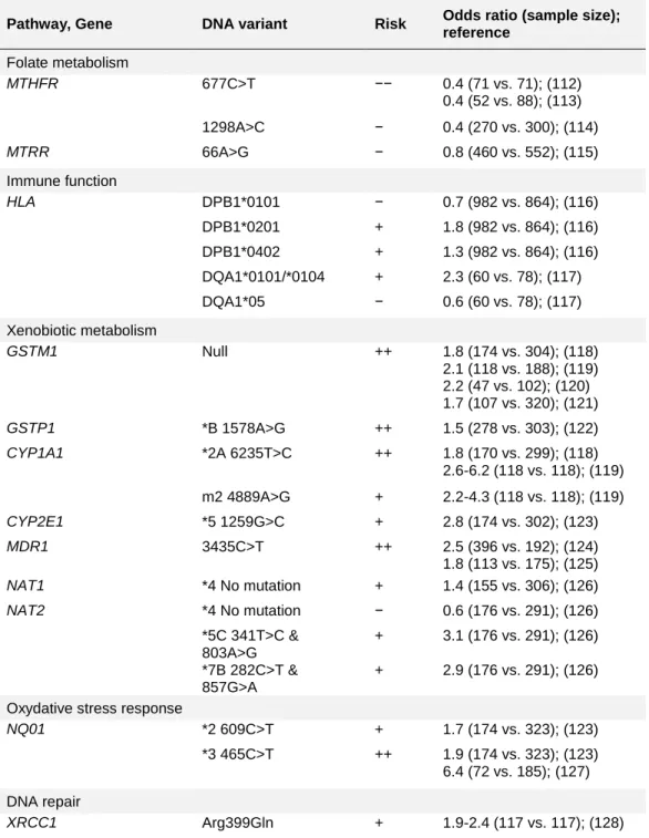
Table 4. Genetic risk factors reported to be significantly associated with childhood ALL
Pathway, Gene DNA variant Risk Odds ratio (sample size); reference Folate metabolism MTHFR 677C>T −− 0.4 (71 vs. 71); (112) 0.4 (52 vs. 88); (113) 1298A>C − 0.4 (270 vs. 300); (114) MTRR 66A>G − 0.8 (460 vs. 552); (115) Immune function HLA DPB1*0101 − 0.7 (982 vs. 864); (116) DPB1*0201 + 1.8 (982 vs. 864); (116) DPB1*0402 + 1.3 (982 vs. 864); (116) DQA1*0101/*0104 + 2.3 (60 vs. 78); (117) DQA1*05 − 0.6 (60 vs. 78); (117) Xenobiotic metabolism GSTM1 Null ++ 1.8 (174 vs. 304); (118) 2.1 (118 vs. 188); (119) 2.2 (47 vs. 102); (120) 1.7 (107 vs. 320); (121) GSTP1 *B 1578A>G ++ 1.5 (278 vs. 303); (122) CYP1A1 *2A 6235T>C ++ 1.8 (170 vs. 299); (118) 2.6-6.2 (118 vs. 118); (119) m2 4889A>G + 2.2-4.3 (118 vs. 118); (119) CYP2E1 *5 1259G>C + 2.8 (174 vs. 302); (123) MDR1 3435C>T ++ 2.5 (396 vs. 192); (124) 1.8 (113 vs. 175); (125) NAT1 *4 No mutation + 1.4 (155 vs. 306); (126) NAT2 *4 No mutation − 0.6 (176 vs. 291); (126) *5C 341T>C & 803A>G + 3.1 (176 vs. 291); (126) *7B 282C>T & 857G>A + 2.9 (176 vs. 291); (126)
Oxydative stress response
NQ01 *2 609C>T + 1.7 (174 vs. 323); (123)
*3 465C>T ++ 1.9 (174 vs. 323); (123)
6.4 (72 vs. 185); (127)
DNA repair
XRCC1 Arg399Gln + 1.9-2.4 (117 vs. 117); (128) Only significant genetic risk factor associations as of December 2006, which coincides with the beginning of my study, are shown. This is not an exhaustive
list; other postulated genetic variants have been tested for association with ALL but are not shown here due to inconclusive results.
a
Increased risk (odds ratio > 1) in one (+) or more (++) studies; decreased risk (odds ratio <1) in one (−) or more (−−) studies.
b
The total number of childhood ALL cases versus healthy controls analyzed in each reference.