Effect of Surface Chemistry of Polymeric Nanoparticles
on Cutaneous Penetration of Cholecalciferol
Augustine Lalloza,b,c, Marie-Alexandrine Bolzingera, Jimmy Faivreb, Pierre-Luc Latreilleb,c, Araceli Garcia Acb, Cyrielle Rakotovaob, Jean-Michel Rabanelb,c,1, Patrice Hildgenc*, Xavier Banquyb*,Stéphanie Briançona*
aUniv Lyon, Université Claude Bernard Lyon 1, CNRS, LAGEP UMR 5007, 43
Boulevard du 11 novembre 1918, F-69100, Villeurbanne, France
bCanada Research Chair on Bio-inspired Materials and Interfaces, Faculté de
Pharmacie, Université de Montréal, C.P. 6128, Succursale Centre-ville, Montréal, Québec H3C 3J7, Canada
cLaboratoire de Nanotechnologie Pharmaceutique, Faculté de Pharmacie, Université de
Montréal, C.P. 6128, Succursale Centre-ville, Montréal, Québec H3C 3J7, Canada
*Corresponding authors: stephanie.briancon@univ-lyon1.fr;
xavier.banquy@umontreal.ca; patrice.hildgen@umontreal.ca;
Abstract
We investigated the influence of nanoparticle (NP) surface composition on different aspects of skin delivery of a lipophilic drug: chemical stability, release and skin penetration. Cholecalciferol was chosen as a labile model drug. Poly(lactic acid) (PLA)-based NPs without surface coating, with a non-ionic poly(ethylene glycol) (PEG) coating, or with a zwitterionic poly(2-methacryloyloxyethyl phosphorylcholine) (PMPC) coating were prepared using flash nanoprecipitation. Process was optimized to obtain similar hydrodynamic diameter. Polymeric NPs were compared to non-polymeric cholecalciferol formulations. Cholecalciferol stability in aqueous medium was improved by polymeric encapsulation with a valuable effect of a hydrophilic coating. However, the in vitro
1 Present address : INRS-Institut Armand-Frappier, 531 Boulevard des Prairies, Laval,
Québec H7V 1B7, Canada 5 10 15 20 25
release of the drug was found independent of the presence of any polymer, as for the drug penetration in an intact skin model. Such tendency was not observed in impaired skin since, when stratum corneum was removed, we found that a neutral hydrophilic coating around NPs reduced drug penetration compared to pure drug NPs and bare PLA NPs. The nature of the hydrophilic block (PEG or PMPC) had however no impact. We hypothesized that NPs coating influenced drug penetration in impaired skin due to different electrostatic interactions between NPs and charged skin components of viable skin layers.
Keywords
Nanoparticle surface, flash nanoprecipitation, drug stability, skin penetration, impaired skin, cholecalciferol
30
1.
Introduction
Drug delivery in complex tissues is a challenging task. The in vivo efficacy of pharmaceutical treatments is determined by the transport and the delivery of a sufficient quantity of active therapeutic agent to the target tissue or cells, avoiding any toxic side effects. Polymeric nanoparticles (NPs) carrying drugs have been developed for several decades to this end. Compared to traditional pharmaceutical formulations, these nanocarriers are able to solubilize drugs, to protect them from degradation during their transport in the organism in the host tissue or cells and to control their release (Couvreur, 2013). The design of the most effective carrier is however relative to a fundamental knowledge which is the understanding of the specific interactions of NPs with biological systems, i.e. complex tissues, living cells and various tissues components (Albanese et al., 2012).
An example of such a complex tissue is the skin. Topical and transdermal delivery are particularly challenging tasks due to the specific structure of the skin made of successive stacks of cellular layers of different polarities. Notably, the external lipophilic layer of the skin, the stratum corneum, which is characterized by a compact structure of dead cells embedded in a lipid medium (Elias, 1983), forms a remarkable barrier to the penetration of chemicals. Moreover, the transport of drugs into the skin can be affected by skin pathologies which alter the barrier integrity (Wester and Maibach, 1992) due to various factors such as inflammation, desquamation, defects in lipid distribution or epidermal thickening. Designing efficient formulations to help drug crossing the skin barrier or target specific skin layers according to the pathology is therefore a great challenge. The 40
45
50
55
use of NPs carrying drugs have been proposed to this end. Indeed, besides efficiently solubilizing drugs in their core, NPs can either i) stay on the skin surface, ii) accumulate in skin appendages, iii) penetrate the skin, iv) influence the skin surface or v) favor the drug partition between formulation and skin. These allow to improve, reduce or modulate the skin diffusion of the drug encapsulated (Bolzinger et al., 2011; Prow et al., 2011). Nevertheless, the role of the interactions between the skin and the NPs on the mechanisms of drug delivery is still unclear, as well as the role of the skin physiopathology on these interactions. In fact, most of the studies dealing with the skin penetration of drugs from NPs are proof of concepts studies that only assess the penetration on healthy skin. Rare studies evaluating the penetration of drugs or NPs in damaged skin had however highlighted the importance to study penetration in a skin model close to the skin pathology since penetration can be thoroughly different than in intact skin (Alnasif et al., 2014; Döge et al., 2016; Jensen et al., 2011; Knudsen et al., 2011; Tsai et al., 2003).
Along with size and shape, NPs surface properties are crucial parameters dictating the nano-bio interactions (Albanese et al., 2012; Venkataraman et al., 2011; Verma and Stellacci, 2010). For treatments with intravenous administration, the presence of ligands on the NPs surface affects the recognition of NPs to the targeted site (Zhong et al., 2014) while a hydrophilic and dense coating of polymers hinders the opsonization of the NPs surface by plasmatic proteins, extending the circulation time in blood (Gref et al., 2000; Gref et al., 1994). A faster diffusion of NPs coated with a hydrophilic layer of polymer in complex media was also reported (Forier et al., 2013; Tang et al., 2009; Xu et al., 2015) 65
70
75
al., 2011; Moore et al., 2015). For skin treatments, a lot of studies have shown the influence of NPs surface charge on their penetration in skin. However, conclusions were often contradictory with no consensus obtained. Some studies have shown that cationic NPs penetrate more efficiently the skin or other tissues than anionic or neutral counterparts (Fernandes et al., 2015; Wu et al., 2010) due to favourable electrostatic attractive interactions with negatively-charged cell membranes (Wu et al., 2010). Another study reported a more favourable penetration of anionic NPs, explained by possible repulsions with anionic skin lipids which created temporary channels within the skin facilitating penetration (Kohli and Alpar, 2004). Other reports demonstrated that a PEG coating around gold NPs favored a deeper penetration of the NPs in the skin compared to charged NPs (Hsiao et al., 2016; Mahmoud et al., 2017). It was also shown that surface coating influence cytotoxicity in epidermal keratinocytes (Ryman-Rasmussen et al., 2007). Since NPs surface influences penetration of NPs in skin, it should also control drug delivery to the skin. A thoughtful design of the polymeric NPs surface and a precise assessment of its effects seem therefore crucial in order to optimize and control the interactions skin-NPs and therefore maximize formulation efficiency. However, to date, studies evaluating the impact of the surface composition of polymeric nanocarriers encapsulating drugs are rare and did not systematically assess the penetration of the drug in all skin layers (Abdel-Mottaleb et al., 2012; Wu et al., 2010).
In order to better figure out how surface properties of polymeric NPs can influence drug delivery to the normal or pathological skin, we propose to systematically compare different surface coatings of drug-loaded polymeric NPs commonly used for drug delivery purposes i.e. i) a lipophilic coating of poly(lactic acid) (PLA) polymer, ii) a 85
90
95
100
neutral hydrophilic coating of poly(ethylene glycol) (PEG) surrounding a PLA core, and iii) a zwitterionic hydrophilic coating of poly(2-methacryloyloxyethyl phosphorylcholine) (PMPC) surrounding a PLA core. These polymeric NPs will be compared to iv) pure drug NPs formulated without polymer as control and to v) surfactant micelles. PEG was chosen because it is a well-established polymer coating for NPs, known to provide stealth properties to NPs (Otsuka et al., 2012). Moreover, PEG is now extensively used as NPs coating for topical delivery (Bachhav et al., 2011; Kilfoyle et al., 2012; Knudsen et al., 2012; Lapteva et al., 2014a; Lapteva et al., 2014b; Laredj-Bourezg et al., 2015; Park et al., 2018; Ramezanli et al., 2017; Shim et al., 2004; Vega et al., 2013), but its role on drug delivery is still unclear. Recently, PMPC, a biomimetic zwitterionic polymer, has also demonstrated benefits for NPs surface coating as an alternative to PEG coating (Jin et al., 2014). Originally designed to mimic and have affinities with the phospholipids bilayers of the cellular biomembranes (Ishihara and Nakabayashi, 1991; Kojima et al., 1991), its remarkable hygroscopic properties, arising from the highly hydrophilic nature of the phosphorylcholine head-groups, inhibits biofouling (Feng et al., 2006). In rare reports studying PMPC for skin hydration, it was shown that PMPC can modify the
stratum corneum structure by altering the organization of the lamellar phase of skin lipids
and improving the skin water retention (Chrit et al., 2007; Kanekura et al., 2002; Lee, 2004). Its potential for skin drug delivery has never been studied and we hypothesized that PMPC can modulate skin penetration, since a disorganization of the lipid structure affects the skin barrier function (van Smeden et al., 2014). Cholecalciferol also named Vitamin D3, was chosen as a very lipophilic model drug (LogP 7.5) in the present study. The role of NPs composition on surface was systematically studied in the different 110
115
120
125
aspects of drug delivery i.e. i) the formation of the drug-loaded NPs, ii) the drug stability in NPs, iii) the drug release in a lipophilic medium, and iv) the drug penetration into intact skin and into a model of impaired skin.
2. Materials & Methods
2.1Materials
Analytical grade acetonitrile, methanol, toluene, hexane, tetrahydrofurane (THF), dichloromethane (DCM), dimethylsulfoxide (DMSO) anhydrous sodium sulfate and sodium chloride (NaCl) were purchased from Fisher Scientific (Loughborough, UK). Hexanol was from Laboratoires MAT (Québec, Canada). Acetone was from Biosolve Chimie (Dieuze, France). Absolute ethanol was purchased from Us Technologies (St-Priest, France). Dimethylformamide (DMF) was obtained from Prolabo (Paris, France). Deuterated chloroform, deuterated water, anhydrous toluene, D,L-Lactide (3,6-dimethyl-1,4-dioxane-2,5-dione), poly(ethylene glycol) monomethyl ether (mPEG) (2 000 g/mol), stannous 2-ethylhexanoate (Sn(Oct)2), bypyridine, CuBr, α-bromoisobutyryl bromide, 2-Methacryloyloxyethyl phosphorylcholine (MPC), diethyl ether, cholecalciferol, were supplied by Sigma Aldrich (Steinheim, Germany). Ethylene diamine tetracetique (EDTA) was purchased from par BDH (Dubai, UAE). Deuterated methanol was from Euriso-Top (Saint-Aubin, France). Butylhydroxytoluene (BHT) was from Jan Dekker (Amsterdam, Netherlands). Oleth-20 surfactant (Massocare ® O20) was bought from Comercial Quimica Masso (Barcelona, Spain). Isopropyl myristate was purchased from Cooper (Melun, France)
135
140
145
Solvents were used as received. Dilactide was purified by recrystallization in toluene and dried overnight in vacuum before use. mPEG was dried over anhydrous sodium sulfate in DCM and dried overnight in vacuum before use. Deionized water of 18 MΩ cm resistivity was used throughout the work.
For the preparation of Hank's Balanced salt solution (HBSS), potassium chloride, sodium chloride and D-glucose anhydrous were purchased from Fisher Scientific (Loughborough, UK), potassium dihydrogenophosphate was from Prolabo (Paris, France), sodium bicarbonate was obtained from Cooper (Melun, France) and di-sodium hydrogenophosphate dihydrate was purchased from Sigma Aldrich (Steinheim, Germany).
2.2Polymers synthesis
2.2.1 Polymer characterizationPolymers were analyzed using 1H NMR spectrometry on a Bruker Advanced 400 MHz spectrometer (Bruker, Germany). Samples were dissolved in CDCl3 (PLA, PLA-Br, and PLA-b-PEG) or in a mixture 1:1 CDCl3/CD3OD (PLA-b-PMPC). The molecular weight average by weight (Mw) and by number (Mn) as well as the polydispersity index (PdI, Mw/Mn) of PLA, PLA-b-PEG, and PLA-Br were obtained by gel permeation chromatography using tetrahydrofuran (THF) as solvent. The Breeze® system from Waters (Waters Corp., Milford, MA, USA) equipped with a 717 plus autosampler, a 1525 Binary HPLC pump, 3 consecutive GPC columns (Phenogel 5 μm, Phenomenex, USA) and a refractive index detector, (Waters Corp., Milford, MA, USA) was used. The columns temperature was set at 35°C and the THF flow rate at 1 mL/min. The calibration 155
160
165
was carried out using linear polystyrene standards with molecular weight ranging from
Mw = 600 to 400,000 g/mol.
2.2.2 PLA synthesis
PLA polymer was synthesized by ring opening polymerization (ROP) of D,L-dilactide with hexanol as initiator and Sn(Oct)2 as catalyst, as previously reported (Rabanel et al., 2015b). D,L-dilactide, hexanol (1/250 AL, 5.10-3 eq. AL) and Sn(Oct)
2 (1/5000 AL, 2.10-4 eq. AL)were introduced into a round double neck glass flask and were stirred at 100 rpm with a stirring blade under heating at 150oC and argon atmosphere. After 6 hours, reaction was stopped by cooling down the mixture to room temperature and the polymer was dissolved in DCM. The organic phase was washed twice with water in a separating funnel to remove unreacted D,L-dilactide and oligomers and dried over anhydrous sodium sulfate. Polymer was finally precipitated twice in hexane and dried over vacuum 48 h. The final polymer structured was assessed by 1H NMR (400 MHz, CDCl
3): δ 1.3−1.5 (m, 3H, CH3), 5.2 (m, 1H, CH) and GPC (Table 1).
2.2.3 PLA-b-PEG synthesis
Similarly, PEG-b-PLA polymer was synthesized by ROP of D,L-dilactide with methoxyPEG-OH (2 kDa) as macroinitiator and Sn(Oct)2 as catalyst, as previously reported(Rabanel et al., 2015b). D,L-dilactide, methoxyPEG-OH (1/250 AL, 5.10-3 eq. AL) and Sn(Oct)2 (1/5000 AL, 2.10-4 eq. AL) were introduced into a round double neck glass flask and were stirred at 100 rpm with a stirring blade under heating at 150oC and argon atmosphere. After 6 hours, reaction was stopped by cooling down the mixture to 175
180
185
190
room temperature and the polymer was dissolved in DCM. The organic phase was washed twice with water in a separating funnel to remove unreacted D,L-dilactide, oligomers and unreacted PEG chains. Organic phase was dried over anhydrous sodium sulfate. Polymer was finally precipitated twice in cold methanol to remove remaining unreacted mPEG chains and was collected with a Buchner funnel. Polymer was dried over vacuum 48 h. The final polymer structure was characterized by 1H NMR (400 MHz, CDCl3): δ 1.3−1.5 (m, 3H, CH3), 3.38 (s, 3H, CH3), 3.65 (m, 2H, CH2), 5.2 (m, 1H, CH) and GPC (Table 1).
2.2.4 PLA-b-PMPC synthesis
PLA-b-PMPC polymer was synthesized according to the protocol of Hsiue et al. as described below (Ging‐Ho et al., 2007).
2.2.4.1 Synthesis of poly(D,L-lactide) bromide-terminated (PLA-Br) macroinitiator
Another batch of PLA polymer was synthesized and purified as described above. PLA-Br macroinitiator was prepared by the addition of α-bromoisobutyryl bromide to OH-terminated PLA. Briefly, OH-OH-terminated PLA (20 kDa) was dissolved in 20 mL anhydrous toluene. Argon was bubbled in the reaction medium and the flask was placed in an ice bath. α-bromoisobutyryl bromide (5 eq. OH) was then added dropwise to the reaction medium and the reaction was left for 5 h. The resulting ATRP macroinitiator was purified by three successive precipitations in hexane/diethylether (9:1, v/v). The end-chain modification was confirmed by 1H NMR (400 MHz, CDCl
3): δ 1.98 (s, 6H, CH3). PLA-Br molecular weight was measured by GPC (see Table 1).
200
205
210
2.2.4.2 Synthesis of poly(D,L-lactide)-block-poly(2-methacryloyloxyethyl phosphorylcholine) (PLA-b-PMPC) diblock copolymer using ATRP
PLA-Br (20 kDa) and MPC (40 eq. Br) were dissolved in DMSO/MeOH (1:1 v/v) and bubbled in argon for 20 min. 2,2'-bipyridine (2 eq. Br)) and CuBr (1 eq. Br)) were then added to the reaction medium and the solution was left 48 h under argon. After polymerization termination by exposure of the reaction medium to ambient oxygen, the polymer was dialyzed for 12 h against water and an excess of EDTA to remove unreacted MPC and copper catalyst followed by a 24 h dialysis in water to remove EDTA. The polymer was then freeze-dried for 72 h. The final polymer structure was checked by 1H NMR (400 MHz, CDCl3/CD3OD): δ 1.3−1.5 (m, 3H, CH3), 3.4 (s, 9H, CH3), 3.8 (m, 2H, CH2), 4.1 (m, 2H, CH2), 4.3 (m, 4H, CH2) 5.2 (m, 1H, CH) (see Table 1).
2.3Nanoparticles manufacturing using Flash Nanoprecipitation
2.3.1 Preparation and purification of NPsDrug loaded-polymeric NPs and pure drug NPs were prepared by Flash Nanoprecipitation using a Confined Impingement Jet Mixer (CIJM) fabricated in-house according to Han et al. (Han et al., 2012).
For the fabrication of polymeric NPs, the organic phase was composed of polymer (1 or 2% w/vorganic phase), cholecalciferol (7% w/wpolymer i.e. 0.07 or 0.14 % w/vorganic phase) and BHT (4% w/wpolymer, i.e. 0.04 or 0.08 % w/vorganic phase) dissolved in acetone for PLA or PLA-b-PEG or in a mixture of acetone:methanol (1:1) for PLA-b-PMPC.
For the fabrication of pure drug NPs, the organic phase was composed of cholecalciferol (0.14 % w/vorganic phase) and BHT (0.08 % w/vorganic phase) dissolved in acetone.
220
225
230
For the NPs fabrication setup, two 3mL syringes were loaded with 3mL of the previously described organic phases and 3 mL of distilled water, respectively. Both syringes were connected to the CIJ mixer. The organic and aqueous phases were injected into the mixing chamber with an equal flow rate. The total flow rate was varied between 0.5-200 mL/min. Immediately after mixing, the nanosuspension was diluted in 12 mL of distilled water stirred at 400 rpm to quench particle growth.
After NPs fabrication, the resulting nanosuspension was dialyzed against distilled water under stirring for 3h in a regenerated cellulose membrane bag with a cut-off of 10-12kDa (Spectra Por, Spectrum Laboratories, Rancho Dominguez, USA) to remove organic solvents. Traces of organic solvent were then gently evaporated under reduced pressure. Finally, NPs were purified by centrifugation (5 min at 2300 g) to remove potential aggregates of NPs or polymers.
BHT, an antioxidant, was coprecipitated with cholecalciferol to prevent cholecalciferol degradation. NPs batches without BHT were also prepared.
2.3.2 Characterization
2.3.2.1 Size
NP size and polydispersity (PDI) were determined after dilution in deionized water by dynamic light scattering (DLS) on a Malvern Zetasizer NanoSeries (Malvern, Worcester, U.K.) at a diffraction angle of 173°. Measurements were done in triplicate at 25ºC.
2.3.2.2 Zeta Potential
240
245
250
Zeta Potential (ZP) of the NPs was determined after dilution in 5 mM NaCl solution by electrophoretic light scattering (DLS) on a Malvern Zetasizer NanoSeries (Malvern, Worcester, U.K.). Measurements were done in triplicate at 25ºC.
2.3.2.3 Drug content
Total drug content in formulations after purification was analyzed by UV-HPLC (method described below, paragraph 2.7) after dissolution of 100 µL of formulation in acetonitrile (1:10) and filtration through a 0.2 µm nylon filter.
Drug yield in nanosuspensions after purification and drug loading (DL) of the NPs were then calculated using equations (1) and (2) respectively.
Drug yield (%)=drug mass∈NPs formulation
Totalinitial drug mass ∗100
Eq. 1
DL(%)=total mass of cholecalciferol ∈NP suspension
total mass of NP suspension ∗100 Eq. 2
NPs concentration was determined by freeze-drying and accurate weighting 1 mL of NPs suspension. Measurements were done in triplicate.
2.3.2.4 TEM imaging
Cholecalciferol/BHT drug-loaded NPs were imaged by transmission electronic microscopy. 3 µL droplets of NP suspensions (0.1−1 mg/mL) were deposited on Formvar-Carbon 200 mesh copper grids (Canemco-Marivac, Lakefield, Canada) at room temperature. After 10 minutes, excess liquid was soaked with a wiper and grids were air-dried for 2-3 hours before image acquisition. No staining procedure was used. NP image 260
265
270
were taken in bright-field mode in a JEM-2100F field-emission electron microscope (Jeol Ltd., Tokyo, Japan) operating 200 kV acceleration voltage. The imaging procedure is as follows. The grids were introduced into the microscope column under vacuum and the samples were cooled with liquid nitrogen added in the sample holder (Gatan Inc., Warrendale, PA). A temperature controller (Smart Set model 900 cold stage controller; Gatan Inc.,Warrendale, PA) maintained the grids at -150 °C during the imaging process. Images were recorded with a digital camera at low electron dose to prevent damage to the heat-sensitive particles.
2.3.2.5 Hydrophilic block surface density
Quantification of polymer surface grafting density was performed by 1H NMR following the method proposed in reference (Rabanel et al., 2015b). To enhance NMR signal, cholecalciferol/BHT drug-loaded NPs made of PLA-b-PEG or PLA-b-PMPC were first concentrated up to 5-fold by ultrafiltration (Ultrafiltration disk membranes, 10 kDa, 76 mm, Millipore®, Sigma Aldrich, Steinheim Germany) (final NPs concentration 10-15 mg/mL). For the quantification of total hydrophilic block in NPs (PMPC or PEG), NPs mass concentration was determined by freeze-drying and accurate weighting of 300 µL of NPs suspension. Freeze-dried PLA-b-PEG NPs and PLA-b-PMPC NPs were then dissolved respectively in CDCl3 or CDCl3/CD3OD(1:1) and analyzed by 1H NMR (Bruker AV500, Bruker, Germany). For the quantification of surface hydrophilic block, 500 μL of concentrated NPs suspension were diluted with 500 μL of deuterium oxide using DMF as an internal standard (6 mg/g) and analyzed by 1H NMR (Bruker AV500, Bruker, Germany). Quantifications were conducted with reference to the intensity of one peak of 280
285
290
DMF standard at 3.01 ppm (3H, s, CH3). PEG or PMPC chain surface densities measurements were done in duplicate.
2.3.2.6 Percentage of pure drug NPs in polymeric nanosuspensions
The percentage of pure drug NPs, i.e. non-encapsulated in polymers, in cholecalciferol/BHT drug-loaded polymeric nanosuspensions was determined by a centrifugation test. Briefly, 1mL of polymeric nanosuspensions was centrifuged at 60 000 g for 2h. 1mL of pure drug NPs was also centrifuged to serve as control. 100 µL of supernatant were withdrawn and cholecalciferol in supernatant was quantified after dilution in acetonitrile (1:9) by UV-HPLC (method described below, paragraph 2.7). Measurements were done in triplicate.
2.4Cholecalciferol stability in NPs
Formulations with or without BHT antioxidant were studied. NPs were diluted if necessary to achieve a cholecalciferol concentration of 90 µg/mL and a NPs concentration of 1.2 mg/mL. A micellar solution of cholecalciferol at the same concentration was used as control. Surfactant concentration in the control micellar solution was 5 mg/mL. Nanosuspensions or micelles were incubated at 37ºC in a water bath protected from light. After 1, 2, 4, 8 and 24 hours of incubation, 100 µL of formulation were withdrawn. Cholecalciferol remaining in formulations was quantified after dilution in acetonitrile (1:9) by UV-HPLC (method described below, paragraph 2.7). Degradation was tested at least n=6.
300
305
310
315
2.5Cholecalciferol release from NPs
In vitro release of cholecalciferol from the formulations was performed in a
membraneless 2 phases system. Cholecalciferol/BHT drug-loaded NPs NPs were diluted in distilled water if necessary to achieve a cholecalciferol concentration of 90 µg/mL and a NPs concentration of 1.2 mg/mL. A micellar solution of cholecalciferol at the same concentration was used as control. Surfactant concentration in control micelles was 5 mg/mL. Briefly, 100 µL of NPs suspension or micellar solution were mixed with 1 mL of isopropyl myristate (IPM). IPM was used as release medium because of its immiscibility with water and its similarities to the skin properties (Hadgraft and Ridout, 1987). Cholecalciferol is fully soluble in IPM ensuring sink conditions. Release was performed under stirring at 400 rpm in a water bath heated at 37ºC and protected from light. After 1, 2, 4, 8 and 24 hours of incubation in the release medium, samples were centrifuged for 2 min at 2,000g to separate IPM from the aqueous phase. Cholecalciferol was quantified in IPM after dilution in acetonitrile (1:9) by UV-HPLC (method described below, paragraph 2.7). Release study was tested in triplicate for each nanosuspension.
2.6Cholecalciferol skin penetration studies
Skin flanks were obtained from young female pigs (30±2 kg) freshly sacrificed at the École de Chirurgie (Université Claude Bernard Lyon1, Lyon France). Porcine skin was used as a model of human skin in the penetration test due to its similarity in terms of morphology and permeability to human skin(Simon and Maibach, 2000). Skin was stored frozen flat at -20°C on aluminium foil until use. Skin flanks were used within 3 months of storage.
325
330
335
On the day of the experiments, skin was thawed at room temperature and hairs were cut with an electric clipper. The subcutaneous fatty tissue was removed with a scalpel and the excised skin was cleaned up with tap water. Skin thickness was measured with a caliper (Mitutoyo) (1.25 ± 0.10 mm). To evaluate skin integrity, transepidermal water loss (TEWL) was measured on each skin piece using a Tewameter TM300 (Courage + Khazaka Electronic GmbH, Köln, Germany) according to OECD guidelines (OECD). Skin pieces were considered suitable with a TEWL value lower than 15 g.m2.h-1. For penetration studies on highly impaired skin, SC was removed by tape stripping (Standard D-Squame, Monaderm, Monaco) up to reach a TEWL value between 30-40 g.m2.h-1 (Dey et al., 2014; Tsai et al., 2003).
Skin pieces were mounted between the donor and receptor chamber of Franz-type static diffusion cells. The effective skin area of diffusion was 2.54 cm2. The receptor medium was composed of HBSS buffer at pH 7.4 with 0.5%w/w of oleth-20 to insure sink conditions and 0.002%w/w of BHT to avoid drug oxidation. After 30 min of equilibration in a water bath at 37ºC, 1mL of formulations (polymeric NPs, pure drug NPs, surfactant micelles, with BHT as antioxydant) was deposited on the skin surface. NPs were diluted if necessary to achieve a cholecalciferol concentration of 90 µg/mL and a NPs concentration of 1.2 mg/mL. A micellar solution of cholecalciferol at the same concentration was used as control. Surfactant concentration in control micelles was 5 mg/mL. All experiments were performed in a water bath heated at 37ºC under occlusive conditions, protected from light and with stirring of the receptor medium at 400 rpm. After 24 hours of incubation with formulations, the cells were disassembled. All the receptor fluid was removed, and freeze-dried. To remove non-absorbed NPs, the skin 345
350
355
360
surface was washed thrice with 1 mL of fresh receptor fluid solution and wiped with a cotton swab. One tape-strip was also performed on the skin surface to remove the last residues of formulation adhering to the skin.
For intact skin, skin was separated in its three layers: stratum corneum, viable epidermis and dermis. For impaired skin, skin was separated in two layers: viable epidermis and dermis. Stratum corneum was isolated from viable layers by tape stripping (28 strips). The viable epidermis was peeled off from the dermis after heat treatment in water at 60 °C for 45 s. Both epidermis and dermis were cut into small pieces.
Cholecalciferol was extracted from all skin layers as well as from the freeze-dried receptor fluid and residual formulation using acetonitrile containing 0.002 %w/w BHT under ultrasounds for 1 hour. Cholecalciferol was quantified in each extract by UV-HPLC (method described below, paragraph 2.7) after filtration through a 0.2 μm nylon membrane.
Skins from at least 3 different pigs were tested in triplicate each (n = 9 in total).
2.7Drug quantification by UV-HPLC
UV-RP-HPLC was used to quantified cholecalciferol content in the different samples using a previously published method with some modifications (Almouazen et al., 2013). The HPLC set up from Waters (St Quentin en Yvelines, France) was composed of a Waters e2695 separation module, a reverse phase column XSelect® HSS C18 (4.6 × 150 mm, 3.5μm) and a Waters 2998 photodiode array UV detector. The wavelength was set up at 266 nm and the column was heated at 30 °C. Cholecalciferol was eluted using an isocratic method at a flow rate of 1 mL.min−1 with a mobile phase of 370
375
380
of samples were injected. Calibration curves were prepared in ACN, water:ACN (90:10) or IPM :ACN (90:10). The linear range was 20 to 0.03 μg.mL−1 (R2=0.999). Data were analyzed with Empower 3 software.
2.8Statistical analysis
Comparison of formulations in skin penetration tests was performed with a Student-t test. Significance level was set at p<0.05
3. Results and Discussions
3.1
Polymers characteristics
390PLA and PLA-b-PEG polymers were synthesized by ring opening polymerization of dilactide with either hexanol or mPEG-OH (2kDa) as chain initiator. PLA-b-PMPC was synthesized by chain elongation of PLA with MPC according to an ATRP polymerization after a PLA chain-end functionalization with α-bromoisobutyryl bromide. The polymers were characterized by GPC and 1H NMR. The chemical structures of PLA, PLA-b-PEG,
and PLA-b-PMPC polymers are presented in Scheme 1. Mass molecular weight, Mw, and
molecular weight distribution, D, are presented in Table 1. The three polymers have a PLA block of Mw = 20 (PLA-b-PEG and PLA-b-PMPC) and 36 kDa (PLA) with a low
dispersity. The length of the PLA block was chosen to facilitate the formation of NPs able to encapsulate lipophilic drugs by nanoprecipitation (Rabanel et al., 2015a). PMPC length was adjusted in order to have the same degree of polymerisation of PEG 2 kDa.
Table 1. Polymer properties using 1H NMR and GPC. (1H NMR spectra of polymers and
details of calculations are reported in Supporting Information (see Figure S-1 and S-2))
Polymer PLA PLA-b-PEG PLA-b-PMPC
Hydrophobic block Mw [g/mol] * 36 300 20 300 20 100
Hydrophilic block Mw [g/mol] ** - 2 000 12 300
Total Mw [g/mol] *** 36 300 22 300 32 400
Polydispersity Index * 1.36 1.52
-Polymerisation degree of hydrophilic block - 45 41 Hydrophilic block content [%w/wt] *** - 8.9 37.9
* calculated from GPC analysis
** calculated from 1H NMR and GPC analysis for PMPC *** calculated from 1H NMR and GPC analysis
400
405
410
3.2
Optimisation of the NPs physicochemical properties
Particle size is a critical factor affecting drug penetration into skin (Alvarez-Roman et al., 2004; Patzelt et al., 2011; Shim et al., 2004). It is also known that, in the case of amphiphilic polymers, NPs size depends strongly on the ratio of hydrophilic to hydrophobic blocks length (Rabanel et al., 2015b). In order to separate the impact of surface composition of NPs from NPs size, it is therefore crucial to optimise NPs preparation method to obtain NPs of similar hydrodynamic diameter independently of polymer composition.
Nanoprecipitation is the most common and versatile method for the preparation of polyester-based NPs (Fessi et al., 1989). Its principle is based on the precipitation in water of a polymer initially dissolved in organic solvent through the solvent displacement phenomenon. Recently, micromixing devices, such has the CIJ mixer or microfluidic mixing systems, have been developed to afford a better control over the mixing time of the organic and aqueous phase and therefore over the properties of the produced NPs (Han et al., 2012; Hickey et al., 2015; Johnson and Prud'homme, 2003; Valencia et al., 2012; Zhigaltsev et al., 2012) compared to classic batch nanoprecipitation. NPs size can notably be finely tuned by varying the flow rate of the organic and aqueous phases (Rode García et al., 2018). In the present study, polymeric and pure drug NPs were produced using a CIJ mixer. No surfactant was used during the preparation in order to reduce possible misinterpretation of the role of coating due to the presence of surfactant on NPs surface or in solution. NPs hydrodynamic diameter, polydispersity, zeta potential and encapsulation efficiency were studied as a function of total flow rate (Figure 1).
420
425
430
As already reported for particles produced from di-block polymers (Han et al., 2012; Johnson and Prud'homme, 2003; Rode García et al., 2018; Shen et al., 2011), variation of the flow rate and therefore of the mixing time between aqueous and organic phases using a CIJ micromixer finely controls the NPs hydrodynamic size (Figure 1A). Independently of the polymer composition, the resulting NPs size decreased as the injection rate increased up to reach a limit size. When the mixing time is larger than the diffusion/coalescence time scale of the NPs, NPs growth is controlled by diffusion and coalescence (Stepanyan et al., 2012). On the contrary, when mixing time is smaller than the diffusion/coalescence time, the polymer solution become supersaturated before aggregation occurs, limiting particle growth (Johnson and Prud'homme, 2003). We observed that polymer concentration also influenced NPs size and the range of potential variation of NPs size as a function of flow rate. PLA NPs were first prepared at a polymer concentration of 20 mg/mL in the organic phase. At this concentration, PLA NPs size varied only between 200 and 170 nm. A lot of macroaggregates were also visible to the naked eye at the exit of the CIJ mixer and in the final suspension, indicating a low stabilization in aqueous medium of the bare PLA NPs. With a lower PLA polymer concentration (10 mg/mL), no aggregates were observable, and NP size was reduced in the range of 160-90nm. As expected, NPs size and limit size were relative to polymer composition. At the same initial polymer concentration, NPs produced with amphiphilic PLA-b-PEG or PLA-b-PMPC polymers were significantly smaller than NPs produced with lipophilic PLA. PEG or PMPC hydrophilic end chains contributed to a steric stabilization of the NPs which limited the growth of the particles during the solvent exchange. However, PLA-b-PMPC produced larger particles than PLA-b-PEG at the 440
445
450
455
same flow rate and concentration even if the percentage of hydrophilic content was higher than for PLA-b-PEG.
Highly monodisperse particles were formed using pure drug without any polymer (PDI always < 0.1). Addition of a polymer tend to increase the dispersity of the NPs due to the inherent polydispersity of polymeric chains. Moreover, we observed a PDI increase for all polymeric NPs as a function of flow rate (Figure 1B). Smaller particles produced at high flow rate tend to be more polydisperse than larger ones.
Zeta potential was also measured as a function of flow rate and polymer composition (Figure 1C). On the one hand, PLA NPs and control pure drug NPs exhibited a negative zeta potential (< -20 mV) providing strong electrostatic interactions between particles in aqueous media. On the other hand, the presence of neutral PEG or zwitterionic PMPC moiety attached to PLA chain tend to shift ZP toward neutrality (~ - 10 mV) as expected due to a shielding of the PLA core surface charges by the PEG or PMPC layer (Gref et al., 1999). Contrarily to the other NPs systems, bare PLA NPs exhibited a significant variation of ZP as a function of flow rate increase from -43 to -24 mV, indicating a gradual compositional change of the NPs surface.
Fabrication drug yields after purification were higher than 90% for total flow rates between 6 and 200 mL/min (Figure 1D). This trend is commonly observed for very lipophilic molecules not soluble in water (Almouazen et al., 2013; Laredj-Bourezg et al., 2015). In the present case, the composition of the polymer had no impact on the drug content, but it has been reported that for more water-soluble lipophilic drug, such as curcumin (logP = 3.6), the hydrophilic / lipophilic ratio of the blocks forming the polymer may influence the encapsulation (Rabanel et al., 2015a). Between 6 and 200 465
470
475
480
mL/min, the flow rate did not impact drug content. These results were expected since it has been previously shown that drug encapsulation was more dependent of the molecule log P than on the process of NPs production (Rode García et al., 2018). However, when flow rate was very low (< 2mL/min), a decrease of the drug content was observed. We hypothesized that some large aggregates of particles were formed in the CIJ micromixer at this mixing rate and were either trapped in the CIJ chamber or removed from the nanosuspensions during the centrifugation step.
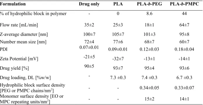
These observations allowed to select fabrication conditions leading to NPs with a hydrodynamic diameter, DH, of 100 nm independently of the polymer or formulation composition (selected data points in Figure 1A and Figure 2). The physicochemical characteristics of the NPs library eventually chosen are presented in Table 2. Monodisperse nanosuspensions of identical hydrodynamic diameters were produced by adjusting the flow rate of the injected phases depending on the polymer composition. Process was highly reproducible as shown by the small standard deviations of the NPs size. Drug loading and drug yield after purification were found identical whatever the NPs suspension (DL ~7 % and drug yield > 90%). The surface properties, i.e. the ZP and the hydrophilic block surface density, of the NPs were the only properties which varied as a function of the polymer composition. As discussed above, 100 nm-pure cholecalciferol and PLA NPs exhibited a negative surface whereas PLA-b-PEG and PLA-b-PMPC NPs were closer to neutrality. The hydrophilic block surface density was characterized by 1H NMR in D2O. PLA solid core which is not soluble in water did not give a NMR signal; only mobile polymer chains in water are visible in the NMR spectrums, i.e. the 490
495
500
Information). PLA-b-PEG and PLA-b-PMPC displayed the same grafting density of hydrophilic block (~ 0.3 chains/nm2) or monomer density (~ 15 units/nm2) assuming a brush conformation of the hydrophilic chains at the NPs surface.
Table 2. Physicochemical characteristics of 100 nm-diameter NPs under study. Data are
reported as mean over three or more separate formulations ± SD. (1H NMR spectra of
NPs and details of density calculations are reported in Supporting Information)
Formulation Drug only PLA PLA-b-PEG PLA-b-PMPC
% of hydrophilic block in polymer - 0 8.6 44
Flow rate [mL/min] 35±2 25±3 18±1 64±7
Z-average diameter [nm] 100±7 105±7 101±3 95±8
Number mean size [nm] 72±4 77±6 68±7 60±7
PDI 0.07±0.01 0.09±0.01 0.12±0.03 0.18±0.04
Zeta Potential [mV] -21±5 -32±7 -13±1 -14±1
Drug yield [%] 90±5 93±7 95±4 93±6
Drug loading, DL [%w/w] - 7.3 ±0.3 7.4 ±0.3 6.7 ±0.3
Hydrophilic block surface density
[PEG or PMPC chains/nm2] - - 0.34±0.05 0.33±0.07
Monomer surface density [EO or
MPC repeating units/nm2] - - 15±2 14±1
The morphology of the NPs is known to be dependant of the % of hydrophilic block in the polymer (Rabanel et al., 2015a; Rabanel et al., 2015b). TEM imaging was performed to assess the internal structure of the NPs used in this study. Solid-core NPs with a round-shaped morphology and a low polydispersity were observed irrespectively of the used polymer (Figure 2).
Before testing the drug release, degradation of the drug and penetration from the polymeric NPs, we first evaluated if the drug in polymeric suspensions was really encapsulated in the polymeric particles or if precipitated aside from the polymer particles. 510
515
This is an important point to solve since cholecalciferol and the polymers can independently form NPs of similar size under similar manufacturing conditions. Indeed, pure drug NPs manufactured without the use of polymer with a size of 100 nm were found stable as colloidal suspension in aqueous medium. A centrifugation test was performed to separate polymer NPs from pure drug NPs. After centrifugation of the nanosuspensions at 60 000 g for 2 hours, the quantity of drug in supernatant was determined. No pellet of pure drug NPs was formed, proving the high colloidal stability of these formulations. As shown in Figure 3, pure drug NPs remained in the supernatant after centrifugation. In contrast, when the drug-loaded polymeric NPs were centrifuged, a solid pellet of NPs was observed and less than 5% of the total quantity of drug was present in the supernatant, indicating that drug sedimented along with the polymer. As a control, a mix of pure PLA NPs (no drug) and pure drug NPs (no polymer) of identical size were centrifuged. All the drug was recovered in the supernatant, proving no possible fortuitous coaggregation between polymer and drug particles during the centrifugation. Since cholecalciferol sedimented during centrifugation when formulated in polymeric nanosuspensions but did not sediment when formulated in pure drug nanosuspensions, theses tests therefore confirm that the drug cholecalciferol was entirely encapsulated in the polymeric NPs.
3.3
Cholecalciferol stability
Cholecalciferol is known to be sensitive to several environmental factors such as light, oxygen, temperature, humidity and acidic conditions (Grady and Thakker, 1980; Mahmoodani et al., 2017). Exposure to these environmental factors leads to the formation of previtamin-D, transvitamin-D, pyrovitamin-D, isopyrovitamin-D, suprasterols or 525
530
535
540
isotachysterols metabolites (Grady and Thakker, 1980) and decreases the bioavailability of the drug. It was demonstrated that cholecalciferol encapsulation inside polymeric particles was a way to reduce its degradation in aqueous medium (Almouazen et al., 2013; Luo et al., 2012; Ramezanli et al., 2017). However, to the best of our knowledge, the influence of NPs physical parameters and notably the composition of the surface coating on the chemical stability of any drug has never been studied. Clarifications about the role of the nanocarrier composition on drug stability may help to design or formulate more efficient vectors to protect drug from degradation.
To this purpose, 100 nm-size pure drug NPs, polymeric NPs with different coatings and control micellar suspensions were formulated with or without BHT as an antioxidant and were incubated at body temperature (37 ºC) protected from light. Degradation kinetics of the drug in these formulations was quantified at different time points by HPLC-UV. The pH of the formulations ranged between 5.5 and 6.
As presented in Figure 4A, encapsulation of cholecalciferol in polymeric NPs or solubilisation in surfactant micelles without any antioxidant significantly improved the stability of the drug compared to pure drug NPs. Only 40 % of drug was remaining after 24 h of incubation when no polymer was used to protect the drug while more than 60% of the initial drug was still present after 24 h in polymeric nanoparticle suspensions. The protection against degradation imparted by encapsulation in polymeric NPs can be attributed to a reduction of the contact between water and drug thanks to the polymeric lipophilic core. The presence of a hydrophilic coating on PLA surface had a small but positive effect on drug protection. Compared to bare PLA NPs, PMPC and PEG coatings increased cholecalciferol stability (60, 71, and 79 % of initial dose remaining after 24 h, 550
555
560
565
respectively). Since drug was theoretically entrapped in the PLA core, we hypothesized that the coating physically protected the PLA core and formed therefore a second physical layer of drug protection against water contact.
Contrary to expectations, surfactant micelles better protected the dispersed drug from degradation than bare PLA NPs and equally well to the coated polymeric NPs, even though they are more dynamic than solid polymeric NPs. Accelerated degradation of the encapsulated drug in polymeric NPs can occur due to a local acid condition in the PLA core arising from the presence of acidic carboxylic groups. In fact, some PLA chain with carboxylic extremities are inevitably present in polymer batches due to uncontrolled initiation of the PLA chains with water or/and lactic acid impurities instead of hexanol or PEG, or transesterification reactions of the polymeric chains (Gupta and Kumar, 2007). The presence of such anionic moieties in PLA was confirmed by the negative ZP of PLA NPs. Since no acidic group was present in the micelle core of oleth-20, drug degradation originating from acid conditions was not possible and was consequently reduced. Our results show therefore that drug encapsulation in a solid polymeric core is not a guarantee of drug protection against degradation.
When BHT was introduced during the preparation of NPs (Figure 4B), drug degradation was hindered, demonstrating that BHT was coencaspulated with cholecalciferol within the particles.
3.4
Cholecalciferol release
In order to study the effect of NPs surface composition on cholecalciferol release, release profiles were performed in vitro. Cholecalciferol release from all formulations was 575
580
585
polymer concentration, if applicable, (1.2 mg/mL) over 24 hours. This duration of release was chosen because it represents the contact time with skin during the cutaneous penetration tests.
First, release studies were conducted by Franz diffusion cell method through a cellulose membrane and using the same receptor medium as the skin penetrations experiments (HBSS with 0.5%w/w oleth-20 and 0.002% w/w BHT, pH 7.4). In any instance, no cholecalciferol was found in the release medium, even if sink conditions were respected (data not shown). This absence of release from polymeric particles in buffer medium was already observed for cholecalciferol and was attributed to the very low solubility of the drug in water(Ramezanli et al., 2017). The membrane itself can also hinder the diffusion of the free drug in the receptor medium due to cholecalciferol adsorption on cellulose.
Drug release was then performed in a biphasic system which did not require the use of a porous membrane. Isopropyl myristate was chosen as lipophilic artificial membrane representative of skin outer layers due to its polar and non polar properties mimicking skin lipids (Hadgraft and Ridout, 1987). Figure 5 shows the release profiles of cholecalciferol from NPs or surfactant micelles in IPM at 37ºC. Release behaviour of drug from oleth-20 surfactant micelles was significantly different from solid NPs (p<0.05). All the drug initially present was released in 12 hours from micelles according to a first-order kinetic while release from solid NPs was 2-fold slower following a zero-order kinetic with no initial burst release. This release behavior from polymeric NPs in lipophilic medium was different from conventional release in aqueous medium. A zero-order kinetic reflected a limited dissolution rate of cholecalciferol from NPs to the medium. Even though cholecalciferol is fully soluble in IPM, IPM phase is not miscible 595
600
605
610
with water phase. When the two phases are mixed by magnetic stirring, a coarse emulsion is formed but contact between NPs and IPM was not optimal. Only NPs in direct contact and wetted with IPM may have released their content, limiting the release rate. An aqueous film may have also remained around the NPs acting as a secondary membrane which could slow down the diffusion of the drug in IPM. On the contrary for the surfactant micelles, droplet size of the water: IPM emulsion was smaller than for particles due to a reduction of the interfacial tension. The bigger transfer area between aqueous and lipophilic phase led to faster drug diffusion to the IPM phase from surfactant micelles. Moreover, the more dynamic structure of the micelles compared to solid NPs may also favor drug release.
Release profiles of cholecalciferol from NPs followed the same trend independently of the nature of surface composition of the NPs. These results were unexpected since it has been previously shown that presence of a PEG corona on polyester NPs surface slowed down the release of a hydrophobic molecule (Peracchia et al., 1997; Rabanel et al., 2015a). Interestingly, even the presence of a polymer protecting the drug in its core did not slow down the release of the drug, since no release difference was found between drug particles made with or without polymer. The sole factor controlling cholecalciferol delivery in IPM was therefore the surface contact between NPs and IPM and it seems that the composition of the NPs coating did not alter the interfacial tension between water and the lipophilic release medium. After 24 hours of release, a layer of aggregated NPs was also observed for NPs formulation at the water-IPM interface, independently of the NPs coating. Rancan et al, already observed a loss of colloidal stability of polyester NPs at the 620
625
630
interface between water and IPM which was explained the reduction of electrostatic interactions between NPs due to the low dielectric constant of IPM (Rancan et al., 2009).
It should be noted that this release test was not discriminative to evaluate the interactions between drug and NPs matrix since the release of the cholecalciferol in an aqueous or lipophilic medium was rather dictated by the rate of solubilization of the drug in the medium than by the diffusion of drug through the polymeric matrix.
3.5
Cholecalciferol skin absorption
The influence of NPs coating on the delivery of cholecalciferol to intact skin was evaluated by the Franz cell method, the gold standard for percutaneous studies (OECD). Penetration in an impaired skin model (stripped skin) was also investigated to determine if the composition of the skin surface, i.e. the presence or not of the SC barrier, influences the interactions skin-NPs. On intact skin, NPs were deposited on the stratum corneum surface, which is a stratified layer of cornified keratinocytes embedded in a lipid rich medium, forming a relatively impermeable layer. On impaired skin, stratum corneum was removed by tape stripping. The majority of the stratum corneum was removed when the TEWL value was comprised between 30-40 g.m2.h-1 (Dey et al., 2014; Tsai et al., 2003). NPs were in this case in contact with a layer of living cells (the viable epidermis), more hydrophilic and permeable than the stratum corneum. The penetration of the drug from pure drug NPs (no polymer) and surfactant micelles was assessed as control. Cholecalciferol absorption in all layers of intact and impaired skin after 24 hours of incubation with formulations is presented in Figure 6. 640 645 650 655 660
Results showed that the distribution of drug in the different skin layers was dependent on both skin condition and NPs surface coating. In intact skin, drug was poorly absorbed (less than 300 ng/cm2, i.e. less than 1% of the applied dose) and accumulated significantly in the stratum corneum (up to 210 ng/cm2) and, to a lesser extent, in the viable epidermis (up to 80 ng/cm2). Such very low penetration in intact skin is well reported for this drug (Alsaqr et al., 2015; Ramezanli et al., 2017) and the affinity of cholecalciferol to the lipophilic layer of the stratum corneum was expected due to the strong hydrophobic character of the drug. In addition, drug lipophilicity inhibited the permeation of the active compound to the receptor medium.
As can be seen in Figure 6, solid NPs (with or without polymer) exhibited a significantly higher skin absorption of cholecalciferol compared to the oleth-20 micelles (up to 2.8-fold, p<0.05). The ability of polymeric NPs to improve drug penetration compared to micelles and to accumulate in the outer layers of the skin was already reported for retinol, another lipophilic drug (Laredj-Bourezg et al., 2015). We hypothesized that the better absorption of the drug in the skin was relative to a better accumulation or a deeper penetration of solid NPs in the stratum corneum than surfactant micelles.
However, the encapsulation of drug in polymer did not modulate its penetration within skin layers. Interestingly, the same quantity of drug was recovered in skin treated with solid NPs made with or without polymer (~ 250 ng/cm2), confirming that polymer did not hinder or favor cholecalciferol diffusion and/or NPs penetration when NPs were in contact with the stratum corneum. The composition of NPs surface had also no influence on the total drug absorption into intact skin, indicating that the stratum corneum acts as a 665
670
675
IPM surface during the release studies, we can suppose that they may also aggregated when they were in close contact with the lipophilic stratum corneum, i.e. on its surface or deeper if they penetrated through the SC.
When deposited on impaired skin and independently of the formulation, cholecalciferol was better absorbed in all viable skin layers compared to intact skin (up to 3.4-fold, Figure 6). As expected, drug diffusion was facilitated due to the removal of the stratum
corneum (Döge et al., 2016; Jensen et al., 2011; Knudsen et al., 2011). In this case and
contrary to intact skin, absorption of cholecalciferol into impaired skin depended on the NPs surface composition. At equivalent drug content, the addition of a hydrophilic coating of a neutral layer (PEG) or a zwitterionic layer (PMPC) on PLA core decreased penetration in both epidermis (~ -70 %) and dermis (~ -130 %), which decreased the overall penetration 1.8-fold compared to bare PLA (p<0.05, Figure 6). We had however found that the nature of the hydrophilic coating (neutral PEG or zwitterionic PMPC) did not influence total skin absorption (440 ng/cm2 for PLA-b-PEG NPs and 410 ng/cm2 for PLA-b-PMPC NPs). Contrary to initial expectations, PMPC coating did not offer any advantages over the other NPs surfaces for skin delivery, at least at these studied density and NPs concentration. It acted similarly to the PEG coating. Interestingly, we also found that encapsulation of the drug in a polymer core did not offer substantial improvement of the percutaneous delivery. The penetration of cholecalciferol from pure drug NPs was 1.3-fold lower than penetration from bare PLA NPs but was better than penetration from PLA-based NPs coated with PEG or PMPC (~1.3-fold). Compared to non-polymeric aggregates, the presence of the hydrophilic coating decreased drug penetration 1.3-fold with a lower accumulation in dermis.
685
690
695
700
NPs displaying a negative ZP (pure drug and bare PLA NPs) improved cholecalciferol penetration compared to globally non-charged coated NPs (PEG and PLA-b-PMPC NPs) only on impaired skin. Accordingly, enhancement of drug penetration is rather relative to the solid particulate and charged structure than to the drug encapsulation in a polymeric core. We suggested that these differences of drug penetration were the results of different rates of NPs accumulation on the skin surface or NPs penetration in the skin which are dependent of electrostatic interactions between skin and NPs. In fact, NPs surface charge already proved its importance in penetration of NPs themselves, even if results are often contradictory (Fernandes et al., 2015; Hsiao et al., 2016; Kohli and Alpar, 2004; Mahmoud et al., 2017; Wu et al., 2010). The significance of these electrostatic interactions seems depended of the composition of the skin surface. Abdel-Mottaleb et al. already reported that skin pathology may condition the accumulation of NPs in skin depending on their charges. The authors noticed no difference of cutaneous penetration between anionic, neutral and cationic ethylcellulose NPs in healthy mice ear skin. However, in inflamed skin conditions, an improved therapeutic effect of bethamethasone by charged NPs compared to neutral NPs was observed (Abdel-Mottaleb et al., 2012). In our case, we propose that negatively charged NPs are not attracted with skin cells since they have slightly negative membranes, but rather may interact with extracellular charged skin elements of the epidermis, such as proteins or salt species, which can trigger their accumulation on the skin. The quasi neutral charge of PEG and PMPC-coated NPs did not allow such electrostatic interactions with skin elements from the viable epidermis, reducing their adhesion/interactions with the skin and so the absorption of the drug.
710
715
720
4. Conclusions
The modifications of the surface properties of NPs resulted to various impacts on the different steps of the delivery of a lipophilic labile drug into the skin. Drug encapsulation in a polymeric core improved the protection of the drug from degradation, with a slight positive effect of the hydrophilic coatings, but did not alter the in vitro drug release kinetics into a lipophilic medium mimicking skin lipids. Regarding cholecalciferol penetration within healthy intact skin, all solid NPs displayed the same efficiency to carry cholecalciferol in the skin, irrespective of the NPs surface properties, highlighting the non-selective role of the stratum corneum barrier. However, the surface properties of the NPs carrying the drug clearly affected cholecalciferol penetration when stratum corneum was removed to resemble to diseased skin, pointing out the importance to consider the influence of skin pathology when designing topical formulations. Detailed characterizations on the mechanisms of NPs-skin interactions depending of the NP surface will be discussed in a further publication.
Supporting information
NMR spectrum and methods of calculations of quantity of PEG and PMPC in polymers and on NPs surface are available free of charge at www.x.com
Acknowledgments
AL is grateful to the Région Auvergne-Rhône-Alpes for their mobility grant (Explo’RA Doc) and the Faculty of Pharmacy of Université de Montréal for their financial support. JF is grateful to the Arthritis Society and the University of Montreal for PhD grants. PLL 730
735
740
745
acknowledges financial support from NSERC, the GRUM and the Faculty of Pharmacy. CR acknowledges the Région Auvergne-Rhône-Alpes for support (mobility grant, Explo’RA Sup). AGA is supported by the TransMEdTech Institute. PH is thankful for the financial support of FRQNT. XB acknowledges the financial support from the Canada Research Chair Program and NSERC.
TEM imaging was performed at the “Centre de Caractérisation Microscopique des Matériaux” of the École Polytechnique (Montréal, Québec, Canada) with the help of Jean-Philippe Massé. Alexander Cunningham is acknowledged for his help in GPC analysis.
Declaration of interest
Authors declare no competing interest.
References
Abdel-Mottaleb, M.M., Moulari, B., Beduneau, A., Pellequer, Y., Lamprecht, A., 2012. Surface-charge-dependent nanoparticles accumulation in inflamed skin. J. Pharm. Sci. 101, 4231-4239. Albanese, A., Tang, P.S., Chan, W.C., 2012. The effect of nanoparticle size, shape, and surface chemistry on biological systems. Annu. Rev. Biomed. Eng. 14, 1-16.
Almouazen, E., Bourgeois, S., Jordheim, L.P., Fessi, H., Briançon, S., 2013. Nano-encapsulation of vitamin D3 active metabolites for application in chemotherapy: formulation study and in vitro evaluation. Pharm. Res. 30, 1137-1146.
Alnasif, N., Zoschke, C., Fleige, E., Brodwolf, R., Boreham, A., Rühl, E., Eckl, K.-M., Merk, H.-F., Hennies, H.C., Alexiev, U., Haag, R., Küchler, S., Schäfer-Korting, M., 2014. Penetration of normal, damaged and diseased skin — An in vitro study on dendritic core–multishell nanotransporters. J. Controlled Release 185, 45-50.
Alsaqr, A., Rasoully, M., Musteata, F.M., 2015. Investigating Transdermal Delivery of Vitamin D3. AAPS PharmSciTech, 1-10. 755 760 765 770 775
Alvarez-Roman, R., Naik, A., Kalia, Y.N., Guy, R.H., Fessi, H., 2004. Skin penetration and distribution of polymeric nanoparticles. J. Controlled Release 99, 53-62.
Bachhav, Y., Mondon, K., Kalia, Y., Gurny, R., Möller, M., 2011. Novel micelle formulations to increase cutaneous bioavailability of azole antifungals. J.Controlled Release 153, 126-132.
Bolzinger, M.A., Briançon, S., Chevalier, Y., 2011. Nanoparticles through the skin: managing conflicting results of inorganic and organic particles in cosmetics and pharmaceutics. Wiley Interdiscip. Rev.: Nanomed. Nanobiotechnol. 3, 463-478.
Chrit, L., Bastien, P., Biatry, B., Simonnet, J.T., Potter, A., Minondo, A.M., Flament, F., Bazin, R., Sockalingum, G.D., Leroy, F., Manfait, M., Hadjur, C., 2007. In vitro and in vivo confocal Raman study of human skin hydration: Assessment of a new moisturizing agent, pMPC. Biopolymers 85, 359-369.
Couvreur, P., 2013. Nanoparticles in drug delivery: past, present and future. Adv. Drug Delivery Rev. 65, 21-23.
Dey, S., Rothe, H., Page, L., O'connor, R., Farahmand, S., Toner, F., Marsh, R., Wehmeyer, K., Zhou, S., 2014. An in vitro Skin Penetration Model for Compromised Skin: Estimating Penetration of Polyethylene Glycol [14C]-PEG-7 Phosphate. Skin Pharmacol. Physiol. 28, 12-21.
Döge, N., Hönzke, S., Schumacher, F., Balzus, B., Colombo, M., Hadam, S., Rancan, F., Blume-Peytavi, U., Schäfer-Korting, M., Schindler, A., Rühl, E., Skov, P.S., Church, M.K., Hedtrich, S., Kleuser, B., Bodmeier, R., Vogt, A., 2016. Ethyl cellulose nanocarriers and nanocrystals differentially deliver dexamethasone into intact, tape-stripped or sodium lauryl sulfate-exposed ex vivo human skin - assessment by intradermal microdialysis and extraction from the different skin layers. J. Controlled Release 242, 25-34.
Elias, P.M., 1983. Epidermal lipids, barrier function, and desquamation. J. Invest. Dermatol. 80, 44s-49s.
Feng, W., Zhu, S., Ishihara, K., Brash, J.L., 2006. Protein resistant surfaces: Comparison of acrylate graft polymers bearing oligo-ethylene oxide and phosphorylcholine side chains. Biointerphases 1, 50-60.
Fernandes, R., Smyth, N.R., Muskens, O.L., Nitti, S., Heuer-Jungemann, A., Ardern-Jones, M.R., Kanaras, A.G., 2015. Interactions of skin with gold nanoparticles of different surface charge, shape, and functionality. Small 11, 713-721.
Fessi, H., Puisieux, F., Devissaguet, J.P., Ammoury, N., Benita, S., 1989. Nanocapsule formation by interfacial polymer deposition following solvent displacement. Int. J. Pharm. 55, R1-R4.
Forier, K., Messiaen, A.S., Raemdonck, K., Deschout, H., Rejman, J., De Baets, F., Nelis, H., De Smedt, S.C., Demeester, J., Coenye, T., Braeckmans, K., 2013. Transport of nanoparticles in cystic fibrosis sputum and bacterial biofilms by single-particle tracking microscopy. Nanomedicine 8, 935-949.
Ging-Ho, H., Chun-Liang, L., Ching-Hao, C., Che-Ping, L., Chun-Kai, H., Hung-Hao, C., 2007. Preparation and characterization of poly(2-methacryloyloxyethyl phosphorylcholine)-block-poly(D,L-lactide) polymer nanoparticles. J. Polym. Sci., Part A: Polym. Chem. 45, 688-698.
Grady, L.T., Thakker, K.D., 1980. Stability of solid drugs: Degradation of ergocalciferol (vitamin D2) and cholecalciferol (vitamin D3) at high humidities and elevated temperatures. J. Pharm. Sci. 69, 1099-1102.
Graf, C., Gao, Q., Schütz, I., Noufele, C.N., Ruan, W., Posselt, U., Korotianskiy, E., Nordmeyer, D., Rancan, F., Hadam, S., 2012. Surface functionalization of silica nanoparticles supports colloidal stability in physiological media and facilitates internalization in cells. Langmuir 28, 7598-7613. Gref, R., Lück, M., Quellec, P., Marchand, M., Dellacherie, E., Harnisch, S., Blunk, T., Müller, R.H., 2000. ‘Stealth’ corona-core nanoparticles surface modified by polyethylene glycol (PEG): 780 785 790 795 800 805 810 815 820 825
influences of the corona (PEG chain length and surface density) and of the core composition on phagocytic uptake and plasma protein adsorption. Colloids Surf., B 18, 301-313.
Gref, R., Minamitake, Y., Peracchia, M.T., Trubetskoy, V., Torchilin, V., Langer, R., 1994. Biodegradable long-circulating polymeric nanospheres. Science 263, 1600-1603.
Gref, R., Miralles, G., Dellacherie, E., 1999. Polyoxyethylene-coated nanospheres: effect of coating on zeta potential and phagocytosis. Polym. Int. 48, 251-256.
Gupta, A.P., Kumar, V., 2007. New emerging trends in synthetic biodegradable polymers – Polylactide: A critique. Eur. Polym. J. 43, 4053-4074.
Hadgraft, J., Ridout, G., 1987. Development of model membranes for percutaneous absorption measurements. I. Isopropyl myristate. Int. J. Pharm. 39, 149-156.
Han, J., Zhu, Z., Qian, H., Wohl, A.R., Beaman, C.J., Hoye, T.R., Macosko, C.W., 2012. A simple confined impingement jets mixer for flash nanoprecipitation. J. Pharm. Sci. 101, 4018-4023. Hickey, J.W., Santos, J.L., Williford, J.-M., Mao, H.-Q., 2015. Control of polymeric nanoparticle size to improve therapeutic delivery. J. Controlled Release 219, 536-547.
Hsiao, P.F., Peng, S., Tang, T.-C., Lin, S.-Y., Tsai, H.-C., 2016. Enhancing the in vivo transdermal delivery of gold nanoparticles using poly(ethylene glycol) and its oleylamine conjugate. Int. J. Nanomed. 11, 1867-1878.
Ishihara, K., Nakabayashi, N., 1991. Specific interaction between water-soluble phospholipid polymer and liposome. J. Polym. Sci., Part A: Polym. Chem. 29, 831-835.
Jensen, L.B., Petersson, K., Nielsen, H.M., 2011. In vitro penetration properties of solid lipid nanoparticles in intact and barrier-impaired skin. Eur. J. Pharm. Biopharm. 79, 68-75.
Jin, Q., Chen, Y., Wang, Y., Ji, J., 2014. Zwitterionic drug nanocarriers: A biomimetic strategy for drug delivery. Colloids Surf., B 124, 80-86.
Johnson, B.K., Prud'homme, R.K., 2003. Flash NanoPrecipitation of Organic Actives and Block Copolymers using a Confined Impinging Jets Mixer. Aust. J. Chem. 56, 1021-1024.
Kanekura, T., Nagata, Y., Miyoshi, H., Ishihara, K., Nakabayashi, N., Kanzaki, T., 2002. Beneficial effects of synthetic phospholipid polymer, poly(2-methacryloyloxyethyl phosphorylcholine-co-n-butyl methacrylate), on stratum corneum function. Clin. Exp. Dermatol. 27, 230-234.
Kilfoyle, B.E., Sheihet, L., Zhang, Z., Laohoo, M., Kohn, J., Michniak-Kohn, B.B., 2012. Development of paclitaxel-TyroSpheres for topical skin treatment. J. Controlled Release 163, 18-24.
Knudsen, N.Ø., Jorgensen, L., Hansen, J., Vermehren, C., Frokjaer, S., Foged, C., 2011. Targeting of liposome-associated calcipotriol to the skin: effect of liposomal membrane fluidity and skin barrier integrity. Int. J. Pharm. 416, 478-485.
Knudsen, N.Ø., Rønholt, S., Salte, R.D., Jorgensen, L., Thormann, T., Basse, L.H., Hansen, J., Frokjaer, S., Foged, C., 2012. Calcipotriol delivery into the skin with PEGylated liposomes. Eur. J. Pharm. Biopharm. 81, 532-539.
Kohli, A.K., Alpar, H.O., 2004. Potential use of nanoparticles for transcutaneous vaccine delivery: effect of particle size and charge. Int. J. Pharm. 275, 13-17.
Kojima, M., Ishihara, K., Watanabe, A., Nakabayashi, N., 1991. Interaction between phospholipids and biocompatible polymers containing a phosphorylcholine moiety. Biomaterials 12, 121-124. Lapteva, M., Mondon, K., Moller, M., Gurny, R., Kalia, Y.N., 2014a. Polymeric micelle nanocarriers for the cutaneous delivery of tacrolimus: a targeted approach for the treatment of psoriasis. Mol. Pharm. 11, 2989-3001.
Lapteva, M., Santer, V., Mondon, K., Patmanidis, I., Chiriano, G., Scapozza, L., Gurny, R., Möller, M., Kalia, Y.N., 2014b. Targeted cutaneous delivery of ciclosporin A using micellar nanocarriers 830 835 840 845 850 855 860 865 870