13574
Journal of Applied Biosciences 133: 13574 - 13583
ISSN 1997-5902
Molecular characterization of the main fungi
associated to Bambara groundnut foliar diseases in
Burkina Faso
Adjima Ouoba1,2*, Elisabeth P. Zida3, Romain W. Soalla, 3, Martine Bangratz4,5, Palanga Essowè,
Moussa N’Golo Konaté2, Hervé Nandkangré2 , Mahama Ouédraogo2 and Mahamadou Sawadogo1
1Laboratoire Biosciences, Equipe Génétique et Amélioration des Plantes, Université Ouaga I Pr Joseph Ki-Zerbo, 03 BP 7021 Ouagadougou 03, Burkina Faso
2Laboratoire de Génétique et de Biotechnologies Végétales, Département de Productions Végétales, Institut de l’Environnement et de Recherches Agricoles (INERA), Centre National de la Recherche Scientifique et Technologique (CNRST), 04 BP 8645 Ouagadougou, Burkina Faso
3Laboratoire de Phytopathologie et de Biotechnologies Végétales, Département de Productions Végétales, Institut de l’Environnement et de Recherches Agricoles (INERA), Centre National de la Recherche Scientifique et Technologique (CNRST), 01 BP 476, Ouagadougou, Burkina Faso
4Institut de Recherche pour le Développement (IRD), 911 Avenue Agropolis, 34394 Montpellier, France 5Laboratoire Mixte International Patho-Bios, 01 BP 476, Ouagadougou, Burkina Faso
Original submitted in on 19th September 2018. Published online at www.m.elewa.org on 31st January 2019
https://dx.doi.org/10.4314/jab.v133i1.9
ABSTRACT
Objective: This study aims to update the database of fungi associated to Bambara groundnut foliar diseases in Burkina Faso using both molecular and morphological identification approaches.
Methodology and Results: In this study, molecular approach based on the sequencing of ITS (Internal Transcripted Spacer) region of fungi and morphological approach were used to identify the main fungi associated to Bambara groundnut foliar diseases. The study was performed with universal polymerase chain reaction (PCR) primer ITS1/ITS4. BlastN comparisons between 19 fungal isolates contigs of the 16 major fungi were produced by their DNA sequences assembly and GenBank sequences yielded identity scores of 99 to 100 % with all of them. The degrees of similarity between these contigs and the loci sequences of classified fungi in GenBank indicate that our fungal isolates are the same species with those in Genbank, particularly the first of the list show after the blastN. It is the first report of molecular characterization of the main fungi infecting Bambara groundnut in Burkina Faso.
Conclusion and Application of results: Nineteen fungi associated to Bambara groundnut foliar diseases were identified and can be taken as targets in varietal improvement of Bambara groundnut for resistance to fungal diseases in Burkina Faso.
Key words: Bambara groundnut, fungi, molecular characterization, PCR primer ITS1/ITS4, Burkina Faso. INTRODUCTION
Bambara groundnut (Vigna subterranea (L) Verdc.) is an indigenous African food legume and has advantages over more favoured species in terms
of nutritional value and tolerance to adverse environmental conditions (Mkandawire, 2007; Berchie et al., 2012). The crop is the third most
13575 important food legume in Africa after groundnut
and cowpea (Odongo et al., 2015). It is a potential crop to provide food security in the dry areas of Africa. Its cultivation outside the African continent is very limited and the main world crop production (45 to 50%) is provided by West Africa (Baudoin et Mergeai, 2001; Brink et al., 2006, Hillocks et al., 2012). Africa-wide production is estimated to be over 330,000 t annually (Hillocks et al., 2012) and Burkina Faso is one of the largest producers of Bambara groundnut which is the second most important food legume after cowpea (Cirad-Gret, 2002; Ouédraogo et al., 2008; Hillocks et al.,
2012). Although Bambara groundnut is considered to be generally less affected by diseases and pests than groundnut or cowpea, several diseases and pests can cause serious damage to the crop (PROTA, 1980; Baudoin et Mergeai 2001). The fungal diseases are a major constraint on Bambara groundnut production in Burkina Faso and can cause yield losses of up to 83% in the event of severe attack (Sérémé et al., 1991; Sérémé, 1992). Around sixteen fungal species have been associated with foliar diseases on Bambara groundnut in Africa six of them have been reported from Burkina Faso since for a long time (Table 1). Table 1. List of Bambara groundnut-infecting fungi present in Africa
Fungi/Diseases Country Reference
Fusarium sp/ wilt Botswana, Togo, Zambia,
Zimbabwe
Heller et al., 1997; Mkandawire, 2007; Hillock et al., 2012
Cercospora sp /leaf spot Botswana, Togo Heller et al., 1997; Hillock et al., 2012
Phoma sorghina Burkina Faso Heller et al., 1997
Phomopsis sojae Burkina Faso Heller et al., 1997
Fusarium oxysporum Burkina Faso, Tanzania Heller et al. 1997; Baudoin et Mergeai,
2001
Rhizoctonia solani Burkina Faso, Sierra Leone,
Tanzania Heller et al., 1997; Baudoin et Mergeai, 2001; Hillock et al., 2012
Macrophomina phaseolina Burkina Faso Heller et al., 1997
Puccinia sp Nigeria Heller et al., 1997; Hillock et al., 2012
Colletotrichum sp Nigeria Heller et al., 1997; Hillock et al.,
2012
Colletotrichum capsici _ Baudoin et Mergeai, 2001
Cercospora anescens/leaf spot Tanzania, Zimbabwe Heller et al.,1997; Baudoin et Mergeai,2001;Mkandawire,2007; Hillock et al., 2012
Erysiphe sp/powdery mildew Tanzania Heller et al., 1997
Cercospora leaf blight Zambia Heller et al., 1997; Mkandawire,2007
Phyllosticta voandzeia/ Leaf spot
Zimbabwe Heller et al., 1997
Sclerotium rolfsii Zimbabwe Heller et al., 1997; Mkandawire, 2007,
Hillock et al., 2012
Phomopsis sp/ Leaf blotch Burkina Faso, Zimbabwe Kiwallo, 1991; Heller et al., 1997, Mkandawire, 2007
13576 The limited knowledge of fungi infecting Bambara
groundnut in Burkina Faso hinders the control of fungal diseases and the creation of resistant varieties. In the event of climate changes which have an influence on epidemiology and inoculum survival (Boland et al., 2004), the main objective of this study is to update the database of Bambara groundnut fungal diseases in Burkina Faso. The Molecular approach was used for completing the morphological identification to characterize the
main fungi associated to Bambara groundnut diseases in Burkina Faso. The ITS (Internal Transcribed Spacer) region of fungi rDNA has been investigated for species identification and phylogenetic relationships (Talarmin, 2007; Karsch-Mizrachi et al., 2012). Here, universal polymerase chain reaction (PCR) primer ITS1/ITS4 (White et al., 1990) was used to characterize the main fungi associated to Bambara groundnut foliar diseases in Burkina Faso.
MATERIAL AND METHODS
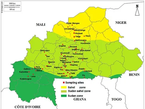
Plant sampling: One hundred and sixty-six (166) leaf samples of Bambara groundnut plants infected by fungal diseases were randomly collected from farmer’s fields in forty-four (44) sites (fig 1) and experimental plots. The sampling was carried out through the three agro-climatic zones (humid Sudan zone, sub-humid
Sudan-Sahel zone and dry Sahel zone) of Burkina Faso (fig 1) from September to October, 2015. Eighteen (18) samples were collected in the humid Sudan zone, one hundred and twenty-nine (129) in the sub-humid Sudan-Sahel zone and nineteen (19) from the dry Sahel zone.
Fig 1. Sites of collection of leaf samples of Bambara groundnut in 2015 in Burkina Faso Isolation of fungi associated with infected leaves of
Bambara groundnut: The collected fresh leaves were surface cleaned in sterilized distilled water followed by immersion in 70% (v/v) ethanol for 1 min. The samples were left to air dry for 30 min. Three to four fresh leaflets of each sample with diseases symptoms were transferred into Petri dishes containing two layers of blotting papers humidified with sterile distilled water. Petri dishes containing samples were incubated at 22 to
25°C under alternating 12 h of light and 12 h of dark for 7 days. After incubation, fungi were identified based on the examination of the acervuli and conidia produced on the infected tissues on the blotter papers under the stereomicroscope and compound microscopes based on the identification key established by Marthur and Kongsdal (2003). A prevalence rate was calculated for each fungus identified as a percentage of samples infected by this fungus.
13577 Single spore production of the main fungi: The
choice of the main fungi has been based on their prevalence (≥25%) in each agro-ecological zone. So, using a sterile loop, isolates were further placed onto potato dextrose agar (PDA) containing streptomycin (0.3 μg/L of PDA). The plates were incubated at 22°C to 25°C for 7 days under ultraviolet (UV) light of alternating 12 h light and darkness to obtain pure culture. For single spore culture production, distilled water (100 ml) was added to Petri dishes containing culture of fungal isolates. Then 200 µl of the suspension was diluted according to its concentration in spores and 200 µl of diluted suspension were spread on AGAR medium and incubated for 24 to 48 h. Five single cells from each isolate were again transferred onto fives new Petri dishes containing PDA. After seven days of growth, single spore pure cultures were obtained and stored at 20°C until used.
DNA extraction: DNA of each isolate was extracted using the cethyltrimethylammonium bromide (CTAB) method used by Sadfi-Zouaoui et al. (2008) with some minor modifications. After adding 600 μl of CTAB extraction buffer [1.4 M NaCl, 2% CTAB (w / v), 0.1 M Tris-Base pH8, 20 mM EDTA and 0.2% (v / v). v) β-mercaptoethanol] and introducing two beads into each tube containing 100 mg of mycelium, the tubes were vortexed and stirred for five to ten minutes. The beads were then removed and the tubes were placed in a water bath at 65 ° C for fifteen minutes. A volume of 450 μl of phenol and 450 μl of alcohol-iso-amyl chloroform (consisting of 49 ml of chloroform and 1 ml of iso-amyl alcohol) was added to each tube and then mixed by inversion of the tubes until the content becomes milky. The mixture was then centrifuged at 13,000 x g for five minutes at 25 ° C. The supernatant (approximately 500 μl) was recovered in a new 1.5 ml tube. After adding 400 μl of alcohol-isoamyl chloroform, the tubes were again centrifuged at 13000 g for two minutes at 25 ° C. The supernatant was further transferred to a new 1.5 ml tube and 0.7 volume (350 μl) of isopropanol was added to the solution to precipitate the DNA. Further centrifugation was performed for 20 minutes at 13000 g at 25 ° C to obtain a DNA pellet at the bottom of the tube. The pellet was rinsed with 500 μl of 96% alcohol and centrifuged for three minutes at 13000 g at 25 ° C. This rinsing step was repeated three times. The pellet was dried under the hood and then dissolved in 30 μl of sterile distilled water. The DNA thus obtained was stored at -20 ° C.
DNA concentration measurement: The concentration of DNA obtained for each isolate was measured by spectrophotometer (Nanodrop 2000) and the initial concentration was evaluated in nanograms per microliter (ng / μl). The different concentrations were diluted case by case so that the DNA volume taken for the PCR contains about 40 ng of DNA.
Polymerase chain reaction (PCR): The amplification reaction was carried out in 25 µl containing 40 ng of DNA (4 µl), 2.5 µl of stained buffer Taq DNA polymerase 10X (Promega), 0.5 µl of dNTP (10 mM), 0.5 μl of each primers (ITS1 at 10 μM and ITS4 at 10 μM), 0.1 μl of Taq DNA polymerase (5 U/ μl) (Promega) and sterile distilled H2O. The amplification reaction was carried out in a thermal cycler. The PCR program was as follows: pre-denaturation at 94 ° C for 5 minutes followed by 30 consecutive cycles of denaturation at 94 ° C for 30 seconds, specific primer hybridization at 58 ° C for 30 minutes and an elongation at 72 ° C for 5 minutes and a post-elongation at 72 ° C for 10 minutes. Agarose gel electrophoresis: A 1% (W / V) agarose gel was prepared with TAE buffer 0.5 X (Tris, acetic acid, EDTA pH 8). A volume of 2 μl of ethidium bromide was added to the agarose once dissolved in the microwave and then cooled. The gel thus prepared was cast in a mini electrophoresis plate in which the buffer TAE (0.5 X) was added after solidification of the gel. The gel wells were then loaded with 12 μl of the PCR product of each sample. A molecular weight marker (100 bp), a water-control and a positive control were separately loaded into the wells of the gel. Electrophoresis was performed at 100 V for 20 minutes. The amplified products were visualized under UV light. Sequencing of amplified ITS: PCR products (amplified ITS) were purified using the MPBiomedicals Geneclean Turbo Kit. The purified DNA was eluted in 30 μl of sterile distilled water and 18 μl was removed for sequencing. Sequencing was performed by Genewiz http://www.beckmangenomics.com/ with primers ITS1 and ITS4
Sequences analysis: The sequences were processed, cleaned and aligned with the Bioedit software. The consensus sequences obtained from the treatments and cleanings were compared to those of the nucleotide sequence database of NCBI (National Center for Biotechnology Information) on the site: https: //blast.ncbi.nlm.nih. gov / Blast.cgi
13578 Several species of fungi are associated with Bambara
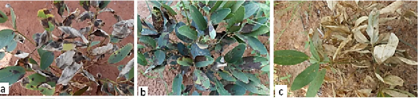
groundnut diseases in Burkina Faso. The analyses of leaf samples of diseased plants (fig. 2), based on morphological characters, detected 35 species of fungi (Table 1) belonging to 19 genera (Macrophomina, Alternaria, Cladosporium, Phoma, Cercospora, Rhizoctonia, Fusarium, Curvularia, Myrothecium, Colletotrichum, Melanospora, Exserohilum, Leptosphaerulina, Didymella, Nigrospora, Aspergillus, Pestalotia, Ulocladium and Microdochium). Fungi were more represented in the Sudan-Sahel zone with 32 species encountered, followed by the Sahel zone with 23 species and finally the Sudan zone with 13 species. The prevalence of fungi varied from 0.77%for Microdochium oryzae and Fusarium moniliforme in Sudan Sahel zone to 83.33% for Cladosporium sphaerospermum and Cercospora sp in Sudan zone (Table 1). Among the 35 fungal species identified using morphological identification approach, sixteen species were the most prevalent (prevalence ≥25%). The fungal species such as Macrophomina phaseolina,
Alternaria sp and Curvulria sp had their prevalence higher than 25% in each climatic zone. While Cladosporium sphaerospermum and Cercospora sp were the most prevalent (83.33%) in Sudan zone, Curvularia sp was the most prevalent (69.77 %,) in sudan-sahel zone. In Sahel zone, Fusarium equiseli and Exserohilum rostiatum were the most prevalent (68.42%). Nineteen isolates constituted with the most prevalent species were identified using molecular identification approach (Table 2).BlastN comparisons between the 19 isolates contigs of the 16 major fungi that were produced by their DNA sequences assembly and GenBank sequences yielded identity scores of 99 to 100 % with all of them(Table 2). The degrees of similarity between these contigs and the loci sequences of classified fungi in GenBank indicate that our fungal isolates are the same species with those in Genbank, particularly the first of the list show after the blastN. Of the nineteen (19) fungi isolates subjected to molecular analysis, fifteen (15) species were identified (Table 2).
Fig 2. Some symptoms of Bambara groundnut leaf diseases observed in the field
a): Light brown spots with or without a yellow halo, b): Reddening spots, c): wilting and drying of the plant. Fungi associated to symptoms about morphological identification a): Cercospora sp., Alternaria sp et Phoma sp., b): Cercospora sp, Fusarium equiseti et Phoma lingam, c): Macrophomina phaseolina, Colletotrichum capsici, Phoma sorghina, Fusarium culmorum et Curvilaria sesami.
The comparison of identities of main fungi with morphological approach between their identities with molecular approach, revealed a concordance with the fungi species Macrophomina phaseolina, Colletotrichum capsici, Exserohilum rostratum, Curvularia lunata and Phoma sp. However molecular approach brought more precisions on the fungi strains. For species such as Curvularia eragrostidis, Fusarium
sp, and Cladosporium cladosporioides, compliance was limited to the genus level. For others, the morphological results did not agree with those at the molecular level. This is the case of the species Corynespora sp., Thielavia terricola and Macrophomina phaseolina previously identified morphologically as Alternaria sp., Fusarium sp. and Rhizoctonia solani, respectively.
13579
Table 2. Fungi associated with diseased Bambara groundnut plants and their prevalence according to climatic zones of Burkina Faso
N° Fungi isolates Prevalence (%) according to climatic zones
Sahel zone Sudan-sahel zone Sudan zone
1 ***Macrophomina phaseolina 31,58 30,23 27,78 2 ***Alternaria sp 31,58 38,76 44,44 3 **Cladosporium sphaerospermum 10,52 65,88 83,33 4 **Phoma sp 5,26 58,14 77,78 5 **Cercospora sp 10,53 43,41 83,33 6 Alternaria alternata 10,53 22,48 11,11 7 *Phoma sorghina 63,15 14,72 00 8 *Rhizoctonia solani 47,37 15,5 16,67 9 *Fusarium equiseli 68,42 19,38 00 10 *Curvularia lunata 52,63 22,48 11,11 11 Myrothecum roridum 26,31 15,5 00 12 *Colletotrichum capsici 52,63 22,48 11,11 13 Phoma lingam 10,52 11,62 00 14 *Melanospora zamia 21,05 31,01 00 15 *Exserohilum rostiatum 68,42 10,85 00 16 *Leptosphaerulina crassiaca 21,05 46,51 00 17 **Fusarium oxysporum 42,1 54,26 00 18 Colletotrichum graminicola 10,52 2,32 00 19 ***Curvularia sp 26,31 69,77 55,56 20 Didymella bryoniae 5,26 00 00 21 Fusarium sp 10,53 14,73 22,22 22 Phoma exigua 5,26 00 00 23 Curvularia eragrolides 5,26 00 00 24 Fusarium moniliforme 00 0,77 00 25 Nigrospora oryzae 00 6,97 11,11 26 Fusarium culmorum 00 1,5 00 27 Cercospora sesami 00 3,1 5,55 28 Curvularia pallescens 00 0,77 00 29 Aspergilus niger 00 1,55 00 30 Aspergilus flavus 00 1,55 00 31 Alternaria sesamicola 00 1,55 00 32 Alternaria brassicicola 00 3,1 00 33 Pestalotia guepini 00 2,32 00
13580
34 Ulocladium consortiale 00 3,87 00
35 Microdochium oryzae 00 0,77 00
*(prevalence ≥25 in one climatic zone); ** (prevalence ≥25 in two climatic zones); *** (prevalence ≥25 in three zone climati zones) Table 3. Results of sequence comparisons of isolates with those available in the NCBI database
Isolates
Size of contig sequences
(bp)
Results of comparisons with NCBI database Accession
numbers Corresponding species in the NCBI database Loci (bp) Identity Query cover Macrophomina phaseolina (S) 522 KF951768.1 Macrophomina phaseolina souche CPC 21498 536 100% 99% Macrophomina phaseolina (SS) 544 KF951780.1 Macrophomina phaseolina souche CPC 21519 554 100% 100%
Macrophomina phaseolina (SD) 511 KU856652.1 Macrophomina phaseolina isolat 171 566 99% 100%
Rhizoctonia solani (S) 544 KU856652.1 Macrophomina phaseolina isolat 171 566 100% 100%
Curvilaria lunata (S) 477 KU856618.1 Curvularia eragrostidis isolat 206 572 99% 100%
Colletotrichum capsici (SS) 510 KY052773.1 Colletotrichum truncatum souche JW-WG-03 531 100% 100%
Fusarium equiseti (S) 535 KU571535.1 Fusarium sp. BAB-4826 559 99% 100%
Phoma sorghina (S) 528 KF493958.1 Uncultured Phoma clone TVD_ITS1F
ITS4_58 579 99% 99%
Exserohilum rostratum (S) 573 KU856642.1 Exserohilum rostratum isolat 204 585 100% 100%
Alternaria sp. (S) 538 KU898065.1 Corynespora sp. isolat OLS1 826 99% 100%
Alternaria sp. (SS) 510 KM979947.1 Phoma sp. F162 552 100% 100%
Curvularia sp. (SS) 552 KP131939.1 Curvilaria lunata souche IP 2328.98 574 100% 100%
Phoma sp. (SS) 507 KX758542.1 Phoma sp. isolat Guangxi-1 541 99% 100%
Cercospora sp. (SD) 543 KU856634.1 Curvularia lunata isolat 214 554 100% 100%
Cladosporium sphaerospermum (S) 521 KX664414.1 Cladosporium cladosporioides isolat F47-03 1087 100% 100% Cladosporium sphaerospermum (SD) 520 KX664414.1 Cladosporium cladosporioides isolat F47-03 1087 100% 100% Cladosporium sphaerospermum (SS) 522 KX664414. 1 Cladosporium cladosporioides isolat F47-03 1087 100% 100%
Alternaria alternata (SS) 538 KU898065.1 Corynespora sp. isolat OLS1 826 100% 100%
Fusarium sp. (S) 539 GU966509.1 Thielavia terricola souche QD02 567 100% 100%
13581 DISCUSSION
Nineteen (19) main fungi isolates associated with Bambara groundnut foliar diseases in Burkina Faso, were characterized using molecular approaches in this study. The identity scores given by the BlastN comparison were 99 to 100 %. According to Raja et al. (2017), molecular identification results obtained by comparing nucleotide sequences of fungi isolates to nucleotide sequences database (GenBank) by the "BLAST" search method on NCBI site, are considered reliable, if comparative sequence cover a rate of 80% at least and a similarity index of 97% at least. The coverage rates and similarity indices obtained in this study are at least 99%. Therefore, we can say that this study results are reliable. However, this study revealed some discrepancies between morphological and molecular approaches concerning the identity of some species. The discrepancies between both approaches can be explained by the flaws that morphological identification approach can include and by the gaps that ITS sequences and the search by "BLAST" on the NCBI site can contain. Indeed, morphological approaches to fungal systematics are problematic because of the lack of useful traits for aggregation and they often fail to provide a robust evolutionary framework, particularly at the species level (Geiser, 2004). Morphological characters can often be misleading due to cryptic speciation and convergent evolution (Harrington and Rizzo 1999, Olson 2002, Hughes et al. According to Raja et al. (2017), until recently, it was a common practice in mycology to make nomenclatural errors by naming the asexual and sexual forms of the same fungus differently (dual nomenclature). In addition, the possible errors of observation or appreciation of biologist could lead to errors of identification. Besides, the ITS region is not a good marker in some very specific genera, such as Aspergillus, Cladosporium, Fusarium, Penicillium and Trichoderma, as these taxa have no gaps in their ITS regions or when they possess them, they are narrow (Schoch et al., 2012). In addition, a variation in the intra-genomic ITS region occurs in some fungal groups (Lindner and Banik, 2011) although more recent studies suggest that this is not widespread in fungi (Lindner et al. 2013). Also, it has been reported that identifications using BLAST search in the nucleotide sequence database (GenBank) should be done with caution as approximately 27% of the GenBank fungal ITS sequences were submitted with insufficient taxonomic identification (Nilsson et al., 2006). At last, to the list of both identification approaches gaps cited below, we
can add the fact that about 20% of the fungal sequences in GenBank may be incorrectly annotated, the fact that the taxonomic names are not up-to-date due to the rapid change in the fungal taxonomy and the fact that most some fungal species described (approximately 70%) have not been sequenced so far (Rossman and Hernandez, 2008). For the present study and for the non-concordant cases, a recovery of both morphological and molecular identification could be considered in order to confirm or invalidate the current results. Among the nineteen (19) main fungi associated to Bambara groundnut foliar diseases in Burkina Faso that this study allowed to characterize, only the species Macrophomina phaseolina and Fusarium sp were previously reported to be associated to Bambara groundnut fungal diseases in Africa (Heller et al., 1997)). In Burkina Faso, only Macrophomina phaseolina was previously reported to be associated to Bambara groundnut fungal diseases (Heller et al., 1997)). However, this study makes it clear that the three isolates of Macrophomina phaseolina from Burkina Faso (isolate from Sahel zone, isolate from Sudan Sahel zone and from Sudan zone) are three different strains. This specie is known to be an important soil- and seed-borne pathogen, has a broad geographic distribution and a large host range (Sarr et al., 2014). Macrophomina phaseolina causes considerable damage in cluster bean, an important legume of arid and semiarid regions in India, Australia, Brazil, South Africa and Pakistan, as well as Oklahoma and Texas in the USA (Purkayastha et al., 2006). Fusarium sp causes leaf blight disease on Bambara groundnut in Northern Nigeria (Tanimu et al., 1997). For others fungi (Curvularia eragrostidi, Colletotrichum capsici, Cladosporium cladosporioides, Curvularia aeria, Phoma sp and Setosphaeria rostrata) characterized in our study, some literature data give some brief news about these fungi. So, Curvularia eragrostidis is a cosmopolitan pathogen that infects hosts from several botanical families (Ferreira et al., 2014). In Brazil, this fungus causes leaf spot on A. comosus, infects Allium sativum, Dioscorea alata, D. cayenensis, Oryza sativa, Sorghum bicolor, Vigna unguiculata, and Zea mays (Ferreira et al., 2014). It also causes postharvest rot disease in pineapple in this country (Ferreira et al., 2014). Colletotrichum capsici is an important plant pathogen that has a wide host range including pepper, eggplant, muskmelon, chickpea, grapes, and many other species of plants (Diao et al., , 2014). C. capsici have been identified as important
13582 pathogens causing anthracnose on cowpea in Burkina
Faso, chili and Andean blackberry (Rubus glaucus) in Thailand and Colombia (Thio et al., 2016; Montri et al., 2009). Cladosporium cladosporioides is a very common, cosmopolitan, saprobic species and often occurs as a secondary invader on necrotic parts of many different host plants (Bensch et al., 2010).
Curvularia aeria causes leaf spot diseases on tomato in Pakistan (Nayab and Akhtar, 2016). It was reported that Phoma sp causes leaf spot disease on Schisandra chinensis in Korea (Choi, 2014). Setosphaeria rostrata is a common plant pathogen causing leaf spot disease, affects a wide range of plant species, mainly grasses (Kusai et al., 2016).
CONCLUSION
The combination of morphological identification and molecular approach based on sequencing of rDNA-ITS region of fungi, has strengthened our knowledge about fungi microflora associated to Bambara groundnut foliar diseases in Burkina Faso. This study reveals the
occurrence of nineteen fungi associated to Bambara groundnut foliar diseases. These fungi can be taken as targets in varietal improvement of Bambara groundnut for resistance to fungal diseases in Burkina Faso.
Conflict of interests
The authors have not declared any conflict of interest Acknowledgements
The authors are grateful to the McKnight Foundation for their financial and technical support REFERENCES
Afanador-Kafuri L, González A, Gañán L, Mejía JF, Cardona N and Alvarez E. 2014. Characterization of the Colletotrichum species causing anthracnose in Andean blackberry in Colombia. Plant Dis. 98:1503-1513.
Baudoin J.P., Mergeai G., 2001. In Agriculture en Afrique Tropicale : Les Légumineuses à graines, Voandzou Vigna subterranea (L.) Verdc.). Raemaekers RH (ed). Bruxelles, Belgique, 397 – 403.
Bensch K, Groenewald JZ, Dijksterhuis J, Starink-Willemse M, Andersen B, Summerell BA, Shin HD, Dugan FM, Schroers HJ, Braun U, Crous PW (2010). Species and ecological diversity within the Cladosporium cladosporioides
complex (Davidiellaceae,
Capnodiales).Studies in Mycology 67: 1–94. http://dx.doi:10.3114/sim.2010.67.01.
Berchie J.N., Adu-Dapaah H.K., Dankyi A.A., Plahar W.A., Nelson-Quartey F., Haleegoah J et al., 2010. Practices and Constraints in Bambara Groundnuts Production, Marketing and Consumption in the Brong Ahafo and Upper-East Regions of Ghana. Journal of. Agronomy 9(3): 111-118.
Brink M., Grubben G.J., Belay G., Agrooh., 2006. Ressources végétales de l’Afrique tropicale 1 : Céréales et légumes secs. Edition M. Brink.Wageningen University, P.O. Box 341, 6700 AH Wageningen, Netherlands. 328p.
Choi IY (2014). First Report of Leaf Spot Caused by a Phoma sp. on Schisandra chinensis in Korea. American Phytopathological Society. Volume 98, Number 1 Page 157. http://dx.doi.org/10.1094/PDIS-05-13-0489-PDN
Cirad-Gret, (2002). Mémento de l’agronome. Editions du GRET, Editions du CIRAD, Ministère français des Affaires étrangères, France, 1691p.
Diao ZC, Zhang, Lin D, and Liu XL (2014). First Report of Colletotrichum truncatum causing Anthracnose of Tomato in China. American Phytopathological Society Volume 98, Number 5 page 687 .http://dx.doi.org/10.1094/PDIS-05-13-0491-PDN.
Ferreira APS., Pinho DB., Machado AR, Pereira OL (2014). First Report of Curvularia eragrostidis Causing Postharvest Rot on Pineapple in Brazil. American Phytopathological Society. Volume 98, Number 9, Page 1277.http://dx.doi.org/10.1094/PDIS-03-14-0288-PDN.
Heller J., Begemann F., et Mushonga J., (1997). Bambara groundnut Vigna subterranea (L.) Verdc. Conservation and improvement of Bambara groundnut (Vigna subterranea (L.) Verdc.). Harare, Zimbabwe: International Plant Genetic Resources Institute. pp 166.
13583 Hillocks RJ, Bennett C and Mponda OM (2012).
Bambara nut: A review of utilization, market potential and crop improvement. African Crop Science Journal, 20(1): 1 – 16.
Jiménez-Díaz, R.M., Blanco-Lópaz, M.A (1983). Incidence and Distribution of Charcoal Rot of Sunflower Caused by Macrophomina phaseolina in Spain. Plant Disease, 67: 1033-10.
Karsch M.I., Nakamura Y., Cochrane G., 2012. The International Nucleotide Database Collaboration. Nucleic Acids Research, 40, 33–37.
Kiwallo L., 1991. Inventaire des maladies cryptogamiques du voandzou (Vigna subterranea (L.) verdc.) au Burkina Faso. Mémoire de fin d’études en agronomie, Institut du Développement Rural, Université de Ouagadougou, Burkina Faso, 54 p.
Kusai NA, Azmi MM, Zainudin NA ,Yusof MT, Razak AA. (2016). Morphological and molecular characterization, sexual reproduction, and pathogenicity of Setosphaeria rostrata isolates from rice leaf spot. Mycologia. 108(5):905-914. Mkandawire C.H., 2007. Review of Bambara Groundnut (Vigna subterranea (L.) Verdc.) production in Sub-Sahara Africa. Agricultural Journal, 2(4):464- 470.
Montri P, Taylor PWJ and Mongkolporn O. 2009. Pathotypes of Colletotrichum capsici, the causal agent of chili anthracnose, in Thailand. Plant Dis. 93:17-20.
Nayab M and Akhtar N (2016). First Report of Curvularia aeria Causing Leaf Spot of Ficus religiosa in Pakistan. American Phytopathological Society Volume 100,
Number 12 Page 2530.
http://dx.doi.org/10.1094/PDIS-05-16-0650-PDN
Ouédraogo M., Ouédraogo J.T., Tignéré J.B., Balma D., Dabiré B.C., Konaté G., 2008. Characterization and evaluation of accessions of Bambara groundnut (Vigna subterranea (L.) Verdc.) from Burkina Faso. Sciences & Nature., 5 (2): 191 – 97.
Odongo FO, Oyo ME, Wasike V, Owuoche JO, Karanja L and Korir P (2015). Genetic diversity of Bambara groundnut (Vigna subterranea (L) Verdc.) landraces in Kenya using microsatellite markers. African Journal of
Biotechnology, 14(4): 283:291. https://doi.org/10.5897/AJB2014.14082. PROTA., 1980. Céréales et légumes secs : Vigna
subterranea (L.) Verdc. Kew Bull. 35(3):474. Purkayastha S, Kaur B, Dilbaghi N and Chaudhury A
(2006). Characterization of Macrophomina phaseolina, the charcoal rot pathogen of cluster bean, using conventional techniques and PCR-based molecular markers. Plant Pathology (2006) 55, 106–116
Sarr MP. Ndiaye M, Groenewald JZ. and CROUS PW.2014. Genetic diversity in Macrophomina phaseolina, the causal agent of charcoal rot. Phytopathologia Mediterranea 53, 2, 250−268.http://dx.doi.org/10.14601/Phytopath ol_Mediterr-13736
Sérémé P., Kiwallo L., Zida E., 1991. Amélioration de la culture du voandzou (Vigna subterranea (L.) Verdc.) au Burkina Faso par la lutte contre ses Principaux pathogènes. In : séminaire régional IFS- CTA, Wageningen, Ouagadougou, Burkina Faso, pp. 23-28.
Sérémé P. 1992. Les contraintes pathologiques à l’amélioration de la culture du voandzou au Burkina Faso : cas des maladies transmises par les semences. In institut du sahel, eds. La lutte intégrée contre les ennemis des cultures vivrières dans ie sahel. Bamako, Mali :
éditions John libbey eurotext, paris. pp 320-324.
Talarmin J.P., 2007. Développement d’une méthode de diagnostic moléculaire (PCR TGGE) pour l’identification des champignons associés à la mucoviscidose. Thèse de diplôme d’état de docteur en médecine. Faculté de médecine, Université de Nantes. France.pp 18-34. Thio, I G, E P Zida, M Sawadogo, and P Sérémé
(2016). Current Status of Colletotrichum Capsici Strains, Causal Agents of Brown Blotch Disease of Cowpea in Burkina Faso.” African Journal of Biotechnology 15(5): 96– 104.
White TJ, Bruns T, Lee S, and Taylor J. 1990. A Guide to Methods and Applications, Academic Press, San Diego, 315-332.

![Agro-morphological characterization of Bambara nut accessions [Vigna subterranea (L) Verdcourt] from Burkina Faso](data:image/gif;base64,R0lGODlhAQABAIAAAP///wAAACH5BAEAAAAALAAAAAABAAEAAAICRAEAOw==)