UNIVERSITÉ DE MONTRÉAL
TRANSFORMATION RÉGIOSÉLECTIVE DU CHITOSANE PAR THIOACÉTYLATION POUR APPLICATIONS BIOMÉDICALES
VINCENT PICKENHAHN INSTITUT DE GÉNIE BIOMÉDICAL ÉCOLE POLYTECHNIQUE DE MONTRÉAL
THÈSE PRÉSENTÉE EN VUE DE L’OBTENTION DU DIPLÔME DE PHILOSOPHIAE DOCTOR
(GÉNIE BIOMÉDICAL) DÉCEMBRE 2016
UNIVERSITÉ DE MONTRÉAL
ÉCOLE POLYTECHNIQUE DE MONTRÉAL
Cette thèse intitulée :
TRANSFORMATION RÉGIOSÉLECTIVE DU CHITOSANE PAR THIOACÉTYLATION POUR APPLICATIONS BIOMÉDICALES
présentée : PICKENHAHN Vincent
en vue de l’obtention du diplôme de : Philosophiae Doctor a été dûment acceptée par le jury d’examen constitué de : M. FAVIS Basil, Ph. D., président
M. BUSCHMANN Michael, Ph. D., membre et directeur de recherche M. DE CRESCENZO Gregory, Ph. D., membre et codirecteur de recherche M. LAVERTU Marc, Ph. D., membre et codirecteur de recherche
M. ZHU Julian, Ph. D., membre
DÉDICACE
REMERCIEMENTS
Je tiens tout d’abord à remercier mon directeur de recherche, le Professeur Michael Buschmann, de m’avoir ouvert les portes de son laboratoire afin que je puisse m’y épanouir, personnellement et scientifiquement. De par son esprit critique, il a su me pousser et me remettre en question dans le but de me faire progresser. J’ai appris énormément à ses côtés. Sur un plan plus personnel, je remercie également Mike d’avoir toujours répondu présent en m’accordant sa confiance lors des moments difficiles.
Je remercie tout autant le Docteur Marc Lavertu, mon co-directeur et superviseur direct, pour nos intenses séances de brainstorming devant le tableau blanc de son bureau. Tous deux armés de feutres et de café Nespresso, nous sommes finalement venus à bout de ce projet qui paraissait beaucoup plus simple qu’il ne l’a été. Marc est un passionné de la Recherche, il a non seulement la faculté de pouvoir toucher à plusieurs domaines scientifiques et d’y exceller, mais aussi de transmettre cette passion.
Je remercie également le Professeur Gregory de Crescenzo, mon second co-directeur de recherche, pour son implication au sein de ce projet. Greg m’a soutenu tout au long de ma thèse, en répondant à mes nombreuses questions et mes doutes, me permettant de voir le côté positif des choses et donc d’avancer. Je le remercie également de m’avoir permis d’assister aux réunions de son groupe, moments enrichissants qui m’ont donné le recul nécessaire dont j’avais besoin sur mon projet. Je remercie naturellement le Conseil de recherches en sciences naturelles et en génie du Canada ainsi qu’ANRis Pharmaceuticals, institutions qui ont rendu ce projet possible.
J’apporte toute ma gratitude à Sylvie Bilodeau, Antoine Hamel et Cédric Malveau appartenant au service de Résonance Magnétique Nucléaire de l’Université de Montréal (UdeM). Par la même occasion, je remercie Marie, Louiza et Karine du service de Spectrométrie de Masse de l’UdeM. Leur expertise et leur grande expérience ont fait aboutir ce projet, je suis honoré d’avoir pu collaborer avec ces personnes. Je garde une attention toute particulière pour Alexandra Furtos qui fut ma confidente dans les moments de doute et qui a toujours été disponible pour me faire remonter à la surface.
Je tiens à adresser une pensée à tous les membres du Biomaterials and Cartilage Laboratory pour avoir partagé des moments aussi intenses qu’inoubliables. Je remercie particulièrement Julie
Tremblay, notre maman à tous qui a toujours répondu avec le sourire à mes nombreuses requêtes de dernière minute (et Dieu sait s’il y en a eu!). Je remercie également Vincent Darras pour ses compétences en chimie analytique et nos nombreuses confrontations à propos de ce projet. Je témoigne également une attention particulière à Ashkan Tavakoli pour nos discussions interminables sur le Mont-Royal; Ainsi qu’à Nicolas Tran-Khanh (allias Pepito, le Roi du gâteau) pour son humour (parfois douteux!) et sa faculté à me changer les idées pendant le lunch.
J’ai également une pensée affectueuse pour mes stagiaires que j’ai encadrés lors de toutes ces années; notamment Florian Dziopa qui poursuit sa brillante carrière chez Pfizer, Jean Cacheux (allias le zoulou) qui fait maintenant son Doctorat au CNRS et Marine Grange qui est consultante chez Ayming. Je suis fier de leur succès (dont je n’ai jamais douté), moi qui ai tout mis en œuvre pour leur transmettre mes connaissances et leur inculquer ma manière de travailler.
Je remercie tout aussi intensément mes amis, les vrais, ceux qui ont soutenu mes choix et qui ont écouté attentivement mes confidences. Je pense à Matthieu qui est en Allemagne, à Anne qui est à Singapour, à Steffen qui est en Australie; mais aussi à Mr. Roland, à Jocelyn, à Yves et aux autres qui sont ici, à Montréal.
Je tiens maintenant à remercier mes familles, d’ici et d’ailleurs pour leur soutien inconditionnel : Annick et Daniel, mes parents, pour m’avoir encouragé tout au long de ce long processus que furent mes études à rallonge. Je remercie aussi ma sœur, Bilou et ma nièce Mathilde, qui s’évertuent à combler ma boite email avec tout article ayant une quelconque connotation scientifique! Tout ce petit monde me manque terriblement, moi qui me suis expatrié outre-Atlantique, à 6000 km de chez moi, …, de chez eux, …, je ne sais plus exactement. Je remercie également ma famille d’ici, au Québec, Hélène et Serge, Yan et mes deux loulous (Will et Alex) qui m’ont accueilli à bras ouverts. Je ne pense pas qu’ils soient au courant d’Ô combien ils comptent pour moi et d’à quel point ils m’apportent l’équilibre dont j’ai besoin, moi qui suis loin de mes origines.
Mes dernières pensées se tournent naturellement vers Véro, celle qui partage ma vie, mes humeurs, mes choix, mes joies et mes doutes. Ma chérie, tu m’as appris à développer mes rêves, à surmonter les impossibles et à me libérer les possibles. Merci d’être là, Merci d’être toi. Je suis à la fois heureux et fier que nous soyons parents d’un petit Léo et je n’ai aucun doute que nous serons capables de lui transmettre ce dont il a besoin : des racines et des ailes.
RÉSUMÉ
Depuis le séquençage du génome, la thérapie génique est devenue une approche de traitement potentielle pour un vaste éventail de pathologies, jusqu’alors considérées comme incurables. Ce procédé consiste en l'introduction de matériel génétique sous la forme d’acide nucléique (AN) chez un patient, afin d'insérer un allèle fonctionnel des gènes déficients responsables de sa maladie, voire de modifier l'expression du ou des gènes délétères impliqués dans sa pathologie. En théorie, après leur administration les ANs doivent pénétrer dans la circulation sanguine et rentrer à l’intérieur des cellules du tissus désiré afin d’y exercer leur activité thérapeutique. Dans la pratique, l’administration d’ANs n’est pas si aisée car ces macromolécules polyanioniques sont extrêmement sensibles aux enzymes contenues dans le plasma sanguin et leurs charges négatives limitent leur passage au travers des membranes biologiques. Ainsi, l’utilisation de transporteurs ou vecteurs s’est avérée nécessaire pour protéger les ANs dans la circulation et de leur permettre de traverser les barrières cellulaires. Au sein de notre laboratoire (Biomaterials and Cartilage Laboratory, Polytechnique Montréal), le chitosane (CS) qui est un polymère naturel a été choisi en tant que vecteur d’ANs pour sa biocompatibilité et ses caractères biodégradables et non-toxiques.
Le CS, en tant que polymère cationique, se complexe avec l’AN via interactions électrostatiques pour former des polyplexes. Ces polyplexes portent une charge globale positive, ce qui leur permet dans des conditions in vitro, de conserver une stabilité colloïdale, de passer facilement les membranes cellulaires, de pouvoir sortir de l’endosome et de libérer le matériel génétique dans le cytoplasme. Cependant, lors de leur administration in vivo, les polyplexes sont soumis au pH sanguin (pH 7.4), aux fortes concentrations en sels (150 mM) et à l’adsorption des protéines plasmatiques (PPL). Il en résulte une agglomération irréversible de ces nanostructures, suivie de leur reconnaissance accrue par le système réticulo-endothélial résultant à leur élimination prématurée de la circulation sans pouvoir y exercer un effet thérapeutique. Une solution pour augmenter la demi-vie des polyplexes CS-AN in vivo est d’y ajouter un bouclier hydratant sur la surface, à savoir une couche de poly(éthylène glycol) (PEG). Le PEG en tant que polymère extrêmement hydrophile et non-interpénétrant, aura ainsi la capacité d’assurer une stabilité colloïdale aux polyplexes, de limiter l’adsorption des PPL sur leur surface et donc de prolonger leur temps de circulation dans l’organisme.
Parmi les stratégies de PEGylation de polyplexes existantes, notre choix s’est porté sur la formation de structures interpolyélectrolytes ayant un comportement micellaire, les « Block ionomer complexes » (BICs). Les BICs seraient formés par auto-assemblage via la complexation d’ANs et de copolymère en bloc CS-b-PEG, le cœur des particules devenant ainsi hydrophobe entouré d’une couche protectrice de PEG hydrophile. Une telle structure à base de CS n’a pas encore été proposée dans la littérature et permettrait d’ouvrir de nouvelles voies au CS en tant que système de livraison. De ce fait, l’objectif principal de ce projet de Doctorat est de former un copolymère en blocs CS-b-PEG et de l’engager dans la formation de BICs.
Les chimies de conjugaison de l’extrémité terminale du CS ont été rarement exploitées dans la littérature. Bien que le dernier monomère réducteur du CS offre une fonction aldéhyde par ouverture de cycle, la façon la plus aisée de générer des extrémités réactives de CS est de procéder à sa dépolymérisation à l’acide nitreux (HONO), produisant ainsi une unité 2,5-anhydro-D-mannose (M-Unit), portant également une fonction aldéhyde. Cependant, la littérature suggère que cette fonction est majoritairement présente sous sa forme hydratée et non-réactive, dite gem-diol, en conditions aqueuses à faible pH. Dans des études similaires, la chimie click-oxime a été utilisée avec plus ou moins de succès afin de réagir avec de tels aldéhydes, mais l’addition d’un co-solvant ainsi que d’agents chimiques additionnels sont nécessaires à l’obtention d’une structure stable. Dans ce projet, la disponibilité de la forme réactive de l’aldéhyde terminal du CS traité au HONO ainsi que sa réactivité envers les thiols en conditions acides ont été évaluées. En effet, ces conditions (aqueuses à faible pH) sont nécessaires à la solubilisation du CS de par la protonation de ses amines; ses dernières étant chargées, elles ne seront donc pas réactives envers l’aldéhyde terminal porté par la M-Unit. Dans ces conditions, le choix des thiols s’est imposé de par leur plus haute réactivité envers les carbonyles que leurs homologues hydroxyles ou amines. Des études de spectrométrie de masse (LC-MS) et de résonance magnétique nucléaire (1H et DOSY NMR) ont confirmé la thioacetylation régiosélective de l’aldéhyde terminal du CS avec des taux de conversion variant de 55 à 70% dépendamment de la molécule engagée. Il a également été démontré que la stabilisation de l’intermédiaire hémithioacétal en thioacétal est facilitée en lyophilisant le milieu réactionnel. En tant qu’application directe de cette nouvelle chimie de conjugaison, la PEGylation en bout de chaine du CS a été effectuée avec succès par thioacetylation. Les copolymères, notés CS-b-PEG2, ont ensuite été engagés avec de l’ADN plasmidique pour
électronique ont révélé que les nanoparticules obtenues ont une morphologie uniformément sphérique alors qu’un mélange de bâtonnets/torroïdes/sphères est en général obtenu avec du CS non-modifié.
En dépit du fait que l’objectif principal de ce projet ait été atteint à ce niveau, le procédé de thioacétylation régiosélective décrit ci-dessus demeure incompatible avec l’introduction de groupements sensibles à l’hydrolyse acide et son efficacité est limitée par l’encombrement stérique occasionné par le fait que deux attaques de thiols successives au même endroit sont nécessaires à l’obtention d’une structure stable, le thioacétal. De plus, la purification des CS PEGylés s’est avérée extrêmement difficile, notamment à cause de la polydispersité du CS utilisé dans la réaction. Nous avons alors développé une autre approche de modification régiosélective de l’extrémité du CS par thioacétylation, impliquant la conjugaison d’un linker portant trois fonctions thiols. Ce linker trivalent, le Triskelion, a été conçu pour activer le mannose terminal du CS (M-Unit) par une de ses deux extrémités portant un crochet-thiol au travers d’un mécanisme intramoléculaire. Son extrémité thiol restante, situé au bout d’un espaceur, sera engagée a posteriori avec d’autres espèces réactives aux thiols. La synthèse en trois étapes du Triskelion a été effectuée avec succès et sa réactivité avec la M-Unit a été étudiée de manière semi-quantitative par LC-MS, révélant la prédominance d’un mécanisme de thioacétylation intramoléculaire (89%). Cette dérivatisation régiosélective a également été accomplie sur l’aldéhyde terminal de chaines de CS avec plus de 85% d’efficacité, tel que confirmé par spectroscopie RMN (1H et DOSY). En tant qu’application finale de ce nouveau procédé, la réactivité de l’extrémité thiol des conjugués CS-b-Triskelion a été démontrée par leur conjugaison chimiosélective sur la surface de billes magnétiques, par déplacement de ponts disulfures avec 50% d’efficacité.
La thioacétylation et plus particulièrement l’activation terminale du CS avec le Triskelion ouvrent de nouvelles perspectives dans le domaine des applications biomédicales, spécialement au niveau des modifications de surfaces et autres formations de copolymères au travers de la formation de liens labiles et/ou non-labiles.
ABSTRACT
Gene therapy, as potential treatment of incurable pathologies, arose from the sequencing of the human genome several decades ago. This process is based on the introduction of nucleic acids (NAs) into a patient, to replace or to modify the expression of an abnormal gene which causes a particular disease. Theoretically, after their administration, such NAs would be able to circulate within the blood vessels until their entrance into the cells of a specific tissue to observe a therapeutic effect. Practically, as highly sensitive polyanionic macromolecules, NAs are easily degraded by plasma components and their negative charges also limit their transit through biological membranes. These limitations can be overcome using carriers, also named vectors, that protect NAs through the circulation while ensuring their internalization into the desired cells. In our laboratory (Biomaterials and Cartilage Laboratory, Polytechnique Montréal), chitosan (CS) has been chosen for gene vectorization since it affords biocompatibility, biodegradability and low toxicity.
As a natural cationic polymer, CS can interact with NA though electrostatic interactions forming polyplexes. In the case of in vitro assays, these polyplexes which carry a positive net charge, are sterically stable, can easily enter cells and their genetic material can be released within the cytoplasm. However, in the case of in vivo assays, the polyplexes are subject to the physiological pH (pH 7.4), to high salt concentrations (150 mM) and to the adsorption of plasmatic proteins (PlPs). Thus, the polyplexes irreversibly aggregate and are easily recognized by the reticuloendothelial system, resulting in their early elimination from the body prior carrying out any therapeutic effect. PEGylation was proposed as a solution to increase the polyplexes life-time in vivo, by recovering their surface with poly(ethylene glycol) (PEG), forming a hydrating shield. Indeed, PEG is a non-penetrating and highly hydrophilic biocompatible polymer that affords colloidal stability to polyplexes and that can limit PlPs adsorption onto their surface, hence prolonging their circulation time within the blood vessels.
Among all the polyplexes PEGylation strategies described in the literature, we opted for the formation of interpolyelectrolyte complexes having a micellar structure, namely block-ionomer complexes (BICs). The BICs are formed by self-assembly upon electrostatic interactions between NAs and PEGylated CS block-copolymers, namely CS-b-PEG, forming a hydrophobic core surrounded by a protective hydrophilic PEG layer. Such structure has not been proposed with CS
so far and may offer new perspectives to CS as gene-delivery system. Thus, the principal objective of the thesis herein is the formation of a CS-b-PEG block-copolymer that could be engaged with NAs in BICs formation.
Chitosan end-group chemistry is a conjugation strategy that has been minimally exploited in the literature to date. Although the open-chain form of the CS reducing extremity bears a reactive aldehyde moiety, the most common method to generate a reactive end-group on CS is nitrous acid (HONO) depolymerization, which produces a 2,5-anhydro-D-mannose unit (M-Unit) bearing also an aldehyde moiety. However, the availability of the latter might be low, since previous literature suggests that its hydrated and non-reactive form, namely the gem-diol form, is predominant in acidic aqueous conditions. Oxime-click chemistry has been used to react on such aldehydes with various degrees of success, but the use of a co-solvent and additional chemical reagents remain necessary to obtain the desired and stable covalent linkage. In this study, we have assessed the availability of the aldehyde reactive form on chitosan treated with HONO. We have also assessed its reactivity towards thiol-bearing molecules in acidic conditions where CS amino groups are fully protonated and thus unreactive towards aldehyde. LC-MS and NMR spectroscopy methods (1H and DOSY, respectively) confirmed the regioselective thioacetylation of the reactive aldehyde with conversion rates between 55 and 70% depending on the thiol molecule engaged. The stabilization of the hemithioacetal intermediates into the corresponding thioacetals was also found to be facilitated upon freeze-drying of the reaction medium. The PEGylation of the CS M-Unit aldehyde by thioacetylation was also performed as a direct application of the proposed conjugation approach. CS-b-PEG2 block copolymers were successfully synthesized and were used to prepare BICs with
plasmid DNA, as revealed by their spherical morphology vs. the rod-like/globular/toroidal morphology observed for polyplexes prepared using native unmodified CS.
Even though the main objectives of this project were reached at this step, the regioselective thioacetylation process described above is incompatible with acid-labile substituents and the conversion efficiency of CS end-groups could be limited by the size of the thiol species engaged in the reaction, mainly by steric hindrance since two substituents are required to obtain the stabilized thioacetal derivative. Moreover, the purification of such PEGylated CS remained challenging, mainly due to the high polydispersity of the CS chains used as starting material.
In a further study, we developed a novel CS end-group thioacetylation approach relying on a novel and regioselective linker that bears three thiol moieties. This trivalent linker, referred to as the Triskelion, was specifically designed for activation of CS 2,5-anhydro-D-mannose (M-Unit) end-group and consists of a thiol-hook for efficient aldehyde conjugation through an intramolecular reaction and of a thiol-tail that remains available for subsequent end-group functionalization with any thiol-reactive species. The chemical synthesis of this linker afforded the desired material with high yields over three steps. The in situ intramolecular thioacetylation process between the Triskelion linker and 2,5-anhydro-D-mannose (M-Unit, monomeric) was assessed by semi-quantitative LC-MS studies, revealing that the corresponding intramolecular thioacetal largely predominates (89%). This regioselective derivatization was also performed onto M-Unit CS aldehydes and the desired CS-b-Triskelion conjugates were obtained with functionalization degrees over 85%, as confirmed by NMR spectroscopy (1H and DOSY). As a final application and
to assess the CS-b-Triskelion thiol-tail reactivity, such conjugates were successfully engaged with thiol-reactive magnetic beads into disulfide bond displacement with 50% efficiency.
The proposed thioacetylation process, as well as the CS terminal activation with the Triskelion linker opens new perspectives for biomedical applications, especially brush-like surface modifications and other copolymers formation through disulfide linkages or Michael type additions.
TABLE DES MATIÈRES
DÉDICACE ... III REMERCIEMENTS ... IV RÉSUMÉ ... VI ABSTRACT ... IX TABLE DES MATIÈRES ... XII LISTE DES TABLEAUX ... XVI LISTE DES FIGURES ... XVIII LISTE DES SIGLES ET ABRÉVIATIONS ... XXXI LISTE DES ANNEXES ... XXXVII
CHAPITRE 1 INTRODUCTION ... 1
1.1 Cadre de la recherche ... 1
1.2 Objectifs ... 3
1.3 Approche envisagée ... 3
CHAPITRE 2 REVUE DE LITTÉRATURE ... 4
2.1 La thérapie génique ... 4
2.1.1 Les acides nucléiques ... 4
2.1.2 Les contraintes de la thérapie génique ... 5
2.1.3 Les vecteurs utilisés en thérapie génique ... 6
2.2 Le chitosane ... 11
2.2.1 Origines ... 11
2.2.2 Préparation ... 12
2.2.3 Caractérisation physico-chimique ... 13
2.3 Vectorisation d’acides nucléiques par le chitosane ... 17
2.3.1 Propriétés physico-chimiques et caractérisation des polyplexes ... 17
2.3.2 Devenir des polyplexes in vivo après administration ... 21
2.3.3 Avantages et Limitations des polyplexes chitosane-acides nucléiques ... 27
2.4 Poly(éthylène glycol) et nanoparticules PEGylées ... 29
2.4.1 Propriétés physico-chimiques du PEG ... 29
2.4.2 Nanoparticules PEGylées et résistance aux protéines de reconnaissances ... 31
2.4.3 Nanoparticules PEGylées et stabilité colloïdale ... 34
2.4.4 Innocuité des nanoparticules PEGylées ... 36
2.5 Formation des polyplexes chitosane – acide nucléique PEGylées ... 37
2.5.1 PEGylation de surface ... 38
2.5.2 Copolymères à greffons ... 38
2.5.3 Copolymères en blocs ... 40
2.5.4 Polyplexes formés à partir de copolymères CS-PEG ... 45
CHAPITRE 3 DÉMARCHE DE L’ENSEMBLE DU TRAVAIL DE RECHERCHE ET ORGANISATION GÉNÉRALE DU DOCUMENT ... 50
CHAPITRE 4 ARTICLE 1 : REGIOSELECTIVE THIOACETYLATION OF CHITOSAN END-GROUPS FOR NANOPARTICLE GENE DELIVERY SYSTEMS ... 55
4.1 Abstract ... 55
4.2 Introduction ... 57
4.3 Materials and Methods ... 62
4.3.1 Reagents, materials ... 62
4.3.2 Aldehyde availability ... 62
4.3.3 Thiol reactivity towards M-Unit CS aldehyde ... 63
4.4 Results and discussion ... 74
4.4.1 Aldehyde availability ... 74
4.4.2 Mechanisms of conjugation of 2,5-anhydro-D-mannose (M-Unit) and thiol-bearing molecules ... 76
4.4.3 M-Unit chitosan HCl salt reactivity ... 80
4.4.4 Effective CS PEGylation by thioacetylation of the CS M-Unit aldehyde ... 85
4.5 Conclusions ... 89
4.6 Acknowledgements ... 92
CHAPITRE 5 ARTICLE 2 : REGIOSELECTIVE CHITOSAN END-GROUP ACTIVATION: THE TRISKELION APPROACH ... 93
5.1 Abstract ... 93
5.2 Introduction ... 95
5.3 Materials and methods ... 99
5.3.1 Reagents, Materials ... 99
5.3.2 Trivalent linker – Triskelion synthesis ... 99
5.3.3 Intramolecular thioacetylation of CS end-group studies involving molecules bearing a thiol-hook ... 101
5.3.4 CS-b-Triskelion conjugation to magnetic microparticles ... 106
5.4 Results and discussion ... 107
5.4.1 The pure trivalent linker was efficiently synthesized in 3 steps ... 107
5.4.2 The 2,5-anhydro-D-mannose aldehyde derivatization in MeOH with molecules bearing a thiol-hook leads to the intramolecular thioacetal as major product ... 109
5.4.3 Efficiency of M-Unit CS HCl derivatization with molecules bearing a thiol-hook .... 113
5.4.4 The terminally activated CS efficiently and selectively react with thiol reactive surface ... 117
5.5 Conclusions ... 121
5.6 Acknowledgments ... 122
CHAPITRE 6 DISCUSSION GÉNÉRALE ... 123
6.1 Omniprésence du gem-diol en extrémité du CS ... 123
6.2 La thioacetylation de l’aldéhyde de la M-Unit nécessite deux attaques de thiols successives ... 124
6.3 La synthèse du copolymère en blocs CS-b-PEG2 a permis la formation de BICs ... 126
6.4 Activation de l’extrémité terminale du CS ... 128
CHAPITRE 7 CONCLUSION ET RECOMMANDATIONS ... 131
BIBLIOGRAPHIE ... 135
LISTE DES TABLEAUX
Tableau 2.1 : Liste non-exhaustive des polymères cationiques destinés à la livraison d'ADN médicament. ... 10 Table 4.1 : Expected product (Figure 4.6) proportions as deduced from LC-MS analyses. Percentages represent the relative proportion of expected final molecules resulting from each conjugation that were implemented in triplicates (N³3 ±SD): (A) hemithioacetal intermediate, (B) oxathiolane (for b-mercaptoethanol only), (C) thioacetal, (D) a,b-unsaturated sulfide. Calculations are based on chromatogram peak integrations of both proton and sodium adducts of a specific chemical formula. The m/z value given in parentheses represents the thioacetal in-source decomposition observations. Method I refers to direct LC-MS analysis of the reaction medium; Method II corresponds to the direct freeze-drying (FD) of the reaction medium before analysis; Method III corresponds to an increase in pH with acetate buffer pH 4 followed by FD. With both models, the hemithioacetal intermediate is stabilized by FD into the corresponding thioacetal. LC-MS/MS experiments rule out the possible formation (post-FD) of both oxathiolane and a,b-unsaturated sulfide (B and D forms in Figure 4.6, respectively). ... 79 Table 4.2 : Efficiency of conjugation of the M-Unit CS HCl salt to 5 equivalents of thiol-bearing molecules (3-mercaptopropionic acid and b-mercaptoethanol, MPA and BME respectively) per CS end unit for 72 h at pH 1. Reaction media were treated according to the following workups: Workup I (dialysis vs. HCl 1 mM solution + FD); Workup II (FD + dialysis vs. HCl 1 mM solution + FD); Workup III (increase in pH with acetate buffer pH 4 + FD + dialysis vs. HCl 1 mM solution + FD). F below corresponds to the functionalization degree, considering 2 thiol molecules per potential aldehyde and calculated using Eqn (2) for BME and Eqn (3) for MPA with N³3 (±SD). F was also calculated using Eqn (5), considering only the relative proportion of the remaining gem-diol per M-Unit. (* corresponds to the results of the conjugations implemented with 20 equivalents (instead of 5) of thiol-bearing molecule per end unit) ... 82 Table 4.3 : DLS measurements of unmodified CS and CS-b-PEG2 polyplexes prepared with pDNA
(pEGFPLuc, N/P = 3.7). Samples were analyzed in duplicate (N=2, ±(max-min)/2). The size of CS-b-PEG2 polyplexes is smaller as compared to native polyplexes. ... 88
Table 5.1 : Sodium adducts of the 2,5-anhydro-D-mannose / Triskelion derivatization products observed in LC-MS. ... 111
LISTE DES FIGURES
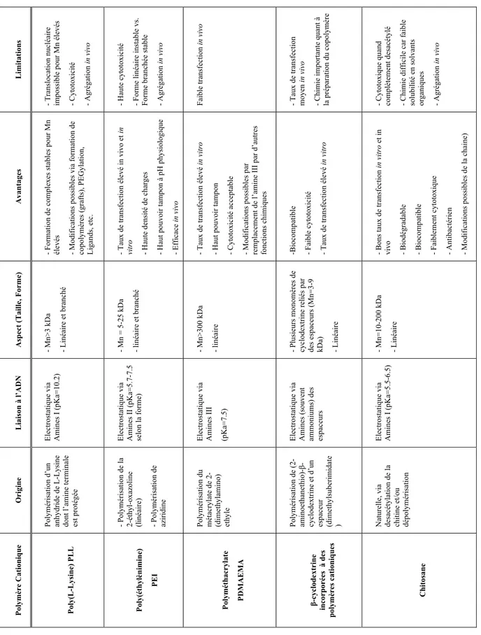
Figure 2.1 : Exemples de polymères cationiques utilisés pour la livraison d’ADN médicament ; a) Poly(L-Lysine) (PLL), b) Poly(éthylènimine) branché (bPEI), c) poly(2-dimethylaminoethyl methacrylate (PDMAEMA), d) Cyclodextrine avec son espaceur cationique diméthylsuberimidate, e) Chitosane (CS). ... 8 Figure 2.2 : Représentation chimique du Chitosane, mettant en scène ses deux unités caractéristiques (2-amino-2-deoxy-D-glucopyranose et 2-acetamido-2-deoxy-D-glucopyranose reliées par une liaison glycosidique de type β(1®4). ... 11 Figure 2.3 : Réaction de dépolymérisation du Chitosane par l’acide nitreux (HONO). La liaison glycosidique y est coupée par l’acide nitreux, libérant ainsi 2 chaines plus courtes, dont l’une porte une extrémité réductrice 2,5-anhydro-D-mannose (GlcN = Glucosamine; GlcNAc = N-acétylglucosamine; Ac = Acétyle). ... 13 Figure 2.4 : Analyse RMN 1H d’un chitosane à 92% de DDA, le pic HOD n’apparait pas car il a été présaturé (= irradié) pour obtenir une meilleure définition des pics. Le chitosane a été préalablement dissout dans un mélange D2O/DCl (20 :1), avant d’être analysé à 70°C afin de
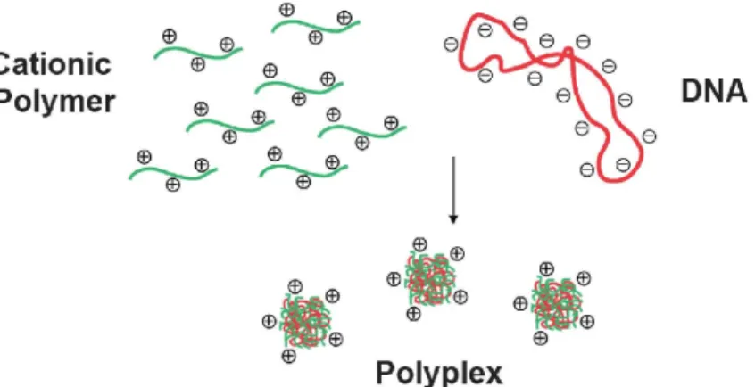
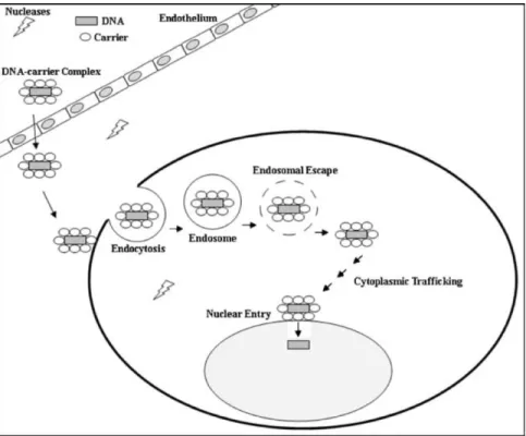
décaler le pic H1D de celui du HOD. 64 scans ont été effectués avec un temps de relaxation fixé à 6s et un temps d’acquisition de 2s, temps nécessaire à la relaxation complète des spins des protons étudiés. ... 14 Figure 2.5 : Représentation schématique des principaux protons du chitosane situés sur les unités acétylées (A unit) et désacétylées (D unit). Le mannose terminal formé suite à la réaction de dépolymérisation à l’acide nitreux porte le nom de M-Unit. ... 15 Figure 2.6 : Représentation schématique de la formation de polyplexes par complexation entre un polymère cationique (CS) et un ADN plasmidique (pDNA) via interactions électrostatiques. Figure inspirée de Kohman et al. (Kohman, 2009). ... 17 Figure 2.7 : Représentation schématique des contraintes rencontrées lors de la livraison d’acides nucléiques in vivo : Au niveau du plasma sanguin (Opsonines, enzymes de dégradation, concentration en sels), de la traversée des tissus biologiques et de la membrane cellulaire, de la sortie de l’endosome, du trafic intracellulaire, de la dissociation des complexes et pour les
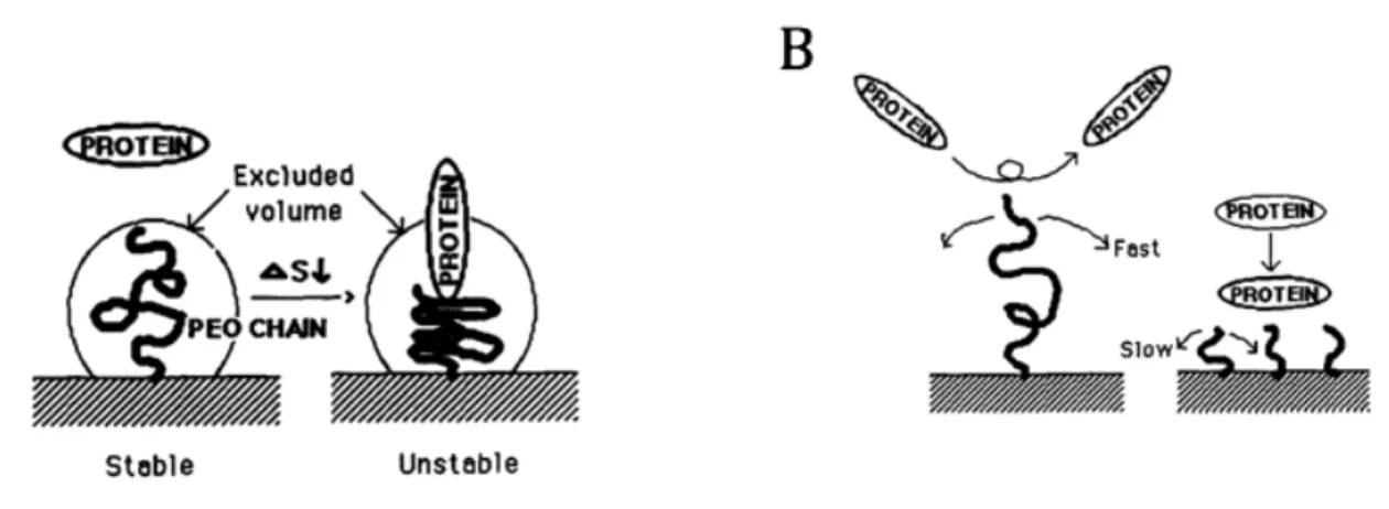
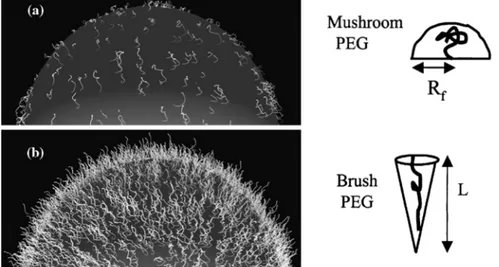
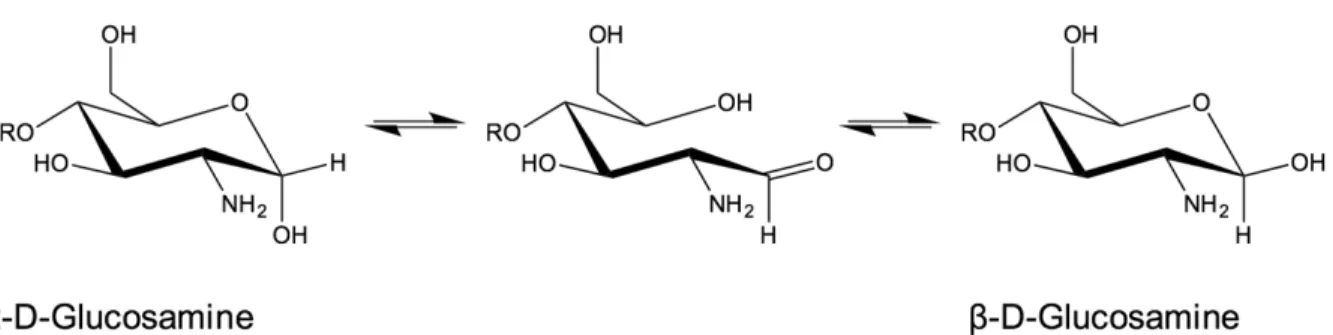
pDNAs seulement, l’entrée du matériel génétique dans le noyau. Figure issue de Al Dosari et al. (Al-Dosari & Gao, 2009). ... 21 Figure 2.8 : Représentation schématique des différentes voies d’internalisation cellulaires des nanoparticules (NPs). Les chemins d’entrée varient principalement en fonction de la taille des particules, de leur charge de surface ainsi que du type de cellule transfectée (CNT= carbon nanotube; MSN= mesoporoussilica nanoparticle; SPION= superparamagnetic iron oxide nanoparticle). Figure tirée de Kunzmann et al. (Kunzmann et al., 2011). ... 25 Figure 2.9 : Représentation schématique du principe d’éponge à protons. L’on y voit l’entrée des ions H+ et Cl-, provoquant une rupture osmolytique de la membrane de l’endosome. Figure tirée de Nguyen et al. (Nguyen, J. & Szoka, 2012). ... 26 Figure 2.10 : Schématisation des mécanismes de résistances du PEG face aux phénomènes d’adhésion protéiques. A) Effet de stabilité stérique expliqué par une diminution de l’entropie ; B) Mobilité de la chaine de PEG en fonction de sa taille. Figure inspirée de Lee et al. (Lee, J. H. et al., 1995). ... 32 Figure 2.11 : Représentation schématique illustrant l’influence de la densité de PEG sur l’adsorption de protéines plasmatiques sur la surface de nanoparticules d’or et sur la séquestration de ces mêmes particules par les macrophages. Figure tirée de Walkey et al. (Walkey & Chan, 2012). ... 33 Figure 2.12. Configurations du PEG en fonction de sa densité sur une surface. (a) Le régime « champignon » où le PEG est considéré comme une demi-sphère de rayon Rf (Rayon de
Flory) posée sur la surface. (b) Le régime « brosse » où le PEG est considéré comme un cône de longueur d’extension L. Figure adaptée de Harris et al. (Harris, J. M. & Chess, 2003). .. 35 Figure 2.13 : Représentation schématique des 2 stratégies de PEGylation recensées dans la littérature ; La stratégie 1 représente la PEGylation des polyplexes chitosane-acide nucléique (CS-AN) après leur formation, alors que la stratégie 2 fait intervenir des copolymères en greffon (CS-g-PEG) ou en bloc (CS-b-PEG) lors de la formation de polyplexes PEGylés avec l’acide nucléique (AN). ... 37
Figure 2.14. Représentation schématique des sites possibles de greffage de PEG sur la chaine latérale du CS, formant des copolymères à greffon, CS-g-PEG. Notons que la position C6 des unités acétylées peut être également occupée par le PEG. ... 38 Figure 2.15 : Mécanisme proposé de dépolymérisation du chitosane par l’acide nitreux (HONO) menant à la coupure de la chaine de chitosane initiale et à la formation d’une unité terminale 2,5-anhydro-D-mannose (M-Unit) sur l’un des deux brins formés (GlcN = glucosamine ; GlcNAc = N-acétylglucosamine ; Ac = acétyle). ... 41 Figure 2.16 : Représentation schématique de la formation d’une base de Schiff par condensation de l’aldéhyde porté par le mannose terminal d’un brin de chitosane (CS) et d’une fonction amine déprotonnée d’un second brin de CS (GlcN = glucosamine ; GlcNAc = N-acétylglucosamine ; Ac = acétyle). ... 42 Figure 2.17 : Représentation schématique de l’équilibre de mutarotation du glucosamine entre ses anomères a et b, passant par la forme ouverte qui présente une fonction aldéhyde. Par souci de clarté, uniquement la forme pyranose du glucosamine est représentée. ... 44 Figure 2.18 : Représentation schématique de la structure attendue de “Block Ionomer Complex” via la complexation entre bloc copolymère CS-b-PEG et acides nucléiques sous un aspect micellaire (Coeur hydrophobe couronné de PEG, appelé ici IPEC pour « Interpolyelectrolyte complex »). Figure adaptée de Pergushov et al. 2012 (Pergushov et al., 2012). ... 47 Figure 3.1 : Représentation schématique du développement du procédé de thioacétylation en bout de chaine du CS (Pickenhahn et al., 2015). Le premier objectif de cette étude est de déterminer la disponibilité de la forme réactive de l’aldéhyde terminal du CS, porté par le 2,5-anhydro-D-mannose (M-Unit) (1), ce dernier étant en équilibre avec sa forme hydratée et non-réactive (2). Bien que dans l’eau, cet équilibre semblerait être déplacé vers la forme hydratée de l’aldéhyde, dite gem-diol, une chimie de conjugaison efficace sur cet aldéhyde serait toutefois possible par catalyse acide en conditions aqueuses. Le second objectif de ce projet est l’étude de la réactivité de l’aldéhyde terminal du CS envers les thiols. Le mécanisme proposé ferait intervenir une première attaque de thiol (ex : b-mercaptoethanol et acide 3-mercaptopropionique, BME and MPA respectivement) sur l’aldéhyde de la M-Unit formant l’intermédiaire hémithioacétal (3) au travers d’un équilibre pH-dépendent. Par analogie avec
la formation d’imines dans laquelle l’équilibre est déplacé lorsqu’on retire l’eau, l’hémithioacétal serait lui stabilisé en thioacétal par lyophilisation (4). ... 51 Figure 3.2 : Représentation schématique du développement de l’activation régiosélective de l’extrémité terminale du CS par thioacétylation par un linker trivalent (1) nommé Triskelion (Pickenhahn et al., 2016). Ce dernier comprend un crochet-thiol (encadré) et une extrémité thiol située au bout d’un espaceur. La dérivatisation de l’aldéhyde terminal porté par la M-Unit par le Triskelion devrait principalement former le thioacetal (2) en promouvant l’attaque du crochet-thiol via un mécanisme de thioacétylation intramoléculaire. Ce procédé serait plus efficace que la stratégie intermoléculaire évoquée dans le Chapitre 4, les deux attaques successives de thiols nécessaires à la stabilisation en thioacétal se faisant alors simultanément. Une fois le CS activé par le Triskelion (formation du CS-b-Triskelion), son extrémité thiol restante serait capable de réagir librement par déplacement de pont disulfure ou encore par addition de Michael (3). ... 53 Figure 4.1 : Production of 2,5-anhydro-D-mannose unit (M-Unit) at the reducing end of chitosan by depolymerization in nitrous acid (HONO): chitosan depolymerization with nitrous acid (HONO) is a rapid, well-understood, and easily controlled method for producing chitosan harbouring a 2,5-anhydro-D-mannose unit (M-Unit) at the reducing end of the cleaved polymer (Allan & Peyron, 1995a). A free aldehyde group (electrophile) is then potentially accessible (1) for reaction with nucleophilic moieties (e.g., CS amine groups, thiols, oxyamines, etc.). Tømmeraas et al. (Tømmeraas, K et al., 2001) demonstrated that the M-Unit aldehyde also exists in its gem-diol hydrated form (2). The neutralization of CS and subsequent freeze-drying of the depolymerization medium induces a Schiff base formation between CS neutralized amines that react with the CS M-Unit aldehyde (3). The rehydration of the imino-adducts in acidic aqueous conditions cleaves the imino linkage between CS chains, transforming the M-Unit into hydroxymethylfurfural (HMF) (4). ... 58 Figure 4.2 : Schematic representation of the equilibria involved in thiol-carbonyl additions. ... 59 Figure 4.3 : Thioacetal conjugation to the chitosan M-Unit formed post-HONO depolymerization: the first objective of this study was to assess the availability of the reactive form of the unhydrated M-Unit aldehyde (2). Although there could be a strong displacement of the equilibrium towards the unreactive aldehyde-hydrated or gem-diol form in water, we
hypothesized that efficient nucleophilic conjugation to the M-Unit was possible in acidic aqueous conditions. The second objective was to assess the M-Unit reactivity towards thiol moieties in aqueous conditions. The proposed reaction pathways between CS end-groups and thiols include the M-Unit CS aldehyde reacting directly with a thiol-bearing model molecule (b-mercaptoethanol and 3-mercaptopropionic acid, BME and MPA respectively) to form a hemithioacetal intermediate (3) through a pH-dependent equilibrium. By analogy with Schiff base formation where the equilibrium displacement occurs by water removal, the hemithioacetal can be stabilized into the corresponding thioacetal (4) by freeze-drying. ... 61 Figure 4.4 : Experimental Design Flowchart. A) Mechanistic studies. Glucosamine (GlcNH2) was
treated with nitrous acid to form the 2,5-anhydro-D-mannose (M-Unit) that was reacted with 2 thiol-bearing molecules (β-mercaptoethanol and 3-mercaptopropionic acid, BME and MPA, respectively). The reaction products were treated using one of 3 methods, i.e. Method I: Direct LC-MS analyses to determine to which extent thioacetal formation occurs in situ; Method II: Freeze-drying (FD) + LC-MS analyses to assess the effect of FD on the thioacetal proportion and to ascertain that no by-products appear post FD; Method III: Acetate buffer pH 4 + FD + LC-MS analyses to determine the effect of an increase in pH prior to FD (this pH increase was included here to prevent any CS acid hydrolysis that could occur when Method II, i.e. FD at pH 1, would be transposed to the polymer). B) Chitosan M-Unit reactivity. CS 92-200 was depolymerized with nitrous acid to produce CS 92-2 HCl salt bearing the M-Unit at the cleaved end of the polymer. M-Unit CS 92-2 HCl salt were reacted with MPA and BME and the reaction products treated with one of 3 workups: Workup I: Dialysis vs. HCl 1mM solution + FD to remove all thiol model excess and to determine the in situ thioacetal formation rate; Workup II: FD + Dialysis vs. HCl 1mM solution + FD to determine the effect of FD on the functionalization rate; Workup III: Acetate buffer pH 4 + FD + Dialysis vs. HCl 1mM solution + FD to determine the effect of an increase in pH prior to FD on the functionalization rate (this pH increase was included to prevent any CS acid hydrolysis that could occur during FD at pH 1 in Workup II). The degree of functionalization of the CS conjugates was determined by 1H NMR, whereas covalent conjugation was assessed by DOSY NMR experiments and Ellman assays in order to rule out the possibility of a simple physical mixture of reagents. 63
Figure 4.5 : Structure of BME (top) and MPA (bottom) chitosan adducts. The protons corresponding to the 1H NMR peaks used for the calculations of the functionalization degree in Eqn (2) and Eqn (3) are highlighted. ... 71 Figure 4.6 : Schematic representation of potential reactions occurring during conjugation of
2,5-anhydro-D-mannose (M-Unit) and 2 thiol-bearing models (3-mercaptopropionic acid and b-mercaptoethanol, MPA and BME respectively) giving the following expected products: product A is the hemithioacetal intermediate that is in equilibrium with its corresponding oxonium, whereas products B and C correspond to the oxathiolane (for BME reactions only) and thioacetal, respectively. Molecule D represents the a,b-unsaturated sulfide. The results of this study suggest that the thioacetal C corresponds to the only stable form observed after freeze-drying. ... 77 Figure 4.7 : Thiol addition to the aldehyde group of the M-Unit CS HCl salt under acidic aqueous conditions: despite the fact that the aldehyde is only present in trace amounts within the reaction medium, the pH- dependent hemithioacetal intermediate formation equilibrium can be displaced by the intermediate stabilization into the corresponding thioacetal at low thiol concentration. ... 83 Figure 4.8 : 1H NMR spectrum of the CS-b-PEG2 block-copolymer after workup II (D2O, T =70°C,
HOD peak was presaturated, number of scans (ns) = 64, relaxation period (d1) = 6 s, acquisition time = 2 s, exponential apodization = 1 Hz). Integration of gem-diol proton peak was used to calculate the functionalization degree (in this particular case, F = 51% according to Eqn (5)). ... 86 Figure 4.9 : Environmental scanning electron microscopy (ESEM) pictures (high vacuum mode, accelerating voltage = 20.0 kV; spot size = 3 and working distance = 5 mm) of polyplexes formed with pDNA and unmodified CS or CS-b-PEG2 block-copolymer (amine to phosphate
ratio = 3.7, N/P = 3.7). (A and B) (x80 000 and x160 000, respectively): polyplexes formed with CS 92-10 are heterogeneous in size and present various morphologies (globular, rod-like and toroidal). Pictures C and D (x80 000 and x160 000, respectively): polyplexes formed with CS-b-PEG2 (CS 92-10 and mPEG-SH 2 kDa), are uniformly spherical. ... 87
Figure 4.10 : Summary of mechanisms elucidated in this study for thiol-based end-group derivatization of chitosans: CS nitrous acid depolymerization induces the formation of
M-Unit that carries an aldehyde moiety at the end of the cleaved polymer (1). The equilibrium between the M-Unit aldehyde and its hydrated form (gem-diol) is strongly displaced towards the latter (2). If the CS depolymerization medium is freeze-dried at pH well below the CS pKa (i.e. pH ~3-4 or below), all the CS amines are protonated and are therefore unable to react with any aldehyde group, maintaining the CS M-Unit integrity at the end of the cleaved polymer (3). Nevertheless, the equilibrium between the M-Unit aldehyde and the corresponding gem-diol is still displaced towards the hydrated form (4). Despite the undetectable aldehyde moieties, thiol molecules and the M-Unit CS aldehyde are engaged in a pH-dependent equilibrium with the corresponding hemithioacetal intermediate (5). The stabilization of the latter into its thioacetal form (6) occurs either by increasing the amount of thiol-bearing reactants in the medium (in situ stabilization), or by freeze-drying the reaction medium when low amounts of thiol are engaged. ... 89 Figure 5.1 : Schematic representation of the CS end-group thioacetylation processes developed by our group: A) Summary of the previously described chitosan end-group thioacetylation process (Pickenhahn et al., 2015). Briefly, CS nitrous acid (HONO) depolymerization induces the formation of 2,5-anhydro-D-mannose (M-Unit) that carries an aldehyde moiety at the end of the cleaved polymer. Despite the fact that this aldehyde is present almost completely in its hydrated and non-reactive form (gem-diol), thiolated macromolecules react with it in strong acidic conditions (pH 1) through a pH dependent equilibrium, forming the corresponding hemithioacetal intermediate (structure 1). The stabilization of the latter into its thioacetal form (structure 2) occurs with a second thiolated macromolecule attack on the same location, an attack that may be sterically hindered by the presence of the first macromolecule. B) The novel HONO-depolymerized CS end-group thioacetylation process. The trivalent linker (structure 3), referred to as Triskelion, comprises a thiol-hook (framed) and a thiol-tail. The derivatization of CS end-groups with Triskelion should predominantly lead to the formation of structure 4 by promoting the thiol-hook attack on the CS aldehyde through a facilitated and more efficient intramolecular thioacetylation process vs. the previously described intermolecular strategy (Pickenhahn et al., 2015). The terminally-activated CS (i.e. the CS-b-Triskelion adduct) can freely react with any thiol-reactive species or structure through its remaining thiol-tail, as represented in structure 5. ... 97
Figure 5.2 : Schematic representation of the Triskelion synthesis pathway. The triol starting material, namely 1,2,6-hexanetriol (1), was activated with methanesulfonyl chloride (MsCl) (2) in an anhydrous 2:1 mixture of dichloromethane (DCM) and tetrahydrofurane (THF) (a). Mesylate leaving groups (-OMs) were displaced by potassium thioacetate (AcSK) in anhydrous dimethylformamide (DMF) (b) to give the acetyl-protected Triskelion linker (3) as major product. The cleavage of acetyl groups was performed using sodium methoxide (MeONa) (c) in order to obtain the desired Triskelion linker (4) in high yields (66% over 3 steps). Compounds (5) and (6) correspond to the elimination products obtained after mesylate group displacement. The detailed synthesis protocols are available in Supp. Info.; Annexe C – Section 1. Summary of conditions: (a) MsCl in anhydrous [2:1] DCM/THF at room temperature for 3h; (b) AcSK in anhydrous DMF at 0-5°C overnight; (c) MeONa in MeOH at room temperature for 5-10 min. ... 100 Figure 5.3 : Experimental design flowchart. (A) Mechanistic studies. M-Unit
(2,5-anhydro-D-mannose) obtained by HONO treatment of glucosamine, was reacted with molecules bearing a thiol-hook (Ethanedithiol -EDT- and Triskelion linker). The products of these reactions were treated using either Method I: Direct LC-MS of the reaction mixture to determine to which extent intramolecular thioacetal formation occurs in situ or Method II: Concentration prior to perform LC-MS analysis to determine the influence of such dehydration/concentration step on the final products proportion and also to detect some by-products formation upon concentration). (B) M-Unit CS HCl salt reactivity. M-Unit CS HCl salt was also reacted with Ethanedithiol -EDT- and Triskelion linkers. After reaction completion, reaction media were treated using either: Workup I: Unreacted thiol-hook molecules removal by reprecipitation + NaOH treatment + reprecipitation in order to determine the in situ conjugation process efficiency or Workup II: Concentration by FD or rota-evaporation + Workup I in order to quantify a potential increase in conjugation efficiency upon concentration. Conjugation process efficiencies were determined by 1H NMR using both Equations 1 & 2 as detailed in the section below. ... 101 Figure 5.4 : Structure of unmodified CS (top) and CS-b-Triskelion adduct (bottom). The protons corresponding to the 1H NMR peaks used for the calculations of the functionalization degree in Equations 1 and 2 are shown in green, blue and red. ... 105
Figure 5.5 : Schematic representation of thiol-activated magnetic beads surface modification. CS-b-Triskelion was attached to thiol-activated magnetic microparticles through pyridyl disulfide bond displacement with the release of the pyridinethione leaving group. ... 106 Figure 5.6 : Stacked 1H NMR spectra (400 MHz, CDCl3, ns = 32 scans, acquisition time = 2s, d1
= 2s) of the trivalent Triskelion linker 3-step synthesis. Step 1 (conversion of OH into Tri-OMs) was obtained with 93% yield, Step 2 (conversion of Tri-OMs into Tri-SAc) led to the desired tri-thioacetate with 75% yield and Step 3 (conversion of Tri-SAc into Tri-SH) gave the deprotected Triskelion linker with 95% yield. The products of the synthesis were also characterized by 13C NMR spectroscopy and Mass spectrometry (Supp. Info; Annexe C – Section 1). ... 109 Figure 5.7 : Schematic representation of potential Triskelion-derivatized 2,5-anhydro-D-mannose (M-Unit) products. Since the Triskelion linker carries three reactive thiol moieties able to engage into the thioacetylation process (“thiol-hook” vs. “thiol-tail”), there are three potential observable structures: Structure A which corresponds to the expected intramolecular thioacetal obtained from thiol-hook reaction with the M-Unit aldehyde; Structure B represents the hemithioacetal intermediate that is formed upon reaction of the thiol-tail with the M-Unit aldehyde. This structure is unstable and it can be stabilized by a second Triskelion attack to form the structure C, the intermolecular thioacetal. All the potential disulfide related products such as A-ss-A, B and C with a disulfide closed hook were considered within the calculations without being represented in this figure. The LC-MS results indicate that the intramolecular thioacetal (structure A) corresponds to the major derivatization product observed by LC-MS when MeOH is used as co-solvent, whereas the intermolecular thioacetal (structure C) is favored when THF is used as co-solvent. ... 110 Figure 5.8 : Relative proportion of the M-Unit / Triskelion conjugation products observed by
LC-MS (See Figure 5.7 and Tableau 5.1). Products A, B and C represent the intramolecular thioacetal, the hemithioacetal intermediate and the intermolecular or linear thioacetal, respectively. Each conjugation reaction (10-20 mM aldehyde, pH 1, T = 50°C, t = 72h, 30% THF or 90% MeOH) was performed in triplicates (N = 3) and the reaction media were treated as follows: Method I refers to a direct LC-MS analysis of the reaction medium whereas Method II stands for a concentration to dryness step prior to performing LC-MS. All the
potential disulfide related products such as A-ss-A, B and C with a disulfide closed hook were considered within the calculations (Tableau 5.1). The results of this semi-quantitative study suggest that the intramolecular thioacetylation (A) is the favored mechanism of M-Unit / Triskelion conjugation when MeOH is used as co-solvent. On the other hand, the intermolecular thioacetal (C) is predominantly formed when THF is used as co-solvent, possibly because of some thiol-hook deactivation occurring due to the presence of peroxides within THF. ... 112 Figure 5.9 : Conjugation efficiency of the M-Unit CS HCl salt reacted with 20 equivalents of molecules bearing a thiol-hook (EDT and the Triskelion linker) per CS end-group for 24-72h at pH 1 and 50°C. 30% isopropanol (iPrOH) and 90% methanol (MeOH) were used as co-solvent for EDT and Triskelion linker solubilization, respectively. The nomenclature used in the graphical representation herein is: “Solvent pH-duration” (i.e., iPrOH 1-72 stands for 30% isopropanol at pH 1 for 72h). Reaction media were treated according to Workup I (alkali treatment + precipitation in THF) and Workup II (concentration to dryness + alkali treatment + precipitation in THF). Functionalization degrees (F) were calculated using Equation 2 (decrease in Gem-diol peak intensity) and confirmed with Equation 1 considering 1 molecule bearing a thiol-hook per purified CS adduct. Functionalization degrees calculated with Equation 1 are not shown here since they were with 2% of those obtained with Equation 2. (* corresponds to the results of the conjugations obtained with 5 equivalents of thiol-hook per M-Unit aldehyde instead of 20). ... 113 Figure 5.10 : 1H NMR, 400 MHz, D2O, T=25°C, ns=64 scans, acquisition time=2s, d1=10s. The
sample herein corresponds to the CS-b-Triskelion conjugate obtained after 72h reaction in 90% MeOH (MeOH 1-72) and processed as per Workup II. Functionalization degrees F of 91% and 92% were obtained using Equations 1 and 2, respectively. ... 115 Figure 5.11 : Proposed mechanism of the Triskelion linker conjugation to M-Unit CS under acidic aqueous conditions. Although the rate limiting step of this conjugation process is the transformation of the gem-diol into its corresponding active aldehyde, the latter is engaged in an equilibrium with the hemithioacetal intermediate (in brackets) upon thiol-hook addition (Triskelion linker shown herein). As suggested by LCMS studies, the hemithioacetal intermediate is readily transformed into its stable thioacetal counterpart with the release of a
water molecule. The equilibrium between the M-Unit aldehyde and the hemithioacetal can be displaced towards the latter by increasing both the amount of thiol-hook engaged in the reaction and the reaction duration. ... 116 Figure 5.12 : DOSY spectrum of the CS-b-Triskelion conjugate (MeOH 1-72, Workup II). 16 gradients between 2.5 and 50.0 gauss.cm-1 with a gradient pulse (δ) of 3 ms, a diffusion time (Δ) of 150 ms. Both CS and Triskelion have the same translational diffusion coefficient at 25°C in D2O, indicating that they are joined together by a covalent bond. ... 116
Figure 5.13 : 1H NMR spectra (400 MHz, D2O, T=25°C, ns=256 scans, acquisition time=2s,
d1=10s) representing A) the initial CS + CS-b-Triskelion mixture that was engaged with the thiol-activated magnetic beads and B) the released CS-b-Triskelion post treatment. Both spectra were normalized using the unmodified CS acetyl peak (d = 2.06 ppm, 5.59 H) for comparison purpose. Both equations 1&2 were used to calculate functionalization degree of CS mixture and released CS calculations, giving 70% (± 2) and 95% (± 2), respectively. This result indicates that only CS-b-Triskelion adducts bind to the thiol-activated beads through disulfide linkage displacement. ... 119 Figure 6.1 : Représentation schématique de l’addition de thiol sur l’aldéhyde terminal du CS porté par la M-Unit en conditions aqueuses. Ainsi, en dépit du fait que l’aldéhyde soit principalement présent sous sa forme hydratée et non-réactive, l’équilibre de formation de l’intermédiaire hémithioacétal peut être déplacé par stabilisation de ce dernier lors d’une seconde attaque de thiol, formant le thioacetal correspondant. ... 124 Figure 6.2 : Résumé graphique du premier article dédié à ce projet de Doctorat (Pickenhahn et al., 2015). A) Représentation schématique de la PEGylation en bout de chaine du CS par thioacétylation: Bien que l’aldéhyde porté par la M-Unit soit majoritairement présent sous sa forme hydratée et non-réactive, une première attaque de PEG-thiol (PEG-SH) sur l’aldéhyde de la M-Unit forme un intermédiaire hémithioacétal instable. Une seconde attaque de PEG-thiol est alors nécessaire afin de stabiliser cette structure, formant le bloc-copolymère CS-b-PEG2 correspondant par thioacetylation. B) Formation de BICs : Les copolymères CS-b-PEG2
ont ensuite été engagés avec de l’ADN plasmidique pour former des structures polyélectrolytes ayant un caractère micellaire, les BICs. ... 126
Figure 6.3 : Représentation schématique de l’activation de l’extrémité du CS par thioacétylation régiosélective. Dans une première étape, l’aldéhyde terminal porté par la M-Unit a été engagé avec un linker trivalent, le Triskelion, via un mécanisme de thioacétylation intramoléculaire. Les conjugués obtenus pourront alors être utilisés pour attacher l’extrémité du CS sur des surfaces, des polymères et des ligands portant des fonctions réactives aux thiols et ce en utilisant des chimies moins agressives. ... 129 Figure 7.1 : Modifications du Triskelion proposées afin d’améliorer le procédé d’activation terminale du CS. La protection sélective de l’extrémité thiol du Triskelion (1), celle qui n’est pas destinée à être engagée avec le CS, permettrait de favoriser l’attaque du crochet-thiol sur l’aldéhyde du mannose terminal. Une fois le CS activé par le Triskelion, cette extrémité serait déprotégée pour permettre d’autres conjugaisons. La flexibilité de l’espaceur étant primordiale quant au succès des futures réactions, le fait d’augmenter sa longueur (2) permettrait d’en accroitre l’efficacité. Enfin, le fait de conjuguer régiosélectivement un PEG de petite taille au bout du Triskelion permettrait de d’effectuer l’activation du CS en conditions aqueuses uniquement. ... 133 Figure 7.2 : Représentation schématique de la fonctionnalisation couche par couche de particules cationiques préformées. Des particules chargées positivement (1) seraient recouvertes via interactions électrostatiques par de l’AN (siRNA et/ou mRNA et/ou pDNA, etc.) (2). Les particules ainsi obtenues pourront alors être protégées dans la circulation sanguine par l’ajout d’une couche de copolymère sur leur surface, toujours par interactions électrostatiques (3). ... 134 Figure 7.3 : 1H NMR spectrum of the CS 92-1 depolymerization medium (D2O/DCl (50 mM),
T=70°C, HOD peak was presaturated, number of scans (ns) = 2000, relaxation period (d1) = 6s, Acquisition time=2s, Exponential apodization = 1 Hz). No aldehyde proton peak was observed around 8-10 ppm, the hydrated gem-diol form remaining predominant. ... 157 Figure 7.4 : 1H NMR spectrum of the CS-BME product after workup II (D2O/DCl, T=70°C, HOD
peak was presaturated, number of scans (ns) = 64, relaxation period (d1) = 6s, Acquisition time=2s, Exponential apodization = 1 Hz). Integration of BME (-CH2-S-) protons peaks was
used to calculate the functionalization degree (F=70% in this particular case, according to Eqn (2)). ... 158
Figure 7.5 : 1H NMR spectrum of the CS-MPA product after workup II (D2O/DCl, T=70°C, HOD
peak was presaturated, number of scans (ns) = 64, relaxation period (d1) = 6s, Acquisition time=2s, Exponential apodization = 1 Hz). Integration of MPA protons peaks was used to calculate the functionalization degree (F=52% in this particular case, according to Eqn (3)). ... 159 Figure 7.6 : 13C solid state NMR (CP-MAS) of an extra-dried CS 99-1 salt. The sample preparation as well as the analysis was performed under inert atmosphere (N2) to avoid contact with water
content in air. (T=25°C, t=6h, v=12kHz). ... 160 Figure 7.7 : DOSY spectrum of the CS HCl salt M-Unit conjugated to MPA. 32 gradients between 11.2 and 358.4 gauss.cm-1 with a gradient pulse (δ) of 1 ms, a diffusion time (Δ) of 60 ms.
Both CS and MPA have the same translational diffusion coefficient at 25°C in 2% DCl in D2O. ... 161
Figure 7.8 : 1H NMR, 400 MHz, CDCl3, T=25°C, ns=32 scans, acquisition time=2s, d1=2s. ... 163
Figure 7.9 : 1H NMR, 400 MHz, CDCl3, T=25°C, ns=32 scans, acquisition time=2s, d1=2s. ... 165
Figure 7.10 : 1H NMR, 400 MHz, CDCl3, T=25°C, ns=32 scans, acquisition time=2s, d1=2s. . 167
Figure 7.11 : Stacked 1H NMR spectra representing the Triskelion linker decomposition which occurs upon longer sodium methoxide deprotection duration (t = 10 min vs. t = 20 min). . 167 Figure 7.12 : 1H NMR, 400 MHz, D2O, T=25°C, ns=32 scans, acquisition time=2s, d1=6s. ... 169
Figure 7.13 : 1H NMR, 400 MHz, D2O, T=70°C, ns=64 scans, acquisition time=2s, d1=10s.
Functionalization degree were calculated with Equations 1 and 2, giving F = 67%. ... 174 Figure 7.14 : Schematic representation of the Thiol-activated surface modification with
CS-b-Triskelion 3-steps procedure. Step 1 refers to Thiol-activated Magbeads preparation, whereas Steps 2 and 3 refer to CS-b-Triskelion reduction and its conjugation onto beads surface, respectively. ... 177
LISTE DES SIGLES ET ABRÉVIATIONS
[M+H]+ Proton adduct (MS)
[M+Na]+ Sodium adduct (MS)
°C Degré Celsius
1H NMR Résonance magnétique nucléaire du proton
A Unit Unité acétylée du chitosane
A2 Second virial coefficient
Ac Acétyle
AN Acide nucléique
ADN Acide désoxyribonucléique Amine II Amine secondaire
ARN Acide ribonucléique
AUC Ultracentrifugation analytique BAM N-tert-butylacrylamide
BIC Bloc ionomer complex
BME Beta-Mercaptoethanol
CMC Concentration micellaire critique
CNTs Carbon nanotubes
COSY correlation spectroscopy (NMR)
CP-MAS Cross Polarization - Magic Angle Spinning (NMR) CS-AN Polyplexe de chitosane - acide nucléique
CS-b-PEG Copolymère en bloc CS-PEG
CS-b-PEG2 Copolymère en bloc CS-PEG portant 2 PEGs par CS
CS-g-(PEI-b-PEG) Copolymère en bloc PEI-PEG greffé latéralement sur le CS CS-g-PEG Copolymère à greffon CS-PEG
CS-pDNA Polyplexe de chitosane-ADN plasmidique D Unit Unité désacétylée du chitosane
DCM Dichloromethane
DDA Degré de désacétylation ddH2O Double deionized H2O
DLS Diffusion de lumière dynamique
DLVO Théorie de Derjaguin, Landau, Verwey, Overbeek
DMF Dimethylformamide
DMSO Dimethylsulfoxide
DNAses Enzymes de dégradation d'ADN DOSY Diffusion ordered spectroscopy DTNB 5,5’-dithiobis-(2-nitrobenzoic acid)
E2 Élimination d'ordre 2
EDC 1-éthyl-3-(3-diméthylaminopropyl)carbodiimide
EDT Ethanedithiol
ESEM Environmental Scanning Electron Microscope
F Functionalization degree
FD Freeze-dry / Freeze-drying
FDA Food and Drug Administration (USA)
GlcN D-glucosamine
GlcNAc N-acétylglucosamine
H1D Proton H1 de l'unité désacétylée du chitosane
HMBC Heteronuclear multiple-bond correlation spectroscopy (NMR)
HMF Hydroxymethylfurfural
HONO Acide nitreux / Nitrous acid
IgG Immunoglobuline G
IgM Immunoglobuline M
IPEC Interpolyelectrolyte complex iPrOH 2-propanol / isopropanol
IR Infrarouge
kDa KiloDalton
L Longueur d'extension du PEG en mode brosse LC-MS Liquid chromatography - Mass spectrometry LC-MS/MS Liquid chromatography-tandem mass spectrometry LC-TOF Liquid chromatography - time-of-flight (MS)
M-Unit 2,5-anhydro-D-mannose
MAL Maléimide
MALS Diffusion de lumière statique
MeOH Methanol
MeONa Sodium methoxide
MHz MegaHertz
mm Millimètre
Mn Masse molaire moyenne en nombre
MPA Mercaptopropionic acid
mRNA ARN messager
ms Millisecond
MsCl Methanesulfonyl chloride MSNs Mesoporous silica nanoparticles MTOC Microtubules organizing center
Mw Masse molaire moyenne en masse
MWCO Molecular weight cut-off
N-2-graft Modification sur l'amine du CS N:P int Ratio amine/phosphate interne
N:P prep Ratio amine/phosphate de préparation
NA Nucleic acid
NHS N-hydroxysuccinimide
NHS-PEG-MAL α-malemidyl-ω-N-hydroxysuccinimidyl poly(éthylène glycol)
NIPAM N-isopropylacrylamide
nm nanomètre
NP(s) Nanoparticule(s)
NSERC Natural Sciences and Engineering Research Council of Canada NTA Nanoparticle tracking analysis
O-6-graft Modification sur l'hydroxyle en C6 du CS ODN Oligonucléotide antisens
OMs Mesylate
pD Potentiel deuterium
PDI Indice de dispersité
pDNA ADN plasmidique PEG Poly(éthylène glycol)
PEG-b-PLL Copolymère en bloc PEG et Poly-L-lysine PEG-NHS N-hydroxysuccinimidyl poly(éthylène glycol)
PEI Poly(ethylènimine)
PEI-pDNA Polyplexe de PEI-ADN plasmidique
pH Potentiel hydrogène pKa Constante d'acidité PLL Poly(L-lysine) PlP Plasmatic proteins PLR Poly-L-arginine PPl Protéines plasmatiques
ppm Parts per million
RES Système réticulo-endothélial
Rf Rayon de Flory
rg Rayon de gyration
RH% Relative humudity
RMN Résonance magnétique nucléaire RNAses Enzymes de dégradation d'ARN
ROP Ouverture de cycle
SAc Thioacetate
SEC Chromatographie d'exclusion stérique
SEC-MALLS Size Exclusion Chomatography - Multi-Angle Laser Light Scattering
SN2 Substitution nucléophile d'ordre 2
SPIONs Superparamagnetic iron oxide nanoparticles
T Température
TCEP Tris(2-carboxyethyl)phosphine
TEM Microscope électronique à transmission
THF Tetrahydrofurane
TIL Tumor-infiltrating lymphocyte
TLC Thin layer chromatography TLR Toll-like receptor
Tris 2-amino-2-hydroxymethyl-1,3-propanediol
UV Ultraviolet
Va Potentiel attractif de van der Waals (DLVO)
vdW van der Waals
VPEG Potentiel répulsif du PEG (DLVO) Vr Potentiel répulsif électrostatique (DLVO)
w/w Masse/masse
ΔGM Énergie libre de mélange
ΔHM Enthalpie de mélange
ΔSM Entropie de configuration de mélange
LISTE DES ANNEXES
Annexe A- Liste non –exhaustive des différentes voies d’administration pour la livraison de gènes médicaments. ... 156 Annexe B – Article 1 : Informations supplémentaires ... 157 Annexe C - Article 2 : Informations supplémentaires ... 162
CHAPITRE 1
INTRODUCTION
Le séquençage du génome humain a, depuis maintenant quelques décennies, ouvert la porte à de nouvelles stratégies thérapeutiques, telles que la thérapie génique, pour traiter des maladies considérées comme incurables. La thérapie génique est une approche de traitement potentielle pour un vaste éventail de pathologies, qu’elles soient héréditaires ou acquises. Il s’agit d’un procédé fondé sur l'introduction de matériel génétique chez un patient, afin d'insérer un allèle fonctionnel du ou des gènes déficients responsables de sa maladie, ou de modifier l'expression du ou des gènes délétères impliqués dans sa pathologie (Naldini, 2009). La livraison d’acide nucléique repose sur deux composantes principales : la première est que le matériel génétique doit pouvoir être exprimé à l'endroit voulu et la seconde est que le matériel génétique utilise un système de livraison efficace et sûr, jusqu'aux cellules cibles. Ce dernier point correspond au thème principal de ce projet de Doctorat, à savoir le développement d’un système de livraison d’acide nucléique de deuxième génération à base de chitosane.
1.1 Cadre de la recherche
Le contexte de cette thèse sera exposé dans le Chapitre 2 qui correspond à la revue de littérature inhérente à mon projet de Doctorat. Brièvement :
Les acides nucléiques (ANs), macromolécules biologiques portant l’information génétique, sont hydrophiles et portent une charge globale négative ce qui limite leur passage au travers des membranes biologiques. D’autre part, les ANs sont extrêmement sensibles et sont ainsi rapidement dégradés par les endonucléases contenues dans le plasma sanguin (Mintzer & Simanek, 2009). Le principal challenge de ces études est le design de transporteurs spécifiques qui permettront une transfection efficace. Ainsi pour placer le gène-médicament, on utilise des vecteurs capables de pénétrer les cellules et d’y libérer leur AN soit dans le cytoplasme, soit dans le noyau (dépendamment du type de matériel génétique à introduire).
On dénombre deux catégories distinctes de vecteurs : les vecteurs viraux et les vecteurs non-viraux. Les vecteurs viraux sont des virus dont on a inhibé le potentiel infectieux, comme les rétrovirus ou les adénovirus. Ces derniers ont un taux de transfection élevé mais présentent une forte immunogénicité (Verma & Somia, 1997). Les vecteurs non viraux tels que les phospholipides et autres polycations sont à l’inverse faiblement immunogènes, moins onéreux mais ont des taux de