UNIVERSITÉ DE MONTRÉAL
HIGH-YIELD PRODUCTION PROCESS OF INFLUENZA VIRUS-LIKE PARTICLES IN HUMAN CELLS TOWARD LARGE-SCALE VACCINE MANUFACTURING
ALINA VENEREO SÁNCHEZ DÉPARTEMENT DE GÉNIE CHIMIQUE ÉCOLE POLYTECHNIQUE DE MONTRÉAL
THÈSE PRÉSENTÉE EN VUE DE L’OBTENTION DU DIPLÔME DE PHILOSOPHIAE DOCTOR
(GÉNIE CHIMIQUE) AVRIL 2017
UNIVERSITÉ DE MONTRÉAL
ÉCOLE POLYTECHNIQUE DE MONTRÉAL
Cette thèse intitulée :
HIGH-YIELD PRODUCTION PROCESS OF INFLUENZA VIRUS-LIKE PARTICLES IN HUMAN CELLS TOWARD LARGE-SCALE VACCINE MANUFACTURING
présentée par : VENEREO SÁNCHEZ Alina
en vue de l’obtention du diplôme de : Philosophiae Doctor a été dûment acceptée par le jury d’examen constitué de :
M. PERRIER Michel, Ph. D, président
M. HENRY Olivier, Ph. D, membre et directeur de recherche M. KAMEN Amine, Ph. D, membre et codirecteur de recherche M. GILBERT Renald, Ph. D, membre et codirecteur de recherche M. DE CRESCENZO Gregory, Ph. D, membre
DEDICATION
“To my mom! For all the love, strength and dedication” “A mi amada mami! Por su amor incondicional y por creer siempre en mi” “To my sister and Mahdi for all the love and support!”
“Just keep swimming” Dory
ACKNOWLEDGEMENTS
I want to express my profoundest gratitude to my supervisor Amine Kamen for giving me this great opportunity, for trusting me, for believing in people’s talent no matter where they come from. His orientations were always on point even if I could not understand them at the moment, then, it resulted the best for the project. I feel so honored having worked with Amine, his vision, wisdom, endless ideas combined with humbleness make him a great human being. Thanks so much.
I would like to thank my supervisor Olivier Henry for orientating me in my beginnings at École Polytechnique de Montréal, for helping me with the courses and for being always available whenever I needed. I learned from him how to be more practical and objective with my work. His advices were priceless for completing this phase of my life.
It is difficult to express in words how grateful I am of having the opportunity to work with Dr. Renald Gilbert during these last years. Renald is a brilliant scientist and has been an excellent supervisor and also a friend to me. He has been involved and has supported every step of this project with great discussions, ideas, and resources. He has facilitated external collaborations such as the experiment in animals. His positivity and good sense of humor encouraged me and kept me motivated in my most hopeless moments. Thank you!
I would also like to thank Melanie Simoneau, Parminder Chahal, Sven Ansorge, and Aziza Manceur for being my mentors in different steps of my project. To Melanie for the expert aid with the molecular biology constructions and cell line development and Aziza for teaching me the analytical techniques. To Sven for guiding me to implement the process optimization strategies, facilitating the bioreactor operations and mediating the collaboration for the LC-MS/MS. To Parminder for teaching me tangential flow filtration, design of experiments, for revising my manuscripts and more.
I acknowledge the employees at NRC that helped me with my project Viktoria Lytvyn (confocal microscope), Melanie Leclerc (cell maintenance and clone selection), Antoine Caron (stable cell line), Lucie Bourget (flow cytometry analysis), Danièle Jacob (bioreactor operation), Alice Bernier (VLPs purification) and Sabo Hrapovic (Electron microscopy).
I want to specially thank Stephane Lanthier for putting time and explaining me in details how to operate a perfusion bioreactor and Johnny Montes for teaching me how to work with insect cells during my former project. I’d also like to thank Marc-Andre Robert for helping me with the French version of the abstract and for the endless discussions.
Thank you to all Master and Ph.D. students and postdoctoral fellows that have shared this journey with me: Emma Petiot, Christine Marie Thompson, Cuitlahuac Chavez-Peña, Ernest Milian, Laura Cervera, Alexandre Audy, Felix Comtois, Eric Karengera and Kahina Mellahi. I wish them all the best in their future careers.
Thanks to my great friend Erick Perera-Medina for helping me with the math during my first courses at École Polytechnique de Montréal.
Finally, for all those who were involved in my education: professors, schools and friends of all life, thank you.
RÉSUMÉ
Le virus influenza a été la cause d’épidémies et de pandémies parmi les plus anciennes et meurtrières rapportées dans l’histoire de l’humanité. La vaccination est le moyen le plus efficace de prévenir les infections. Par contre, la fabrication actuelle des vaccins contre l’influenza se fait dans les œufs, avec des embryons de poulet, un procédé lent et laborieux qui limite la capacité de répondre efficacement en cas de pandémie ou suite à une forte demande pour la grippe saisonnière. De plus, ces vaccins induisent principalement une réponse humorale contre les antigènes dominants hémagglutinine (HA) et neuraminidase (NA), deux protéines du virus, causant un manque de protection croisée contre certaines des nouvelles souches. Leur efficacité est également réduite chez certains groupes plus vulnérables (par ex. personnes âgées et jeunes enfants). Par conséquent, l’industrie se tourne vers le développement d’une nouvelle gamme de vaccins plus immunogéniques et produits à partir de plateformes plus efficaces. Les particules pseudo-virales (Virus-like particles en anglais, VLPs) constituent une alternative intéressante comme vaccin. Les VLPs présentent une structure qui s’apparente à celle des virus de type sauvage, en permettant la présentation à leur surface des antigènes principaux, dans leur conformation native. De plus, les particules pseudo-virales sont non-infectieuses et incapables de se répliquer. Les cellules de mammifères offrent plusieurs avantages comme plateforme d’expression pour la synthèse de nombreux produits biopharmaceutiques; elles sont capables d’effectuer des modifications post-traductionnelles complexes, de croître à haute densité et de produire des VLPs en suspension et en bioréacteur. Jusqu’à maintenant, les études traitant des VLPs influenza (produites avec des cellules de mammifères) se sont concentrées principalement sur l’assemblage du virion et sur le mécanisme de bourgeonnement cellulaire, alors que seulement un nombre limité d’études porte sur leur production à grande échelle et leur emploi potentiel comme vaccin. Dans le cadre de cette thèse, un bioprocédé transposable à grande échelle pour produire des quantités importantes de VLPs chimériques Gag-influenza à partir de cellules HEK-293 (cellules de reins issues d’un embryon humain) a été développé.
En premier lieu, nous avons généré une lignée cellulaire HEK-293 exprimant de façon stable les protéines HA et NA (souche H1N1 du virus Influenza) sous le contrôle d’un système inductible au cumate. Ensuite, la formation des VLPs chimériques a été induite et dirigée par la transfection de plasmides codant la protéine Gag du virus de l’immunodéficience humaine ou la protéine M1,
une composante de la matrice du virus de l’influenza. La protéine Gag a été fusionnée à la protéine fluorescente verte pour faciliter le suivi de la production des VLPs. Les protéines antigéniques ont été produites 7 fois plus efficacement en présence de la protéine Gag, ce qui indique qu’il s’agit d’une meilleure protéine structurale que M1 dans ce contexte. Par conséquent, la production de VLPs contenant HA-NA et Gag (par transfection) a donc été transférée à l’échelle d’un bioréacteur de 3L avec agitation. Les VLPs ont été recueillies par ultracentrifugation sur un coussin de sucrose, puis concentrées par filtration à flux tangentiel en employant une membrane ayant des pores d’une taille limite de 1000 kDa. Plusieurs techniques ont été employées pour caractériser les VLPs produites: immunodiffusion radiale simple, essai d’hémagglutination, immunobuvardage de type Western et dot blot, ainsi que la microscopie électronique à transmission. Par ailleurs, lors d’essais sur des animaux, l’immunisation intranasale à partir de VLPs a permis d’induire une réponse immunitaire spécifique en plus de conférer une protection totale à toutes les souris soumises à l’épreuve (100% d’efficacité) avec une souche homologue de l’influenza.
Après avoir démontré l’efficacité des VLPs comme vaccin in vivo (démonstration de faisabilité), une nouvelle lignée cellulaire inductible a été développée, exprimant cette fois les trois protéines HA, NA et Gag fusionnée à la GFP. Le but de générer une telle lignée était de simplifier le procédé de production en éliminant l’étape de transfection transitoire, qui peut être laborieuse à conduire à grande échelle. Sans sacrifier la production spécifique, le procédé a été optimisé pour obtenir une plus grande production volumétrique en augmentant notamment la densité cellulaire au moment de l’induction et en employant un mode de perfusion. L’opération a été réalisée à l’aide d’un bioréacteur de 3L et d’une filtration à flux tangentiel subséquente permettant d’augmenter les rendements de VLPs de 60 fois (3x10¹¹ Gag-GFP événements fluorescents/L de
culture mesurés par cytométrie de flux) en comparaison à une production sans perfusion (5x10⁹ Gag-GFP événements fluorescents/L). Le procédé a été caractérisé en déterminant la cinétique de production des protéines d’intérêts présentes dans les VLPs (le procédé en amont) ainsi que le taux de récupération pour chaque opération unitaire de la purification (le procédé en aval). L’opération du bioréacteur, en mode de perfusion et avec la lignée stable 293-HA/NA/Gag-GFP, a permis d’obtenir 5 fois plus de protéines antigéniques HA dans les VLPs que le bioréacteur opéré sans perfusion où la transfection transitoire a été employée. Le nouveau procédé développé
a permis de générer des rendements supérieurs à ceux publiés jusqu’à ce jour pour des VLPs influenza produits à partir de cellules de mammifères.
Finalement, dans le but de répondre à certaines préoccupations de biosécurité associées à un usage potentiel de VLPs comme vaccins (parce que ce sont des particules enveloppées qui bourgeonnent d’une cellule hôte et qui renferment des protéines cellulaires, de l’ADN et de l’ARN), nous avons effectué une caractérisation du protéome des VLPs Gag-influenza produites par transfection transitoire et des vésicules extracellulaires produites par des cellules HEK-293 de type sauvage. Les fonctions des protéines identifiées dans les VLPs et dans les vésicules extracellulaires ont été discutées.
Le procédé développé dans le cadre de cette thèse devrait être efficace pour produire des VLPs exposant les protéines HA et NA issues de différentes souches d’influenza. Les VLPs produites pourraient être évaluées dans le cadre d’essais cliniques dans le but de conduire au développement d’un vaccin efficace et sécuritaire pour remplacer ou complémenter le procédé actuel de production dans les embryons poulets.
ABSTRACT
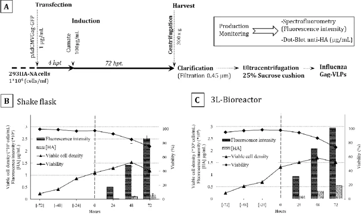
Of the fatal infections noted in human history, influenza epidemics and pandemics are among the most ancient. Vaccination remains the most effective tool to prevent infection. However, the current production of influenza vaccines in embryonated chicken eggs has limited capacity during pandemics or high demand seasons, and is both labor-intensive and time-consuming. Furthermore, the seasonal egg-produced vaccines mainly induce humoral response to the Hemagglutinin (HA)/Neuraminidase (NA) dominant antigens, which leads to a lack of cross-protection against other non-matching novel strains. In addition, the vaccines provide low protection in high risk groups (e.g., elderly and young children). Consequently, the industry is moving toward the development of novel, more immunogenic influenza vaccines as well as more efficient production platforms. Virus-like particles (VLPs) constitute a promising alternative as influenza vaccine. They mimic the particulate structure of wild-type viruses while they are non-infectious, non-replicative particles, and the main antigens repetitively displayed on their surface maintain the native conformation. Mammalian cell culture offers several advantages for the production of biopharmaceuticals such as their ability to perform complex post-translational modifications and the high cell densities and productivities reached in suspension culture bioreactors. Up to now, the production of influenza VLPs from mammalian cells has been mostly addressed to study influenza assembly and budding mechanisms but little attention has been paid to its potential use for large-scale manufacturing of VLPs as influenza vaccine candidate. The aim of this thesis was to develop a scalable process to produce large quantities of chimeric influenza Gag-VLPs from stable human embryonic kidney HEK-293 cells in suspension culture. First, a HEK-293 cell line stably expressing HA and NA proteins of influenza (subtype H1N1) under the regulation of the inducible cumate system was established. Then, the formation of VLPs was mediated by transient transfection of plasmids encoding human immunodeficiency virus (HIV) Gag or M1 influenza matrix protein. Gag protein was fused to the green fluorescent protein (GFP) to facilitate the monitoring of VLPs production. VLP antigenic proteins were produced seven times more efficiently in the presence of Gag, indicating that Gag is a better scaffolding protein than M1 in this context. Subsequently, the production of HA-NA containing VLPs after transient transfection of Gag as scaffold protein was successfully implemented in a 3-L controlled stirred tank bioreactor. V3-LPs were recovered by ultracentrifugation on a sucrose
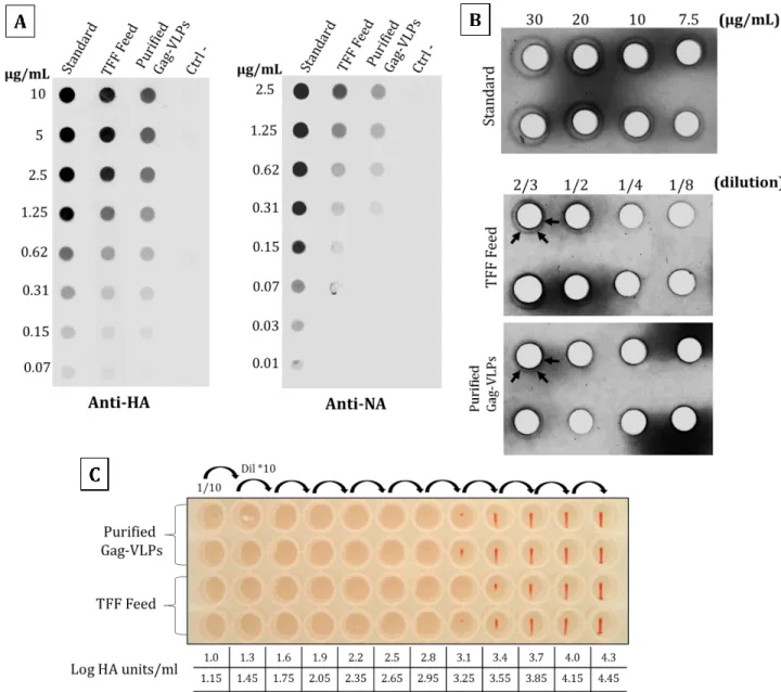
cushion followed by concentration through tangential flow filtration (TFF) using a 1000 KDa cut-off membrane. Different techniques were employed to characterize the produced VLPs: Single radial immunodiffusion (SRID), hemagglutination assay, dot-blot, western-blot, and transmission electron microscopy (TEM). Of great significance, intranasal immunization of VLPs induced specific immunogenic response and provided complete protection in mice challenged with the homologous influenza strain.
Once the proof of concept of VLPs as an efficacious influenza vaccine was demonstrated in vivo, we developed a new inducible cell line expressing the three proteins HA, NA and the Gag fused to GFP. This was performed in an effort to streamline the production process by eliminating the transient transfection step that can be cumbersome at large scale. The process was optimized to reach a high volumetric yield of VLPs by increasing the cell density at the time of induction without sacrificing the cell specific productivity. By operating a 3L-bioreactor in perfusion mode followed by TFF, the yields of VLPs were improved by 60-fold (3x10¹¹ Gag-GFP fluorescent events/L of culture measured by flow cytometry) compared to a standard batch culture (5x10⁹ Gag-GFP fluorescent events/L). The process was characterized for the upstream kinetics of production of VLP proteins and recovery rates for each downstream step. The production of a single bioreactor, operated in perfusion mode, with the stable cell line 293-HA/NA/Gag-GFP yielded 5-fold more total VLP antigenic HA proteins than what was produced with the 3L-batch bioreactor using transient transfection. Our process provided unprecedented yields of influenza VLPs produced from mammalian cells.
Finally, because VLPs are enveloped particles that bud from a host cell potentially enclosing host cell proteins, DNA and RNA, which could pose a safety concern, we performed a proteomic characterization of the influenza Gag-VLPs produced by transient transfection and also extracellular vesicles (EVs) produced from wild-type HEK-293 cells. The functions of all proteins identified within VLPs and EVs were critically discussed.
The process developed in this thesis could support the production of VLPs harboring HA and NA of different strains for clinical trials and could potentially result in a better vaccine candidate with higher efficacy and safety to replace the current labor-intensive egg-produced influenza vaccines.
TABLE OF CONTENTS
DEDICATION ... III ACKNOWLEDGEMENTS ... IV RÉSUMÉ ... VI ABSTRACT ... IX TABLE OF CONTENTS ... XI LIST OF TABLES ... XVI LIST OF FIGURES ... XVII LIST OF SYMBOLS AND ABBREVIATIONS... XVIIICHAPTER 1 INTRODUCTION ... 1
Research problem ... 1
Hypothesis ... 2
Research objectives ... 2
Thesis organization ... 3
CHAPTER 2 LITERATURE REVIEW ... 4
Influenza ... 4
2.1.1 Classification and virus structure ... 4
2.1.2 Infective replication cycle ... 9
2.1.3 Influenza pathogenesis ... 12
Influenza vaccines ... 12
2.2.1 Immune response to current influenza vaccines ... 14
2.2.3 Recombinant influenza vaccines ... 16
2.2.4 Influenza virus-like particles as vaccines ... 17
2.2.5 Gag as a core protein for VLPs ... 20
Mammalian cells as expression system ... 21
2.3.1 Mammalian cells expression regulation systems ... 23
2.3.2 Cell line development ... 25
Challenges associated to the large-scale manufacturing of VLPs for vaccine ... 27
2.4.1 VLPs design ... 27
2.4.2 Upstream process ... 29
2.4.3 Downstream process ... 30
CHAPTER 3 ARTICLE 1: HEMAGGLUTININ AND NEURAMINIDASE CONTAINING VIRUS-LIKE PARTICLES PRODUCED IN HEK-293 SUSPENSION CULTURE: AN EFFECTIVE INFLUENZA VACCINE CANDIDATE ... 33
Abstract ... 33
Introduction ... 34
Materials and Methods ... 36
3.3.1 Cells, plasmids and antibodies ... 36
3.3.2 Generation of 293CymR-rcTA cell line ... 37
3.3.3 Generation of 293HA-NA stable cells ... 37
3.3.4 Immunofluorescence assay ... 37
3.3.5 3L-Bioreactor ... 38
3.3.6 Ultracentrifugation and concentration of VLPs by sucrose cushion 25% ... 38
3.3.7 Tangential Flow Filtration ... 38
3.3.8 Cell lysate using RIPA buffer ... 38
3.3.10 Single Radial Immunodiffusion ... 39
3.3.11 Hemagglutination assay ... 39
3.3.12 Determination of host cell proteins and host cell DNA ... 39
3.3.13 Fluorescence intensity measurement and p24 quantification ... 39
3.3.14 Transmission electron microscopy (TEM) ... 40
3.3.15 Immunization and viral challenge ... 40
3.3.16 Enzyme-linked immunosorbent assay (ELISA) for HA-specific antibodies detection……… ... 40
Results and Discussion ... 41
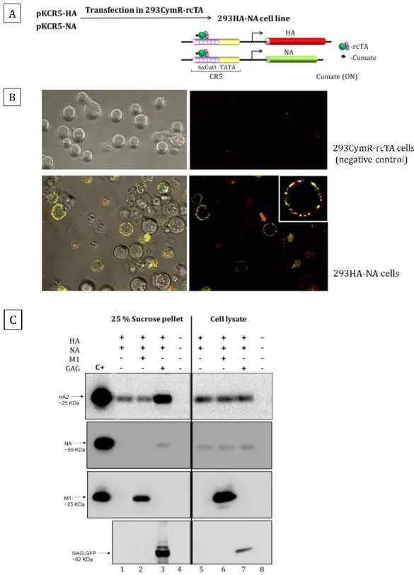
3.4.1 Development of 293HA-NA stable cells ... 41
3.4.2 Comparison of influenza matrix M1 and Gag proteins effect on influenza VLPs production ... 43
3.4.3 Influenza Gag-VLPs production in shake flasks and 3L-Bioreactor ... 44
3.4.4 Tangential Flow Filtration (TFF) of influenza Gag-VLPs ... 46
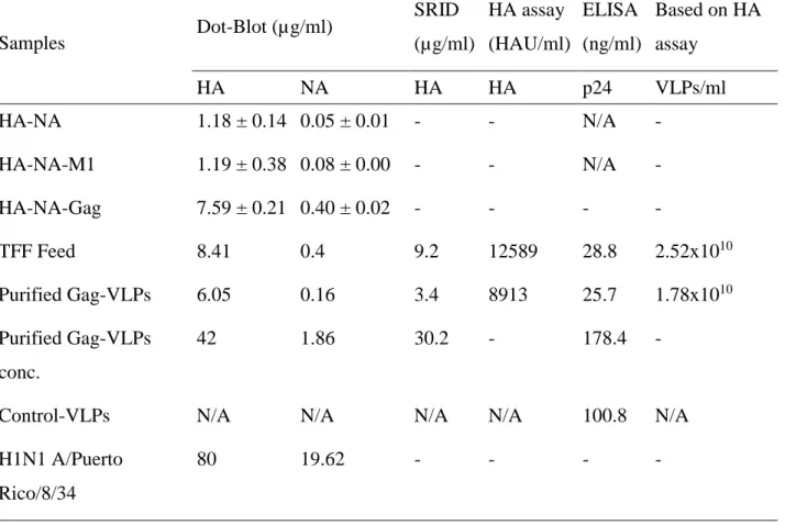
3.4.5 Quantification, characterization and yield of influenza Gag-VLPs ... 49
3.4.6 Immunization and mice challenge protection study ... 52
Acknowledgements ... 54
References ... 54
CHAPTER 4 ARTICLE 2: PROCESS INTENSIFICATION FOR HIGH YIELD EXPRESSION OF INFLUENZA H1N1 GAG VIRUS-LIKE PARTICLES USING AN INDUCIBLE HEK-293 PRODUCING CELL LINE ... 59
Abstract ... 59
Introduction ... 60
Materials and Methods ... 62
4.3.2 Generation of 293-HA/NA/Gag-GFP stable cell line ... 62
4.3.3 Flow cytometry ... 63
4.3.3.1 Cells ... 63
4.3.3.2 Gag-GFP events count by flow cytometry ... 64
4.3.4 Perfusion Bioreactor ... 64
4.3.5 VLP proteins quantification and characterization ... 64
4.3.6 Clarification and Tangential flow filtration (TFF) ... 65
Results and Discussion ... 65
4.4.1 Stable cell line development ... 65
4.4.2 Media evaluation at small scale ... 66
4.4.3 Medium replacement at small scale ... 68
4.4.4 Perfusion Bioreactor ... 71
4.4.5 Tangential Flow Filtration (TFF) ... 73
Declaration of interest ... 76
Acknowledgements ... 76
References ... 76
CHAPTER 5 PROTEOMIC CHARACTERIZATION OF INFLUENZA H1N1 GAG VIRUS-LIKE PARTICLES AND EXTRACELLULAR VESICLES PRODUCED IN HEK-293SF………. ... 79
Introduction ... 80
Methodology ... 81
5.2.1 nLC-MS/MS of Tryptic Digests ... 81
5.2.2 Production of VLPs and EVs ... 82
Results and Discussion ... 83
5.3.2 Extracellular vesicles unique proteins ... 96
Conclusions ... 99
CHAPTER 6 GENERAL DISCUSSION ... 101
CHAPTER 7 CONCLUSION AND RECOMENDATIONS ... 108
Conclusions ... 108
Recommendations for future studies ... 110
LIST OF TABLES
Table 2-1 Influenza A virus strain A/PR/8/34 genome and proteins ... 7 Table 2-2 Virus-like particles in development phase and/or licensed for use as human vaccine. . 19 Table 3-1 Summary of HA, NA and Gag quantification ... 45 Table 3-2 Recovery of the purification by TFF based on anti-HA Dot-Blot results ... 49 Table 4-1 Specific productivity, specific growth rate and productivity improvement factor from
batch to perfusion ... 71 Table 4-2 Recovery of VLPs during the downstream process of the perfusion bioreactor harvest
in terms of Gag-GFP events/ml and HA concentration ... 75 Table 5-1 Proteins identified by n-LC-MS/MS in VLPs sample ... 92 Table 5-2 Proteins uniquely identified in extracellular vesicles produced in HEK-293 cells ... 97 Table 6-1 Summary of the production performances of the two processes employed to produce
LIST OF FIGURES
Figure 2-1 Schematic representation of influenza virions.. ... 6
Figure 2-2 General steps in the infection cycle of influenza virus.. ... 11
Figure 2-3. Structure of Gag protein of HIV-1. ... 21
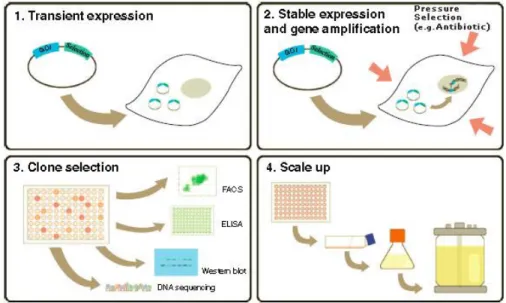
Figure 2-4 General method followed to create a stable cell line for large-scale production of recombinant protein. ... 26
Figure 3-1 Development of the 293HA-NA stable cell clone and analysis of VLPs production efficiency.. ... 42
Figure 3-2 Gag-VLPs production in shake flasks and 3 L-bioreactor. ... 46
Figure 3-3 Tangential flow filtration (TFF) of influenza Gag-VLPs. ... 48
Figure 3-4 Quantification of HA and NA on influenza Gag-VLPs before and after TFF.. ... 51
Figure 3-5 Immunity of influenza Gag-VLPs and mice challenge.. ... 53
Figure 4-1 Development of stable cell line 293-HA/NA/Gag-GFP.. ... 66
Figure 4-2 Media evaluation at small scale.. ... 68
Figure 4-3 Production of VLPs in cultures with medium replacement (MR) at small scale. ... 70
Figure 4-4 Production of VLPs in a perfusion mode 3L-bioreactor.. ... 72
Figure 4-5 Tangential Flow Filtration (TFF) of VLPs produced in the 3-L perfusion bioreactor.. 75
Figure 5-1 Differential and common proteins identified by nano LC-MS/MS in Gag-VLPs and extracellular vesicles (EVs). ... 85
LIST OF SYMBOLS AND ABBREVIATIONS
ALIX apoptosis-linked gene 2-interacting protein X ATP adenosine triphosphate
CCLs continuous cell lines CMV cytomegalovirus
CR5 cumate responsive promoter DNA deoxyribonucleic acid
ESCRT endosomal sorting complexes required for transport EVs extracellular vesicles
FDA Food and Drug Administration Gag core structural protein of retrovirus GFP green fluorescent protein
GOI gen of interest
HA hemagglutinin
HCPs host cell proteins
HEK-293SF human embryonic kidney cells adapted to grow in serum-free medium HIV-1 human immunodeficiency virus type 1
hpt hours post-transfection HSP heat shock protein
IgG immunoglobulin
ILVs intraluminal vesicles
LAIVs live attenuated influenza vaccine LMH flux in L/m2/hour
MDCK Madin-Darby canine kidney cells MHC major histocompatibility complex
MR medium replacement
MVBs multi-vesicular bodies
NA neuraminidase
NADPH nicotinamide adenine dinucleotide phosphate
nLC-MS/MS nanoscale liquid chromatography tandem mass spectrometry p-Cym operon of Pseudomonas putida
PEI polyethyleneimine pfu plaque-forming units
PR Puerto Rico
RBC red blood cells rcTA reverse transactivator RNA Ribonucleic acid
SDS-PAGE sodium dodecyl sulfate polyacrylamide gel electrophoresis SRID single radial immunodiffusion
TCA Tricarboxylic acid
TEM transmission electron microscopy TFF tangential flow filtration
TIVs trivalent influenza vaccine
Vero African green monkey kidney cells VLPs virus-like particles
vRNPs virus ribonucleoprotein WHO World Health Organization
293CymR-rcTA HEK-293SF containing the cumate switch system 293HA-NA HEK-293SF stably expressing HA and NA (H1N1 A/PR/8/34)
CHAPTER 1
INTRODUCTION
Research problem
Influenza is still a major threat to human health causing high morbidity and mortality in humans Influenza outbreaks are among the most ancient and lethal infections in human history. The reason for the persistence of this disease is because the causative agent is a virus with high and fast mutation rates. The mutations occur in the dominant antigenic proteins exposed in the viral envelope, hemagglutinin and neuraminidase. Thus far, there exist 18 types of HA and 11 NA that conform different strain subtypes. The new mutant strains cannot be recognized by the host immune system and therefore can cause severe infections. Vaccination remains the most effective way to combat the disease. Influenza vaccines have been produced in eggs for more than 70 years, but this system carries major drawbacks such as limited production capacity in case of high demand seasons or pandemics, egg allergies in some patients and low adaptability of some strains to eggs, which can result in mismatch with the circulating strains. Furthermore, the seasonal vaccines are highly efficacious in healthy adults but poorly immunogenic in high-risk groups (elderly, young children and pregnant women). With the great progress made in the development of cell culture process to produce biopharmaceuticals, many researches are now focused on switching the manufacturing of influenza vaccines to cell based production. Mammalian cells offer several advantages for the production of complex biomolecules such as superior post-translational modifications and easy adaptability to grow in serum-free suspension cultures. Among mammalian cells, human cell lines provide a glycosylation pattern homologous to endogenous human proteins reducing the risk of reactogenicity of biopharmaceuticals for human use. On the other hand, there is a need for new generation of influenza vaccines that mitigate the limitations of current seasonal vaccines and induce cross-protective immune response. Virus-like particles constitute an attractive platform to present the dominant antigens HA and NA in their native conformational structure on a non-infectious, non-replicative particulate structure similar to wild-type virus. The repetitive high-density arrays of antigenic proteins elicit a strong humoral response while the particulate structure induces the uptake by professional antigen-presenting cells stimulating cellular immune response. The immunogenicity and efficacy of VLPs as vaccine candidates have been extensively proven by in vivo experiments. However, there are still
numerous challenges for the large-scale manufacturing of these nanoparticles and their approval as vaccines. There is lack of robust and scalable processes to produce sufficient amount of VLPs from mammalian cells. This fact has limited the development of efficient methods to purify and characterize influenza VLPs for use in clinical trials. As enveloped particles, VLPs bud from the host cell membrane taking up not only the viral antigenic proteins but also host cell proteins, DNA and RNA. In mammalian cells, VLPs are co-produced with extracellular vesicles (EVs) also containing host cell contaminants and having a similar size to the VLPs, which makes difficult the development of an efficient purification process.
Hypothesis
To address the issues mentioned above, in this thesis we have made the following hypothesis. The design and development of a VLP producer HEK-293SF stable cell line adapted to suspension growth will allow to develop and characterize a scalable process to produce, and further characterize high yields of influenza VLPs that are immunogenic and protective.
Research objectives
To demonstrate the veracity of the hypothesis, the following objectives were defined:
To develop a HEK-293SF inducible stable cell line expressing the main influenza antigenic proteins HA and NA and to compare the VLPs production yields after transient co-expression of M1 influenza matrix protein and Gag structural core protein of HIV-1. To produce VLPs in a laboratory scale bioreactor and partially purify them to assess their
immunogenicity and efficacy by in vivo experiments.
To develop and apply process intensification strategies to achieve high yields of VLPs aiming at large scale manufacturing.
To characterize the protein composition of VLPs and extracellular vesicles by LC-MS/MS.
Thesis organization
Following this introduction, Chapter 2 provides a detailed literature review on the state-of-the-art of influenza vaccines, mammalian cells platform and virus-like particles; Chapter 3 presents the production process of VLPs in a 3L-bioreactor after transient transfection of a gag-containing plasmid and the results of the immunization with VLPs in an in vivo experiment. The methodology and results of this chapter have been published in the Vaccine journal and presented as a poster at the Cell Culture Engineering XV, Vaccine Technology VI and Virus-like particle &
nanoparticle vaccines conferences.
Venereo-Sanchez A, Gilbert R, Simoneau M, Caron A, Chahal P, Chen W, et al. (2016). “Hemagglutinin and neuraminidase containing virus-like particles produced in HEK-293 suspension culture: An effective influenza vaccine candidate”. Vaccine, 34:3371-80. Chapter 4 presents the process intensification strategies performed to increase the yields of VLPs produced from the stable cell line 293-HA/NA/Gag-GFP including a 3L perfusion bioreactor and the concentration of 9.5L harvest by tangential flow filtration. These results are presented in a manuscript entitled “Process Intensification for High Yield Expression of Influenza H1N1 Gag
Virus-Like Particles using an Inducible HEK-293 Producing Cell Line” which has been
submitted to the Vaccine journal.
The proteomic composition of VLPs and EVs as well as a critical discussion of the function of each host cell protein identified in both nanoparticles species are presented in Chapter 5. This chapter is entitled “Proteomic characterization of influenza H1N1 Gag virus-like particles and
extracellular vesicles produced in HEK-293SF”.
Chapter 6 provides a general discussion of all the results obtained in each chapter; and, finally, Chapter 7 presents the main conclusions and recommendations.
CHAPTER 2
LITERATURE REVIEW
Influenza
The name Influenza arose in the fifteenth-century in Italy, from an epidemic attributed to the “influence of the stars” (Barberis, Myles et al., 2016). In the years 1918-1920, influenza A virus caused one of the worst worldwide human pandemics in history. The so-called “Spanish flu” (H1N1) led to the death of at least 20 million people (Taubenberger, Reid et al., 1997). The “Asian flu” (H2N2) in 1957 (70,000 deaths in North America) (Dunn, 1958; Neumann, Noda et
al., 2009; Trotter, Dunn et al., 1959) and the “Hong Kong flu” (H3N2) in 1967 (Cockburn, Delon et al., 1969) caused the second and third most important influenza A pandemics. Recently, the
“swine flu” pandemic of 2009–2010 provoked over 16,000 deaths worldwide (Garten, Davis et
al., 2009). Most people call "flu" the “common cold” which is usually caused by some upper
respiratory viruses such as rhinoviruses and coronaviruses, not by influenza viruses. Influenza is a severe acute disease that can occur in all age groups with symptoms such as sore throat, cough, high fever, headache, and muscular pains (Dermody & Chappell, 2011; Osterholm, Kelley et al., 2012). The virus spreads quite rapidly through the air by droplets released from infected person sneezes or cough, and also by mucosal contact (eyes, mouth) with contaminated hands. The World Health Organization (WHO) reports approximately 3-5 million cases of severe illness
worldwide and up to 500,000 deaths annually
(http://www.who.int/mediacentre/factsheets/fs211/en/). Vaccination remains the best method to control and prevent seasonal and pandemic influenza.
2.1.1 Classification
and virus structure
Influenza viruses have a genome of approximately 13.5 kb and belong to the Orthomyxoviridae family. The viruses are classified into three types: A, B and C (Milián & Kamen, 2015; Noda, 2011; Subbarao & Matsuoka, 2013; Zambon, 2014). Recently, a novel genus termed influenza virus type D has been isolated from cattle in the United States (Collin, Sheng et al., 2015) and in Europe (Ducatez, Pelletier et al., 2015). The main differences between influenza types A, B and C lie in the number of RNA segments, 8 in the case of type A and B and 7 segments for influenza C; unlike influenza A and B, influenza C only contains one envelope protein, the HEF
(hemagglutinin-esterase fusion) that binds to 9-O-acetylneuraminic acid receptors instead of the N-acetylneuraminic acid receptors recognized by influenza A and B (Gao, Brydon et al., 2008; Muraki & Hongo, 2010). Influenza type A viruses are the best-studied orthomyxoviruses causing the most serious infections in humans, and are also widespread in many avian and mammalian species (Steel, 2011). Influenza type B is limited to humans, although occasional infections to other mammalian species such as seals have been documented (Bodewes, Morick et al., 2013; Chen & Holmes, 2008). Two lineages of influenza B have been identified referred to as B/Yamagata and B/Victoria (Shaw, Xu et al., 2002). The influenza type C causes mild infections in both pigs and humans, thereby having less importance for public health (Zambon, 2014). Influenza A viruses are categorized into different subtypes based on the hemagglutinin (HA) or neuraminidase (NA). Recently, novel influenza viruses have been isolated from bats in Central America giving rise to a total of 18 different types of HA (H1-H18) (Tong, Li et al., 2012; Tong, Zhu et al., 2013) and 11 NA (N1-N11) (Jagadesh, Salam et al., 2016) identified to date. Combination of these proteins gives rise to several influenza virus subtypes with different antigenicity. The virus classification refers to the surface antigens exposed (e.g. H3N2, H5N1, H1N1). The nomenclature used to describe each isolated influenza viral strain is descriptive and includes: the type of virus (A, B, C), the original host (this parameter is not indicated in human isolations), the geographic site of origin, the registered strain number and the year of isolation. For influenza A viruses, the corresponding identified HA and NA subtype must appear (e.g. A/H1N1/PuertoRico/8/1934, A/Chicken/Pennsylvania/1370/83 (H5N2) (Horimoto & Kawaoka, 2001).
The influenza virions are enveloped with a negative-sense, single-stranded, segmented RNA genome. Influenza viruses type A contain eight RNA genome segments encoding at least 12 viral proteins. Each one of the first 6 segments mostly gives rise to one protein: PB2, PB1, PA, HA, NP, NA. The PB2 (basic polymerase 2), (PB1 basic polymerase 1), and PA (acidic polymerase) proteins form the RNA polymerase complex (Kawaoka & Neumann, 2012). Hemagglutinin (HA) is a type I transmembrane protein and is the most represented envelope glycoprotein. The nucleocapsid protein (NP) tightly wraps the genomic viral RNA. Neuraminidase (NA), type II transmembrane protein, is the second most represented envelope glycoprotein. There are some exceptions where some of these first 6 segments code for more than one protein. In some influenza A strains, the RNA segment that codes PB1 has a second open reading frame for the
protein PB1-F2 which have been involved in the degree of virulence of the virions (Conenello, Zamarin et al., 2007). Meanwhile, the 6th segment of influenza B virus also encodes a protein
called NB that is not essential for viral replication in vitro (Hatta & Kawaoka, 2003) and structurally resembles the M2 protein of influenza A (Bouvier & Palese, 2008). The segments 7 and 8 are spliced into two messenger RNA (mRNA) encoding two different viral proteins; segment 7 codes for the matrix protein (M1) and the third enveloped protein (M2), whereas segment 8 codes for two nonstructural proteins (NS1 and NS2). The highly conserved 5’ and 3’ ends of the viral RNA segments form a helical hairpin at which is bound the trimeric PB2, PB1, and PA RNA polymerase complex. Arginine-rich nucleoproteins with positive net charge coat the rest of the negative-charged RNA phosphate backbone, approximately one NP per 24 nucleotides. These structures called the viral ribonucleoprotein (vRNP) are exported from the host cell nucleus and packaged at the plasma membrane to form the virion (Noda, Sugita et al., 2012) (Figure 2-1). The shape of the virions can be either roughly spherical or filamentous with an approximated size of 80-120 nm in diameter, and a length of up to 20 μm in the case of filamentous particles. The morphology of the virions appears to be related to the source of isolation. Mostly filamentous structure virions are observed from clinical isolations and more spherical/rounded viruses are found during in vitro productions (eggs passage or cell culture) (Noda, 2011). The Table 2-1 summarizes the main functions of the viral proteins.
Figure 2-1 Schematic representation of influenza virions. Hemagglutinin, Neuraminidase and M2 ion channel are transmembrane proteins exposed in the viral envelope. The M1 matrix protein is found underneath the viral membrane. Inside of the enclosed lipid bilayer are the eight RNA
PB1 PB2 PA 5’ 3’ virus Ribonucleoprotein (vRNP) viral RNA (-) sense Polymerase complex (PB1, PB2, PA) Hemagglutinin (HA) Ion channel (M2) Neuraminidase (NA) Matrix protein (M1) Lipid bilayer NS2(NEP) Nucleocapsid protein (NP)
individual segments associated to the nucleocapsid protein and the RNA polymerase complex (PB1, PB2 and PA) forming the 8 helical vRNPs (adapted from (Dermody & Chappell, 2011)). Table 2-1 Influenza A virus strain A/PR/8/34 genome and proteins
RNA segment Length (nucleotide) Encoded Proteins Protein size (aa) Protein function 1 2 3 2341 2341 2233 PB2 PB1 PA 759 757 716
These three proteins form the heterotrimeric RNA-dependent RNA polymerase complex. The PA endonuclease generates the primers; the PB2 binds the capped RNA and PB1 subunit performs the RNA synthesis (Stevaert & Naesens, 2016).
4 1778 HA 566 The hemagglutinin is a homotrimeric
transmembrane glycoprotein and the main antigenic determinant; it is crucial for the binding to sialic acid receptors at cell virus entry and membrane fusion at low pH in the endosome.
5 1565 NP 498 The nucleoprotein encapsidates the viral RNA, control functions during RNA synthesis and is also involved in the virion morphology, probably through interaction with M1 (Bialas, Bussey et
6 1413 NA 454 The neuraminidase is a tetrameric surface glycoprotein and the second antigenic determinant. It cleaves terminal N-acetyl neuraminic acid (sialic acid) mediating the release of the new virions progeny. It also facilitates hemagglutinin-mediated fusion and viral spread in the respiratory epithelium (Jagadesh et al., 2016). Mostly, the HA:NA ratio in wild type virus is approximately 1:4.
7 1027 M1
M2
252
97
The matrix protein (M1) interacts with the cytoplasmic tails of HA, NA, M2 (Chen, Leser
et al., 2007) and also with the nucleocapsids in
the vRNPs, and NS2 (Noda, 2011). M1 is involved in the virion assembly, budding and morphology as well as the nuclear export of the vRNPs.
Integral membrane protein forming an ion channel crucial in the un-coating of virions inside the endosome and scission membrane for viral budding. The HA:M2 ratio is around 10 to 100 M2 per HA molecule (Bouvier & Palese, 2008).
8 890 NS1
NS2(NEP) 230
121
The nonstructural protein 1 is an interferon-antagonist that down-regulates host cell mRNA processing; reduces apoptosis of infected cells. The nonstructural protein 2 mediates the export of viral nucleocapsids from the nucleus; interacts with M1.
2.1.2
Infective replication cycle
Influenza infection begins with the binding of hemagglutinin glycoprotein to the N-acetylneuraminic (sialic) acid-containing receptors exposed on the host cell membrane. The term hemagglutinin arise from the protein’s ability to agglutinate red blood cells. HA is synthesized as a homotrimeric molecule (HA0) that is sensitive to cleavage from cellular serine-proteases. After cleavage, polypeptide HA0 is excised in two subunits: the immunodominant globular head HA1 subunit that is responsible for the binding to sialic acid and the stalk domain HA2 that contains the fusion peptide involved in membrane fusion inside the endosome (Carr & Kim, 1993; Lorieau, Louis et al., 2013). The virus enters the cell via clathrin-mediated endocytosis. The low endosomal pH causes an important conformational change in the HA protein which is comparable to the opening of a coiled spring (Baker & Agard, 1994; Dermody & Chappell, 2011; Lorieau et al., 2013). The fusion peptide hidden within a hydrophobic region of HA moves out toward the membrane of the endosome causing the fusion of the virus envelope with the endosomal membrane (Ivanovic, Choi et al., 2013). After membrane fusion, the next step includes the release of the helical nucleocapsids from the virion to the cytoplasm mediated by the M2 ion channel present in the virus envelope. The hydronium protons enter into the virion through the M2 channel protein and this acidic environment in the inner virion causes weakening of the interaction between M1 and the helical nucleocapsids, provoking their release across the previously fused membrane to the host cell cytoplasm (Bouvier & Palese, 2008).
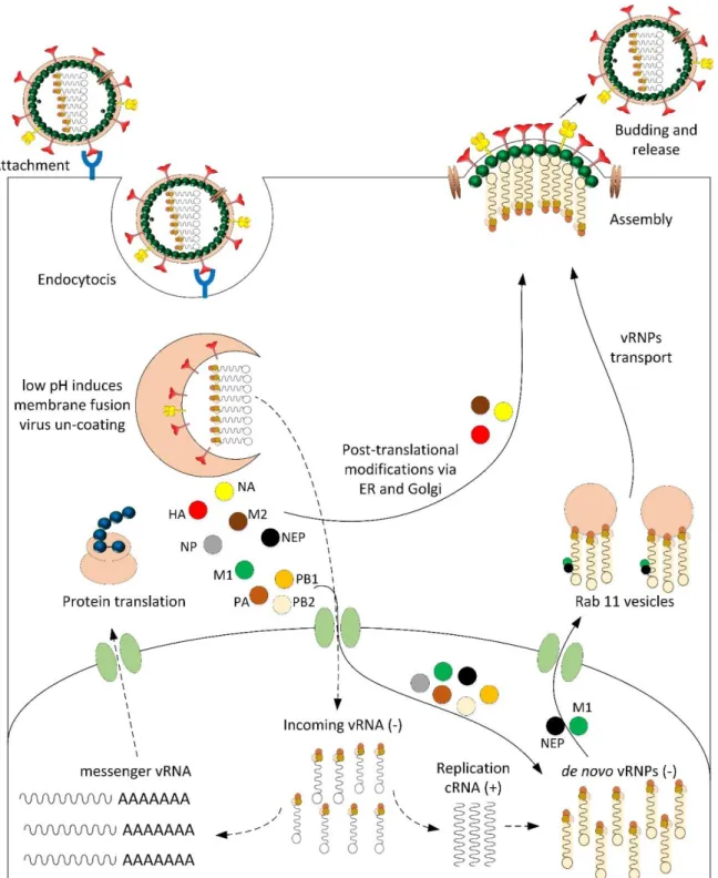
Unlike most other RNA-containing viruses, influenza virus replication occurs in the nucleus (Pohl, Lanz et al., 2016). The RNA segments of infecting virions liberated to the cytoplasmic lumen are transported into the nucleus via the classical importin-α– importin-β1 (IMPα–IMPβ1)-dependent nuclear import pathway (Eisfeld, Neumann et al., 2015). This is facilitated by nuclear localization signals present in the vRNPs. Once the viral genome segments get into the nucleus the negative-sense viral RNAs are transcribed to messenger RNAs. The subunit PB2 of the vRNP complex binds to cellular capped pre-messenger RNA while PA subunit cleaves them to generate primers for RNA synthesis. The PB1 polymerase transcribes the RNA terminating at 5’ poly U sequence found at the end of the RNA. These mature mRNAs are exported to the cytoplasm for translation in cellular ribosomes (Fodor, 2012). The polymerase also replicates the vRNA to a complementary RNA (cRNA) which serves as template for de novo vRNAs synthesis. Because of
their nuclear localization signals, NP and the trimetric polymerase are redirected again to the nucleus for the assembly of the ribonucleocapsids. Hence, the new progeny of vRNAs are assembled with NP and the RNA polymerase complex proteins newly synthesized inside the nucleus and then transported to the cytoplasm. This process involves the interaction with M1 and NEP. The transportation of mature vRNPs to the budding site is mediated by Rab11 vesicles (Eisfeld et al., 2015).
The proteins of the viral envelope (hemagglutinin, neuraminidase and M2) take the secretory pathway through the endoplasmic reticulum (ER) and Golgi apparatus to the cell surface facilitated by the protein-trafficking machinery. HA and NA head towards cellular membrane zones enriched in cholesterol, named lipid rafts, whereas M2 is not associated to these domains (Rossman & Lamb, 2011). M1 is one of the most abundant proteins in the virion and it is located throughout the inner side of the viral envelope. M1 interacts directly with the cytoplasmic tails of HA, NA and M2 and also binds to virus nucleocapsids giving it an important structural role in maintaining the morphology of the virus (Chen et al., 2007; Thompson, Petiot et al., 2015). The exact mechanism for viral budding initiation remains unknown. Studies on the formation of influenza virus-like particles revealed that the accumulation of HA at the lipid-rafts patches is sufficient to mediate membrane curvature and release of HA-containing vesicles (Chen et al., 2007; Rossman & Lamb, 2011). Viral budding occurs on the apical membrane previously filled up with HA, NA, and M1 associated to the vRNP segments and M2 could mediate the membrane scission and viral release. The review of Rossman, and Lamb (2011) proposed that, considering the inconsistencies encountered in the viral budding studies done with VLPs, there is not one single protein totally responsible of the influenza viral budding but rather it is a reduntant cooperation among them. Figure 2-2 shows the major steps in the infection cycle of influenza virus.
Figure 2-2 General steps in the infection cycle of influenza virus. First, the binding of virions to the cellular membrane and internalization in endosomes take place. Subsequently, there is release of the RNA genomes through the lipid bilayer after virus and endosome membrane fusion. Nucleocapsids enter into the nucleus for transcription and replication of the RNA segments. Finally, translation of the viral proteins, virus ribonucleoprotein assembly in the nucleus and viral budding at the host plasma membrane take place.
2.1.3 Influenza pathogenesis
The pathogenicity of influenza is determined by both viral and host factors (Behrens, Stoll et al., 2006). The amount of host cell receptors, the abundancy of enzymes responsible of virus entry and replication (e.g. availability of protease mediating the cleavage of HA0), and the immunocompetency of the individual are among the factors related to the host individual. Viral factors are determined by the affinity and ability of the virus strain subtype to bind to the host cell receptors, the susceptibility to antigenic mutation and reassortment with other co-infecting viral strains, and the capability to attenuate the host immune response, etc.
In humans, the influenza virus infects and replicates in epithelial cells of both upper and lower respiratory tract (Taubenberger & Morens, 2008). Infected persons usually show asymptomatic to mild illness. The symptoms persist for 7-10 days and include fever, inflammation of the respiratory tract, cough, prostration, headache, etc. An experiment in healthy volunteers showed that a peak of influenza virus replication occurred 48 hours post-inoculation in the nasopharynx and little to no virus shedding was observed after 6 days (Barnard, 2009; Carrat, Vergu et al., 2008). However, these results cannot be generalized since children or older persons were not included in the experiment, in whom the illness evolves differently. Since influenza virus infects epithelial cells, complications of the respiratory tract including tracheitis, bronquitis, bronchiolitis, otitis media, and sinusitis are commonly developed. Pneumonia and acute respiratory distress syndrome (ARDS) are the most severe complications of influenza infection (Li & Cao, 2016). The major reason of the high mortality during influenza pandemics is due to secondary bacterial infections mainly caused by Streptococcus pneumonia (Stegemann, Dahlberg
et al., 2009). A strong overactivated immune response due to an uncontrolled production of
cytokines have been reported in severe influenza infections causing lung damage (Li & Cao, 2016).
Influenza
vaccines
The source of variation in influenza viruses arises from two phenomena: 1) the accumulation of point mutations in the coding RNA segments, which is known as antigenic drift and 2) the reassortment between RNA segments of two or more different virus subtypes co-infecting the same host cell, which is termed antigenic shift. The major variations introduced due to the
antigenic drift occur in the main antigenic determinants, the globular domain of HA (HA1) and NA. Consequently, the new mutant virions progeny cannot be recognized by the host immune system causing severe infections. The antigenic shift occurs occasionally but can be extremely dangerous. By genome segments interchange, strains originally infecting one species may pass to another one appearing new to the immune system making the infection potentially highly fatal (Dermody & Chappell, 2011).
These viral strategies used to escape the host immune system have made it very difficult for researchers to find an efficient vaccine to totally eradicate the disease. The first human influenza virus was isolated after propagation in ferrets in 1933 (Hannoun, 2013). The discovery of the causative agent of influenza triggered several investigations to develop an effective vaccine. The progress in the development of methods allowing large-scale cultivation in embryonated chicken eggs and methods for virus quantification by hemagglutination assay, made possible the production of the first inactivated vaccines in 1942, which showed protective efficacy against influenza A and B after evaluation in trials among US military recruits (Francis, 1953). Currently, after more than 70 years, the basic technology and principles of influenza vaccine production remain much the same (Sambhara & Stephenson, 2009).
Thus far, influenza vaccines have to be annually updated to be effective against new emerging strains. Every year, through a surveillance system, the World Health Organization (WHO) identifies newly evolved antigenic strains most likely to predominate in the following influenza season. Twice a year, a committee assembled by WHO provides guidance to vaccine manufacturers on the viruses that will be included in the trivalent or quadrivalent influenza vaccines for the next seasons in the northern and southern hemispheres (Barr, 2014). Currently, vaccine companies have 6 months to produce their vaccines and provide them to public health systems. Two types of trivalent vaccines are approved for use: trivalent inactivated vaccine (TIV) and live attenuated virus vaccine (LAIV). These vaccines are composed of the 3 currently circulating seasonal influenza virus strains: two subtypes of influenza A virus (H3N2 and H1N1) and one strain of influenza B. The vaccine update is carried out by genetic reassortment between the field strains chosen by WHO surveillance and the backbone of A/PR8/34 master strain that has been shown to grow to high titers in embryonated eggs (HA and NA genes are exchanged). For influenza B, the field isolates are the seed strains for vaccine production because there is no evidence thus far of any master influenza B with high growth in the egg-based production
system. Once the seed strains are confirmed to be the reference strains by identity testing and sequence analysis, vaccine production begins. The TIVs can be prepared as whole inactivated virus- and split virus or subunit vaccines. Viruses are grown in the allantoic cavity of embryonated eggs, harvested, chemically inactivated by formalin or -propriolactone and purified by ultra-centrifugation (whole inactivated virus vaccine), then, viruses can be split using a nonionic detergent and the HA and NA are purified (split virus or subunit vaccines) (Gerdil, 2003; Milián & Kamen, 2015). The LAIVs were produced in the 1960s by serial passage of the virus in eggs under suboptimal conditions at low temperature, selecting for variants that could grow in the cool nasopharynx but could not grow in the lung. Master donor strains were established for vaccine production (Shaw, 2012; Wong & Webby, 2013). Recently, quadrivalent inactivated vaccines have been approved by the Food and Drug Administration (FDA) including another influenza B-lineage leading to superior immunogenicity against the added B strain (Greenberg, Robertson et al., 2013; Tinoco, Pavia-Ruz et al., 2014).
2.2.1 Immune response to current influenza vaccines
The immunogenic efficacy of the licensed seasonal influenza vaccines is associated with the patient age, displaying less protection in young children and elderly. Current conventional TIVs stimulate strong serum antibody responses against the major envelope glycoproteins HA and NA but elicit poorly mucosal IgA influenza-specific antibody and cellular immunity (Cox, Brokstad
et al., 2004). Due to the antigenic drift that takes place in influenza A viruses, the main surface
proteins mutate almost every year leading to new strains that cannot be neutralized by the host immune system. Thus, the immunity induced by seasonal TIVs vaccines is subtype-specific providing short-time protection.
The primary immune response to LAIV vaccines consists in serum IgA and IgM that peak two weeks post-vaccination while the IgG serum can last for at least 1 year post-vaccination. The nasal fluid IgA elicited is significantly higher than those elicited with TIVs. The LAIVs induce secretion of interferon-γ, proliferation of T lymphocytes including cytotoxic T-cells. Both vaccines have shown high rate of protection in human, but LAIVs can be more immunogenic in young children since a prior natural infection is needed for eliciting a potent humoral response against TIVs (Cox et al., 2004). Both vaccines offer low protection in the elderly.
Considering that cytotoxic T-cell immunity is responsive to more conserved internal proteins which could be efficient for mediating cross-protection and fast recovery from the disease, vaccine candidates stimulating influenza A virus-specific CD8+ T cells are very promising (Grant, Quiñones-Parra et al., 2016; Johansson & Brett, 2007; Kang, Yoo et al., 2009; Trollfors, 2006).
2.2.2 Limitations of current vaccine manufacturing process
The egg-embryonated technology that supports production of seasonal influenza vaccines carries some drawbacks that are pushing the scientific community to find novel and more efficient vaccine production platforms. Disadvantages associated with the egg-based production platform include: 1) adaptation of virus to eggs can introduce mutations that alter the composition of the hemagglutinin and negatively affect its antigenicity, 2) some virus strains cannot be efficiently grown in eggs which leads to a time-consuming process with the risk of developing undesired reassortants 3) the supply of eggs in the cases of pandemics or high demand seasons can be a limiting factor (one or two high quality eggs are needed per dose of vaccine). Also, diseases affecting chickens such as avian influenza virus outbreak can disrupt the supply eggs for vaccine production; 4) local or systemic allergic reactions to egg-derived vaccine components in vaccinated persons and 5) the requirement of biosafety level 3 to manipulate highly pathogenic viral strains or non-human influenza viruses (Genzel, Behrendt et al., 2013; Quan, Steinhauer et
al., 2008; Tree, Richardson et al., 2001).
Therefore, there is a need to develop a new generation of influenza vaccines and a robust production system that takes advantage of recent progress in bioengineering, biotechnology, molecular and cellular biology. Most of the available expression systems have already been explored for the production of influenza vaccines: mammalian cell culture (Chu, Lugovtsev et al., 2010; Le Ru, Jacob et al., 2010; Paillet, Forno et al., 2011); insect cells (Cox & Hollister, 2009; Pushko, Tumpey et al., 2005); plants (Landry, Ward et al., 2010); bacteria (Rudolph & Ben-yedidia, 2011). An excellent review from Milián, and Kamen (2015) provides a very complete state-of-the-art on current influenza vaccines that have been approved by the FDA along with the expression system used. For example, the Flucelvax influenza vaccine, originally developed by Novartis and recently acquired by SeqirusTM (a CSL company), is produced in Madin Darby Canine Kidney (MDCK) suspension cells and the Flublok, manufactured by Protein Sciences
Corp., is produced in insect cells and both vaccines have been licensed by FDA, demonstrating the potential of cell culture technology as a modern and robust influenza vaccine production system.
2.2.3 Recombinant influenza vaccines
Influenza vaccines design is a key issue for achieving broader humoral and cellular immunity combined with improved safety. Different influenza vaccine strategies have been examined so far mainly looking for a universal vaccine that can prevent infections from new strains, and also for vaccine presentation formats and adjuvants to increase immunogenicity; and induce a long-lasting response. The subunit vaccines mainly based on the engineering of the HA antigen have been extensively approached (He, Leyrer et al., 2016; Song, 2016). Toward a universal vaccine based on HA, chimeric globular HA domain containing the main conserved epitopes from different clades/subtypes based on computationally optimized broadly reactive antigens (COBRA) (Ducatez, Bahl et al., 2011; Giles, Bissel et al., 2012; He, Prabakaran et al., 2014) and headless HA vaccine candidates containing conserved epitopes of the stalk domain able to elicit broadly protective antibodies (Krammer & Palese, 2013; Neu, Dunand et al., 2016) constitute promising approaches. In the same direction of finding universal vaccines targeted to conserved epitopes of viral proteins, the highly conserved extracellular domain of the ion channel M2 protein (M2e) has been studied (Deng, Cho et al., 2015). Considering the role of M2 during the viral infection, the antibodies raised against this protein could inhibit the virus growth. However, since this protein is the least represented in the viral envelope, little M2e-specific humoral response have been detected by natural infection. In general, vaccine candidates based on M2e do not induce neutralizing antibodies; they have to be enhanced with strong adjuvants and/or advanced antigen presentation formats to be efficacious (Krammer & Palese, 2015; Shaw, 2012). As an alternative approach, DNA- and viral vector-based vaccines have shown to preserve the native structure of the antigens efficiently (Chen, Cheng et al., 2008; Laddy, Yan et al., 2008; Smith, Wloch et al., 2010; Van Kampen, Shi et al., 2005). Several other studies have focused on peptide-based vaccines by combining different conserved epitopes as synthetic peptides to enhance T-cells response (Alexander, Bilsel et al., 2010). Finally, virus-like particle vaccines are becoming a promising way to safely elicit both humoral and cellular response in the host immune system.
2.2.4 Influenza virus-like particles as vaccines
Influenza virus-like particles (VLPs) are usually formed by recombinant expression of the major viral proteins, e.g., HA, NA, M1, and M2. These proteins mediate the budding of particles that resemble native influenza virus as observed by electron microscopy. The VLPs do not contain viral genetic material; they are non-replicating and non-infectious particles displaying HA and NA antigens in the lipid envelope. The displayed viral glycoproteins keep their native conformational structure and intact biochemical functions (Haynes, 2009). VLPs are more immunogenic than subunit vaccines and induce an immune response similar to the wild-type virus, without the associated safety concerns. The repetitive display of the main antigens on VLPs and their particulate structure enhance phagocytosis such that they are easily taken up by antigen-presenting cells, especially dendritic cells. The antigenic epitopes are cross-presented through MHC I molecules resulting in the activation of cytotoxic T lymphocytes. Furthermore, VLPs can induce strong activation of B cell response in a T cell-independent mode. The high-density of epitopes on the VLPs envelope induces the crosslinking of the B-cell receptor promoting the B cells proliferation, T-helper activation by MHC II presentation, immunoglobulins secretion and generation of long-lasting memory B cells (Chen & Lai, 2013; Rynda-Apple, Patterson et al., 2014).
The viral proteins required for the formation of VLPs have varied in many studies. VLPs have been assembled from the co-expression of HA/NA/M1/M2 (Pan, Wei et al., 2010), HA/NA/M1 (Pushko et al., 2005), HA/M1 (Song, Hossain et al., 2010), or by the single expression of M1 (Gómez-Puertas, Albo et al., 2000), M2 (Rossman, Jing et al., 2010), NA (Lai, Chan et al., 2010), and HA (Chen et al., 2007; D’Aoust, Couture et al., 2010). These inconsistencies observed in the production of VLPs have brought to light that the mechanisms of viral budding of influenza viruses are not completely understood. As mentioned previously, it appears that there is not a single protein responsible for the viral budding but rather there is a certain redundancy among them (Rossman & Lamb, 2011). It is worth mentioning that most of the studies done to elucidate viral budding mechanism have been performed in different expression systems and using different vectors to deliver the recombinant genes. The expression system may influence the budding mechanism, since the host cell provides the cellular machinery for the virus replication and budding.
The baculovirus/insect cell-expression system is the most widely used to produce influenza VLPs (Vicente, Roldão et al., 2011). Several studies producing influenza VLPs in insect cells have shown the efficacy of the VLPs to induce protective immunity against influenza virus challenges (Bright, Carter et al., 2007; Galarza, Latham et al., 2005; Kapczynski, Tumpey et al., 2016; Pushko et al., 2005; Pushko, Tumpey et al., 2007; Quan, Huang et al., 2007; Smith, Flyer et al., 2013). Moreover, infuenza VLPs have been generated in Nicotiana benthamiana (a close relative to the tobacco plant) providing cross-protection in ferrets and promising immunogenicity in humans (Landry et al., 2010). Generation of influenza VLPs has also been achieved in different types of mammalian cells, including: HEK-293T (HEK-293 containing the SV40 large T-antigen), HeLa (human cervical cancer derived cells), Cos-1 (monkey kidney cells), and Vero cells (derived from the kidney of an African green monkey) resulting in efficacious protection in
in vivo challenge experiments (Carter, Darby et al., 2016; Tang, Lu et al., 2011; Wu, Yeh et al.,
2010). Various strategies have been followed to deliver influenza genes in mammalian cells to generate VLPs. Tang et al. (2011) carried out the production of VLPs using a unique BacMam containing individual expression cassettes of HA, NA, M1 under the control of the CMV promoter. In this work, very basic optimization of the transduction conditions was performed. The MOI, incubation time, and the addition of sodium butyrate (an additive used for enhancing gene expression in baculovirus transduced cells) were established. Following sucrose gradient purification, baculoviral DNA was detected but there was no detectable baculovirus in the purified VLPs. A positive hemagglutination assay and protective immunity in animals showed the efficacy of the produced VLPs. Wu et al. (2010) obtained VLPs from a stable Vero cell line expressing M1, M2, NA and HA using plasmids with the inducible system based on tetracycline operon. The work was focused on the characterization of VLPs recovered by ultracentrifugation. A detailed analysis of the VLP proteomic composition, glycosylation profile and demonstration of VLPs efficacy in animals was presented. These studies demonstrated the immune-protective efficacy of influenza VLPs derived from mammalian cells. However, more efforts are needed in order to develop and optimize a process to scale-up the influenza VLP production in mammalian cells with the ultimate goal that this approach can become seriously considered for large-scale vaccine manufacturing.
Table 2-2 shows different types of VLPs, including influenza-like, that have been licensed and/or are in the process of acceptation as vaccines.
Table 2-2 Virus-like particles in development phase and/or licensed for use as human vaccine (Effio & Hubbuch, 2015). The expression system used is specified for influenza VLPs.
Development stage Virus/pathogen/disease
Preclinical Chikungunyavirus (Metz, Martina et al., 2013), Coxsackievirus B3 (Koho, Koivunen et al., 2014), Cytomegalovirus (Vicente, Burri et al., 2014), Dengue virus (Schmitz, Roehrig et al., 2011), Enterovirus 71 (Chung, Chen et al., 2010; Ku, Liu et al., 2014), Group A Streptococcus (Chuan, Wibowo et al., 2014), Human B19 parvovirus (Chandramouli, Medina-Selby et al., 2013), Human immunodeficiency virus (Buonaguro, Tagliamonte et al., 2012; Negrete, Pai et al., 2014), Human papillomavirus (Gissmann, 2009), Rotavirus (Li, Lin et al., 2014)
Phase 1 Allergenic rhinitis/Asthma (anti IgE Qβ-VLP), Chikungunyavirus (Chang, Dowd et al., 2014), Ebola virus (Mohammadi, 2014), Influenza H1N1 (Low, Lee et al., 2014; Ward, Landry et al., 2014) (produced in bacteria and plant Nicotiana benthamiana, respectively), Influenza H7N9 (Fries, Smith et al., 2013) (produced in insect cells (HA,NA and M1), Respiratory syncytial virus (Glenn, Smith et al., 2013)
Phase 2 Alzheimer’s disease (amyloid Qβ-VLP) (Bachmann & Whitehead, 2013; Caputo, Graf et al., 2014), Human B19 parvovirus (Bernstein, Sahly et
al., 2011), Influenza H1N1 (López-Macías, Ferat-Osorio et al., 2011)
(produced in insect cells HA, NA and M1), Influenza H5N1 (Ward et al., 2014) (produced in plant Nicotiana benthamiana) , Norovirus (Sundararajan, Sangster et al., 2015)
Phase 3 Human papillomavirus (Erickson, Landers et al., 2014; Hildesheim, Wacholder et al., 2014), Malaria P. falciparum (RTS,S) (Umeh, Oguche
et al., 2014)
Licensed Human papillomavirus (Gardasil®, Cervarix®) (Dochez, Bogers et al., 2014), Hepatitis B virus (Recombivax HB®, Engerix-B®) (Lacson, Teng