Université de Montréal
L’efficacité du CD154 monomérique dans le traitement des
complications thrombotiques
Par
Mourad DANDACHLI
Département de Sciences biomédicales Faculté de Médecine
Mémoire présenté á la Faculté des études supérieures en vue de l’obtention du grade de Maitrise (M.Sc.)
en sciences biomédicales
Mars, 2016
Abstract
CD154 has emerged as an important player in the pathogenesis of several autoimmune diseases, as well as vascular dysfunctions. CD154 is a member of the tumour necrosis factor family of pivotal importance in humoral immunity. However, CD154 also shares critical inflammatory functions through its interaction with its classical CD40 receptor or recently identified binding partners, namely αIIbβ3, α5β1 and αMβ2. These responses imply CD154 as a key factor in chronic inflammatory disorders including autoimmune diseases and thrombosis. Disrupting the interaction of CD154 with its receptors through anti-CD154 Abs significantly inhibits the development of these diseases, albeit serious side effects have been associated with these therapies. To overcome these adverse effects, other approaches such as knockout, antisense oligonucleotide and siRNA targeting were developed. These approaches were focused on the CD154/CD40 interaction and did not address the interaction of CD154 with its other receptors. Thus, there is a need for novel CD154 treatments for the prevention/abrogation of inflammatory or autoimmune diseases that address all CD154 receptors. Our group profoundly investigated the structural/functional interaction of CD154 with its various receptors. Given the importance of the trimeric structure of CD154 for its biological activity, we generated monomeric form of the molecule that can specifically bind to one receptor without inducing intracellular activation. This agent represents a potential therapeutic tool for the treatment of CD154-mediated diseases such as thrombotic events.
Résumé
CD154 joue un rôle important dans la pathogenèse de plusieurs maladies auto-immunes, ainsi que des dysfonctionnements vasculaires. CD154 est un membre de la famille du facteur de nécrose tumorale (tumor necrosis factor, TNF) d'une importance cruciale dans l'immunité humorale. Cependant, CD154 partage également des fonctions critiques inflammatoires grâce à son interaction avec son récepteur CD40 ou des partenaires de liaison récemment identifiés, à savoir αIIbβ3, α5β1 et αMβ2. Ces réponses impliquent CD154 comme un facteur clé dans les maladies inflammatoires chroniques, notamment les maladies auto-immunes et la thrombose. L’interruption de l'interaction de CD154 avec ses récepteurs par anticorps anti-CD154 inhibe de manière significative le développement de ces maladies, bien que des effets secondaires graves ont été associés à ces traitements. Pour remédier à ces effets indésirables, d'autres approches comme des souris defficientes (Knockout), oligonucléotide antisens et ARNi ciblage ont été développés. Ces approches ont été axées sur l’interaction de CD154 avec CD40 et ne traitent pas l'interaction de CD154 avec ses autres récepteurs. Par conséquent, il existe un besoin de nouveaux traitements pour la prévention/abrogation des maladies inflammatoires ou auto-immunes qui cibles tous les récepteurs de CD154. Notre groupe a profondément étudié l'interaction structurelle/fonctionnelle de CD154 avec ses différents récepteurs. Étant donné l'importance de la structure trimérique de CD154 pour son activité biologique, nous avons généré une forme monomère de la molécule qui peut se lier spécifiquement à un récepteur sans induire l'activation intracellulaire. Cet agent est un outil thérapeutique potentiel pour le traitement de maladies reliées au CD154, tels que les événements thrombotiques.
Contents
Abstract ... iv
Résumé ... v
Contents ... vi
List of tables ... viii
List of figures ... ix
List of abbreviations: ... x
Chapter 1: Introduction ... 1
1. Structure of CD154 and its different receptors. ... 3
1.1. CD154 ... 3
1.2. The receptors of CD154 ... 8
1.2.1. CD40 ... 8
1.2.2. Integrins ... 11
1.2.2.1. The integrin αIIbβ3 ... 12
1.2.2.2. The integrin α5β1 ... 13
1.2.2.3. The integrin αMβ2 ... 14
2. The biological role of the interaction of CD154 with its different receptors: ... 15
2.1. The CD154/CD40 interaction ... 15
2.2. The CD154/αIIbβ3 interaction ... 19
2.3. The CD154/α5β1 interaction ... 20
2.4. The CD154/ αMβ2 interaction ... 22
3. Thrombosis and cardiovascular diseases. ... 24
Chapter 3: Materials and Methods. ... 30
2.5. Cell lines. ... 31
2.6. Antibodies and Reagents. ... 31
2.7. Plasmids and mutagenesis. ... 32
3.4. Cell culture, transfection and protein purification. ... 33
3.5. Flow cytometric analysis. ... 34
3.6. Western Blotting. ... 34
3.7. Animals. ... 35
Chapter 4: Results. ... 36
4.1. CD154 mutants (Y170 or G227) fail to induce intracellular signalling. ... 37
4.2. CD154 mutants capable of binding to all cells ... 37
4.3. CD154 binding to BJAB and U937 was inhibited by CD154 mutants. ... 38
4.4. Inhibition of thrombus formation in mice. ... 38
Chapter 5: Discussion. ... 45
Chapter 6: Conclusion and Perspectives ... 51
List of tables
Chapter 3 : Materials and Methods
List of figures
Chapter 1: Introduction
Figure 1. Schematic representation of the CD154 structure. ... 4
Figure 2: The-three dimensional structure of soluble CD154 structure. ... 6
Figure 3: Gene structure of human CD154. ... 7
Figure 4. Structure of CD40 protein. ... 9
Figure 5: Different integrin subunits and their αβ associations. ... 12
Figure 5 : CD154 and its receptors. ... 15
Figure 6: The interaction of CD154 with the αIIbβ3. ... 20
Figure 7: Interaction of CD154 with α5β1. ... 21
Figure 8 : Binding affinity and kinetic analysis of the interactions between sCD154 and α5β1 and between sCD154 and αIIbβ3, as determined by the surface plasmon resonance approach. ... 22
Figure 9: CD154 interaction with αMβ2 (Mac-1). ... 23
Chapter 4: Results. Figure 10. Coomassie Blue staining (A) and Western blot analysis (B) of purified CD154 mutants. ... 39
Figure 11. CD154 mutants (Y170, G227 and Y170/G227) are unable to induce intracellular signaling . ... 40
Figure 12. CD154 mutants (Y170, G227 and Y170/G227) are capable of binding to CD154 receptors. ... 41
Figure 13. CD154 mutated at its Y170 and/or G227 residues is capable of inhibiting the binding of CD154WT. ... 42
Figure 14.Thrombosis in mice in the presence or absence of CD154-interfering molecules. ... 43
List of abbreviations:
cDNA: complementary DNA.CTLA4Ig: cytotoxic T-lymphocyte-associated protein 4 immunoglobulin. ECM:extracellular matrix.
EAE: experimental autoimmune encephalomyelitis.
TNF: tumour necrosis factor.
TNFR: tumour necrosis factor receptor. sCD154: soluble CD154.
TRAF: TNF receptor-associated factor. PTK: protein tyrosine kinase.
Jak3: janus kinase 3.
NF-kB:nuclear factor kappa-light-chain-enhancer of activated B cells. PI-3K: phosphoinositide 3-kinase.
JNK: c-Jun N-terminal kinase.
MAPK: mitogen-activated protein kinase. ICAM-1 : intracellular adhesion molecule 1. MAC1:macrophage-1 antigen.
RA: rheumatoid arthritis. RF: rheumatoid factor.
MMP: matrix metalloproteinase. SLE: systemic lupus erythematosus. MS: multiple sclerosis.
HIM: hyper IgM.
MAP: mitogen-activated protein. RGD: Arginine-Glycine-Aspartic acid. ERK: extracellular signal regulating kinases. siRNA : synthetic RNA.
HEK293: human embryonic kidney 293. ATCC: american type culture collection. DMEM: dulbecco’s modified eagle medium. PSG: penicillin, streptomycin, glutamine. mAb: monoclonal antibodies.
Anti-TSST-1: anti toxic shock syndrome toxin 1. Y170: tyrosine 170
G227: glycine 227
IgG-HRP: immunoglobulin G horseradish peroxidase. GAM: goat anti-mouse.
GAH: goat anti-hamster.
PCR: polymerase chain reaction. DEC: drosophilia expression vector.
Drosophila-SFM: drosophilia serum free medium. RMPI: Roswell Park Memorial Institute medium
BSA: bovine serum albumin. PVDF: polyvinylidene fluoride. ECL: enhanced chemiluminescence.
I dedicate this thesis to my valuable treasure in
life:
To my Beloved parents
Imad and Faika
My brothers
ACKNOWLEDGMENTS
Firstly, I would like to express my sincere gratitude to my research director Prof Walid Mourad for the continuous support of my master study and related research, for his patience, motivation, and immense knowledge. His guidance helped me in all the time of research and writing of this thesis. Moreover I want to give gratitude to Dr Ghada Hassan who offers her unreserved help and guidance and lead me to finish my thesis step by step.
I want to give gratitude to co-director Prof Yahye Merhi for his insightful comments and encouragement. I also would like to thank the members of the jury for having accepted to assess my thesis.
During these two years, many people and friends have provided their help and support to me in every way. My sincere thanks to all my Lab-mates who guided me through the laboratory and research facilities. Without their support it would not be possible to conduct this research.
Finally, I must express my very profound gratitude to my parents for providing me with unfailing support and continuous encouragement through out my entire life. Thank you.
Chapter 1: Introduction
Synchronization between the innate and adaptive immune response is paramount to an effective and vigorous defense against any infection. Innate immunity is the first line of defense, which is generally based on rapid and less specific answer. Adaptive immunity is our second line of defense, since it requires the generation of effector cells which act specifically against pathogens. This specificity may be obtained in a straightforward manner cellular immunity that requires contact between the different immune cells such as dendritic cells, macrophages, B and T cells. This specific action can also be induced in an indirect manner, the humoral response via antibody secretion. In addition, adaptive immunity also includes the formation of memory cells, which results in more efficient and faster responses during a second infection with the same pathogen.[1]
Regulation of the immune response is crucial for the maintenance of homeostasis that is important for good body functioning. As discussed below, CD154 and its classical receptor, CD40 play an important role in the maintenance of homeostasis and are critically involved in both innate and adaptive immune responses. Any disruption of the CD154/CD40 interaction results in the development and/or progression of several inflammatory and autoimmune diseases.
1. Structure of CD154 and its different
receptors
1.1. CD154
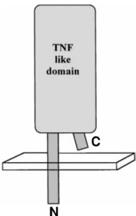
CD154, also known as CD40 ligand, is a member of the tumour necrosis factor family. It’s a type II transmembrane glycoprotein, since the N-terminal part of the protein is intracellular, while its C-terminal portion is extracellular Although preliminary studies on murine CD154 proposed a protein of 39kDa [2], the molecular weight of the protein in most cell types is 32-33 kDa (human and mouse) [3]. Like other members of the tumor necrosis factor (TNF) family, the overall configuration of CD154 consists of a short cytoplasmic tail, a transmembrane domain, an extracellular rod and a globular C-terminal region, responsible for the biological activity. The globular C-terminal region has a three-dimensional structure similar to that of TNF, encompasses amino acids 116 (glycine) to 261 (leucine), is rich in cysteine residues and is able of binding the receptor [4] (Fig 1). Regarding the other domains of CD154, the extracellular rod is composed of 75 amino acids the transmembrane domain of 24 amino acids and the intracellular tail includes 22 amino acids [3].
CD154 was initially reported on the surface of activated T lymphocytes, but was subsequently shown to be expressed on a wide variety of cells including activated platelets, dendritic cells, mast cells, monocytes, and macrophages as well as non-hematopoietic cells such as epithelial cells, smooth muscle cells and endothelial cells [5, 6].
Figure 1. Schematic representation of the CD154 structure.
The CD154 is a type II transmembrane protein with an extracellular domain showing structural homology with other members of the TNF family [3].
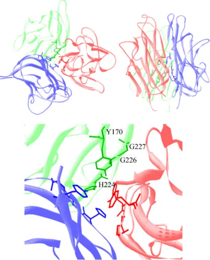
The crystallographic structure of sCD154 reveals a sandwich of two B sheets with a jelly-roll topology, capable of forming a homotrimer. Indeed the structure of soluble CD154 (sCD154) and its folding is crucial for a proper activity as demonstrated in patients with Hyper-IgM (HIM) syndrome where mutations in CD154 affecting the folding of the protein are shown to disrupt the interaction of the molecule with its receptor, CD40 leading to the disease initiation [7]. Indeed, the importance of the three-dimensional structure of CD154 in its interaction with CD40 was also demonstrated in various studies [8]. Researchers have cloned the extracellular domain of CD154 in Escherichia coli to thereby generate the full-length 29 kDa protein, and two fragments of 18 and 14 kDa, that they purified [9]. Only the soluble fraction containing the 18 kDa protein exhibited an activity on B cells and could form a trimer. Interstingly, Fanslow et al., produced three soluble forms of CD154: a monomeric, a dimeric form supported by the hinge region of human immunoglobulin and a trimeric form where the N-terminal domain of the extracellular portion of sCD154 is fused to a pattern “leucine zipper”. Even though all three forms of CD154 can bind to CD40, only the trimeric form can induce B cell proliferation in the absence of stimulus [10]. Moreover, it is known that the sCD154 released from activated T cells is trimetric [11].
Figure 2: The-three dimensional structure of soluble CD154 structure.
Ribbon diagram of human soluble trimeric CD154. Panel A) bottom and side view of the molecule. Panel B) a magnification of the core region showing residues (Y170, H224, G226, G227) that might be implicated in trimer formation[12].
The gene coding for CD154 is located at chromosome Xq26.3-Xq27.1[13], where it occupies almost 12-13 kb of DNA [3]. Figure 3 illustrates the gene structure of CD154 human. The gene consists of five exons and four introns. The first exon encodes the intracellular region of the protein, transmembrane region and part of its extracellular domain (6 amino acids), while exons 2 to 5 encode the rest of the extracellular region of CD154 [3]. Coding regions are lined with non-coding regions consisting in the 5’ of 45 base pairs (bp) and in 3’ has 975 bp, including a poly-A tail [14]. Northern blot analysis of activated T cells revealed the presence of two RNA messengers of 2.1 and 1.4 kilobase (kb) that differs by the length of their untranslated region 3’ [15]. The cDNA of CD154, with approximately 1.8 kb length encodes a polypeptide membrane of 261 amino acids containing a cytoplasmic domain of 22 amino acids, a hydrophobic transmembrane domain of 24 amino acids and an extracellular domain of 215 amino acids [2, 14].
Figure 3: Gene structure of human CD154.
The gene of CD154 located at chromosome Xq26.3-Xq27.1 consists of 5 exons and 4 introns. (VanKooten,and Banchereau 2000) [3]
1.2. The receptors of CD154
The first characterized receptor for CD154 [16], CD40, is a 45–50 kDa integral membrane glycoprotein that belongs to the tumour necrosis factor receptor (TNFR) superfamily. For almost two decades, CD40 was considered the sole receptor for CD154. Recently, it has been reported that CD154 also binds to αIIbß3 integrin [5], α5β1 [17] and αMβ2 (also known as Mac-1) [18]. Although all the newly identified receptors belong to the integrin superfamily, each of them interacts with CD154 in a specific manner. While only inactive α5β1 and active αMβ2 interacts with CD154, αIIbβ3 interact with CD154 in both its inactive and active forms [17-19].
1.2.1. CD40
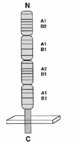
CD40 is a 48-kDa type I transmembrane glycoprotein composed of 277 amino acids including an extracellular domain (N-terminal) of 193 amino acids, a transmembrane domain of 22 amino acids and a cytoplasmic tail of 62 amino acids. The CD40 gene is located at chromosome 20q11-2-q13-2 [15]. Its extracellular domain is rich in cysteine residues and is similar to that of other TNFR family members (Fig.3), giving CD40 the property of forming disulfide bonds and allowing protein folding [15]. Earlier studies showed that CD40 can exist as a homodimer [20], however little is known in this matter until one report stated that CD40 homodimers are a consequence of cell activation [21]. More recently, we demonstrated that low levels of disulfide bond-CD40 (db-CD40) homodimers are constitutively expressed on the cell surface, and that ligation of CD40 with sCD154 leads to a rapid and significant increase in the level of CD40 homodimers [22]. The db-CD40 dimers are predominantly localized within lipid rafts, and disruption of lipid raft integrity abolishes CD40 homodimer formation. Cysteine at position 238 (Cys238) of the CD40 cytoplasmic tail is required for its disulfide-linked homodimer formation, and for some
but not all CD40-induced responses (Fig. 1) [23]. Indeed, CD40-triggered CD23 expression and CD40 enhancement of toll-like receptor (TLR)-induced responses is totally dependent on CD40 homodimer formation [24].
Figure 4.Structure of CD40 protein.
The CD40 is a type I transmembrane protein. Its extracellular region has 4 cysteine-rich sub-domain [3].
CD40 is expressed on antigen presenting cells (APC) like B lymphocytes, macrophages and dendritic cells [25]. It is also constitutively expressed on other hematopoietic and non-hematopoietic cells, such as eosinophils, endothelial cells, epithelial cells, smooth muscle cells, fibroblasts as well as platelets. These cells could also exhibit an inducible higher expression of CD40 in pathological conditions [3, 26].
The intracellular domain of CD40 has no enzymatic activity and is not bound to a kinase/phosphatase [27]. Since CD40 is deprived of a kinase domain, it employs associated cytoplasmic molecules for intracellular signal transduction. Tumor necrosis factor receptor-associated factors (TRAFs) werefirst identified as intracellular proteins associatedwith TNFR2, a member of the TNF receptor (TNFR) superfamily. There are currently six known TRAF familymembers (TRAF1-6) that initiate signals transduced by members of the TNFR family, members of the TLR family, as well as the IL-1R. TRAFs have been implicated in a vast array of critical cellular events induced by CD40 engagement. For example, TRAF2 is involved in CD40-induced JNK activation and, together with TRAF5 and TRAF6, triggers NFκB activation. Furthermore, TRAF3 and TRAF5 are involved in CD40-induced B7 and CD23 upregulation on B cells [28, 29]. CD40 engagement induces tyrosine phosphorylation and activation of Jak3, which is constitutively associated with CD40. This interaction is required for the expression of CD23, ICAM-1, and LTα genes in B cells [30].
According to the co-crystal structure of CD154/CD40 [8] and the results generated using scanning site-directed mutagenesis [31], CD154 residues involved in CD40 binding are centered on at least two clusters of residues (K143, Y145, Y146 and R203, Q220) [32]. Interestingly, while determining the crystal structure of the CD154 and CD40 complex, it was reported that hydrophilic and charge interactions are implicated in the formation of such complex [8], and showing again that Y145 and R203 residues of CD154 are among those implicated in the ligand-receptor interaction [8].
1.2.2. Integrins
Integrins are membrane receptors that form the dynamic link between the cell cytoskeleton and the extracellular matrix (ECM). The integrin family of proteins consists of alpha (α) and beta (β) subtypes, which form transmembrane heterodimers [33]. Eighteen α subunits and 8 β subunits have been identified and can form 24 different integrins named according to their composition (eg: α4β1,: α5β1,: αvβ3…) [34]. The extracellular part of these integrins has a folded configuration, which can unfold under the influence of some intracellular signals allowing molecule to become active. These integrins provide a bidirectional transfer of information playing an important role in cell-cell adhesion as well as in signal transduction to the cytoskeleton, also activating different signalling pathways. Indeed, an inside-out signalling will permit the passage of intracellular messages, activates the integrin and induce its binding to the ECM while an outside-in signalling will convey the message of proliferation or survival depending on the ECM [35-37]. Thus, integrins are capable of triggering a bidirectional signalling [38].
Integrins are essentials for the maintenance of tissue integrity, cell migration, cell morphology, proliferation and differentiation. They are also implicated in thrombosis, immune response, hemostasis as well as cancer development [39].
Figure 5: Different integrin subunits and their αβ associations.
Eight β subunits can assort with 18 α subunits to form 24 distinct integrins, blue recognize the tripeptide sequence (RGD), purple mediates adhesion to basement membrane laminins, yellow in the case of β2 and β7 integrins are only expressed on white blood cells, grey are collagen receptors with I/A domains, green are restricted to chordates [40].
1.2.2.1. The integrin αIIbβ3
The αIIbβ3 also known as GPIIb/IIIa or CD41/CD61, is a member of the integrin superfamily that recognizes a tripeptide RGD sequence within molecules such as fibronectin and vitronectin. The expression of αIIbβ3 is mainly restricted to platelets and megakaryocytes [41]. Platelets are important for maintaining blood vessel integrity through hemostasis and are the first line of defense against bleeding therefore platelets
must be activated consecutively and in coordinated manner to properly fulfill their role. Upon the activation of platelets, they are able to adhere, secrete and aggregate
[42]. The αIIbβ3 integrin plays an important role in hemostasis; Kato et al. have shown that bleeding and hemostasis problems seen in patients suffering from
Glanzmann's thrombasthenia are due to mutations in genes encoding the αIIb or β3 chains [43].
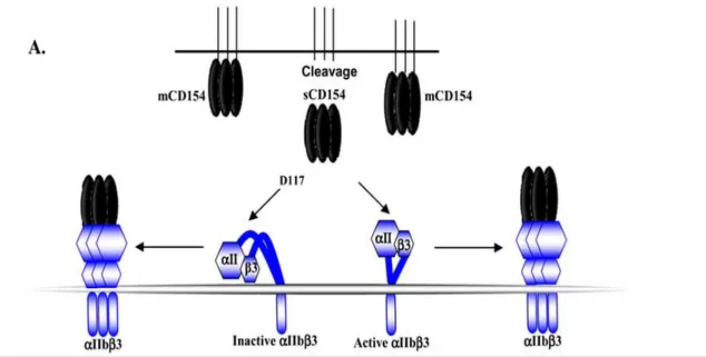
The αIIbβ3 has been identified as a receptor for CD154. Indeed, Andre et al. reported that CD154/αIIbβ3 interactions are mediated by the RGD residues of murine CD154 and the KGD residues of human CD154 (at positions 115-117) [5]. Residue D117 seems to be the major binding residue, as its substitution by alanine (A) abolishes the CD154/αIIbβ3 interaction [44, 45].
1.2.2.2. The integrin α5β1
The α5β1 integrin is expressed on the surface of numerous cell types such as B cells, T cells, monocytes, platelets, epithelial cells, endothelial cells, smooth muscle cells and fibroblasts. Like αIIbβ3, the α5β1 naturally binds fibronectin and vitronectin at their RGD tripeptide sequences [40]. This integrin is involved in cell adhesion, cell migration, cell proliferation and the survival of different cell types [46].
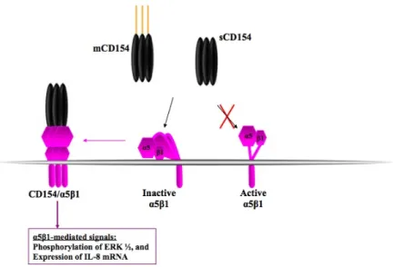
Léveillé et al. showed that CD154 is able to bind to a CD40/αIIbβ3- monocytic cell line via α5β1 [17]. CD154, unlike the natural ligand of α5β1, binds the inactive form of the integrin. Indeed, it has been demonstrated that the CD154/α5β1 binding is lost after the activation of α5β1 with Mn2+ or dithiothreitol. More recent studies reported that residues asparagine at position 151 (N151) and Glutamine at position 166 (Q166) of CD154 are implicated in its binding to α5β1. Moreover, our group also showed that CD154 is capable of simultaneously interacting with CD40 and α5β1 [45].
1.2.2.3. The integrin αMβ2
The αMβ2 (also known as Mac-1) integrin is mainly expressed on neutrophils and monocytes/macrophages [47].The integrin αMβ2 is highly involved in neutrophil functions due to its ability to identify numerous protein and non-protein ligands [48]. Like other integrins, αMβ2 naturally binds fibrinogen [49] and vitronectin [50] but also binds the C3bi [51], adhesion molecules such as ICAM-1[52] and heparin. Its main function is to allow the adhesion and transmigration of leukocytes to the site of inflammation [53].
Recently, αMβ2 has also been identified as a receptor for CD154 [18]. CD154 binds only to the active form of the αMβ2. This connection is established with the αMβ2 I-Domain, a sequence of 220 amino acids in the αM chain of the integrin, and more specifically with a EQLKKSKTL motif within this domain [53]. In addition, our group has recently demonstrated that CD154 interacts with αMβ2 via the same residues involved in its interaction with CD40 (Y145 and R203) [45].
2. The biological role of the interaction of CD154
with its different receptors:
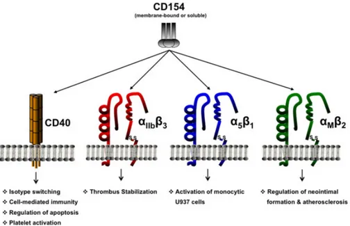
Figure 5 : CD154 and its receptors.
Binding of CD154 to its classical CD40 counter-receptor regulates numerous critical biological responses [54].
2.1. The CD154/CD40 interaction
Although CD154 and CD40 have no signaling motifs in their cytoplasmic tail, it is well established that their engagement with specific mAbs or with their binding partner activates various cellular events and modulates cell functions.
2.1.1. Signal triggered via CD154
The first evidence was the finding that injection of CD40-Ig into CD40 Knockout (KO) mice induces small but quantifiable Germinal Centers after immunization [55]. Following this observation, it has been demonstrated that ligation of CD154 with a specific monoclonal antibody (mAb) induces a variety of signaling pathways including PTK, JNK, p38, Rac-1, and p56lck activation [56-61]. Along the same line of evidence, we reported that relocalization of CD154 into lipid rafts, via its transmembrane domain is necessary and specific for PKCα, PKCδ, p38, and IL-2 secretion but not ERK1/2 activation [62, 63], reinforcing the notion that lipid rafts play a critical immunological role in T-cell activation. In the course of these studies, we observed a decrease in CD154 expression when CD154 expressing cells were co-cultured with CD40-positive cells or stimulated with soluble CD40. This process was shown to be due to CD154 release from the cell surface. We then focused on elucidating the mechanisms by which CD154 is shed from the cell surface. Our recent results indicate that ligation of CD154-positive T cells with CD40 leads to the shedding and release of CD154 from the surface membrane. This process requires the enzymatic activity of the ADAM-10 and ADAM-17 metalloproteinases (MMPs) [64].
2.1.2. Signal triggered via CD40
Engagement of CD40 on immune and non-immune cells with CD154 or specific mAbs triggers several cellular events that modulate cell functions (reviewed in [3, 6, 28]). In resting B cells, ligation of CD40 results in B cell proliferation, differentiation, Ig production [65], and prevents the apoptotic death of germinal center B cells which contributes to the development of B cell memory [66, 67]. In dendritic cells, CD40 signaling induces an increased surface expression of MHC, co-stimulatory, and adhesion molecules, and induces high levels of cytokine synthesis such as IL-12P40 and IL-6 [68]. CD40 is also a crucialelement in the process of lung
fibroblast activation, as ligation of CD40 induces their proliferation, an increased expression of adhesion molecules, and their production of inflammatory mediators [69]. During the inflammatory response, expression of CD40 is increased on fibroblasts, thereby allowing greater interactions with inflammatory CD154+ cells such as T cells and mast cells. The cellular responses result from the activation of signaling pathways that include PTKs such as Lyn, Fyn, and Syk, phosphatidylinositol-3-kinase (PI-3K), PLCγ2, Jak3, p38, JNK and ERK1/2 [28, 29]. CD40 employs associated molecules for adequate transduction of intracellular signals [28, 70]. Several members of the tumor necrosis factor receptor-associated factor (TRAF) family (TRAF2, 3, 5, and 6) have been identified and shown to be associated with various residues of the CD40 cytoplasmic domain [28, 29, 71, 72]. In addition, CD40 engagement induces tyrosine phosphorylation and activation of Jak3, which is constitutively associated with CD40, and this association is required for CD40-induced expression of CD23, ICAM-1, and LTα genes in B cells [30].
2.1.3. Involvement of CD154/C40 interaction
in disease development
The primary function of CD154 is linked to the thymus-dependent humoral response via its interaction with CD40 [16]. The importance of this interaction was demonstrated by the fact that point mutations or deletions in the gene coding for CD154, cause the X-linked Hyper IgM syndrome (HIM) [73]. Patients suffering from this syndrome have normal counts of circulating B cells, but low levels of serum IgGs, IgAs and IgEs, while they exhibit elevated levels of IgMs. As a result, these individuals are susceptible to various infections.
Today, it is well established that CD154 shares a broader role than originally described, as it is involved in a variety of inflammatory immune responses [3, 31], some of which lead to the pathogenic processes of chronic inflammatory disorders
and autoimmunity [74, 75]. Studies approved the implication of CD154 with multiple immune and inflammatory conditions such as rheumatoid arthritis (RA). Indeed, RA patients show high expression of CD154 on different vascular and synovial cell types. Moreover, CD154 interact with B cells via CD40 achieving B cell survival and production of rheumatoid factor (RF), the mediator in RA [47, 76]. Further immune-inflammatory functions of CD154 include the release of cytokine/chemokine, the production of MMPs and the up-regulation of adhesion molecules in synovial monocytes/macrophages, fibroblasts and articular chondrocytes [77]. Indeed, the administration of antibodies against CD154 to mouse models of RA was shown to attenuate inflammatory events and disease severity, supporting the role of CD154 in the pathogenesis of this disease [77, 78]. In other cases such as systemic lupus erythematosus (SLE), the administration of antibodies against CD154 in mice suffering from syndrome similar to SLE, significantly reduce symptoms including inflammation and extends survival. The administration of CTLA4Ig combined with antibodies against CD154 inhibits the production of autoantibody [79]. Furthermore, administration of antibody against CD154 in mice suffering from an experimental allergic encephalomyelitis (EAE), a condition that mimics multiple sclerosis (MS) in human, reduces clinical symptoms and inhibits the production of auto-antibodies [80]. The effect of antibody against CD154 is more significant once it is combined with CTLA4Ig, demonstrated by a complete cessation of cellular infiltration in the central nervous system [81]
Other CD154-driven pathological conditions include vascular disorders such as thrombosis, throughout which CD154/platelet interactions are central. Inwald and colleagues reported that CD154 triggers platelet P-selectin expression and dense granule release, critical steps in platelet activation, through its interaction with CD40 [82]. The role of CD40 in CD154-mediated platelet activation has been further shown in other studies [83, 84]. Interestingly, our group reported that sCD154 is capable of enhancing platelet activation in vitro, and thrombus formation in vivo in a CD40-dependent mechanism [85]. These findings identify CD154/CD40 interactions
as enhancers rather than inducers of platelet function, but nevertheless provide further evidence as to the important pro-thrombotic role of CD154.
2.2. The CD154/αIIbβ3 interaction
CD154 was shown to stabilize arterial thrombi by its binding to αIIbβ3, the major platelet integrin and a receptor for fibrinogen and soluble von Willebrand factor [19, 41]. Knowing that sCD154 contains a KGD motif (lysine-glutamate-aspartate), a specific binding motif for αIIbβ3, André and colleagues were interested in the arterial occlusion phenotype in mice deficient in CD154 (CD154-/-). They found that the absence of CD154 affected the stability of arterial thrombus and delayed the occlusion in vivo, although the initial formation of thrombus is not affected. Indeed, sCD154 acts as an agonist for platelet activation and aggregation. Indeed, the binding of CD154 to αIIbβ3 induces the phosphorylation of tyrosine-759 at the intracellular portion of the chain β3 [41]. This activation of αIIbβ3 mediates the transition of the integrin from low affinity state to a high affinity one, allowing αIIbβ3 to bind soluble fibrinogen. The binding of fibrinogen then permits the formation of bridges between platelets and eventually the phenomenon of aggregation.
Figure 6: The interaction of CD154 with the αIIbβ3.
CD154 interacts with the active and inactive form of integrin αIIbβ3 via the D117 residue [86].
2.3. The CD154/α5β1 interaction
Experiments have demonstrated the binding of recombinant sCD154 marked with Alexa-488 (rsCD154-A) to CD40/αIIbβ3-negative myelomonocytic cells, U937 cells [17]. This interaction was blocked by purified soluble α5β1 integrin, a fibronectin receptor constitutively expressed on monocytes [87] and by a monoclonal antibody directed against α5β1, PID 6. Binding of rsCD154 to U937 cells induced the activation of MAP kinases, ERK1/2 and p38, the gene expression of IL-8 and the translocation of the integrin α5β1 to the insoluble membrane fraction in Triton X-100, confirming that the integrin α5β1 is a functional receptor for CD154, at least in monocytes [17].
This CD154/α5β1 interaction was later shown to synergize with that of CD40 leading to pro-inflammatory cytokine and MMP production [44].
Figure 7: Interaction of CD154 with α5β1.
Membrane and soluble CD154 is capable of binding to the inactive form of the α5β1 inducing the activation of some signaling cascades [86].
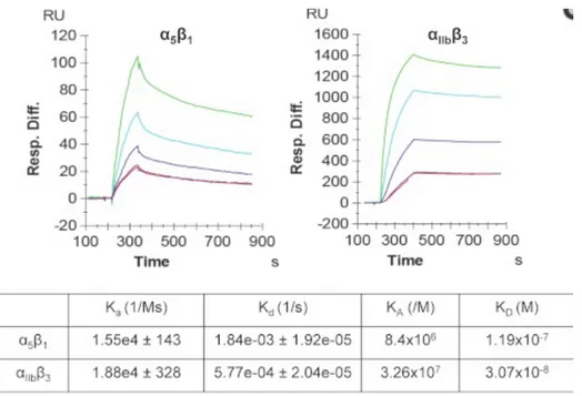
Figure 8 : Binding affinity and kinetic analysis of the interactions between sCD154 and α5β1 and between sCD154 and αIIbβ3, as determined by the surface plasmon resonance approach.
The binding affinity of CD154 for αIIbβ3 is 4 fold higher than for α5β1[44].
2.4. The CD154/ αMβ2 interaction
The CD154/αMβ2 interaction is capable of activating monocytes in vitro and promoting adhesion between ECs and monocytes at the site of atherosclerotic lesions in vivo. This interaction mediates the production of necessary cytokines including MIP-2, interleukin-1β, interleukin-8, the procoagulant tissue factor, and the proinflammatory transcription factor nuclear factor κ-B,, chemokines and
myeloperoxidases that lead to the development of inflammation and atherosclerosis [18] .
In 2011, Wolf et al. demonstrated that inhibition of the binding CD154 to αMβ2 decreased the accumulation of monocytes and significantly reduces the formation of an atherosclerotic plaque, further confirming the role of the CD154/αMβ2 dyad in atherosclerosis development [53]
Figure 9: CD154 interaction with αMβ2 (Mac-1).
3. Thrombosis and cardiovascular diseases
Thrombosis is a clot forming also called thrombus in a blood vessel that blocking it. The clot can form in a vein or an artery. it is called venous thrombosis respectively (or phlebitis) and arterial thrombosis [88]. The most serious complication of deep venous thrombosis is pulmonary embolism when the clot breaks off from where it was formed and migrated to the pulmonary artery, which can lead to sudden death. Complications of arterial thrombosis are myocardial infarction (heart attack), stroke or other vascular events in eg the lower limbs or intestines. There are several treatments to prevent thrombus formation or dissolve.
Two types of thrombosis:
Venous thrombosis or phlebitis: superficial it may be affecting a surperficial veins, or affecting the deep veins, particularly in the thigh. It can be triggered by the injection of a intravenous drug, trauma, inflammation, bacterial infection, varicose veins, cancer, an oral contraceptive [89].
Arterial thrombosis blocking an artery, which is more serious if the artery in question is the only irrigate a specific area of the body, It can be triggered by an injury to the wall of an artery resulting in clot formation in contact with the damaged area. This is what occurs in atherosclerosis often implicated in heart attacks or strokes, mainly caused by toxic substances either to the walls of arteries, or for blood clotting: tobacco, hormonal conception [90].
Platelet activity is particularly involved in the thrombotic complications following percutaneous coronary intervention, as in stents investments. When the vessel wall is damaged or the endothelium is altered, collagen in the subendothelial matrix becomes exposed leading to the formation of a thrombus and triggering the accumulation and activation of platelets, in contrast to tissue factor that initiates the secretion of thrombin which activates platelets [90, 91].
4. Platelet CD154 and its proinflammatory and
prothrombotic functions
Platelets constitutively express CD40 on their surface. Following their activation, platelets will also exhibit surface CD154 expression [92]. Beside being involved in homeostasis and thrombus formation, platelets are considered as mediators and effector cells in inflammation, by the secretion of many proinflammatory substances and by their direct interaction with leukocytes [93].
CD154 of platelets may interact with CD40 expressed on other cell types. It can induce several inflammatory responses and promotes coagulation as it interacts with positive CD40 cells such as vascular endothelial cells or monocytes. CD40/CD154 dyad stimulates cytokine secretion such as IL-1, IL-6, IL-12, and TNF-α and increases the expression of adhesion molecules in endothelial cells including intercellular adhesion molecule (ICAM)-1, vascular adhesion molecule (VCAM)-1, and E-selectin, secretion of matrix metalloproteinases (MMPs), such as MMP-1, -2, -3 and -9 [26] and that of tissue factor in monocytes [94], in addition it modulates adaptive immunity by enabling the maturation of dendritic cells, amplifying CD8 + T cells response and inducing the switching of Ig classes in B cells [95].
Different studies targeted the role of CD154 in platelet function. André et al. have shown that recombinant sCD154 binds to the αIIbß3 integrin on activated platelets, thereby inducing platelet spreading, as well as promoting platelet aggregation under high shear rates [19]. On the other hand, Inwald and colleagues reported that CD154 triggers platelet P-selectin expression and dense granule release, critical steps in platelet activation, through its interaction with CD40 [82]. The role of CD40 in CD154-mediated platelet activation has been further shown in other studies [83, 84]. Our group has reported that soluble CD154, by interacting with CD40, is capable of
shape change and actin polymerization. In addition, our data revealed the signaling pathway downstream of CD154 stimulation to involve TNF receptor-associated factor-2 (TRAF-factor-2), Rac1, p38 MAPK [85], and NFκB [96]. Thus, these data elegantly show that sCD154 is capable of enhancing platelet activation in vitro, and thrombus formation in vivo in a CD40-dependent mechanism [85]. More recently, CD154 was shown to enhance collagen-induced platelet aggregation and thrombus formation independently of CD40 in a mechanism involving PI3K/Akt activation [97].
Chapter 2: Rationale and Hypothesis
CD154 has been identified on the surface of activated platelet and shown to play an important role in the stabilization of the thrombus [19]. Therefore, CD154 could represent a therapeutic target for cardiovascular diseases. Indeed, anti-CD154 mAb therapy was effective in various mouse models of autoimmune disease, and even showed beneficial results in the treatment of human patients [75, 98] ; however, serious adverse effects such as thromboembolic complications have been reported. These events could be explained by the simultaneous interaction of the Ab with CD154 and Fc receptors on the surface of platelets, or by the ability of the Ab to trigger activation of CD154+ platelets, thereby promoting platelet aggregation and thrombosis [19, 99]. To overcome these adverse effects, other approaches such as knockouts, antisense oligonucleotides and siRNAs for CD40 were developed. These approaches were focused on the CD154/CD40 interaction and did not address the interaction of CD154 with the other receptors. Thus, there is a need for novel CD154 treatments for the prevention/abrogation of CD154-mediated diseases such as thrombosis that take into account all receptors of CD154 and that do not involve antibodies in order to minimize or completely block treatment complications. In this matter, recombinant DNA technology have been widely used to manipulate and modify protein structure and thus synthesize therapeutic antagonist proteins [100]. In the case of CD154, we took advantage of the fact that CD154 is biologically active only in its trimeric form and we generated CD154 monomers as potential antagonists of CD154-mediated responses.
Given the implication of CD154 in multiple inflammatory diseases such as vascular disorders, and the fact that a biologically active CD154 is an absolute trimeric molecule, we hypothesize that CD154 monomers not capable of trimerization would inhibit the development of CD154-mediated thrombotic events.
To verify this hypothesis, we proposed the following objectives: 1. Study the role of residues in trimerization of CD154.
2. Study the specificity of these mutants in binding different receptors.
3. Investigate the effect of these CD154 interfering molecules on thrombus formation in mice.
Chapter 3: Material and Methods
2.5. Cell lines
The monocytic cell line U937 (Human leukemic monocytic lymphoma), BJAB (Human Burkitt Lymphoma B) and the HEK 293 (Human embryonic Kidney) were obtained from the ATCC (American Type Culture Collection , Manassas, VA, USA). Cells were cultured in RPMI 1640 or DMEM supplemented with 5% heat-inactivated FBS (Fetal Bovine Serum) (Wisent, St-Bruno, QC, Canada) and 1% of PSG (10,000 U of Penicillin, 10,000 29.9 µg of Streptomycin and 29.9 µg/µL L-Glutamine) (GIBCO, Burlington, ON, Canada).
2.6. Antibodies and Reagents
Anti-CD154 hybridoma 5C8 (IgG2a) and anti-CD40 hybridoma G28.5 (IgG1) were obtained from ATCC. The isotype controls anti-TSST-1 mAb2H8 (IgG1) and anti-SEB mAb 8C12 (IgG2a) were developed in our laboratory. The following antibodies were purchased: rabbit anti-phospho-p38, anti-p38Abs and anti-α5β1 (JBS5) mAb (Cell Signaling Technology, Beverly, MA,USA), goat anti-mouse (GAM) IgG-fluorescein isothiocyanate (FITC) Ab (Sigma, St.Louis, MO,USA), anti- αMβ2 mAb (ICRF44; BD Biosciences, Mississauga, ON, Canada); and goat anti-rabbit (GAR) IgG-HRP Ab and GAM IgG-HRP Ab (Santa Cruz Biotechnology, Santa Cruz, CA, USA). The GAM-Alexa488 (Goat Anti-Mouse Alexa-fluor 488), the Streptavidin-Alexa fluor 488 and the goat anti-hamster (GAH)-Alexa 488 were obtained from Invitrogen Life Technology, Burlington, Ontario, Canada.
2.7. Plasmids and mutagenesis
The pMTBip/V5-His-(A) vector was purchased from Invitrogen Inc. pMTBip/V5-His-(A)-hsCD154wt (human soluble CD154 wild type) was generated by subcloning sCD154wt at BgI Il and Xhol in pMTBip/V5-His-(A) vector. Prior to the cloning, the 450bp sequence encoding the soluble form of wild type CD154 (333bp to 783bp; M113 to L261 SEQ ID NO.X) was first amplified by PCR from pSecTag2A/CD154 (gift from Dr Daniel Jung) using a 5' and 3' sCD154 primers containing respectively a BgIII and Xhol restriction site (5'sCD154-Bglll: 5' CGGGAGATCTATGCAAAAAGGTGATS' SEQ ID NO.X, 3'sCD154-Xhol:5' TAGACTCGAGTT TGAGTAAGCC 31 SEQ ID NO.X).
To generate monomeric forms of both murine and human sCD154, single and double mutants of soluble CD154 were constructed using PCR-overlap method (Table.1) and generated by PCR-overlap extension methods. Briefly, a 5' PCR product was generated from pMTBip/V5-His-(A)-sCD154wt using the 5' sCD154-Bglll primer describe above and a reverse mutagenic primer. A 3'PCR product was also generated, using a forward complementary mutagenic primer and the 3'sCD154-Xhol. The two overlapping PCR products were mixed and a final PCR was performed using the 5' and 3'sCD154 external primers. The PCR product was then subcloned in pMTBip/V5-His-(A) at BgIII and Xhol. The four plasmid constructs pMTBip/V5-His-(A)-hsCD154Y170A and pMTBip/V5-His-(A)G227A and the two murine equivalents were then sequenced to confirm mutation. To generate human sCD154 double mutant Y170/G227 plasmid construct of Y170 single mutant was used as template with the different mutagenic primers G227 as described for generating single mutant. Murine double mutant were also generate in the same manner. All double mutant constructs were also sequenced to confirm mutation.
Mutants Primers
Y170A CD40L-Y170A-C 5" caa gga etc tat get ate tat gee 3' CD40L-Y170A-B 5" ggc ata gat age ata gag tec ttg 3' G227A CD40L-G227A-C 5' cac ttg gga gca gta ttt gaa ttg 3
CD40L-G227A-B 5' caa ttc aaa tac tgc tec caa gtg 3'
Table 1: Primers used for generation of monomeric form of CD154
3.4. Cell culture, transfection and protein
purification
Schneider's (S2) cells were cultured in complete DES Schneider's Drosophila medium (Invitrogen Corp.), supplemented with 10% heat inactivated Fetal Bovine Serum (F.B.S) (Wisent Inc.), 100 Units of Penicillin G Sodium, 100 mg/ml of Streptomycin Sulfate and 0.25 mg/ ml of amphotericin B as fungicide (Invitrogen Corp.) at a cell density between 1.5 - 3 X10⁶ cells/ ml at 26°C without CO2. To produce recombinant soluble CD154 protein with
different point mutations, cells were co-transfected with pMT-BiP/V5-6x HisA carrying sCD154 DNA sequence and pCoHygro vectors (Invitrogen Corp.) at a weight ratio of 19:1 , respectively, by the method of calcium phosphate. The clones were then selected with a final concentration of 300 mg/ml of hygromycin B (Wisent Inc.) The expression of recombinant sCD154 proteins were tested by seeding 1-2 X 10⁶ cells in 100 mm petri dish followed by
induction with 100 mM of copper sulfate for 24 hours, cell debris were removed and further Western blot analysis of supernatant generated were performed. A stable population of hygromycin resistant S2 cells was obtained after 20 days period. The selected cultures were initiated to a large scale-up in Drosophila-SFM serum free medium (Invitrogen Corp.) supplemented with antibiotics- antimycotic (Invitrogen Corp.) without addition of hygromycin B. Upon induction, the proteins remaining in the supernatant were recovered by centrifugation of supernatant at 3000g for 20 min. The samples were filtered through a 0.22 mm filter and concentrated to 1/20 initial volume with Centricon (Amicon 8400) concentrator. The
used to purify sCD154 proteins on HisTrap-HP column (Amersham-Pharmacia Biotech). Concentration of the proteins were determined using bradford protein assay (Thermo Scientific Pierce BSA Protein Assay Standards)
3.5. Flow cytometric analysis
Cell-surface analysis was performed with specific mAbs or labeled CD154-Alexa-488 (sCD154-A).Washed cells were then analyzed by FACS LSR-II (BD Biosciences, Mississauga, Ontario, Canada) and Flowjo program (BD Biosciences).For binding analysis, cells were incubated with CD154WT, CD154-Y170, CD154-G227, CD154-G/Y in staining medium for 30 min at 4ºC for BJAB cells, and at 37ºC for U937 and HEK-293 cells. Washed cells were the incubated with biotinylated anti-CD154 (αv5) for 30 min at 4ºC, followed by streptavidin-phycoeruthin (Sigma) for 30 min, washed , and analyzed on FACSort. For competition binding assay U937 or BJAB cells (2x10⁵ cells/100µL) were incubated in binding assay medium (RPMI 1640, HEPES 10mM, bovine serum albumin (BSA 1%) containing 20ng of labeled Alexa in the absence or in the presence of Y170, G227, or CD154-Y170/G227, for 1h at 37ºC in a humidified incubator and a 5%C02 atmosphere. Washed cells were analyzed on a FACSort (BectonDickinson, Mountain View, CA, USA)
3.6. Western Blotting
Polyvinylidene fluoride (PVDF) membranes were blocked in blotto ( 5% skim milk in Tris saline pH 7.5, 0.15% Tween 20 (Fischer Scientific) for 1 h at room temperature. The phosphorylation of p38, and ERK1/2 was assessed by immunoblotting using specific Abs according to the manufacturer’s instructions. Membrane were stripped (62mM,Tris-HCL pH 6.8, 2% SDS/100mM,2-ME) for 30
min and reprobed with antibodies recognizing total p38, or ERK1/2. Antigen/antibody complexes were revealed with ECL( CE Healthcare, Mississauga, ON, Canada). Imaging western blot using X-ray Film ( Thermo Scientific CL-Xposure Film)
3.7. Animals
We first developed an in vivo approach to induce thrombosis in a mouse model
female 12 to 14 week-old C57BL/6. Carotid thrombosis was induced using ferric chloride (FeCl3). This model, widely used to assess arterial thrombosis in mice, was validated in our laboratory [85]. Briefly, after dissection of the left carotid artery, blood flow is measured with a pulsed Doppler flow system (Transonic Systems) and carotid injury is induced by placing a filter paper saturated with 10% FeCl3 solution on the adventitial surface proximal to the flow probe for 3 min, after which blood flow and time to thrombotic occlusion (blood flow of 0 ml/min) are measured. In addition, blood flow is monitored for an additional 10 minutes post-thrombosis for spontaneous reflow, which is indicative of thrombus instability. Mice (6 mice/group) received IV treatments (monomeric CD154 mutants 60 µg/mouse or a control anti-αIIbβ3 antagonist) 10 min prior to the FeCl3 injury of the left carotid.
Chapter 4: Results.
4.1. CD154 mutants (Y170 or G227) fail to induce
intracellular signalling
The trimeric state is primordial for the biological activity of CD154. By examining the crystal structure of CD154 (83), it was hypothesized that residues located at position Y170 and G227 in human CD154 sequence may be involved in trimer formation. Mutagenesis of each of these residues was performed by mutating them to alanine residues. Recombinants were then produced, purified, and first analyzed for their purity by Coomassie Blue and Western blot showing one single band with no contamination detected (Fig. 10). Results suggest a monomeric form for Y170 and G227, Functional analysis was then undertaken by stimulating BJAB (CD40-positive cells) and U937 (α5β1-(CD40-positive) for 5 min with CD154WT or mutants CD154 at 300ng and activity was monitored by detection of p38 phosphorylation. Fig. 11 shows a significant decrease in the activity of Y170 and G227 mutants, suggesting that these monomeric CD154 mutants fail to induce cell activation.
4.2. CD154 mutants capable of binding to all cells
These two CD154 mutants, as well as the CD154 double mutant G/Y (CD154 mutated at both Y170 and G227) were tested for their binding capacities in BJAB, U937, and HEK293 cells transfected with αMβ2 mutated at the GFFKR region within the α chain, rendering the molecule constitutively active. As shown in Fig. 12, these mutants are capable of binding to CD40, αIIbβ3, α5β1, or αMβ2. Thus, these CD154 mutants maintain their ability to bind receptors, but fail to induce intracellular signaling as mentioned above.4.3. CD154 binding to BJAB and U937 was inhibited
by CD154 mutants
Herein, we want to investigate the capacity of monomeric CD154 mutants to modulate the binding of sCD154 to CD40, α5β1, or αMβ2 expressed on cell surface. To reach our aim BJAB and U937 cells were used as model cell lines. Cells were incubated with CD154-Alexa in the absence or the presence of CD154 (Y170), CD154 (G227) or CD154 (Y170/G227) mutants.
Results indicate that the binding of CD154-Alexa to BJAB and U937 was inhibited by both monomeric single mutants but most importantly by the double mutant CD154 (Y170/G227) (Fig. 13). These data support the monomeric status of these mutants and their ability to bind to their receptors and to block the binding of endogenous CD154.
4.4. Inhibition of thrombus formation in mice
We will assess herein, the usefulness of monomeric CD154 mutants that bind to their receptors without triggering any intracellular signal (Fig. 10 and Fig. 11) as inhibitors of CD154 interactions for the prevention of thrombosis.
In this experiment, soluble monomeric CD154 were used. Results shown in Figure 13, demonstrate that thrombosis was impacted by treatment with the sCD154 monomeric form, Y170 or the monomeric form, G227 as reflected by the amplitude (%) of blood flow plotted against time. Interestingly, the CD154 double mutant (Y170/G227) completely inhibited the FeCI3-induced thrombosis. Our data
demonstrate that monomeric CD154 represents an efficient therapeutic tool of great value in the treatment of CD154 related various inflammatory disorders in general and thrombosis in particular.
Figure 10. Coomassie Blue staining (A) and Western blot analysis (B) of purified CD154 mutants.
For Western blot, 100 ng of protein was loaded on 10% SDS-PAGE gel, transferred to membrane, and blotted with rabbit anti-CD154 Ab, followed by anti-rabbit HRP conjugated Ab.
Figure 11. CD154 mutants (Y170, G227 and Y170/G227) are unable to induce intracellular signaling .
BJAB (CD40+) or U937 (α5β1+) cells were stimulated for 5 min with CD154WT or mutants or with medium alone and activity was monitored by detection of p38 phosphorylation. Total p38 was used to control for sample loading. Data shown are representative of three independent experiments (each experiment was performed in duplicate)
Figure 12. CD154 mutants (Y170, G227 and Y170/G227) are capable of binding to CD154 receptors.
BJAB (CD40+), U937 (α5β1+) and HEK293 (α5β1+) transfected with vector alone or expressing active αMβ2 (αMΔβ2) were incubated with CD154WT or CD154 monomers then with biotin-conjugated anti-CD154 Ab followed by streptavidin-PE. Cells were then analyzed by Flow cytometry (empty plot= anti-CD154; grey plot= negative isotype control). Data shown are representative of three independent experiments (each experiment was performed in duplicate)
Figure 13. CD154 mutated at its Y170 and/or G227 residues is capable of inhibiting the binding of CD154WT.
BJAB (CD40+) or U937 (α5β1+) cells were incubated for 1h with CD154-Alexa in the absence or presence of CD154 monomer, then analyzed by Flow cytometry. The dotted empty plots indicate the binding of CD154-Alexa in the presence of CD154 monomers and the black plots represent the CD154-Alexa binding in their absence (The negative
control is represented in the empty continuous line-plot). Data shown are representative of three independent experiments (each experiment was performed in duplicate)
Figure 14.Thrombosis in mice in the presence or absence of CD154-interfering molecules.
Thrombosis formation was induced by FeCl3 in carotid arteries of C57Bl6 mice, and blood flow was measured with a pulsed Doppler flow system, Mice (6 mice/group) were treated, prior to the injury with saline (control), F(ab) fragments of an isotype control mAb, anti-CD154 mAb, or the GR144 (a αIIbß3 antagonist used here as a positive control), in (A), and with monomeric CD154 (Y170, G227 or Y170/G227) in (B). The graph represents the amplitude (%) of blood flow plotted against time for the different treatment groups.
Chapter 5: Discussion
Over the last five years, The laboratory of doctor Mourad and collaborators provided significant insights into the structural/functional interaction of CD154 with its receptors(s). Indeed, based on the crystal structure of CD154 [7], they generated monomeric forms of CD154, which retain their ability to bind their receptors without inducing intracellular signals. These agents represent a potential therapeutic tool for the treatment of CD154 related diseases.
The CD154/CD40 dyad, initially known for its role in humoral responses, was later shown to be implicated in cell-mediated immunity and thus playing a critical part in the development of several inflammatory and autoimmune diseases such as atherothrombosis, rheumatoid arthritis, lupus erythromatosis, lupus nephritis, oophoritis, and Crohn’s disease [101]. Several pre-clinical studies have demonstrated the promise of agents capable of modulating the CD154-mediated responses for the prevention/treatment of immune-mediated diseases [75, 98]. Indeed, the disruption of CD154/CD40 interactions using anti-CD154 monoclonal antibodies (mAbs) was
therapeutically successful in animal models and in patients with autoimmune/inflammatory diseases but was discontinued due to life-threatening complications [75, 98]. Therefore, there is a need for new methods and reagents based on CD154 for the prevention and treatment of CD154-mediated conditions such as thrombosis.
Different mAbs have been used to imitate the functions of human proteins or just hindering these functions, yet the recent biopharmaceutical approaches focus on manipulating and modifying protein structure through different methods such as recombinant DNA technology in order to synthesize therapeutic antagonist proteins [100]. Indeed, studies demonstrated that TNF monomers engineered with point
dominant-negative heterotrimers that neither bind or induce signalling through TNF receptors [102]
The function of CD154 is based on two different aspects of protein-protein interactions. First, the interaction between CD154 monomers allowing CD154 trimer formation [103] as discussed in the next paragraph, and which is an essential process for the subsequent interaction of CD154 with its receptor, CD40, and trimerization of this latter. This CD154/CD40 crosstalk will thus induce intracellular signalling by engaging TRAF adaptor proteins to the cytoplasmic domain of CD40 [104]. The interaction between CD154 and its receptors involves specific amino acids. According to the co-crystal structure of CD154/CD40 [8] and the structural analysis of scanning site-directed mutagenesis [6], the CD154 residues involved in CD40 binding are centered on at least two clusters of residues (K143, Y145, Y146 and R203, Q220) [32] whereas CD154/αIIbβ3 interactions are mediated by the KGD residues of human CD154 (positions 115-117) [5]. Using scan-directed mutagenesis and structural analysis, our group reported that CD154 binds to the α5β1 receptor via distinct residues than those implicated with CD40 and αIIbβ3 suggesting a possible simultaneous binding of CD154 to its receptors [44]. Based on experiments assessing the competitive binding of CD154 to its various receptors [44], and the protein sequence and structure alignment between TNFα and CD154, our group generated several CD154 mutants, and demonstrated that CD154 R203/Y145 mutant lost its ability to interact with CD40- and αMβ2-positive cells, while maintaining its binding to α5β1. The CD154 N151/Q166 mutants lost its ability to interact with α5β1-positive cells but not to CD40-positive or αMβ2-positive cells. Taken together, these data indicate that residues N151 and Q166 of CD154 are required for α5β1 binding whereas CD154 residues required for the interaction with the αMβ2 integrin, Y145 and R203 are shared with those involved in CD40 binding [45].
Different residues of CD154 were also shown to be involved in its homotrimer formation. Indeed, CD154 similar to other TNF family proteins, is identified as a
domain of human CD154, suggested that CD154 residues Y170 and H224 are implicated in its trimeric stucture [7, 105]. Data obtained from our current study also demonstrated a role for the residue Y170 of CD154 in its trimerization as substitution of Y170 by alanine rendered the molecule biologically inactive while still capable of binding to receptors. It is worth noting at this point that we also undertook single-point mutations of residues H224 as well as residue G226 and generated their corresponding recombinants, produced and purified them. However, CD154 mutants H224 and G226 were not analyzed further as they were insoluble under biological conditions (data not shown).
These above findings strongly indicate that mutations of certain amino acid residues could affect the structure and the function of CD154 as is the case in X-linked Hyper IgM syndrome (HIM) [106], a condition characterized by mutations in the gene coding for CD154. Some mutations at the level of the CD154 gene affected the expression level of the protein [107-109]; however other CD154 mutations affected the functional aspect of the protein by reducing its capacity to trimerize and/or to interact properly with CD40 [105, 107]. Similarly, different mutations in the human TNFα affected its activity depending on the site of the mutation, i.e.: mutations involving internal residues inhibited trimerization, while most mutations of surface residues affected receptor binding but not trimer formation. However all TNFα mutants incapable of forming trimers, showed little cytotoxic activity towards the L929 fibroblast cell line, suggesting the importance of trimer formation for a biologically active TNFα [110]. These results are in harmony with our current study, where we generated CD154 mutated at Y170 and/or at G227 residues, that are incapable of forming trimers. While these mutants maintain their binding capacities to all CD154 receptors, their biological activity is abrogated. Thus, these mutants can be used as CD154 antagonists, binding to CD154 receptors without activation while inhibiting the binding of endogenous CD154.
are essential. Indeed, André et al. have demonstrated that recombinant soluble CD154 binds to the αIIbß3 integrin on activated platelets, inducing platelet aggregation [19]. On the other hand, Inwald and colleagues reported that CD154 triggers platelet P-selectin expression and dense granule release, important steps in platelet activation, through its interaction with CD40 [82]. The role of CD40 in CD154-mediated platelet activation has been more identified in other studies [83, 84]. In addition, our group has previously demonstrated that sCD154 is capable of enhancing platelet activation in vitro, and thrombus formation in vivo in a CD40-dependent mechanism [85]. CD154 showed also a significant role in the pathology of rheumatoid arthritis (RA) and vascular events such as thrombosis. Indeed studies showed an association between thrombotic events and increased serum levels of CD154 in RA patients [111, 112]. These data reveal CD154/CD40 interactions as enhancers of platelet function and provide more evidence as to the important prothrombotic role of CD154. Given the role of CD154 in vascular disorders, we wanted to investigate, in the current study, the ability of CD154 monomeric mutants to prevent thrombus formation. Our data showed the capacity of our CD154 double mutant (Y170/G227) to completely inhibit thrombus formation in a mouse model of thrombosis. In this matter, we used the FeCl3-induced thrombosis in mice which is a well established model for this vascular condition and has been used and well documented in several studies by our group as well as others [113-116]. Interestingly, the morphology of the thrombi induced by FeCl3 application is similar to those found in humans [115], thus allowing our results to be translated to human settings in future clinical studies.
Our group has previously reported that CD154 residues involved in the interaction with the CD40 and αIIβ3 are different from those involved in the interaction with α5β1. This suggests that the trimetric CD154 could simultaneously engage both receptors[44].These observations surge the need for generating CD154 mutated at a specific residue implicated with binding and activating of one receptor, without affecting the binding of another. Interestingly these mutants may provide the development of novel therapeutic strategies aiming at inhibiting a particular
