THE ROLE OF INTERCELLULAR COMMUNICATION AND
OXIDATIVE METABOLISM IN THE PROPAGATION
OF IONIZING RADIATION-INDUCED
BIOLOGICAL EFFECTS
by
Narongchai AUTSA V APROMPORN
Département de Médecine Nucléaire et de Radiobiologie Thcsis Submitted to the Fm.:ulty of Medicine and Health Sciences
for the Dcgrce ofDoctor of Philosophy (Ph.l>.) in Radiobiology
Thesis Examiner Thesis Examiner Thesis Examiner Thesis Supervisor Thesis Supervisor May 2011
Doctoral Committee:
Prof. Carmel Mothershill, McMaster University Prof. Benoit Paquette, Université de Sherbrooke Prof.
Éric
Rousseau, Université de Sherbrooke Prof. Jean-Paul Jay-Gerin. Université de Sherbrooke Prof. Edouard I. Azzam, University of Medicine and Dentistry of New Jersey - New Jersey Medical School
,_,,-NOTICE:
Published Heritage Branch
395 Wellington Street Ottawa ON K1A ON4 Canada
The author has granted a
non-exclusive license allowing Library and Archives Canada to reproduce, publish, archive, preserve, conserve, communicate to the public by
telecommunication or on the Internet, loan, distrbute and sell theses
worldwide, for commercial or non-commercial purposes, in microform, paper, electronic and/or any other formats.
The author retains copyright ownership and moral rights in this thesis. Neither the thesis nor substantial extracts from it may be printed or otherwise reproduced without the author's permission.
ln compliance with the Canadian Privacy Act some supporting forms may have been removed from this thesis.
While these forms may be included in the document page count, their removal does not represent any loss of content from the thesis.
C d ...
ana.a
Direction du
Patrimoine de l'édition 395, rue Wellington Ottawa ON K1A ON4 Canada
AVIS:
Your file Votre référence ISBN: 978-0-494-89663-1 Our file Notre référence ISBN: 978-0-494-89663-1
L'auteur a accordé une licence non exclusive permettant
à
la Bibliothèque et Archives Canada de reproduire, publier, archiver, sauvegarder, conserver, transmettre au public par télécommunication ou par l'Internet, prêter, distribuer et vendre des thèses partout dans le monde,à
des fins commerciales ou autres, sur support microforme, papier, électronique et/ou autres formats.L'auteur conserve la propriété du droit d'auteur et des droits moraux qui protege cette thèse. Ni la thèse ni des extraits substantiels de celle-ci ne doivent être imprimés ou autrement
reproduits sans son autorisation.
Conformément
à
la loi canadienne sur la protection de la vie privée, quelques formulaires secondaires ont été enlevés de cette thèse.Bien que ces formulaires aient inclus dans la pagination, il n'y aura aucun contenu manquant.
List of illustrations (Tables and Figures) Acronyms, abbreviations and symbols Abstract
Résumé 1. Introduction
I. l. Physics of radiation biology I.1.1. Ionizing radiation I.1.2. Linear energy transfer
I.1.3. Relative biological effectiveness I.2. Biological effects of ionizing radiation
I.2.1. The action of ionizing radiation on DNA I.2.2. DNA damage and biological effects I.2.3. Cellular responses to DNA damage
I.2.4. Ionizing radiation-induced reactive oxygen species I.2.4.1. Radiolysis of water
I.2.4.2. Oxidative stress and antioxidant enzymes
I.2.4.3. Effects ofionizing radiation on the cell membrane I.2.5. Repair of radiation damage
I.3. Targeted and non-targeted effects of ionizing radiation 1.3.1. Targeted effects
1.3.2. Non-targeted effects 1.3.3. Mechanisms
1.3 .3 .1. Role of intercellular communication 1.3.3.2. Role ofoxidative metabolism I.4. Objectives of the research project
1.4.1. Hypothesis 1.4.2. Experimental strategy Page üi vii ix X 1 4 4 5 6 7 7 9 11 14 14 17 18 19 23 23 24 27 27 33
40
40
40
II. Article No.1
"The role of gap junction communication and oxidative stress in the
propagation oftoxic effects among high-dose a.-particle-irradiated human cells" N. Autsavaprompom, S.M. de Toledo, J.B. Little, J.-P. Jay-Gerin, A.L.
Harris and E.I. Azzam. Radiation Research 175:347-357 (2011)
III. Article No. 2
Page 42
80
"Intercellular communication amplifies stressful effects in high-charge, high-energy (HZE) particle-irradiated human cells" N. Autsavaprompom, SM. de Toledo, M. Buonanno, J.-P. Jay-Gerin, A.L. Harris and E.I. Azzam.Journal of Radiation Research (In press)
IV. Article No. 3
"Permeability of connexin channels mediates human cell responses to ionizing radiation" N. Autsavaprompom. S.M. de Toledo, J.-P. Jay-Gerin,
A.L. Harris and E.I. Azzam.
Journal of Radiation Research (In preparation)
103
V. Discussion 126
V .1: The role of gap junction intercellular communication in the responses of 126 human cells to high LET radiation
V .2. The role of intercellular communication and oxidative metabolism in 133 the propagation of stressful effects in human cells exposed to a.-particles
V.3. The role ofselective permeability of gap junctions in the nature of the 139 biological effect propagated among human cells exposed to low- or high-LET radiations V .4. Future direction VI. Conclusion Acknowledgements Bibliography 143 151 153 154
List of illustrations (Tables and Figures)
CHAPTER 1 - Introduction
Fig. l Projections over the XY plane of track segments calculated (at -10·13 s) 6
for (a) 1H+ (0.15 MeV), (b) 4He2+ (1.75 MeV/nucleon), (c) 12C6+ (25.5
MeV/nucleon), and (d) 2°Ne1o+ (97.5 MeV/nucleon) impacting ions.
Fig. 2 Variation ofRBE with LET 7
Fig. 3 Mode of action ofradiation on a cell 8
Table 1 Estimation of the number of early physical and biochemical changes that 10 occur when mammalian cells are irradiated with 1 Gy oflow LET radiation.
Fig. 4 DNA damage response pathway 11
Fig. 5 Activation of ATM in response to DSB and downstream effects 14 Fig. 6 Time scale of events in the low LET radiolysis of water 16 Fig. 7 Oxidative and antioxidant systems in mammalian cells 18 Fig. 8 Fraction survival of C3H lOTl/2 cells exposed to two equal doses from
X rays or neutron and incubated at 37" C for various intervals between
the doses 20
Fig. 9 The fràction survival response of confluent human fibroblast AG 1522 22 cells exposed to
y
rays (4 Gy) ora
particles (0.85 Gy) and held inconfluence for different times_ at 3 7°C prior to subculture.
Fig.10 Induction of sister chromatid exchanges (SCE) by low doses of
a
24 particlesFig.11 Gap junctions structure 28
Fig.12 Gap junction intercellular communication mediates the propagation 30 of stressful effects from irradiated to non-irradiated cells.
Table 2 Cloning efficiencies for four cell lines exposed to 5 Gy dose 31 or recipient of medium from cell cultures irradiated with 5 Gy
Fig.13 W estem blot analysis of p21 Wafllevels in AG 1522 human 34 cultures exposed to low fluence
a
particle in the presenceor absence of antioxidant enzymes CHAPTER II- Article No. 1
Fig.1 Potentially lethal damage repair in confluent AG 1522 cells exposed to 137Cs y rays or 3.2 MeV a particles
Fig. 2 Oxidative stress in a particle-irradiated AG1522 cells
Fig. 3 Role of gap-junction intercellular communication in the propagation of stressful effects among a particle-irradiated confluent cells: Effects of the gap~junction inhibitor 18-a-glycyrrhetinic acid (AGA)
Fig.4 The effect of connexin43 knockdown in the propagation of stressful effects among a particle-irradiated cells
Fig. 5 The role of oxidative metabolism in the propagation of a particle-induced stressful effects
CHAPTER Ill - Article No. 2 Fig.1
Fig. 2
Modulation ofstressful effects in confluent AG1522 normal human fibroblasts exposed to energetic protons or iron ions as a function oftime after irradiation
Role of gap junction intercellular communication in the propagation of stressful effects among energetic proton or iron ion-irradiated confluent AG 1522 cell cultures
Table 1 Dosimetry parameter
for
confluent AG 1522 cells irradiated with 1 GeV protons or 1 GeV/u iron ions.CHAPTER IV - Article No. 3 Fig.1
Fig. 2
Clonogenic survival ofHeLa cells expressing connexin26 or
connexin32 assayed within 5-10 min after exposure to different types of ionizing radiation ·
Micronucleus formation in HeLa cells that express connexin26 or
66 67 68 69 70 96 97 98 119 120 connexin32 after exposure to different types of ionizing radiation. The cells were manipulated for the assay within 5-10 min after irradiation.
V
Fig.3 Viability ofHeLa cells that express either connexin26 or 121 connexin32 following exposure to different types of ionizing radiation CHAPTER V - Discussion Fig.1 Fig. 2 Fig.3 Fig. 4 Fig. 5 Fig. 6' Fig. 7 Fig. 8 Fig. 9
The RBE, relative to y rays, of protons,
a
particles or iron ions with different LET values as determined at the 10% survival lev el in irradiated confluentAG1522 cellsClonogenic survival of confluent or sparse AG 1522 cells exposed to low LET y rays or high LET
a
particles assayed within 5-10 min or a 3 h incubation after irradiationRole of gap junction intercellular communication in the
propagation of stressfu.1 effects among irradiated confluent cells: Effects of the gap junction inhibitor AGA on Clonogenic survival. Role of gap junction intercellular communication in the
propagation of stressful effects among irradiated confluent ce Ils: Effects of the gap junction inhibitor AGA on micronucleus formation The role of gap junction communication and oxidative metabolism
127
129
130
131
136 in the propagation of a particle-induced stressful effects. The effect of ectopie over-expression of glutathione peroxidase on clonogenic survival of AG 1522 ce Ils that were incubated at 3
r
C for 3 h post-irradiationThe role of_gap junction communication and oxidative metabolism 137 in the propagation of
a
particle-induced stressful effects. The effect of ectopie over-expression of glutathione peroxidase on clonogenic survival of AG 1522 cells that were incubated at 3r
C for 3 h post-iITadiation.The relationship between RBE and LET determined at the 10% survival 140 level in HeLa cells that express either connexin26 or connexin32
following exposure to protons, a. particles, iron·ions ory rays and assay within 5-10 min after irradiation.
Induction of apoptosis in irradiated HeLa cells that express either connexin26 or connexin32
Clonogenic survival of confluent NB 1 RGB normal human
fibroblasts exposed to low- or high-LET carbon ions at isosurvival dose 142 147
levels and subcultured for assay within 5-10 min after irradiation or following 3 and 24 h at 37°C in the presence or absebce of a gap junction inhibitor.
Fig. 10 Fraction of micronucleated bystander cells in cultures exposed in the 149 presence or absence of a gap junction inhibitor to X rays from
microbeam irradiator as a function ofràdiation dose
Fig. 11 Fraction of micronucleated bystander cells from cultures exposed 150 in the presence or absence of a gap junction inhibitor to microbeam argon ions: The effect of dose
î a.
a
p s min hµm
AGA
ATM
ATP
cGy Cx DNADSB
keV LET MeV fs Ge V GJICGPX
Gy kDa HNE HR HZE IR MN NHEJ OERAcronyms, abbreviations and symbols
Gamma Alpha Delta Probability of an event Second Minute Hour Micrometer 18-a.-glycyrrhetinic acid Ataxia-telangiectasia mutated Adenosine triphosphate Ceiitigray Connexins Deoxyribonucleic acid Double strand break
Kilo-electronvolt Linear energy transfer M ega-electronvo 1 t Femtosecond Giga-electronvolt
Gap junction intercellular communication Glutathione peroxidase
Gray Kilo-dalton Hydroxynoneal
Homologous recombination
Higb charge and high energy particles Ionizing radiation
Micronucleus formation Non-homologous end joining
Acronyms, abbreviations and symbols (continued)
pllwan Cyclin-dependent kinase inhibitor lA p53 Tumor suppressor gene 53
pKa Acid dissociation constant
PE
Plating efficiencyPLDR
Potentially lethal damage repairRBE
Relative biological effectivenessRIT Radio-immunotherapy
ROS
Reactive oxygen speciesRNS
Reactive nitrogen speciesSCE
Sister chromatid exchangesSF
Survival fractionsiRNA Small interfering ribonucleic acid
SLDR
Sublethal damage repairSOD
Superoxide dismutaseSSB
Single strand breakAbstract
Coordinated interactions of specific molecular and biochemical processes are likely involved in the cellular responses to stresses induced by different ionizing radiations with distinctive linear energy transfer (LET) properties. Here, we investigated the roles and mechanisms of gap junction intercellular communication and oxidative metabolism in modulating cell killing and repair of potentially lethal damage (PLDR) in confluent AG1522 human fibroblasts exposed to 1 GeV protons (LET-0.2 keV/µm), 137Cs y rays {LET-0.9 keV/µm), 241Am a particles (LET-122 keV/µm) or 1 GeV/u iron ions (LET-151
keV/µm) at doses by which all cells in the exposed cultures are irradiated. As expected, a.-particles and iron ions were more effective than protons and y rays at inducing cell killing. Holding y-or proton-irradiated cells in the cônfluent state for several hours after irradiation promoted increased survival and decreased chromosomal damage. However, maintaining a-particle or iron ion-irradiated cells in the confluent state for various times prior to subculture resulted in increased ratber than decreased lethality, and was associated with persistent DNA damage and increased protein oxidation and lipid peroxidation. lnhibiting gap junction communication with 18-a-glycyrrhetinic acid or by knockdown of connexin43, a constitutive protein of junctional channels in the se cells, protected against the toxic effects expressed in these cells during confluent holding. Up-regulation of antioxidant defense by ectopie over-expression of glutathione peroxidase, protected against cell killing by a-particles when cells were analyzed shortly after exposure. However, it did not attenuate the decrease in survival during confluent holding. Together, these findings indicate that the damaging effect of a particles results in oxidative stress, and the toxic effects in the bours following irradiatfon are amplified by intercellular communication, but the communicated molecule(s) is unlikely to be a substrate of glutathione peroxidase.
To further understand the rote of GJIC, we tested the effect of specific connexin channel permeabilities on radiation-induced cell killing and induction ofDNA damage. We used human adenocarcinoma (HeLa) cells in which specific connexins can be expressed in the absence of endogenous connexins. When exposed to protons, y rays, a particles, or iron ions, connexin26 and connexin43 cbannels mediated the propagation oftoxic effects among irradiated cells; in contrast, connexin32 channels conferred protective effects. ·
Collectively, these studies provide a novel mechanistic understanding of the molecular events that mediate the fate of cell populations exposed to different types of ionizing radiation. They show that the LET of the radiation significantly impacts these events. The enhancement of cell killing in the hours after exposure oftumor cells to high charge and high energy particles and or a. particles support the use of these particles in cancer radiotherapy. Characterization of the molecules that are communicated through junctional channels from tumor to normal cells would help fonnulate countermeasures to protect normal tissues during radiotherapy. Future in vivo research would contribute to validating these concepts.
Résumé
Les interactions coordonnées des processus moléculaires et biochimiques sont probablement impliquées dans la réponse au stress cellulaire induit par des rayonnements ionisants de transfert d'énergie linéaire (TEL) différent. Ici, nous avons étudié le rôle des jonctions de type gap et le métabolisme oxydatif dans la modulation de la mort cellulaire et la réparation des dommages potentiellement létaux (PLDR) dans des cultures a confluences de fibroblastes humains AG1522. Ces cultures ont été exposées à des protons d'énergie 1
GeV (TEI.r-0,2 keV/µm), des rayons y de 137Cs (TEL-0,9 keV/µm), des particules a.
d'241Am (TEL-122 keV/µm) ou des ions fer d'énergie 1 GeV/u (TEL-151 keV/µm) et à
des doses où toutes les cellules exposées sont irradiées. Comme attendu, les particules a et les ions fer ont été plus à même d'induire la mort cellulaire que les protons et les rayons y. Le maintien des cellules à confluence pendant plusieurs heures après irradiation aux rayons
y et aux protons favorise la survie cellulaire et diminue les dommages chromosomiques. En revanche, le fait de maintenir dans un état de confluence des cellules traversées par des particules a. pour des temps donnés différents aboutit à un accroissement de la mort cellulaire, ce qui a été associé à une augmentation des lésions de l'ADN, à l'oxydation des protéines et à .la peroxydation lipidique. D'autre part, l'inhibition des jonctions gap par l'acide 18-a-glycyrrhétinique ou la diminution de l'expression de la protéine connexine 43, protéine constitutive des canaux jonctionnels de ces cellules, ont un effet protecteur contre les effets toxiques des radiations. Une sur-régulation des protections anti-oxydantes par une surexpression anormale de la glutathion peroxydase protège les cellules contre les effets toxiques lorsque celles-ci sont analysées peu après l'exposition. Toutefois, cela n'a pas atténué la diminution de la survie lors du maintien de cellules à l'état de confluence. L'ensemble de ces données indiquent que les dommages induits par les particules a. résultent du stress oxydant et que ces effets toxiques sont amplifiés par la communication intercellulaire dans les heures suivant l'irradiation. Cependant, la (les) molécule(s) transmise(s) n'est probablement pas un substrat de la glutathion peroxydase.
Pour mieux comprendre le rôle des jonctions gap, nous avons testé l'effet de la perméabilité d'une connexine (Cx) spécifique sur la mort cellulaire radio-induite et l'induction de dommages de l'ADN. Nous avons utilisé des cellules issues d'adénocarcinome humain (HeLa) dans lesquelles des connexines spécifiques peuvent être exprimées en l'absence des connexines endogènes. Suite à l'exposition aux protons, aux rayons y, aux particules a ou aux ions fer, nous avons constaté que les canaux formés de Cx26 et ceux formés de Cx43 jouent un rôle dans la propagation des effets toxiques parmi les cellules irradiées tandis que les canaux formés de Cx32 confèrent des effets protecteurs.
Collectivement, ces études apportent une compréhension mécanistique nouvelle des événements moléculaires qui interviennent dans le devenir des populations de cellules exposées à différents types de radiations ionisantes. Elles montrent que le TEL des radiations peut avoir des répercussions importantes sur ces événements. L'induction de la mort cellulaire des cellules tumorales dans les heures suivant l'exposition à des radiations de haut TEL ou de particules a. est en faveur de l'utilisation de ces particules en radiothérapie. La caractérisation des molécules transmises des cellules tumorales aux cellules normales via les canaux jonctionels pèrmettrait de formuler des mesures pour protéger les tissus sains durant la radiothérapie. D'autres recherches sont nécessaires pour étudier la pertinence de ces conclusions in vivo et valider ces concepts.
1. Introduction
Ionizing radiation (IR) is an effective method of cancer radiotherapy, diagnostic radiology, and nuclear medicine (SAUNDERS et al, 1985; BLAK.EL Y and KRONENBERG, 1998; HALL .and G~CCIA, 2006; DURANTE and LOEFFLER 2010). It consists of particulate or electromagnetic types with low- or high-linear energy transfer (LET) properties (TUBIANA et al., 1990; HALL and GIACCIA, 2006). The ionization and excitation events produced as a result of cellular traversai by different types of IR are dependent on the energy and mass of the ionizing particle. LET is defined as the energy transferred per unit h:ngth of the track (TUBIANA et al., 1990; HALL and GIACCIA, 2006). Extensive studies indicate that radiation-induced _biological effects are dependent on LET (ELKIND 1984; TODD et al., 1985; GOODHEAD et al., 1993; TSURUOKA et al., 2005; HAMADA et al., 2006; FRANKENBERG et al., 2006; WHALEN et al., 2008). In comparing the biological effects of different radiations, it is customary to use X-rays as the standard. The relative biological effectiveness (RBE) of high LET radiation such as a particles, neutrons, or heavy ions is significantly greater than that of low LET radiation such as high energy protons (TUBIANA et al., 1990; BLAKEL Y and KRONENBERG, 1998; HALL and GIACCIA, 2006). Various biological end points such as cell killing, chromosome aberration, induction of DNA damage, mutation induction, cell transformation and change in gene expression support the LET dependence of radiation effects (see, for example: YANG et al., 1985; HEI et al., 1988; KASTAN et al., 1991; CHEN et al., 1993; BELYAK.OV et al., 1999; KAWATA et al., 2004; GUIDA et al., 2005; DESAI et al., 2005; DING et al., 2005; WHALEN et al., 2008; TSURUOKA et al., 2008, Autsavaprompom et al. 2011 ). However, the mechanisms underlying LET effects remain unclear.
Recently, human exposure to IR has been on the increase. In addition to exposure to background radiation, humans are exposed to radiation from industrial applications, fallouts from weapons testing and significantly from medical applications such as diagnostic radiology or nuclear medicine procedures. In addition, with the expansion of the space program and the initiation of long-term space flights, there is great concem by the National Aeronautics and Space Administration (NASA) in the biological effects ofhigh charge (Z) and high energy (E) ions known as HZE particles (e.g., iron ions) and ofhigh energy protons (CUCINOTTA et al., 2006).
Cell traversal by a single HZE or a particle results in the deposition of a large amount of energy along the particle tracks, with the potential of producing clustered DNA damage (up to 25 lesions per cluster) and damage to other molecules such as proteins and lipids. In contrast, cellular exposure to comparable doses from low LET radiation (e.g. y rays or energetic protons) generates sparse ionizations that result in clusters with a maximum 10 lesions (CUCINOTTA et al., 1998; SUTHERLAND et al., 2001; SEMENNENKO and STEWART, 2004). Therefore, it is important to understand how a biological response is produced by low- or high-LET radiations and how the effect is processed in cells.
Recent evidence has suggested that gap junction intercellular communication (GJIC) and oxidative metabolism are involved in the propagation of stressful effects from irradiated to non-irradiated bystander cells in an exposed population to
a
particles (AZZAM et al., 1998, 2001, 2002, 2003, 2004; Zhou et al., 2001; SHAO et al., 2003b). The involvement of these mechanisms in the propagation of signaling events among irradiated cells has not been investigated previous to this project. Gap junctions are dynamic intercellular membrane channels that are critical for diverse physiological fonction implicated in the control of cell homeostasis, proliferation and death. They allow the direct exchange of small molecules (-1 kDa) that are well above the size ofmost secondary me~sengers between adjacent cells. They are composed of connexins (Cx), which are members of a large family of proteins. Different Cxs form channels with specific permeability properties (KOREEN et al., 2004; KING and BERTRAM, 2005; SHAO et al., 2007; HARRIS and LOCKE, 2008) and play a critical role in cellular responses to IR (AZZAM et al. 1998, 2001, 2003). The nature of the signaling molecule(s) communicated through gap junction channels linking irradiated cells with bystander cells remains unknown.
Two mechanism of transmission of molecules among irradiated cells and between irradiated and unirradiated neighbor cells have been proposed. They consist either in diffusible factors éxcreted into the cell culture medium or factors that are directly transmitted by GJIC (AZZAM et al., 1998, 2001, 2002, 2003, 2004; MOTHERSILL and SEYMOUR, 1997, 1998, 2001). It is thought that IR-induced formation of teactive oxygen species (ROS) is the messenger that triggers damage to cellular constituents, including proteins, lipids and DNA. It contributes to the biological effects of both low- or high-LET radiation. Though a burst of excess ROS is initially produced at the time of irradiation and is believed to persist for only microseconds or less (SPITZ et al., 2004), radiation-induced oxidative stress on the cell may be prolonged due to persistent long-term effects on oxidative metabolism (AZZAM et al., 2001, 2004). Exposure to IR may affect mitochondrial and membrane oxidases (Burdon, 1996) leading to excess ROS production, and may also disrupt antioxidant activity. ROS produced at the time of irradiation or subsequently as a resuft of perturbations in oxidative metabolism modulates the expression of signaling pathways in irradiated cells, regulates intercellular communication, including GJIC, as well as cell growth, differentiation and apoptosis (HARRIS and LOCKE, 2008; UPHAM and TROSKO, 2009). However, the mechanisms by which intercellular communication and oxidative metabolism contribute to low- or high-LET radiations
have not been clearly elucidated. A better understanding of the mechanisms of radiobiological effects of low- or high-LET radiation on human cells and tissues is of significance to radiation therapy and radiation protection.
1.1. Physics of radiation biology
1.1.1. Ionizing radiationEnergetic particles or electromagnetic ionizing radiations (IR) transfer their energy when they interact with matter, thus causing ionization (i.e., emission of electron from atom) or excitation (i.e., an interaction that transfer energy, but does not completely displace an electron). Examples of electromagnetic radiation are X or y
rays. The latter rays consist of a spectrum of waves, like other electromagnetic radiations that are non-ionizing such as radio waves, microwaves, infrared, visible light, and ultraviolet light. However, X and y rays are distinctly characterized by their short wavelengths, high frequency, and high energy.
Particulate ionizing radiations, on the other band, include energetic electrons, protons,
a
particles, neutrons, and heavy charged ions. Like X and y rays, particulate radiations induce significant biological effects when they traverse living matter. However, depending on the specific physical characteristics, such as energy and mass, of each type of particulate radiation, the concentration of induced biochemical effects in the traversed matter varies due to unique patterns of energy deposition and ionization events. Unlike the sparse ionization events produced by X rays, y rays, and highly energetic electrons, certain charged particles such asa
particks produce dense ionization columns along the particle path. This is due to the vast difference (-8,000 fold) in the charge-to-mass ratio ofa particles and electrons. Therefore, heavy chargedelements mainly cause clustered DNA damage that results in DNA double strand breaks.
1.1.2. Linear energy transfer
Radiation quality or linear energy transfer (LET) is a term used to describe the different ionization densities produced by IR along the track of the irradiating particle. LET is a measure of the ionization density, and the LET concept is defined as the energy transferred per unit length ofparticle track, in keV/µm. Typically, X or y rays are considered low LET (sparsely ionization) radiations, while energetic neutrons, protons and heavy charged particles are high LET (densely io.nization) radiations (reviewed in HALL and GIACCIA, 2006). Note that the demarcation value between low- and high-LET radiations is at about 10 keV/µm (PODGORSAK, 2005).
Exposure to IR results in the deposition of energy events that lead to DNA damage. The level of DN A damage is believed to increase with increasing LET values of the radiation. Condensed energy deposition results in clusters of ionizing events. Consequently, these ionization clusters can yield numerous lesions in DNA and the site of such lesion is termed clustered DNA damage. High LET radiation is believed to produce high yield of such damage (NIKJOO et al., 2001; reviewed in PODGORSAK, 2005; HALL and GIACCIA, 2006; LEHNERT 2007).
Track structure depends greatly on LET. A high LET radiation such as a
particles will have a thicker and shorter particle track compared to that of low LET radiations such as protons at the same LET (Fig. 1 ). Another issue is the secondary electrons . (ô . rays). Often, the energy deposited in the medium by high LET radiations such as carbon, neon and iron ions is not considered in calculation of LET. However, ô
rays are of low LET radiation and have a torturous track path. Therefore, in reality, certain high LET radiations will have combined high- and low-LET radiation components toits tracks (CUCINOTTA et al., 1998; MUROY A et al., 2006) .
,H+ 1.11 ! 5 (a) j U.S ~ 4 j j ~ 0.6
l'.
:: "i >- 0.4 1 -O.'.! l.:.Her·
(b) (c) \ f {d) I/
t , 0.0 0 ..._ ... ;--.J..._. ... ..._..___,_.._._--''--...1.-..~.J....3 ___.__, -0.20.00.2 ·0.20.0 0.2 -1.5-1.IJ..(J.5 0.0 0.5 -2 .) 0 1 5 X (Inn) X (~;m) X (11m) X (µm)Figure 1. Projections over the XY plane of track segments calculated (at -10·13 s) for
(a) 1H+ (0.15 MeV), (b) 4He2+ (1.75 MeV/nucleon), (c) 12C6+ (25.5 MeV/nucleon), a:nd
(d) 2°Ne10+ (97.5 MeV/nucleon) impacting ions. Ions are generated at the origin and
along the Y axis in liquid water under identical LET conditions (-70 keV/µm). Dots represent the energy deposited at points where an interaction occurred. From MUROY A et al. (2006).
1.1.3. Relative biological effectiveness
The concept of relative biological effectiveness (RBE) has evolved because of the availability of different types of IR that produce a different degree of damage. This is due to the fact that the LET for each type ofradiation is different. RBE is the ratio of the doses of l_ow and high LET radiations that would give the same radiobiological effect. RBE is dependent on LET, dose, dose rate and the biological endpoint investigated. Generally, RBE increases with LET, mainly due to track structure. High
LET radiation is more effective at inducing biological damage that is less repairable than low LET radiation. In general the RBE ofradiation increases with its LET up to a value of about 100 keV/µm and above this value starts to decline due to energy deposition in excess of that needed to cause the biological effect (over kill) (Fig. 2),. At an LET of 100 keV/µm, IR can most effi.ciently produce double-strand breaks by a single track (reviewed in HALL and GIACCIA, 2006; LEHNERT 2007)~
X-ra y 100 keV/p 200 keV/µ
RBE
LET
Figure 2. Diagram illustrating why ionizing radiation with a LET of 100 keV/µm bas
the greater RBE for cell killing, mutagenesis, or oncogenic transformation. From HALL and GIACCIA (2006).
1.2. Biological effects of ionizing radiation
1.2.1. The actions of ionizing radiation on DNA
When cells are exposed to IR, the induced biological effects result mainly from damage to the DNA, which is considered to be the most critical target molecule within a cell. The damage to DNA can be inflicted by two processes: direct action and
indirect action of radiation (Fig. 3 ). Due to their unique inherent physical properties and energy deJ?osition patterns, particulate radiations cause biological changes mainly by direct/y damaging critiçal targets in the cells like DNA. Altemately, electromagnetic radiation interacts with other atoms or molecules in the cell, especially water to produce free radîcals (e.g. hydroxyl, superoxide radicals) and other reactive species that go on to damage critical targets in the vicinity; therefore, they cause cellular damage largely by an indirect manner (reviewed in HALL and GIACCIA, 2006; LEHNERT 2007). Ultimately, these direct and indirect effects of IR produce biological and physiological alterations in the cell or organism that manifest in seconds to even decades after irradiation. This thesis will further explore the mechanisms underlying the biological effeéts electromagnetic and particulate radiations in human cells. DIRECT ACTION IONIZATION (AND EXCITATION
!
_
RADIOCHEMICAL'
PROCES SES
-INDIRECT ACTIONFigure 3. Mode of action ofradiation on a cell. In direct action, an electron resulting from absorption of an incident photon enters the nucleus and ionizes or excites the DNA. In indirect action, the ejected electron interacts with water to produce an "OH radical, which diffuses to and reacts with the DNA. From SELMAN (1983).
1.2.2. DNA damage and biological effects
The most :frequent types of DNA damage produced are base and sugar modifications, single-and double-strand breaks, sites of base loss, tandem lesions, DNA-DNA and DNA-protein crosslinks, and various clustered lesions. The so-called "locally multiply damaged sites" (also termed "clusters of damaged sites" or more conveniently "clustered lesions") refer to the formation of two or more lesions in close proximity along the DNA (including different combinations of all possible singly damaged sites produced in opposite strands or in the same strand within about 10-20 base pairs separation) by a single radiation track (WARD, 1988; GOODHEAD, 1994; BOUDAIFF A et al., 2000). These clustered types of lesions, whose complexity depends on the LET of the radiation, result :from the spatial heterogeneity of the energy deposition events that follow the passage of radiation through matter.
Whereas high LET radiation can cause clusters and dense DNA damages and the direct action is dominant process, the indirect action is dominants for low LET radiation (about two third of the damages) which can cause only sparse DNA damages. Moreover, high LET radiation leads to damage with higher complexity, which can be extremely hazardous to the cells involved. Both high and low LET radiations cause dose-dependent lesions in DNA (Table 1 ),. including single strand breaks (SSB) and double strand breaks (DSB). As this thesis and oth.er studies demonstrate, for the same total absorbed dose, high LET radiation is more damaging to cells than low LET radiation.
Among the DNA lesions, the DSB is the most serions and potentially the most lethal. The DNA SSB are of lesser biological significance as they can be repaired easily and accurately using an opposite template strand. Cells that experience DNA SSB can thus carry out normal cellular functions following DNA repair. In contrast,
DNA DSB is complex and requires a cohort ofDNA repair proteins and
a
multitude of signalling events. The DSBs are mainly repaired by two processes. Homologous recombination (HR) which is error-free but occurs in S and G2 phase of the cell cycle,.
or non-homologous end joining (NHEJ) which is the common mechanism for rejoining DNA DSB in inammalian cells and occurs at ail phases of the cell cycle; however, it is error-prone (BURMA and CHEN, 2004; BURMA et al., 2006).
Failure of cells to repair DNA damage correc~y may contribute to mutagenesis and/or genome instability that can lead to carcinogenesis, aging, inherited disease, and cell death (see, for example: BECKER and SEVILLA, 1993; BREEN and MURPHY, 1995; CADET et al., 1997; WALLACE, 1998; SUTHERLAND et al., 2000; 0 ' NEILL, 2001; HALL and HEi, 2006; VON SONNT AG, 2006).
Table 1. Estimation of the number of early physical and biochemical changes that occur when mammalian cells are irradiated with 1 Gy of low LET radiation. From GOODHEAD (1984).
Initial physical damage
Ionization in the cell nucleus Ionization directly in DNA Excitation directly in DNA
Selected biological damage
Damaged bases Damaged sugar
DNA-protein cross-links DNASSB
DNADSB
Selected cellular effects
Lethal events Chromosome aberrations -1000,000 -2000 -2000 -1000-2000 -1200 -150 -1000 -40 -0.2-0.8 -1
1.23. Cellular responses to DNA damage
As described above, IR induces many types of DNA lesions, of which DNA DSB are the most deleterious. The spectrum of DNA lesions is recognized by DNA-damage-response pathways. As shown in Figure 4, cells respond to DSB through the action of systems that detect the DNA lesion and then trigger various downstream events. These systems can be viewed as classical signal-transduction cascades in which a 'signal' (DNA damage) is detected by a 'sensor' (DNA damage binding protein) that then triggers the activation of a 'transducer' system (protein kinase cascade), which amplifies and diversifies the signal by targeting a series of downstream 'effectors' of the DNA-damage response. This DNA-damage response pathway is extremely sensitive and selective; it is triggered rapidly and efficiently by a low number of and maybe just one DNA DSB, and must remain inactive under other conditions (KASTAN et al., 1991; reviewed in LEHNERT, 2007; JACKSON and BARTEK, 2009).
Endogenous agents
IR chemicals ~ ~ replication
meiotic DSBs
Figure 4. DNA damage response pathway. DSB are recognized by different sensor
proteins, which transmit the signal fo a series of downstream effectors molecules
through a transduction cascade, to activate signalling mechanisms for cell-cycle arrest and induction of repair, or cell death if the damage is irreparable. From LEHNERT (2007).
The early activation and recruitment to the damage sites of the protein kinase Ataxia-Ielangiectasia Mutated (ATM) play a major role in signalling the DSB response pathways in mammalian cells. In response to DNA damage ATM kinase is rapidly auto-phosphorylated and in tum phosphorylates various substrates involved in cell cycle regulation or maintenance of genomic instability (SHILOH, 2003; K.AST AN and BAR TEK, 2004; LA VIN et al., 2005). ATM is most renowned for its regulation of DSB-induced cell cycle arrests that include G1/S, intra G2, and G2/M arrests (SHILOH, 2001; KURZ et al., 2004). ATM also contributes to the regulation of apoptosis in response to DSB (BROWN et al., 2009). ATM deficiency leads to the human cancer predisposition and neurodegenerative syndrome Ataxia Telangiectasia (AT). At the -cellular level, ATM deficiency is manifested by increased sensitivity to IR
(BARZILIAI et al., 2002; reviewed in HALL and GIACCIA, 2006).
Inactive ATM exists as a dimer that undergoes autophosphorylation on serine 1981 in a response to DSB to become an active monomer (BAKKENIST et al., 2003). Recent data suggest that ATM is recruited and activated at sites ofDSB (ANDEGEKO et al., 2001). Activated ATM is also intimately linked with numerous substrates (Fig. 5). Downstream substrates for ATM include Murine double minute-2 (MDM2), Tumour suppressor gene 53 (TP53 or p53), two serine/threonine protein kinase (CHKl or CHK2), Breast cancer type 1 susceptibility protein (BRCAl), Nibrin (NBSl), nonrecepter protein tyrosine kinases (c-Abl) and Stress-active protein kinases (SAPK), which are involved in cell cycle progression, DNA repair or apoptosis. Collectively, these proteins function as key regulator of the DNA damage response and a clear interdependency exist among them as inactivation of any renders cells hypersensitive to DSB (DE TOLEDO et al., 2000, PETRIN! et al., 2003; SEDELNIKOVA et al., 2003: SHILOH, 2003; reviewed in LEHNERT, 2007).
Currently, there is substantial evidence that ATM acts upstream of p53 in a signal transduction pathway initiated by IR. ATM bas intrinsic protein kinase activity. and phosphorylates p53 at serine-15 in response to DNA damage (BANIN et al., 1998; CANMAN et al., 1998). Fu~ermore, ATM, p53 and a p53 downstream effector, the cyclin-dependent kinase inhibitor IA (CDKNIA), known also as p21 wan, have been implicated in the cell cycle arrest (G1) that occurs in human fibroblast cells exposed to low or high LET radiations (DE TOLEDO et al., 2000; AZZAM et ·al. 2000). However, a comprehensive characterization of these effects in cells exposed to high LET radiation bas not been as established. It may be dependent on many factors, such as cell line, types of IR, and dose. For example, relative to low LET X, human TK6 lymphoblastoid cells exposed to iron ions (LET -1000 keV/µm) expressed substantially greater inhibition of S-phase progression as a result of higher frequency clustered DNA damage blocking DNA replication (GOTO et al., 2002). In this project, we use ATM/p53/p21 wan · signaling events to characterize the propagation of stress among irradiated cells and between irradiated and neighboring non-irradiated bystander cells.
..
\.:~, .~,,~~r _. ~'1:JIP~·;!Jl.'i. .1 ATMiATR activation,:B~Lë.: \~
:f>· Mdm2 - 1 P53 - hk2 - .. BRCA 1 NBS1 c·Abl ~·I
c Il') .t~
..
\~
rad51~
i~.~
p21 14
3.3~
13
\
j~~/~o:
rad51 SAPKcdk/2 ·• cdk!2
l
cyclinE cyclin81
+
i
Figure 5. ATM is activated in response to DSB and signais the presence of DNA damage by phosphorylating downstream targets including p53. Downstream effectors of p53 are p21/clipl and 3cr. P21 inhibits the activity of cdk2/cyclin E and 14-3-3 cr inhibits the activity of cdc2/cyclin B causing cell-cycle arrest, which is also mediated by activation of Chkl and Chk2. c-Abl activates SAPK for transcriptional regulation of stress-response gene. Other proteins (BRCAl, NBSI) are involved in DNA repair. From LEHNERT (2007).
1.2.4. Ionizing radiation-induced reactive oxygen species 1.2.4.1. Radiolysis of water
Water is the most predominant molecule in living organisms (about 80% of the mass of a living cell in water) (SELMAN, 1983; reviewed in LEHNERT, 2007), therefore, a major proportion of the radiation energy deposited will be absorbed in cellular water and one must understand the radiolysis of water to understand the early stages in the complicated chain of radiobiological events that follow the passage of radiation and ultimately lead to the observation of a biological response. In this context, IR provokes the decomposition reaction of water producing a variety of ROS or
reactive nitrogen species (RNS) in the cellular environment. These species are likely to be major contributors to the induction of chemical modifications and changes in cells that may subsequently act as triggers of biological damage or signalling effects (MUROYA et al., 2006; MEESUNGNOEN, 2007).
The interaction of IR with water causes ionization and excitation of the water molecules. The sequence of events that occurs chronologically during water radiolysis is usually divided into three characteristic stages: (i) the physical stage (-10·15 s), (ii) the physicochemical stage (-10·12 s), and (iii) the nonhomogeneous chemical stage (-10-6 s) (PLATZMAN, 1958). · For example, following exposure to low LET radiations such as X- or y-rays, the water decompositions are e;q (hydrated electron),
Ir,
OH-, H' (hydrogen atom), 'OH (hydroxyl radical), Hi (molecular hydrogen), H202 (hydrogen peroxide), H02'/02- (hydroperoxyl/superoxide anion radicals, pKa = 4.8) etc., (the time scale of events that occur in the radiolysis of wa~er is shown in Fig. 6). In contrast, under 4igh LET radiations, the general trend is that by increasing the LET of the radiation, the lower the free-radical is (e.g., e;q, 'OH, and H') and the higher the molecular (e.g. Hi) primary yields are. This behavior is explained by the increased intervention ofbiradical reactions as the local concentration ofradicals along the track of tpe radiation increases (A UT SA V APROMPORN et al., 2007; MEESUNGNOEN, 2007).Geminate recombinadon (< 10 fs) "OH+ H30+ Proton transfer (-10 fs) Event H20.-e-tb (<10-40fs) H0 + "OH (-10-40 fil)
1 l
fr+"OH H1~-roo
rs>
l
H20l
H,+OJr _ . Dissociative electron e aq attachment Electron hydration (-240-660 fs)l
Approximate time sca/e -.- -10-16 s H2+ 0(1L>) Nonradiative decay back to ground-state water- - -10·12 s
Formation of free radicals and molecular products in the spurs and diffusion ()f species out of the spurs
--. -10-6s
Figure 6. Time scale of events in the low LET radiolysis of water. From MEESUNGNOEN (2007)
1.2.4.2. Oxidative stress and antioxidant defence
To consider the radiolysis of water, IR leads to the formation of ROS .(e.g., 'OH, H202, Oi) or RNS (e.g., "NO) that are believed to persist for milliseconds or less. A high level of ROS/RNS that might result from disruption in the balance between oxidant production and cellular antioxidant defence produces a state of
oxidative stress, which leads to oxidative damage to biomolecules such as DNA, proteins and lipids that contributes to the radiobiological effect of IR. For example (Fig.
7), the oxidation of polyunsaturated fatty acids in membrane induced by ROS is called lipid peroxidation (ALBANESE and DAINIAK, 2003).
Organisms have protective systems against cellular oxidative stress. Central to these systems is endogenous antioxidant enzymes. Superoxide dismutases (Mn-SOD in mitochondria and Cu-Zn-SOD in the cytosol), glutathione peroxidase (GPX) and catalases (CAT) constitute primary enzymatic defence system. Mn-SOD or Cu-Zn-SOD interact with 02- to form H202and 02. CAT catalyzes the dismutation ofH202to H20 and 02. GPX catalyze the reduction of H202 to H20 using glutathione (GSH) as substrate (HALLIWELL and GUTTERIDGE, 1985; RILEY, 1994;LEHNERT, 2007).
There is much evidence to support the concept that exposure to IR results in the formation of ROS/RNS within minutes of exposing cells to ionizing radiation, which causes oxidative damage to biomolecules. For example, in the dose range between 50-400 cGy from
r
rays, the amount of ROS/RNS detected per cell increased with dose and was accompanied by a decrease in the level of GSH (MORALES et al., 1998). For high LET a particles, the intracellular produétion of ROS/RNS was 50· fold greater than the extracellular production and the effect was inhibited by diphenyleneiodonium (DPI), an inhibitor of flavoproteins, suggesting the involvement of plasmamembrane-bound NADPH oxidase and possible other enzymes (e.g. niric oxide synthases) containing flavoproteins (NARAY ANAN et al., 1997).
DNA/protein damage
Upid-peroxldatlon
Figure 7.
Oxidative and antioxidant systems in mammalian cells. Superoxide anion (02-) is produced in cytosol and mitochondria. Two molecules of 0 2- rapidly dismutase, either spontaneously or via superoxide dismutases (SOD) to dioxygen and hydrogen peroxide (H202), the latter permitting flux of ROS between cellular compartments. H202 can be enzymatically metabolized to 02 and H20 by a number of different enzyme systems or converted to the hydroxyl radical ("OH), which is extremely reactive, via a chemical reaction catalyzed by transition metals. From . LEHNERT (2007).1.2.4.3. Effects of ionizing radiation on the cell membrane
The physicochemical structure of biological membrane makes them peculiarly susceptible to oxidative damage, and consequently a target of IR-generated ROS. In this context, the generation of ROS inducing protein and lipids modifications seems to bè most possible mechanism representing an alternative target to DNA in radiation induced cell damage. For a long time, the biological membranes have been consideredas inactive semi-permeable lipid bilayers consisting of amphipathic phospholipids, sterol and proteins allowing the segregation of molecules in different cell compartments. Polyunsaturated lipids of the plasma membrane contain double bonds between some of their carbon atoni that are susceptible to attack by 'OH and 02. These · radicals act on polyunstaturated fatty acids and irradiation of plasma membrane lipids in the presence o~ 02 results in lipid peroxidation (HALLIWELL and GUTTERIDGE, 1985; LEHNERT, 2007; CORRE et al., 2010).
Lipid peroxidation causes cell damage by its decomposition in breakdown toxic products that are bifunctional aldehydes. These aldehydes can act as bioactive molecules in physiological and/or pathological conditions. They can affect and modulate several cell functions at very low and non toxic concentrations including signais transduction, gene expression, cell proliferation and other cell responses. The most abundant aldehyde that has been identified is 4-Hydroxynoneal (4-HNE). This toxic product has been reported to be involved in cell cycle control, mutagenesis and the regulation of expressiop of a multitude of gene (FENG et al., 2004; LEONARDUZZI et al., 2004). Furtheimore, carbonyl derivatives of proteins are also formed by the interaction of protein amino acid side chains with lipid peroxidation products including 4-HNE. Carbonyl derivatives of proteins constitute suitable biomarkers of ROS;.mediated. protein oxidation (ST ADTMAN, 2006). Covalent modifications of proteins due to radiation-induced ROS may serve as an indicator of oxidative stress induced by IR (AUTSAV APROMPORN et al., 2011 BUONANNO et al., 2011)
1.2.S. Repair of radiation damage
When mammalian cells are exposed to IR, two phenomena have been observed: sublethal damage repair (SLDR) and potentially lethal damage repair (PLDR). In
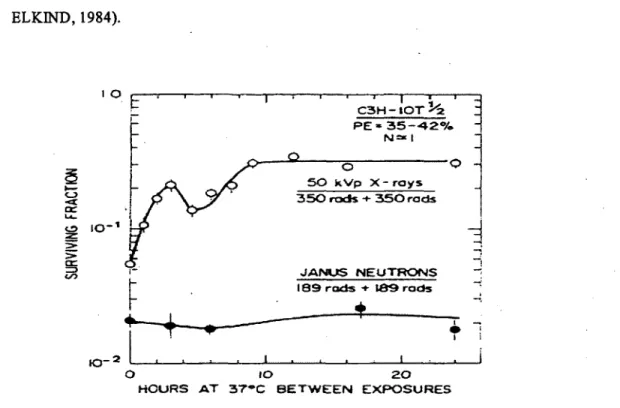
Figure 8, SLDR or split-dose recovery is characterized by restoration of the shoulder on the survival curve and is measured as the increase in survival when the time interval between two dose fractions from X rays is increased to allow for repair of radiation damage. Compared to low LET X rays, a smaller component of SLDR is associated with high LET neutrons. SLDR is largely completed in about 1-2 h, -but it may be longer in the late-responding normal tissues in vivo. (ELKIND and SUTTON, 1960; ELKIND, 1984). 10
~
' 1 1~
C3H- IOT!-2
rt
PE • 35-420/o N"* 1~
0 0 ~.r
50 kVp X-roys...
~ 350 rads + 350 rads < cr...
10-1 \1
<.!) ;z:>
>
cr -! => 1- .JANUS NEUTRONS </)~
189 rads + 189 rads.
~+.
+
----
1 1 I0-2 '•
•
1'
1 0 10 20HOURS AT 37•c BETWEEN EXPOSURES
Figure 8. The fraction survival response of 1 OTl/2 cells, derived from mouse embryo,
exposed to equal of dose from X rays or neutron. The X rays were generated by 50 kV set at 1500 rad/min and the dose rate of neutron is 37.8 rad/min (~E; plating. efficiency). From ELKIND (1984).
Experiments involving fractionation survival curves determined at various times after a conditioning radiation dose, and repeat doses after intervals long enough
for full repair, as well as experiments with a variety of different types of cells irradiated in vitro and/or in vivo led to the following general conclusions.
• Low LET radiation kills mammalian cells as ~ result of a damage accumulation · process.
• Surviving cells following a low LET exposure are sublethally damaged.
• Cells rendered hypoxie by a change in ambient conditions rapid enough to
avoid metabolic consequences due to sustained hypoxia are fully able to repair sublethal damage (ELKIND and SUTTON, 1960).
• Qualitatively, normal cells and tumour cells have similar properties with respects to the accumulation and the repair of sublethal damage.
• A smaller component of sublethal damage accumulation is associated with a. high LET radiation, compared to low LET radiation. Accordingly, cells repair Jess SLD after high LET exposures (NGO, et al., 1979) (e.g. results in Fig. 8). • The repair of sublethal damage reflects the repair of DNA breaks before they
can interact to form lethal chromosome aberrations.
PLDR is characterized by a change in the survival curV-e slope and is measured by the increase in survival in the post irradiation period as the cells are treated under
.
. different conditions such as incubation at suboptimal temperature, in minimal medium, treatment with · inhibitors of protein synthesis, or holding in density-inhibition (confluent) state (PHILLIPS, and TOLMACH, 1966; LITTLE, 1973; RAJU et al., 1977). Examples are shown in Figure 9 where cells in stationary phase cultures are held in a particular metabolic state in G1 that favours the efficient repair of PLD lesion. Conditions which allow the cells to leave this state and progress into S favour fixation of damage and reduced survival (LITTLE, 1973). However, this situation is less clearwith cells exposed to high LET radiation such as a. particles. This is often explained by the hypothesis that repair mechanism is less effective or non-functional with lesions generated by densely ionizing radiations. The experiments in this thesis were designed to characterize the mechanisms underlying defective PLDR in human cells exposed to high LET radiations.
The relevance of PLD to radiotherapy became much more obvious when it was shown that it occurs in irradiated human tumours (HAHN et al, 1974). It has been suggested that the radioresistance of certain types of human tumours is linked to their ability to repair PLD; that is, radiosensitive tumours repair PLD inefficiently, but radioresistant tumours have efficient mechanisms to repair PLD (HALL and GIACCIA, 2006). 0.4 c 0 :;:; 0.3 0
e
•
·r (4Gy) u.. .Il:»...
a(0.85Gy) c ·s; 0.2 ·~ :J U) ~t
+
+
0.1+
0.0 -~.~~~...-~~~...--~~--.~~~-...~~~~ 0 5 10 15 20 25 Incubation time (h)Figure 9. The fraction survival response of confluent human fibroblast AG 1522 cells exposed to y rays (4 Gy) or a. particles (0.85 Gy) and held in confluent at 37°C prior to subculture. From AZZAM. et al., (2000).
1.3. Targeted and non.:.targeted effects of ionzing radiation
This thesis examines targeted and non•targeted effects of IR, and the following is a brief synopsis of the progress that occurred in this field in the past two decades.
1.3.1. Targeted effects
It bas been long considered that the important deleterious effects of IR result from the deposition of energy in the cell nucléus cau~ing damage to genomic DNA, the critical radiation-sensitive "volume" or pr.imary target for induced effects. Thus, IR-induced cell death, by mitotic failure and/or apoptosis, is attributed to failure to repair DNA damage. lJpon repair of DNA damage, the progeny of an irradiated cell would be expected to be normal, but if the damage is misrepaired or unrepaired, the progeny of irradiated cells would be expected to show JR-induced genetic changes in all descendent cells (i.e., the change is clonai). As malignant transformation is generally regarded as being initiated by a gene mutation or a chromosomal aberration, the initiating lesion for malignant transformation bas been similarly attributed to DNA damage in the directly irradiated cells (LEÀ, 1946; LETT et al., 1961; MARSHELL et al., 1970; LITTLE, 2003; HALL and GIACCIA, 2007; reviewed in LEHNERT, 2007). The evidence implicating the DNA, as the sensitive target for IR-induced damage is supported by the following:
• The viscosity of DNA in vitro was found to decrease by exposure _to X rays, an effect they attributed to a reduction in the molecular weight of the DNA and, by implication, the introduction ofDNA strand breaks (TAYLOR et al., 1948). · • The sensitivity of cell nuclet to the lethal effects of X rays were 2-4 times
• ln contrast to radioisotopes incorporated into the cytoplasm, radioisotope incorporated into DNA cause cell killing (MUNRO, 1970; W ARTERS et al., 1978)
• IR-induced DNA damage as the cause of cell death is supported by the finding that incorporation of thymidine, particularly the halogenated pyrimidines into DNA increases radiation sensitivity (SZYBALSKI, 1974).
• DNA DSB plays an important role in the production ofmutagenic lesions by IR (LEENHOUTS and CHADWICK, 1978; LITTLE, 1991)
• Chemical agents that interfere with DNA repair processes affect cell survival following exposure to IR (WALDREN and RASKO, 1978).
• The levels of chromatid and chromosomal aberrations following IR correlate well with cell killing (NAT ARAJAN et al., 1980).
• The relative abilities of cells to repair DNA damage relate closely with cell survival (THACKER and WILKINSON, 1995).
1.3.2. Non-targeted effects
Recently, the traditional dogma ofradiation biology that IR-induced deposition of energy in the nucleus of an irradiated cell is the sole cause of adverse consequences has been challenged by observations in which effects of IR arise in cells that are not themselves irradiated, but are in the vicinity of irradiated cells. The bystander effect has been considered to refer to the occurrence of biological effects in non-irradiated cells as a result of exposure of other cells to IR (N AGAS A W A and LITTLE, 1992; MOTHERSILL and SEYMOUR, 1997, 1998, 2001; MOTHERSILL et al., 2001; AZZAM et al., 1998, 2001, 2002, 2003, 2004; SAW ANT et al., 2001; LITTLE et al., 2002; SHAO et al., 2002, 2003a, 2003b, 2008a, 2008b; MITCHELL et al., 2004;
ZHOU et al., 2000a, 2000b, 2002, 2004; HU et al,. 2006; PRISE and O'SULLIV AN, 2009). Several protocols have been used to detect IR induced bystander effects: cultures consisting of sparse-or density-inhibited .cells were exposed to low fluences of
a
particles generated frdm conventional broad ... or microbeam irradiators, the transfer of medium from irradiatèd onto non-irradiated cells, and radiolabeled cells were mixed with non-labeled cells and assembled in multicellular clusters .(NAGASAW A and LITTLE, 1992; MOTHERSILL and SEYMOUR, 1997, 1998; AZZAM et al., 1998, 2001; BISHAYEE et al., 1999; SAWANT et al., 2001; HOWELL and BISHAYEE, 2002).The discovery of bystander effects can be traced back to the early 1950's
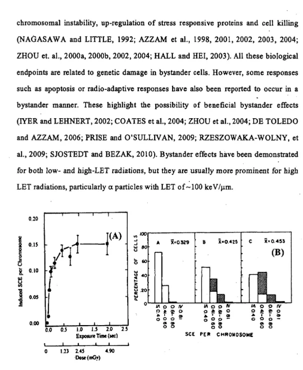
(P ARSON et al., 1954). But, these effects were first identified clearly in 1992 (Fig. 10) when NAGASAWA and LITTLE (1992), found that when only 0.1-1% of the cell population was traversed by low fluences of
a
particles, 20-40% of the cells in the . exposed population had chromosomal damage in . the form of sister chromatid exchanges (SCE). These results indicated that the· target of genetic damages bya
particles was much larger than the nucleus or in fact than the celJ itself. These results were subsequently confirmed by others using the same endpoint in human fibroblast cells (DESHPANDE et al,, 1996). Subsequently, experiment using gene expression as an endp~int have indicated that signais for stressful effects are transmittable from irradiated to bystander cells in response of exposure of cell populations to low fluence a. particles irradiation (AZZAM et al., 1998, 2001, 2002, 2003, 2004; LITTLE et al., 2002).An increasing amount of data from IR-induced bystander effect studies bas Ied to the proposai that damage-inducing signais can be transmitted from irradiated to non-irradiated ce Ils, leading to a variety of biological effects. Bystander responses include SCE formation, gene mutation, micronucleus formation, cell cycle arrest,
chromosomal instability, up-regulation of stress responsive proteins and cell killing (NAGASAWA and LITTLE, 1992; AZZAM et al., 1998, 2001, 2002, 2003, 2004; ZHOU et. al., 2000a, 2000b, 2002, 2004; HALL and HEi, 2003). Ali these biological endpoints are related to genetic damage in bystander cells. However, some responses such as apoptosis or radio-adaptive responses have also been reported to occur in a bystander manner. These highlight the possibility of beneficial bystander effects (IYER and LEHNERT, 2002; CO ATES et al., 2004; ZHOU et al., 2004; DE TOLEDO and AZZAM, 2006; PRISE and O'SULLIVAN, 2009; RZESZOWAK.A-WOLNY, et al., 2009; SJOSTEDT and BEZAK., 2010). Bystander effects have been demonstrated for both low- and high-LET radiations, but they are usually more prominent for high LET radiations, particularly a. partie les with LET of -100 ke V /µm.
0.20 ~
il~
r)
SI 0.JS SI c 0t
..1: u...
0.10'
K. i.I ~l
1
0.05I
l
•
0.00 0.0 0.5 1.0 1.5 2.0Exposure Tune (sec)
0 l.ll 2.45 4.90 ·Dost(mG!) "'100 .... .... tJ 80
5
60...
~ 40...
:z...
~ .20...
CL 0 2S A X•0.329 8 X•0.425 c X•0.453 111oorv !') 4" ~ 1°'
T 0 CD 0 0 p -OI CD 0 0 SCE li\ o o IV 0 ;e !!! 0 .. • f at 0 0 p -Cii .. oo(B)
1.11 0 o IV 0 ~ Cii 0 ... ï Ca 0 0 p -Cii -Cii 0 0 PElt CHROMOSOMEFigure 10. Induction of sister chromatid exchanges (SCE) by low doses of a. particles. (A) Induction of SCE by irradiation with plutonium-238 a particles. (B) Distribution of frequencies of SCE among individual cells. A, nonirradiated cells; B, 0.31 mGy; C, 2.45 mGy. The hatched bars in the histograms represent a particles induced SCE while
the open bars represent background frequencies of SCE based on the distribution observed in nonirradiated cells. From NAGASA W.(\ and LITTLE (1992 ).
1.3.3. Mechanisms
The mechanisms underlying the propagation of stressful or protective effects from irradiated to non-irradiated bystander cells are not fully understood. Though, research to date bas provided some hints. In particular, some evidence suggests that multiple signal transduction pathways are involved. Gap junctiOn intercellular communication (GJIC), secreted diffusible factors, and oxidative metabolism have been proposed to mediate radiation-induced bystander effects (AZZAM et al.,_ 1998, 2001, 2002, 2003; MOTHERSILL and SEYMOUR, 1997, 1998, 2001; IYER and LEHNERT, 2002; SHAO et ~l., 2003a, 2003b, 2007; DE TOLEDO and AZZAM, 2006; ZHOU et al., 2000a, 2000b, 2002, 2004). :Iwo major pathways will be briefly described below.
1.3.3.1. Role of intercellular communication
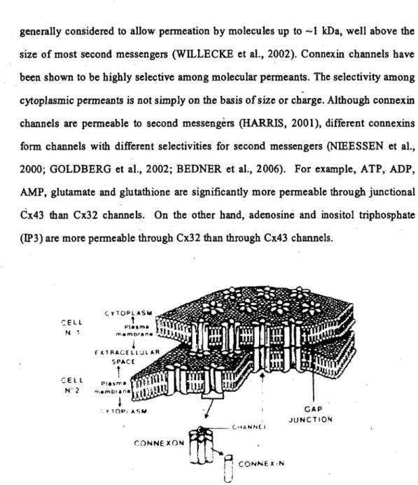
The :first one is GJIC, which is a cell-to-cell gap junction's route and requires the irradiated and non-irradiated cells to be in physical contact. Figure 11 shows gap junctions which are dynamic structures that are critical for diverse physiological function (YAMASAKI et al., 1995; YAMASAKI and NAUS, 1996; TROSKO and RUCH, 1998). By allowing direct intercellular transfer of cytoplasmic molecules, they provide a powerful pathway for direct molecular signalling between cells. Bach of the -20 isoform of connexins (Cx) forms channels with distinct penneability properties (HARRIS, 2001). The channels are formed by two apposed subunits called
hemichannels or connexons, one contributed by each cell. Bach hemichannel is a heiamer of connexin and can be composed of more than one connexin isoform.
generally considered to allow permeation by molecules up to -1 kDa, well above the size of most second messengers (WILLECKE et al., 2002). Connexin channels have been shown to be highly selective among molecular permeants. The selectivity among . .
'
cytoplasmic permeants is not simply on the basis of size or charge. Although connexin channels are permeable to second messengèrs (HARRIS, 2001), different connexins form channels with different selectivities for second messengers (NIEESSEN et al., 2000; GOLDBERG et al., 2002; BEDNER et al., 2006). For example, ATP,. ADP, AMP, glutamate and glutathione are significantly more permeable through junctional èx43 than Cx32 channels. On the other band, adenosine and inositol triphosphate (IP3) are more permeable through Cx32 than through Cx43 channels.
C:Ell N 1 CEl L N'·2 .. (,dANNU CONNE X ON .
A
CONNEX1N ; ! i~' GAP JUNCTIONFigure 11. The structure ~fa gap junction. From Y AMASAK.I and NAUS (1996).
Evidence for the involvement of GJIC in propagation of targeted and non-targeted effects has been derived from studies with high- or low-LET radiations. The involvement of GJIC was confirmed by the modulation of stress responsive proteins
(e.g., p53 and p21 Wafl) and the induction of micronucleus formation in GJIC-proficient cells in confluent, . density-inhibited cultures of human fibroblasts exposed to low fluences of a particles (AZZAM et al., 1998, 2001, 2002, 2003). Treatment with gap ·junction inhibitors such ~ lindane resulted in a marked reduction of cells expressing stress responsive proteins (Fig. 12). These finding indicate that the inhibition of gap junctions by lindane had a significant effect on reduction of these responses, which may have resulted from change in gap junction penneability and Cx43 expressiorl (GUAN et al., 1995; GUAN and RUCH, 1996; AZZAM et al., 1998; KE et al., 2005). Other studies have also showed that irradiation of even l0% of confluent human-hamster hybrid A(L) cells with a single a particle per cell through the nucleus results in a mutant yield similar to that observed when all cells in the population are irradiated. This effect was siginificantly eliminated by chemical inhibition of gap junction mediated intercellular communication, .or when exposed cells expressed a dominant negative Cx43 vector (ZHOU et al., 2001 b ).
Other experiments identifi.ed the importance of the propagation distance of a particle-induced bystander effects. When confluent human skin fibroblasts were exposed to low fluence a particles, the stress-responsive protein p21 wan was induced in bystander cells within 100 µm from irradiated cells. The mean propagation distance ranged from 20 to 40 µm around the intranuclear