Science Arts & Métiers (SAM)
is an open access repository that collects the work of Arts et Métiers Institute of
Technology researchers and makes it freely available over the web where possible.
This is an author-deposited version published in:
https://sam.ensam.eu
Handle ID: .
http://hdl.handle.net/10985/18339
To cite this version :
François LOISEL, Stan DURAND, Sylvain PERSOHN, Sébastien AUBRY, Daniel LEPAGE,
Xavier BONNET, Wafa SKALLI - Scapholunate kinematics after flexible anchor repair - Medical
Engineering & Physics - Vol. 75, p.59-64 - 2020
Any correspondence concerning this service should be sent to the repository
Administrator :
[email protected]
Scapholunate kinematics after flexible anchor repair
François
Loisel
a, b, ∗,
Stan
Durand
a,
Sylvain
Persohn
a,
Sébastien
Aubry
c,
Daniel
Lepage
b,
Xavier
Bonnet
a,
Wafa
Skalli
aa ENSAM, Institut de Biomécanique Humaine G. Charpark, 151, Boulevard de l’Hôpital, 75013 Paris, France
b Service de Chirurgie Orthopédique, Traumatologique, Plastique et Reconstructrice, SOS Main, CHU J. Minjoz, 3 Bd A. Fleming, 250 0 0 Besançon, France c Service de Radiologie Ostéoarticulaire, CHU J. Minjoz, 3 Bd A. Fleming, 250 0 0 Besançon, France
Keywords: Kinematics Scapholunate ligament Flexible anchors Biplane X-ray
a
b
s
t
r
a
c
t
Thescapholunatejointisoneofthekeystonesofthewristkinematics,anditsstudyisdifficultduetothe carpalbonessizeandtherichnessofsurroundingligaments.Weproposeanewmethodofquantitative assessmentofscapholunatekinematicsthroughbonemotiontrackinginordertoinvestigatescapholunate ligamentlesionaswellasrepairtechniques.On6intactwrists,steelbeadswereinsertedintothebones ofinteresttotracktheirmotions.Experimentalsetupallowedwristflexionextensionand radio-ulnar deviationmotions.Low-dosebi-planarradiographswereperformedeach10° ofmovementfordifferent configurations:1)intactwrist,2)scapholunateligamentdivision,3)repairbysoftanchorsatthe poste-riorthen4)anteriorpart.Beads’3DcoordinateswerecomputedateachpositionfrombiplanarX-Rays, allowingaccurateregistrationofeachwristbone.TheMonteCarlosensitivitystudyshowedaccuracy be-tween0.2° and1.6° forthescaphoidandthelunateinmotionsstudied.Themaximumflexion-extension rangeofmotionofthescaphoidsignificantlydecreasedafteranteriorrepairfrom73° ininjuredwristto 62.7°.
Theproposedprotocolappearsrobust,andthetrackingallowedtoquantifytheanchor’sinfluenceonthe wristkinematics.
1. Introduction
Thewristcanbeschematizedintoaduallinkagesystem includ-ingtheproximalandthedistalcarpalrow,inwhicheachbonein agivenrowmovesinthesamedirectionduringwristmotion.The scapholunate interosseus ligament(SLIL) isa complexanatomical structure connecting the scaphoid to the lunate within the first rowof carpalbones. Therefore,duringwrist flexion orradial de-viation, the distal part ofthe scaphoid flexes which leadslunate intoflexionthroughtheSLIL.Thisligamentisalsocriticalfor com-plex wristmovementsusefulindailylivingfunctions,such asthe dartthrowingmotion.Whenthisligamentiscompletelytorn and thescapholunatejointbecomesdissociated,thedorsalandlateral
∗ Corresponding author at: Service de chirurgie Orthopédique, Traumatologique,
aspectofradioscaphoidfossabecomesstressedbecausetheforces crossingthewristcannotbedistributednormally.
Sooner or later, this instability leads to a predictable course of degenerative arthritis called scapho-lunate advanced collapse (SLAC).
AlthoughthedorsalcomponentoftheSLILiswidelyconsidered asthemostresilient [1,2],recentworkhighlightedthat theloads to failure betweendorsal and volarpart of the ligamentare not statisticallydifferent [2].
Froma mechanicalperspective,theaimofa surgicalrepairor ligamentoplastyis thesubstitution of the SLILusing biomaterials (bone-ligament-boneautografts or other solution [3–6]) allowing wristmotionsrecoveryandphysiologicalloaddistribution.
The studyof scapholunate joint kinematics is challenging be-cause the carpal bones are small and the surrounding ligament structuresarecomplex.
There are only few studies investigating the effect of the surgeryfortherestorationofthescapholunatekinematics [7,8].
Those previous studies are based on different motion track-ing methods making any comparison difficult. For example, two studies [9,10] computedtheirgeometrical parameterssuch asthe Plastique et Reconstructrice, SOS Main, CHU J. Minjoz, 3 Bd A. Fleming, 250 0 0 Be-
sançon, France.
E-mail addresses: [email protected] (F. Loisel), [email protected]
(S. Durand), [email protected] (S. Persohn), [email protected] (S. Aubry), [email protected] (D. Lepage), [email protected] (X. Bonnet),
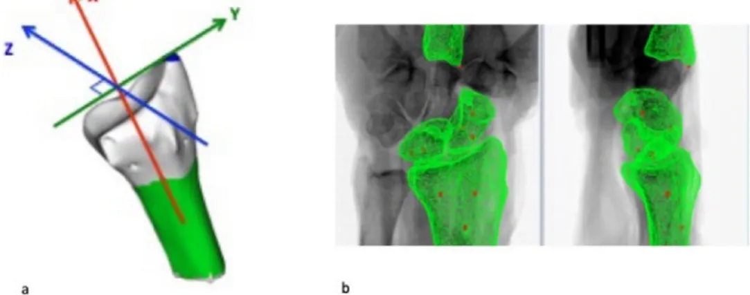
Fig. 1. a) Radius local frame: red axis = prono supination axis, green axis = Flexion Extension axis, blue axis = Radio Ulnar Deviation axis. A cylinder is fitted with a least square condition to the green area to obtain the prono supination axis and the radius styloid is manually selected and corresponds to the barycenter of the blue area. b) Registration of the three bones considered using the bead 3D coordinates (in red the beads). (For interpretation of the references to color in this figure legend, the reader is referred to the web version of this article.)
gapbetweenthelunateandthescaphoid usinga single2D X-ray posteroanterior (PA) radiograph of the wrist, which means that theirresultsmaybeaffectedby theprojectionof3D shapesonto a 2D plane compared to other studies measuring the same dis-tancein3Dusingbiplanarradiographs [7]orelectromagnetic(EM) sensors [8].
In addition, one could questionthe impact ofthe EMsensors onthewristkinematics.
Recent approaches use biplanar X-Rays with embedded ra-diopaquemarkersforbonetracking [11–13].
Thus, we propose a new method of quantitative assessment ofscapholunate kinematics through bone motion tracking allow-ing the investigation ofscapho-lunate ligamentlesion aswell as techniquesofrepair.Theobjectivesofthisstudyaretwo-fold:the evaluationoftheaccuracyofthisnewmethodandtheanalysisof theeffectofthemodelizationofanadditionalvolarrepair ofthe scapho-lunateligament.
2. Methods
2.1.Dissectionandinclusionofthewrists
Six arms from 3 female cadavers were collected from the anatomylaboratoryofouruniversityhospitalinafrozenstateand subsequentlythawedatroomtemperature.
Wrist arthroscopy were performed to check the integrity of theSLIL: we classifiedthe scapho-lunate joint instability accord-ing to the Geissler and the European Wrist Arthroscopy Society (EWAS) classification [14,15] (wrists having a scapho-lunate joint statesmallerthan2areconsideredtobestable).
The morphology ofthelunatewasclassifiedaccordingto Vie-gas [16].Alltheanatomicalstructureshavebeenpreserved.A lon-gitudinaldorsalandpalmarincisionfromtheentireforearmtothe baseofthethird fingeraswell asretinaculumof theflexorsand extensorswasperformedtoallowaccesstothejointcapsule.The setofepidemiologicalcharacteristicsofthewristsaresummarized in Table1.
2.2.Settingupmarkers
Steel beads were introduced under fluoroscopic control (SiemensSiremobilCompact,SiemensAG,Munich,Germany)three by three, within the lunate, the base of the third metacarpal (1mmbeads).thescaphoidandthedistalradiusepiphysis(1.5mm beads). These beads were placed manually after making a bone orificeusingaKirchner wireofdiameter 1.2and1.6respectively.
Table 1
Epidemiological characteristics of anatomical parts.
Age Side Arthroscopic assessment Viegas classification Geissler EWAS 91 R I 1 2 L I 2 2 89 R I 2 1 L I 2 1 80 R II 2 1 L II 2 1
Because wrist bones are very small,the robustnessof each local framewasinvestigatedasdefinedin Section2.4.
2.3. Imagingand3Dmodeling
Eachincludedwristwasscannedwithabonedensityphantom using a Philips Brilliance 64 scanner (120kV, 500mA, Thornton, CO, USA), 0.5mm thick, allowing for an accurate 3D reconstruc-tionofwristbonesandsteelbeadsusingMITKsoftware.RadiusCT reconstruction allowed todefine thewrist Flexion-Extension(FE), Radio-Ulnar Deviation (RUD), and Prono-Supination (PS) axes as describedintheliterature [17](Fig.1a).X-axiswasdefinedasthe axisofthebestfitcylinderdefinedfromtheradiusdiaphysis.The
Y-axiscorrespondedtotheaxiscrossing theradiusstyloid secant andperpendicular to the X-axis. Z axis wasdefinedby the cross product.
2.4. Kinematicsanalysisandexperimentalsetup
Bone tracking was performedusing low dose biplanar X-rays allowing thecomputationofthebeads3D coordinates.First,each segmentedbone waspositioned onto the first X rays acquisition using a rigidregistration. Then, the bead positions ateach wrist position were carefully checked. Since any bead sliding within the bone was not measured, for each step, the position of the bonewasreadjustedby rigidregistrationusingthenewbeads3D coordinates.
Customsoftware(Fig.1b)wasusedtoautomaticallyrecordthe beads 3D coordinates ateach step, and custom MATLAB (Natick, USA) routines were used to compute the new position of each bone.
Given the small size of the bones, a Monte Carlo sensitiv-itystudywasperformedto quantifythe robustnessofeach local frame.AGaussian noise witha rangeofmotion(RoM)of0.1mm
Fig. 2. Test bench. Legend: the asterisk ( ∗) represents the 3D printed base fixed
to the reference plane. The crosses ( + ), the pulleys guiding the cables with 500 g masses (red arrow). The white arrow indicates the plate on which are fixed the fingers and the palm with the aid of a clamp. An intermediate piece connects it to the motor.
was applied to the bead coordinates usingthe MATLAB function randn. 500 “noisy” frameswere compared to theinitial frame to quantifytheangulardeviationinducedbythenoise.Thestandard deviation(1SD)oftheangulardeviationoverthe500iterations al-lowedustoestimatethereliabilityofeachlocalframe.
The radius andthird metacarpal positions were used to com-putethe wristmotion.From theradiuslocalframe,scaphoid and lunate rotations and translations along wrist FE, RUD and PS in eachwristpositionwerecomputed.
Based on morphometric data provided by the 3D reconstruc-tion,thedimensionsofthetestbench couldbe adjustedforeach specimen.Thetestbench consistedofa 3Dprintedbaseattached to the referenceplane. In this base, the forearm was positioned ina neutralprono-supination positionby two 35mm screws and fixedbyaPMMAsurgicalcement.Allthewristandfingertendons weresecuredinthreegroupsatthevolarandtheposteriorpartof thewristtowhichaforce of5Nwastransmittedthroughpulleys (Fig.2).
Thehandwasfixatedtoaperforatedplateusingstrapsaround each fingerandthe wristto removeanyslidingeffectduringthe imposedmotion.
The hand motion was controlled in displacement. The test benchwasspecificallydesignedtoimposepureplanarmotions.
The RoMs considered are FE (y-axis)ranging from30° flexion to60° extension,andRUD(z-axis)from20° radialdeviationto30° ulnardeviation.
Table 2
Monte Carlo sensivity study (1 SD, degrees) for each bone according the 3 axes. Lunate Scaphoid 3rd metacarpal X axis (Pronation – Supination) 1.4 3.0 1.0
Y axis (Flexion - Extension) 1.5 0.2 1.0 Z axis (Radial - Ulnar deviation) 1.6 0.2 1.0
AnEOSbiplanarX-Raysacquisition(EOSimaging,Paris,France) wasperformedevery10° afterperformingacyclingtestof5cycles. Foreachwrist,movementswereperformedwhenthewristwas intact,afterSLILlesion,dorsalrepairandbothdorsalandvolar re-pair.
Injuryandrepairswereperformedbyaseniorhandsurgeon. Injuryconsistedinsectioningthedorsalandthevolarpartsof theSLILaswell asthedorsalradiocarpalligament(DRC)and dor-salintercarpalligament(DIC)withablade.
Finally, the modelling of the ligament was performed us-ing1mm softanchors (Juggerknot,ZimmerBiomet, Warsaw,USA) (Fig.3).Forthethirdconfiguration:oneanchorwasinsertedatthe posteriorandproximal partofthescaphoidandanotherone par-allel inthe lunate.The threads ofthe two anchors were knotted together witha Nicky’s knot. Forthe last configuration, an addi-tional volarrepair was modelized by the same procedure at the anteriorpartofthescaphoidandthelunate.
2.6.Statistics
Foreach subject, the kinematics has beenstudied on the in-tactwrist injured, after repairof thedorsal part ofthe ligament and finally after further repair of the anterior part of the liga-ment.Friedman’sstatisticaltestsweregenerated,anda Wilcoxon testwasperformedforthesignificativevaluesasapost-hocstudy usingSPSSStatistics® softwareforMac(Version25,SPSS,IBM,NY, USA).
ThethresholdofsignificancewasP<0.05.
3. Results
Theresultsofthesensitivitystudyhaveshownameasurement accuracybetween0.2° and1.6° concerningthemotionsofinterest (FEand RUD)(Table 2).There wasa significant differencewithin the different configurations during FE of the wrist for the FE of the scaphoid (p=0.03), and the translation along the RUD axis (p=0.01).DuringRUDofthewrist,therewerealsosignificant dif-ferencesfortheRUDofthescaphoid(p=0.01)andthetranslation along the FE axis (p=0.003) and the RUD axis (p=0.02). There were no significant changes for the lunate motions duringFE or RUDofthewrists.AlltheresultsoftheRoMofthescaphoidand the lunate concerning the configuration andthe direction of the displacementofthewristarepresentedintheannex.
Thepost-hocstudythatfocusedontherotationofthescaphoid
aroundtheY-axisduringFEofthewrist(Fig.4a)hasshowna sig-nificantincreaseofthemotionofthescaphoidbetweentheintact configurationandtheinjured one(63.8° to 73°) andasignificant decreasebetweentheinjuredandtheadditionnalvolarrepair con-figuration (73° to62.7°). Forthe Z-axis, duringRUD of thewrist (Fig. 4b), we found the same behavior (29.3° to 33.3° and 33.3° to 30.7°, respectively). For the scaphoid/lunate translation along
Z-axis, for the FE of the wrist (Fig. 5a) or along Y-axis for RUD ofthewrist (Fig.5b), theresults haveshowna significant differ-encebetweentheintactandtheinjuredwrist (1.9mmto4.1mm, and1.1mm to 2.8mm) butno differencesamong the two repair modalities.
Fig. 3. Ligament injuries and repairs. Legend: Intact configuration: Dorsal Radio Carpal (DRC) ligament in orange, Dorsal Intercarpal (DIC) ligament in blue, posterior and volar part of the SLIL in red. Injured configuration: realization of the lesion of the DRC, DIC and the posterior and the volar part of the SLIL. “Dorsal repair only” configuration: placement of anchors at posterior part of the scaphoid and the lunate and fixed together. Additional volar repair configuration: same additional procedure on the volar part of these bones.
Fig. 4. a) Mean maximum RoM of the rotation ( °) of the scaphoid around the Y - axis during FE of the wrist for the four configurations.
b) Mean maximum RoM of the rotation ( °) of the scaphoid around the Z -axis during RUD of the wrist for the four configurations. ∗correspond to P < 0.5.
4. Discussion
Quantitative assessment of physiological kinematics, the ef-fect of a potential lesion or even surgical restoration is difficult withrespectto thisjointduetotheuncertaintyoftrackingwrist bonemovements.Our original approachwas toperform the anal-ysis of the scapholunate kinematics in vitro using a low dose biplane x-ray with intraosseous markers tracking the bone. Fur-thermore, different configurations of the wrist were compared: when the SLIL was intact, then divided and repaired by soft anchors.
A significantincrease ofthe RoMofthescaphoid between in-tactandinjured configurations along FE (during FEof the wrist) andRUD(duringRUDofthewrist)wasobserved.
The modeling of an additional volar repair significantly de-creasedtheRoM.
The reported trends concerning the scaphoid RoMbefore and afterthelesionareconsistentwiththeliterature.Inparticular, Wa-tersetal. [18]highlightedthesameincreaseinscaphoidflexionin injuredwrists.
Regarding the lunate, while the literature reported that SLIL lesion increases lunate extension from 5.4° to 15°, we found a greater variabilityranging from11.8° in extension to 7.4° in flex-ion(Table3).
ThewiderangeofRoMreportedintheliterature [19,20] under-linesthe multiplechallengesofthistype ofinvestigation suchas great inter-individualvariationsorthereproducibility ofthe liga-mentouslesion [18].
Fig. 5. a) Mean maximum RoM of the translation (mm) of the scaphoid/lunatum along the Z -axis during FE of the wrist for the four configurations.
b) Mean maximum RoM of the translation (mm) of the scaphoid/lunatum along the Y -axis during RUD of the wrist for the four configurations. ∗correspond to P < 0.5. Table 3
Comparative study of the literature comparing FE RoMs of the scaphoid and lunate before and after ligament injury in FE of the wrist. SLIL = Scapho Lunate Inter osseus Liga- ment, ST = Scapho Trapezial Ligaments, RSC = Radio Scapho Capitate Ligament, DRC = Dorsal Radio Carpal Ligament, DIC = Dorsal Inter Carpal Ligament, EM = ElectroMagnetic sensors. + = flexion; - = extension.
Autors, year Number of wrists
Method Lesion(s)/ Repair Kinematics ( °) FE Scaphoide effect of the lesion ( °)
FE Lunate effect of the lesion ( °)
Short, 1995 [21] 6 EM sensors/ active wrist simulator SLIL FE: 50–30 + 3.6 - 5.4
Short, 2002 [22] 8 EM sensors/ active wrist simulator SLIL + ST + RSC FE: 40–30 + – Short, 2005 [23] 24 EM sensors/ active wrist simulator SLIL + ST + RSC FE: 50–30 + > 2 -> 2
Short, 2007 [24] 24 EM sensors/ active wrist simulator SLIL + DRC + DIC FE: 50–30 + 3.9 - 6.4 Short, 2009 [8] 8 EM sensors/ active wrist simulator SLIL + DRC + DIC FE: 50–30 + – Stilling, 2010 [20] 12 tantalum beads, preformed plate,
biplanar radiographs
ST 0 /E 30 + Not studied
Eschweiler, 2016 [26] 8 EM sensors/ passive wrist simulator
SLIL FE: 30–30 + 2 No difference [ < 1]
Waters, 2016 [18] 16 EM sensors/ active wrist simulator SLIL FE: 50–30 + 9 - 15
Current Study, 2018 6 wrists, passive wrist simulator SLIL + DIC + DRC FE: 30–60 + 9.2 [6.5 - 12.5] + 2.9 [ −11.8 - 7.4]
From Table 3, itis noticeable that mostofthe previous stud-ies [8,18,21–26]usedanactivemotionsimulatorcoupledwithEM sensors.
Althoughactive motionsimulators allowtoproducehand mo-tionsclosertothephysiologicalbehaviorbystretchingthetendons whichhelpstoemulatewrist rotations,theuseofEMsensors fix-atedtothedorsalfaceofthewrist bymeansofcarbonrodcould interfere with the joint kinematics by exerting non-physiological forcesontothesofttissues.Inaddition,theuseofEMsensors in-ducesanasymmetricRoMmoreimportantinflexion(50°)thanin extension (30°)due tothe presenceof thesensors on thedorsal side.
Ourworkisinterestedinstudyingtheeffectofasurgical treat-mentonscapholunatekinematics,whichunfortunatelyisnotwell documentedintheliterature [7,8].
Slater etal. [7]have shownthat the lesion increases the gap betweenscaphoidandlunatein clenchedfistposition (2.1mmin intactversus8mm ininjuredwrist)andtherepairdecreasedthe diastasis: between 3.1mm and 5.8mm according to the type of capsulodesis.
Similarly, Pollock et al. [9] highlighted an increase in the scapho-lunate gapfrom2.9mmwhen theligamentsare intactto 5.0mm when the ligaments are sectioned. The repair decreased thepathologicalgapfrom2.6mmto4.6mmaccordingtothetype ofcapsulodesis.
Althoughasimilarincreasewasobserved,thelower values re-portedinthispaper,around1mmfortheintactconfigurationand
about3mmforthe injuredone,are mostlikelydueto thewrist configuration(passiveRoMversusclenchedfistposition).
From ourresults,although the lunateseems lessaffected, the volarrepair oftheligament tends toimprove thescaphoid kine-maticsandreducethedistancebetweenthescaphoidandthe lu-nate.Thus,thisstudysuggeststhata combinedposteriorand an-teriorrepairmaybehelpfulinimprovingscapholunatekinematics afterinjury.
Basedon theMonteCarlo sensitivitystudy,uncertainty ofthe carpal bone displacements was quantified, with values ranging from0.2° to1.6° concerningtheFEandtheRUDaxis.Thescaphoid andlunateframesweremoresensitivetothebeaddisplacements because the beads were closer to each other compared to the beads inside the radius or the third metacarpal. While the re-sultswere all processed,the analysisfocused onrotations in the
Y andZ-axis corresponding to the displacement imposed by the motor. Indeed,for the scaphoid, the estimated uncertaintyalong the Z-axis wasrelatively high (3°). This uncertaintycould be re-duced by positioning the beads further away from each other. Nevertheless,although thecarpal bonesarevery small,we man-agedtogetrobustlocalframesaswell aspreservingtheligament integrity. In addition, the reported changes in scaphoid and lu-nate RoM along wrist FE and RUD after injury and ligament re-pairarebothhigherthantheuncertainty,andconsistentwiththe literature.
Our study has some limitations: the relative low number of wristsandtheuse ofapassive wrist movementsimulator which
doesnotconsiderthetransmissionofforceswithinthejoint pro-ducedbyanactivecontractionofthewristtendons.
Despitetheselimitations,theproposedprotocolhasseveral ma-jor strong points: minimally invasive intraosseous radio-opaque makersthat both preservethe integrityof ligamentsandprevent fromextrinsicsensor limitations.The integrity ofthe SLIL exam-inedin situunderprior arthroscopyis alsoa pledgeofrigor be-cause the proportion of ligamentous lesions on the cadaveric is significant:43% inour study,rangingfrom16% to 50%in the lit-erature [27–29].
This preliminary study seeks to accuratly understand the scapholunate kinematics. Our results about the wrist kinematics are consistent withpreviously reportedvalues. The scapholunate kinematicsanalysishighlightshowthelesionaltersthebone mo-tionsandhowtheanchorstendtorestore thephysiological kine-matics. Further investigation on larger samples would be highly valuable to track subject-specific variabilities andstrengthen the trendsreportedinthispaper. Ourprotocolcanalsobe easily ex-tendedtootherwristbonestoinvestigatetheimpactofthelesion atalargerscalewithinthewrist.
DeclarationofCompetingInterest
Non-financial support fromZimmerBiomet, non-financial sup-port from Stryker, non-financial support from Arthrex, financial supportfromFH orthopedics(travelfellowship grant)outsidethe submittedwork.
Aknowledgement
Dr QuentinLepiller forthevirologicalanalysisofthecadaveric parts,Pr.TatuandPr.Parrattefortheprovisionofcadaveric speci-men,HuguesGrandin,EmmanuelLaurentandMartialBulleforthe technicalhelp,ThomasJoubertforhistechnicalsupport.,David Jin-seongKimforhisproofreadingandEnglishcorrection.
Funding
ZimmerBiomet, Biomecam Chair program on subject specific musculoskeletalmodelling.
EthicalApproval
[1] Berger RA , Imeada T , Berglund L , An KN . Constraint and material properties of the subregions of the scapholunate interosseous ligament. J Hand Surg 1999;24:953–62 .
[2] Nikolopoulos FV , Apergis EP , Poulilios AD , Papagelopoulos PJ , Zoubos AV , Ke- falas VA . Biomechanical properties of the scapholunate ligament and the im- portance of its portions in the capitate intrusion injury. Clin Biomech Bristol Avon 2011;26:819–23 .
[3] Hofstede DJ , Ritt MJ , Bos KE . Tarsal autografts for reconstruction of the scapholunate interosseous ligament: a biomechanical study. J Hand Surg 1999;24:968–76 .
[4] Shin SS , Moore DC , McGovern RD , Weiss AP . Scapholunate ligament recon- struction using a bone-retinaculum-bone autograft: a biomechanic and histo- logic study. J Hand Surg 1998;23:216–21 .
[5]Cuénod P , Charrière E , Papaloïzos MY . A mechanical comparison of bone-liga- ment-bone autografts from the wrist for replacement of the scapholunate lig- ament. J Hand Surg 2002;27:985–90 .
[6]Ehsan A , Lee DG , Bakker AJ , Huang JI . Scapholunate ligament reconstruc- tion using an acellular dermal matrix: a mechanical study. J Hand Surg 2012;37:1538–42 .
[7]Slater RR , Szabo RM , Bay BK , Laubach J . Dorsal intercarpal ligament capsulode- sis for scapholunate dissociation: biomechanical analysis in a cadaver model. J Hand Surg 1999;24:232–9 .
[8]Short WH , Werner FW , Sutton LG . Dynamic biomechanical evaluation of the dorsal intercarpal ligament repair for scapholunate instability. J Hand Surg 2009;34:652–9 .
[9]Pollock PJ , Sieg RN , Baechler MF , Scher D , Zimmerman NB , Dubin NH . Ra- diographic evaluation of the modified brunelli technique versus the blatt capsulodesis for scapholunate dissociation in a cadaver model. J Hand Surg 2010;35:1589–98 .
[10]Lee SK , Zlotolow DA , Sapienza A , Karia R , Yao J . Biomechanical compar- ison of 3 methods of scapholunate ligament reconstruction. J Hand Surg 2014;39:643–50 .
[11]Tsai T-Y , Dimitriou D , Hosseini A , Liow MHL , Torriani M , Li G , et al. Assess- ment of accuracy and precision of 3D reconstruction of unicompartmental knee arthroplasty in upright position using biplanar radiography. Med Eng Phys 2016;38:633–8 .
[12]Fayyazi AH , Ordway NR , Park S-A , Fredrickson BE , Yonemura K , Yuan HA . Ra- diostereometric analysis of postoperative motion after application of dynesys dynamic posterior stabilization system for treatment of degenerative spondy- lolisthesis. J Spinal Disord Tech 2010;23:236–41 .
[13]Garner MR , Dow M , Bixby E , Mintz DN , Widmann RF , Dodwell ER . Evaluat- ing length: the use of low-dose biplanar radiography (EOS) and tantalum bead implantation. J Pediatr Orthop 2016;36:e6–9 .
[14]Geissler WB , Freeland AE , Savoie FH , McIntyre LW , Whipple TL . Intracarpal soft-tissue lesions associated with an intra-articular fracture of the distal end of the radius. J Bone Joint Surg Am 1996;78:357–65 .
[15]Messina JC , Van Overstraeten L , Luchetti R , Fairplay T , Mathoulin CL . The ewas classification of scapholunate tears: an anatomical arthroscopic study. J Wrist Surg 2013;2:105–9 .
[16]Viegas SF , Wagner K , Patterson R , Peterson P . Medial (hamate) facet of the lunate. J Hand Surg 1990;15:564–71 .
[17]Wu G , van der Helm FCT , Veeger HEJD , Makhsous M , Van Roy P , Anglin C , et al. ISB recommendation on definitions of joint coordinate systems of various joints for the reporting of human joint motion–Part II: shoulder, elbow, wrist and hand. J Biomech 2005;38:981–92 .
[18]Waters MS , Werner FW , Haddad SF , McGrattan ML , Short WH . Biomechan- ical evaluation of scaphoid and lunate kinematics following selective sec- tioning of portions of the scapholunate interosseous ligament. J Hand Surg 2016;41:208–13 .
[19]Stromps JP , Eschweiler J , Knobe M , Rennekampff HO , Radermacher K , Pallua N . Impact of scapholunate dissociation on human wrist kinematics. J Hand Surg Eur Vol 2018;43:179–86 .
[20]Stilling M , Krøner K , Rømer L , Van De Giessen M , Munk B . Scaphoid kinematics before and after scaphotrapeziotrapezoidal ligament section. Assessment by ra- diostereometric analysis and computed tomography in a cadaver study. J Hand Surg Eur Vol. 2010;35:637–45 .
[21]Short WH , Werner FW , Fortino MD , Palmer AK , Mann KA . A dynamic biomechanical study of scapholunate ligament sectioning. J Hand Surg 1995;20:986–99 .
[22]Short WH , Werner FW , Green JK , Masaoka S . Biomechanical evalua- tion of ligamentous stabilizers of the scaphoid and lunate. J Hand Surg 20 02;27:991–10 02 .
[23]Short WH , Werner FW , Green JK , Masaoka S . Biomechanical evaluation of the ligamentous stabilizers of the scaphoid and lunate: part II. J Hand Surg 2005;30:24–34 .
[24]Short WH , Werner FW , Green JK , Sutton LG , Brutus JP . Biomechanical evalua- tion of the ligamentous stabilizers of the scaphoid and lunate: part III. J Hand Surg 2007;32:297–309 .
[25]Werner FW , Sutton LG , Allison MA , Gilula LA , Short WH , Wollstein R . Scaphoid and lunate translation in the intact wrist and following ligament resection: a cadaver study. J Hand Surg 2011;36:291–8 .
[26]Eschweiler J , Stromps JP , Rath B , Pallua N , Radermacher K . Analysis of wrist bone motion before and after SL-ligament resection. Biomed Tech 2016;61:345–57 .
[27]Viegas SF , Yamaguchi S , Boyd NL , Patterson RM . The dorsal ligaments of the wrist: anatomy, mechanical properties, and function. J Hand Surg 1999;24:456–68 .
[28]Overstraeten LV , Camus EJ , Wahegaonkar A , Messina J , Tandara AA , Binder AC , et al. Anatomical description of the dorsal capsulo-scapholunate septum (DCSS)-arthroscopic staging of scapholunate instability after DCSS sectioning. J Wrist Surg 2013;2:149–54 .
[29]Loisel F , Cohen G , Marès O , Garret J , Clavert P . Kystes mucoïdes de la face dor- sale du poignet : lésions anatomiques, place de la prise en charge chirurgicale et technique opératoire. Rev Chir Orthopédique Traumatol 2017;103:S185–92 .