HAL Id: hal-02905699
https://hal.mines-ales.fr/hal-02905699
Submitted on 8 Jan 2021
HAL is a multi-disciplinary open access
archive for the deposit and dissemination of
sci-entific research documents, whether they are
pub-lished or not. The documents may come from
teaching and research institutions in France or
abroad, or from public or private research centers.
L’archive ouverte pluridisciplinaire HAL, est
destinée au dépôt et à la diffusion de documents
scientifiques de niveau recherche, publiés ou non,
émanant des établissements d’enseignement et de
recherche français ou étrangers, des laboratoires
publics ou privés.
Adsorption of xyloglucan and cellulose nanocrystals on
natural fibres for the creation of hierarchically
structured fibres
Estelle Doineau, Guillaume Bauer, Léo Ensenlaz, Bruno Novales, Cécile
Sillard, Jean-Charles Benezet, Julien Bras, Bernard Cathala, Nicolas Le
Moigne
To cite this version:
Estelle Doineau, Guillaume Bauer, Léo Ensenlaz, Bruno Novales, Cécile Sillard, et al.. Adsorption
of xyloglucan and cellulose nanocrystals on natural fibres for the creation of hierarchically structured
fibres. Carbohydrate Polymers, Elsevier, 2020, 248, pp.1-10. �10.1016/j.carbpol.2020.116713�.
�hal-02905699�
Adsorption of xyloglucan and cellulose nanocrystals on natural
fibres for the
creation of hierarchically structured
fibres
Estelle Doineau
a,b,c,*
,1,2, Guillaume Bauer
c,1, Léo Ensenlaz
b,1,2, Bruno Novales
c,d,1,
Cécile Sillard
b,1,2, Jean-Charles Bénézet
a,1, Julien Bras
b,1,2, Bernard Cathala
c,1,
Nicolas Le Moigne
a,*
,1aPolymers Composites and Hybrids (PCH), IMT Mines Ales, Ales, France bUniv. Grenoble Alpes, CNRS, Grenoble INP2, LGP2, F-38000 Grenoble, France cINRAE, UR BIA, F-44316, Nantes, France
dINRAE, BIBS Facility, F-44316 Nantes, France
Keywords: Cellulose nanocrystals Xyloglucan Flaxfibres Adsorption isotherm A B S T R A C T
Green treatment of natural fibres is a major issue in paper, textile and biocomposites industries to design in-novative and eco-friendly products. In this work, hierarchical structuring of flax woven fabrics by the adsorption of xyloglucan (XG) and cellulose nanocrystals (CNC) is studied. Indeed, CNC have high mechanical properties, high specific surface area and great potential for functionalization. The adsorption of XG and CNC has been investigated in terms of localization by confocal and scanning electron microscopy (SEM) and quantification through adsorption isotherms. Adhesion force measurements have also been performed by Atomic Force Microscopy (AFM). XG and CNC are homogeneously adsorbed on flax fabric and adsorption isotherms reach plateau values around 20 mg /gfibres for both. The pre-adsorption of XG on flax fabric influences the amount of adsorbed CNC in the high concentrations and also creates entanglements and strong interactions between XG and CNC with the formation of an extensible network.
1. Introduction
Surface treatments of natural fibres (chemical, physical, enzy-matic...) are commonly used in a wide range of industrial sectors like textile, paper or biocomposites to bring them specific properties ac-cording to the targeted applications (Kalia, Thakur, Celli, Kiechel, & Schauer, 2013). For instance, in textile applications, surface treatments primarily concern the dyeing and thefinishing while in paper industry, surface treatments target functional properties such as printability, bleaching, hydrophobicity, gas barrier, softness, strong mechanical properties. In the more recently developedfield of biocomposites, most of the natural fibres surface treatments are developed to improve wettability and compatibility with polymeric matrices. In all these ap-plications, environmental concerns prompt the search and the devel-opment of environmental friendly surface treatments procedures in order to decrease the use of polluting chemical products (Araújo, Casal, & Cavaco-Paulo, 2008;Faruk, Bledzki, Fink, & Sain, 2012;Haider, Cho, Moon, & Kim, 2019;Le Moigne, Otazaghine, Corn, Angellier-Coussy, &
Bergeret, 2018;Rastogi & Samyn, 2015;Sun, 2016;Wei, 2009). In the search of innovative and greener surface modifications of natural fibres, functionalization with nanoparticules or biopolymers knows a growing interest (Hajlane, Kaddami, & Joffe, 2017;Dai & Fan, 2013; Hajlane, Joffe, & Kaddami, 2018; Idumah, Ogbu, Ndem, & Obiana, 2019;Lee, Bharadia, Blaker, & Bismarck, 2012;Malik, Kumar, Ghosh, & Shrivastava, 2018;Oksanen, Timo, Retulainen, Salminen, & Brumer, 2011;Zhou, Baumann, Brumer, & Teeri, 2006;Zhuang et al., 2011). With this long term goal, this work proposes to combine ad-sorption capacity of xyloglucan (XG) on cellulosic substrates with the outstanding properties of cellulose nanocrystals (CNC) to develop hierarchically structured assemblies at the surface offlax woven fabrics. XG is an hemicellulose found in the primary cell wall of plants, which is involved in the wall extensibility and its integrity, through its strong affinity with cellulose microfibrils (Fry, 1989;Fry et al., 1993;Hanus & Mazeau, 2006;Hayashi, Ogawa, & Mitsuishi, 1994; Yong BumPark & Cosgrove, 2015; Pauly, Albersheim, Darvill, & York, 1999). On the other hand, CNC are used in many applications thanks to their amazing
⁎Corresponding authors.
E-mail addresses:doineau.estelle@orange.fr(E. Doineau),Nicolas.le-moigne@mines-ales.fr(N. Le Moigne).
1
Members of the European Polysaccharide Network of Excellence (EPNOE),http://www.epnoe.eu.
2Institute of Engineering Univ. Grenoble Alpes.
combination of XG and CNC adsorption may yield different to final organization to achieve hierarchical materials combining microscale fibres displaying nanostructured surfaces. Moreover, this study has been achieved on substrates of industrial interest, i.e.flax woven fabrics that present additional levels of complexity when compared to model substrates or unitary naturalfibres.
The adsorption of XG and CNC onflax woven fabrics is investigated in terms of localization by confocal and scanning electron microscopy. In order to quantify the adsorbed amount of XG and CNC onflax woven fabrics, adsorption isotherms are established by an UV spectroscopy method. Different adsorption sequences on flax woven fabrics are evaluated: (i) adsorption of XG or CNC (ii) successive adsorption of XG then CNC, (iii) successive adsorption of CNC then XG. Finally, Atomic Force Microscopy (AFM) with XG and CNC modified tips was used to analyze the adhesion between XG, CNC andflax fibres.
2. Materials and methods 2.1. Materials
A twill 2 × 2 flax woven fabric 200 g/m² was provided by SF Composites (France). Cellulose nanocrystals (CNC) were produced by acid hydrolysis of wood pulp and purchased in spray-dried powder from CelluForce (Quebec, Canada). CNC surface charge density : 0.023 mmol/g; Crystalline fraction : 0.88; CNC lateral dimensions : 2−5 nm; CNC length : 50−110 nm. Xyloglucan Glyloid 6 C was ob-tained from tamarind seed gum and purchased from DSP Gokyo Food & Chemical. Mw= 840 000 g/mol; Mw/Mn= 1.24, Rg 72 nm;
mono-saccharide composition : Glucose 50.7 %; Xylose 31.7 %; Galactose 16.0 %; Arabinose 1.6 %. Fluorescent agents RBITC (Rhodamine B iso-thiocyanate) and FITC (Fluoresceine isoiso-thiocyanate) were purchased from Sigma-Aldrich.
2.2. Labelling of XG and CNC with RBITC and FITC
Thefluorescent labelling of XG and CNC were performed according to the method reported by Belder A & Granath K (de Belder & Granath, 1973). The reaction occurring is the formation of covalent bonds be-tween hydroxyl groups of polysaccharides and isothiocyanate groups of thefluorescent agents (Iwakura & Okada, 1962). First, 1 g of CNC or XG was solubilized in 40 mL of DMSO under stirring and heated at 65 °C. After 15 min, few drops of pyridine, 50μL of dibutyltin dilaurate and 7.5 mg of RBITC (Rhodamine B Isothiocyanate) or FITC (Fluoresceine isothiocyanate) were added. The reaction was heated under stirring at 65 °C during 2 h. XG-RBITC and CNC-RBITC (or CNC-FITC) were re-covered by precipitation in ethanol/acetone mixture for XG and NaCl 1 M solution for CNC followed by centrifugation (10 min; 10,000 g). Three dispersion/centrifugation cycles were achieved and final pre-cipitates were dissolved / dispersed in deionized water at average concentration of 1 wt% and dialyzed (12–14000 MWCO) during one week against ultrapure water.
2.3. Adsorption experiments onflax woven fabrics
The different steps of adsorption experiments are illustrated inFig. 1 and detailed below.
2.3.1. Simple adsorptions
Small pieces offlax fabrics between 8–12 mg were cut and put in test tubes. To avoid the quenching of RBITC or FITC, all samples need to be in the dark (tubes were wrapped in aluminiumfilm). XG-RBITC or CNC-RBITC solutions of 0.5 g/L were diluted with water in test tubes (final volume 5 mL) to obtain final concentrations ranging between 0.05 g/L and 0.45 g/L. Reference samples were prepared in the same way withoutflax woven fabrics. Test tubes were gently shaken during 16 h. Other samples were prepared with CNC-FITC in the same properties (Habibi, Lucia, & Rojas, 2010). They have outstanding
me-chanical properties, i.e high Young’s Modulus estimated between 100 and 150 GPa, a high specific surface area (Dufresne, 2017; Habibi, Chanzy, & Vignon, 2006; Nagalakshmaiah, El Kissi, & Dufresne, 2016; Rusli & Eichhorn, 2008; Yue et al., 2018) and can be chemically func-tionalized through substitution of their hydroxyl groups. Xyloglucan interactions with cellulosic surfaces have been intensively studied in vitro and in vivo (1994, Dick-Pérez et al., 2011; Hayashi, Marsden, & Delmer, 1987; Morris, Hanna, & Miles, 2004; Young B. Park & Cosgrove, 2012; Vincken, de Keizer, Beldman, & Gerard Joseph Voragen, 1995), and are known to be an entropy driven process (Benselfelt et al., 2016). From the physico-chemical point of view, glucan chains will interact each other through Hydrogen bonds, van der Waals and polar bonds with some reorganizations of the conformation of XG depending on the polymer concentration (Villares, Moreau, Dammak, Capron, & Cathala, 2015). The final architecture of the cel-lulose/xyloglucan assemblies is related to concentrations of the building blocks, the molar mass and other adsorption processing parameters (Benselfelt et al., 2016; Dammak, Quémener et al., 2015; Hanus & Mazeau, 2006; Hayashi & Maclachlan, 1984; Hayashi et al., 1987; Lima, Loh, & Buckeridge, 2004; Lopez et al., 2010; Stiernstedt, Brumer, Zhou, Teeri, & Rutland, 2006; Stiernstedt, Nordgren et al., 2006; Vincken et al., 1995). In particular, the strong affinity of XG for cellulose surfaces has attracted researchers for the elaboration of cel-lulose-based materials as natural cross-linker. For instance, XG and CNC have been already used for the implementation of thin films displaying structural colors or for the reinforcement of nanocellulose aerogels (C. Cerclier, Cousin, Bizot, Moreau, & Cathala, 2010, 2011; C. V. Cerclier et al., 2013; Dammak, Moreau et al., 2015; Jean, Heux, Dubreuil, Chambat, & Cousin, 2009; Sehaqui, Zhou, & Berglund, 2011; Sehaqui, Zhou, Ikkala, & Berglund, 2011).
Ligno-cellulosic fibres properties have also been improved by the adsorption of either XG or nanocellulose. For example, addition of XG by spraying on a bleached birch kraft pulp increases its wet web strength, tension holding, drying tension and also its smoothness (Oksanen et al., 2011). Similarly, when XG is used as a wet end ad-ditives in paper making process, paper formation is facilitated and final paper strength is increased (Christiernin et al., 2003; Yan, Lindström, & Christiernin, 2006). In fact, fibre bonds in pulps treated with XG seem to give high adhesion levels. To go further in this way, friction mea-surements have been performed by AFM between cellulose and XG (Stiernstedt, Brumer et al., 2006; Stiernstedt, Nordgren et al., 2006). These studies show the irreversibility side of the adsorption of XG to cellulose. Moreover, the adhesion between the cellulose surfaces in water, which is often very small, is increased with the presence of XG on the surfaces. It appears that specific bonds are formed between XG and cellulose surfaces, creating a strong bridging between cellulose fi-bres. Nanocellulose, and more especially bacterial cellulose, has also demonstrated its capacity to increase the strength of the interfacial adhesion between natural fibres and polymeric matrices as reported by Bismarck’s group (Fortea-Verdejo, Lee, Zimmermann, & Bismarck, 2016; Juntaro, Pommet, Mantalaris, Shaffer, & Bismarck, 2007; Lee, Bharadia et al., 2012; Lee, Ho, Schlufter, & Bismarck, 2012; Pommet et al., 2008). For example, the creation of “dense” or “hairy” coated sisal fibres via the dipping of sisal fibres in a slurry of bacterial cellulose (BC) has been proposed to reinforce PLA based biocomposites (Lee, Bharadia et al., 2012). The results of this study were promising with an increase of the specific surface area up to 800 % compared to neat sisal fibres. In terms of mechanical properties, the storage modulus of the PLA- BC coated sisal fibres- biocomposite was increased by around 20 % in comparison with PLA-neat sisal fibres. However, up to our knowl-edge there is no report of combined adsorption of XG and nanocellulose on (ligno-)cellulosic substrates. In this work, the challenge addressed is to characterize the adsorption of XG and CNC on flax fibres and the potential synergistic effect of the combination of the two biobased building blocks. Indeed, similarly to the plant wall architecture,
conditions for confocal microscopic observations. At least triplicate was performed.
2.3.2. Successive adsorptions
For successive adsorptions of XG / CNC-RBITC and CNC / XG-RBITC onflax woven fabrics, similar protocol as for labeled-CNC or -XG ad-sorption was used preceded by adad-sorption of XG or CNC. A solution of non-labeled XG or CNC at 0.4 g/L was added in test tubes. After 5 h, the flax woven fabrics were washed with deionized water. Labeled CNC or XG were added at concentrations ranging between 0.05 g/L and 0.45 g/ L, during 16 h. Other samples were prepared with both XG-RBITC and CNC-FITC in the same conditions for confocal microscopic observations. At least triplicate was performed.
After adsorption experiments, the supernatant was collected and the absorbance of Rhodamine at 558 nm for XG-RBITC or CNC-RBITC was measured by UV spectroscopy. The amount of adsorbed XG-RBITC and CNC-RBITC on the fibres was calculated by the difference between measured absorbance in test tubes withflax woven and the absorbance of reference tubes (withoutflax woven).
2.4. Scanning electron microscopy
Elementary flax fibres extracted from the raw and treated flax woven fabrics were observed with an Environmental Scanning Electron Microscope (ESEM, FEI Quanta 200 FEG, Netherlands). The samples were pasted on an adhesive wafer and metallized in high vacuum by depositing a thin layer of carbon on their surface using a BALZERS CED 030. SEM micrographs were obtained under high vacuum at an accel-eration voltage of 3 keV, a working distance of 9 mm and magnification up to 50 000 × . At least 10 micrographs per sample were performed and the most representative pictures were conserved for the discussion.
2.5. Confocal microscopy
The confocal microscopy images were obtained using an inverted Nikon A1 laser scanning confocal microscope. Several pieces of yarns (raw or treated) were deposited between slide and slip cover with one spacer and deionized water, avoiding the prolonged light contact. The samples were viewed with a Plan Fluor 4× or with a Plan Apo 20× Nikon objective by scanning using a laser beam at a wavelength of 488 nm and 561 nm, for imaging FITC and RBITC fluorescences re-spectively. The emissions offluorescence were recorded via a photo-multiplier through 500–530 nm and 570–620 nm band-pass filters, re-spectively. Images were processed using the NIS-Element software (Nikon). At least triplicate was performed.
2.6. Atomic Force Microscopy (AFM)
AFM force curves (Bruker Icon dimension) between the tip and the substrate have been performed in the air to study the adhesion between xyloglucan (XG), cellulose nanocrystals (CNC) andflax fibres. Shortly, during the adhesive force measurements, the cantilever tip approached the surface of the substrate at a speed of 1.4μm/s until contact, and then separated. Before all these measurements, calibrations have to be performed on the cantilever Sharp Nitride Lever (SNL-10, Bruker) with a tip radius below 10 nm. First, the stiffness of the cantilever was de-termined by the thermal tune method and gave a spring constant of 0.098 N/m. Secondly, the sensitivity of the cantilever has to be mea-sured. To achieve this calibration, force curves have been performed on a sapphire in contact mode. By measuring the yield of the curve, made possible by a functionality of the AFM device, the sensitivity of the cantilever can be determined. Then, the different force curves between cantilever tips and substrates were measured (Cappella & Dietler, 1999;
Fig. 1. Illustration of the different steps of adsorption processes on flax woven fabric with labeled cellulose nanocrystals (CNC) and xyloglucan (XG). For clarity reasons, XG, CNC andflax fibres are not scaled.
Ralston, Larson, Rutland, Feiler, & Kleijn, 2005). The raw data were converted by a calculation algorithm detailed inFig. 2. The method is based on the conversion of the raw data graph (deflection of the can-tilever vs piezo position) into a force-distance curve representing the force vs the tip-substrate distance (D). The baseline was done and the zero separation was obtained from the gradient of the constant com-pliance region. Different parameters are required like the cantilever rest position (Z), the deflection of the cantilever (δc), the deformation of the
surface of the substrate (δs) and the constant spring of the cantilever
(kc).
In order to perform the experiments, two different tips functiona-lization have been done. First, one consisted on the dipping of the SNL-10 cantilever tip in a drop of XG solution at 20 g/L during 1 min and then a rinsing in water during 1 min before a drying at 60 °C during 30 min. This functionalized tip is named XG-tip within this article. The second one consisted on a tip’s functionalization with CNC. The pre-coating of the tip by polycations poly(ethylene imine) (PEI, Mw = 750 000, Sigma Aldrich) was made to decrease the time of dipping and obtain a uniform coating of CNC (Kolasinska & Warszynski, 2005; Podsiadlo et al., 2007). The different steps of the dipping are described in the study ofJean, Dubreuil, Heux, and Cousin (2008)). This func-tionalized tip is named CNC-tip within this article. Finally, a mica substrate was also spin coated (POLO SPIN150i, SPS-Europe) by PEI and CNC following the protocol of Aulin et al. (2009), and is named CNC-surface throughout the article. Adhesion measurements were taken in contact mode at three localizations, 35 on each, i.e. 105 measurements in total for each configuration. Outliers were identified and rejected considering the interquartile range, i.e. values inferior to Q1– 1.5 x (Q3-Q1) or superior to Q3 + 1.5 x (Q3-Q1). No obvious irreversible deformation or damaging of the substrates or the tip was detected during the measurements.
3. Results and discussions
3.1. Adsorption behavior of XG and CNC onflax woven fabrics
In order to assess XG and CNC adsorption behaviors qualitatively and quantitatively, CNC and XG were labeled withfluorescent dies. In a first approach, confocal microscopy was used to study the localization and homogeneity of XG and CNC adsorption onflax fibres. Using the UV absorbance of the dies, the amount of XG and CNC adsorbed onflax fabrics were then used to determine adsorption isotherms.
3.1.1. Microscopic observations
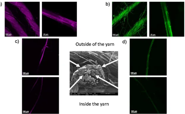
Confocal microscopy images onFig. 3present two different views of XG-RBITC (Fig. 3a) and CNC-FITC (Fig. 3b) adsorbed onflax woven
fabrics, at the scale of a yarn and an elementaryfibre. Both adsorption of XG-RBITC and CNC-FITC is homogeneous with no aggregates on the surface of yarns and elementaryfibres. Some microfibrils functionalized by XG-RBITC or CNC-FITC were also observed at the surface of ele-mentaryfibres. These microfibrils probably originate from the peeling offibre cell walls and/or the presence of impurities and residues at the surface offlax fibres generated during the manufacturing process of flax woven fabrics.
Some elementary flax fibres were extracted from the yarns of treatedflax woven fabrics to compare qualitatively the homogeneity of XG-RBITC (Fig. 3c) and CNC-FTIC (Fig. 3d) adsorption acrossflax yarns diameter. The elementaryfibres collected at the outer part of flax yarns show higher intensities for both CNC-FTIC and XG-RBITC, compared to fibres extracted within the yarns. These results highlight differences in penetration of CNC-FTIC and XG-RBITC within the complex and multi-scale architecture offlax woven fabrics. Indeed the elementary fibres are twisted together to formflax yarns that are then woven to form the fabric. This puts forwards the issues related to the homogeneous treatment of industrial ligno-cellulosic substrates as natural woven fabrics having complex architectures and possibly impurities.
The surface offlax fibres treated with CNC was imaged by SEM. Cellulose nanocrystals at the surface of treatedflax fibres are clearly visible inFig. 4b (see also insert), compared to rawflax fibres (Fig. 4a) with no visible rod-like shaped nanoparticles at their surface. Adsorbed CNC appear homogeneously distributed and randomly oriented at the surface offlax fibres. This clearly demonstrated the creation of hier-archical structuration at the surface of thefibres by the formation of a nanostructured layer. This microscale structuration echoes the organi-zation of the cellulosic elements of theflax fibres. It should be noticed that CNC were not present on all elementaryfibre surfaces. This has to be related to the accessibility of fibre surfaces during adsorption treatments. Indeed, the twisted structure offlax yarns and physical contacts between elementary fibres likely limit the exposure of flax surface upon adsorption. As observed by confocal microscopy (Fig. 3), flax fibres from the inside of the yarns are less accessible and hence likely treated to a lower extent.
3.1.2. Adsorption isotherms
Xyloglucan (XG) and cellulose nanocrystals (CNC) adsorption pro-cesses are complex phenomena, and the final adsorbed amount and conformation over substrate’s surface are related to several parameters such as the molar mass, concentrations, solubility, etc. (Ahola, Myllytie, Österberg, Teerinen, & Laine, 2008;Villares et al., 2015). Additionally, flax woven fabrics are industrial materials and as such are far from being model substrates. For instance,flax fibres swelling and destruc-turation of the fabrics occurring during treatment might influence the
adsorption kinetics. Indeed it changes the available surfaces for ad-sorption and their accessibility, as well as the particular microstructure of naturalfibres with its porosities and the lumen. Based on preliminary adsorption trials, an adsorption time of 16 h was chosen. These trials also highlighted that further investigations on the adsorption kinetics of XG and CNC onflax fibres would be of interest.
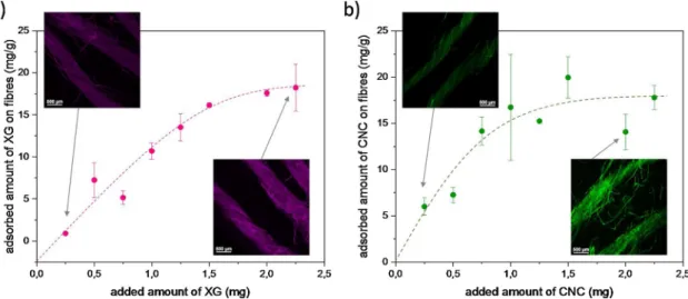
The obtained adsorption isotherms are displayed inFig. 5, for XG and CNC on flax woven fabrics. Adsorption was determined by com-paring the UV absorbance of reference labeled CNC-RBITC or XG-RBITC solution to those incubated withflax woven fabrics. First, theFig. 5a represents the adsorption isotherm of XG onflax woven fabrics. The insert shows the related confocal images of the yarns at low and high adsorption ratios. The difference in intensity of XG-RBITC on confocal images is nicely correlated with the increase of adsorbed amount of XG onflax fibres depending on initial concentrations of XG in solutions. A
plateau is observed at average 15−20 mg/g. In literature, the adsorp-tion of xyloglucan has already been studied on different ligno-cellulosic substrates. For example, paperfibres like bleached softwood kraft pulp (BSKP) (Moser, Backlund, Lindström, & Henriksson, 2018) or chemical and mechanical pulp (Zhou et al., 2006). For BSKPfibres, the amount of adsorbed XG was between 4 and 12 mg/g pulp depending on the re-fining time. In the case of thermomechanical pulp (TMP) and chemi-thermomechanical pulp (CTMP), refined or unrefined pine/spruce and birch pulp, an adsorption of XG from 15 to 30 mg/g pulp was reached. In our study, the adsorbed amount of XG onflax woven fabrics seems to be in the same range as TMP and CTMP pulps. Previous studies also reported the adsorption of xyloglucan on cellulose nanocrystals (Dammak, Quémener et al., 2015;Gu & Catchmark, 2013;Lopez et al., 2010) with a maximal adsorption between 98–333 mgXG/gcellulose. The
higher amount of adsorbed XG on cellulose nanocrystals as substrates is
Fig. 3. Confocal microscopy images of (a) XG-RBITC and (b) CNC-FITC adsorption onflax yarns and elementary fibres; and of (c) XG-RBITC and (d) CNC-FITC adsorbed on elementaryflax fibres extracted from different positions across the diameter of flax yarns.
due to the much higher specific surface area of CNC that range from 150 to 800 m²/g (Brinkmann et al., 2016;Habibi, 2014), as compared to that offlax fibres, roughly 1 m²/g (Bismarck et al., 2002;le Duigou, Bourmaud, Balnois, Davies, & Baley, 2012;Legras, Kondor, Heitzmann, & Truss, 2015; Müssig, Fischer, Graupner, & Drieling, 2010), even if fibre swelling could contribute to improve specific surface area.
Fig. 5b represents the adsorption isotherm of CNC on flax woven fabrics. As for XG adsorption, the difference in intensity of CNC-FITC on confocal images is well correlated with the increase amount of adsorbed CNC onflax fibres depending on initial concentrations of CNC in sus-pensions. A plateau is reached at average 15−20 mg/g. Plateau values of the amount of adsorbed XG and CNC are thus similar for the studied flax woven fabric. This points out that both XG and CNC can be ad-sorbed in similar quantities onflax woven fabrics, and suggests that the surface availability of the substrate has more influence on the amount of adsorbed polymers/particles than its nature and physico-chemical characteristics. It should be noticed that the adsorbed amounts of XG and CNC are rather high when considering the low specific surface area of flax woven fabric. It might be suggested that adsorption occurs in multilayered structure and not only in monolayer as usually proposed in adsorption studies based on model substrates. Other parameters than adsorption mechanisms cannot be ruled out to explain ourfindings. As discussed above, other processes such as the swelling offlax yarns and fibres with an increase of the available surface for adsorption, the complex and heterogeneous surface chemistry offlax fibres as well as a significant fibrillation rate due to flax processing (retting, scutching, weaving etc.) should also be considered.
3.2. Successive XG and CNC adsorptions onflax woven fabrics
Two different successive adsorption procedures of XG and CNC have been tested onflax woven fabrics, i.e. adsorption of XG then CNC (XG-CNC) and vice versa (CNC-XG). Both had the aim to observe a possible synergy between XG and CNC during their adsorption onflax woven fabrics.
3.2.1. Microscopic observations
Fig. 6shows confocal microscopy images of XG-RBITC (Fig. 6a) and CNC-FITC (Fig. 6b) on the same elementaryflax fibre.
RBITC and FTIC were successively imaged to investigate their po-tential co-localization. Globally XG and CNC are homogeneously ad-sorbed onflax fibres, recovering the entire surface of the fibre on both Fig. 6a and b, even if some difference can be seen between the two images. As for the simple adsorptions of XG or CNC on flax woven fabrics, some microfibrils around the flax fibre are visible and are likely
originated from the peeling offibre cell walls and/or residues produced during the manufacturing process of the industrialflax woven fabric. These residues display RBITC labeling and seem thus to have a pre-ferential adsorption of xyloglucan (Fig. 6a) than cellulose nanocrystals (Fig. 6b). The confocal plan used allows also the imaging of the inner part of thefibre. It has to be noticed that any fluorescence can be de-tected in the bulk of thefibres indicating that the adsorption of both XG and CNC remain located at the surface of thefibres.
Surface of XG-CNC treatedflax fibres were imaged by SEM. As for simple CNC adsorption (Fig. 4), cellulose nanocrystals are clearly visible at the surface of treatedflax fibres inFig. 7b (see also insert). Adsorbed CNC appear homogeneously distributed and randomly or-iented at the surface offlax fibres. As mentioned above, CNC were not present on all elementaryfibre surfaces which could be explained by differences in fibre surfaces accessibility and chemistry during ad-sorption treatments.
3.2.2. Adsorption isotherms of successive XG and CNC
Adsorption isotherms of the successive adsorptions CNC-XG and XG-CNC have been plotted inFig. 8. The goal was to investigate if the pre-treatment offlax woven fabric by CNC or XG had an influence on the adsorbed amount of the second compound (Fig. 8a; b).
It seems that the pre-adsorption of CNC onflax fibres,Fig. 8a, has no influence on the adsorption behavior of XG. Indeed, the two adsorption isotherms are very similar with the same plateau around 15−20 mg/g. However, it should be noticed that the plateau seems to be reached at lower initial concentrations of XG in solution with the presence of CNC onflax woven fabric. The pre-adsorption of XG on flax fibres,Fig. 8b, appears to have a slight influence on the amount of adsorbed CNC. In fact, the adsorption isotherm for XG-CNC reaches a plateau at around 25−30 mg/g for against 15−20 mg/g for simple CNC adsorption. These results demonstrate that CNC have a good affinity for flax fibres surface, the pre-adsorption of XG increasing the amount of adsorbed CNC at high initial concentration of CNC in the suspension.
3.3. Adhesive force measurements by Atomic Force Microscopy (AFM) In order to get a deeper understanding of the affinity between xy-loglucan (XG), cellulose nanocrystals (CNC) andflax woven fabric, we performed different adhesive force measurements by the atomic force microscopy (AFM). All of these results are detailed inFig. 8. Different configurations tip / substrate were analyzed:
(a) neat-tip or XG-tip / CNC-surface obtained by deposition of CNC on PEI pre-coated mica substrate by spin-coating.
Fig. 5. Adsorption isotherms of (a) XG and (b) CNC onflax woven fabric and associated confocal images of XG-RBITC and CNC-FITC at low and high adsorption ratios.
(b) XG-tip /flax-CNC or XG-tip / raw flax, where flax-CNC corresponds to aflax fibre surface adsorbed with CNC,
(c) CNC-tip /flax-XG or CNC-tip / raw flax, where flax-XG corresponds to aflax fibre surface adsorbed with XG.
The adhesion between the tip and the substrate is due to the che-mical interactions between the two surfaces into contact. The force curves measurements allow the quantification of these interactions. The rupture distance corresponds to the distance tip-substrate needed to completely separate the tip and the substrate. Finally, the work of ad-hesion is the energy required to separate the tip from the substrate.
All the results inFig. 9show high standard deviations, probably due to differences in surface roughness and chemistry between the different surfaces.
First, the configuration (a) with the neat-tip or the XG-tip ap-proaching the CNC-surface was used as a supplementary control of the dipping of the XG-tip. The adhesion force and the work of adhesion between the XG-tip and the CNC-substrate are respectively 7 times and 25 times higher than the measurements with the untreated nitride tip. In this configuration, the rupture distance is unchanged. These results show that the XG-tip has a high adhesion force with the CNC-substrate, which is correlated with the high affinity between XG and CNC with strong hydrogen and van der Waals bonds.
Then force curves measurements were performed with the
configuration (b), XG-tip in contact with the raw flax or the flax-CNC and results are detailed inFig. 9b. We can observe that the adhesive force is the same, around 6 nN for both, with more heterogeneous measurements for the rawflax substrate (between 3 and 15 nN). The work of adhesion is also in the same range with 60–90 nN.nm. However the rupture distance seems to be slightly increased with the presence of CNC on theflax woven fabric with a maximum of 12 nm against 5 nm for the rawflax. This result shows a possible increase of the interactions zone between biopolymers XG and adsorbed CNC with the creation of an extended network. The configuration (c) is the opposite of the configuration (b), with the raw flax or flax-XG as substrates and CNC coated on the AFM tip. The adhesion force and the work of adhesion are higher between the CNC-tip and the rawflax than the flax-XG substrate with respectively 35 nN against 20 nN and 750 nN.nm against 3100 nN.nm. It means that CNC seem to have very good interactions with theflax woven fabrics, without the presence of adsorbed XG on the surface. On the other side, the rupture distance is strongly improved between the CNC-tip and theflax-XG with a median around 23 nm against 5 nm for the rawflax. That means, as observed with the con-figuration (b), that the interactions between CNC and adsorbed XG create specific bonds and entanglements with an extensible network, illustrated inFig. 10. Note that adhesion force, rupture distance and work of adhesion obtained with the CNC-tip are much higher than those obtained with the XG-tip, probably because the tip coated by CNC offers
Fig. 6. Confocal microscopy images of successive adsorption of XG-CNC on an elementaryflax fibre with (a) XG-RBITC (b) CNC-FITC.
more available surface with more available OH groups due to the high specific surface area of CNC (more than 100 g/m²) as compared to the XG biopolymer.
Despite the similar adsorbed amounts are similar for CNC and XG whatever the adsorption process, the AFM results suggest that a change
in the sequence of adsorption and the combination of the two biobased building blocks can be a mean to vary the structuration of the surface organization. Accordingly, future studies will focus on the impact of hierarchically structured surfaces obtained by varying the adsorption sequence on the macroscopic properties offlax fibres based materials.
Fig. 8. Adsorption isotherms of (a) XG and (b) CNC for simple and successive adsorptions (XG-CNC and CNC-XG) onflax woven fabrics and associated confocal images of XG-RBITC and CNC-FITC at low and high adsorption ratios.
Fig. 9. Adhesion force, rupture distance and work of adhesion obtained with force measurements by AFM in different tip / substrate configurations (a) neat-tip or XG-tip / CNC-surface, (b) XG-XG-tip / rawflax or flax-CNC, (c) CNC-tip / raw flax or flax-XG.
4. Conclusions
Adsorption isotherms, confocal microscopy and force measurements by Atomic Force Microscopy (AFM) have been used to characterize different configurations of xyloglucan (XG) and cellulose nanocrystals (CNC) adsorptions on an industrial flax woven fabric. This industrial fabric has a complex architecture with twisted yarns composed of several hundred elementaryflax fibres. Moreover, some residues likely to be generated by the manufacturing process of the fabric are present at the surface and seem to have affinities mainly with xyloglucan and also cellulose nanocrystals. The confocal images show the homo-geneous distribution of XG and CNC on theflax fabric for the simple and successive adsorptions with no aggregates. However, SEM images show non-treated surfaces, which can be explained by the complex architecture of the fabric with porosities, entanglements etc. Moreover, phenomena such as swelling and destructuration occur during the dipping in water suspension and can influence the adsorption. The adsorption isotherms of XG and CNC on theflax woven fabric are very similar for the simple and successive adsorptions with a plateau around 20 mg/gfibres, highlighting the strong influence of available surfaces on
CNC or XG adsorption ontoflax fibres. The pre-adsorption with XG on flax woven fabrics increases the adsorbed amount of CNC in the high concentrations and seems to change the architecture of the biopolymers network. In fact the AFM adhesive force measurements showfirst a good affinity between CNC and XG and also higher adhesive force and work of adhesion between CNC and the neat flax woven fabric. However, it seems that the pre-adsorption of XG on the fabric increases the rupture distance with the CNC-tip. The combined XG and CNC, adsorbed with strong interactions on theflax woven fabric, create an extensible network. These results open perspectives in the treatment of natural fibre fabrics and the possibility to change their surface and architecture with the adsorption of biopolymers like xyloglucan or nanoparticles like cellulose nanocrystals. However, we have to keep in mind the complexity of the hierarchical architecture of the industrial flax woven fabric for such treatments in terms of homogeneity, residual components and swelling effect.
CRediT authorship contribution statement
Estelle Doineau: Methodology, Investigation, Funding acquisition, Writing - original draft, Writing - review & editing.Guillaume Bauer: Formal analysis, Methodology. Léo Ensenlaz: Formal analysis, Methodology. Bruno Novales: Investigation, Methodology. Cécile Sillard: Investigation, Methodology, Supervision, Validation. Jean-Charles Bénézet: Conceptualization, Methodology, Validation, Resources, Writing - review & editing, Supervision, Project adminis-tration. Julien Bras: Conceptualization, Methodology, Validation, Resources, Writing - review & editing, Supervision, Project adminis-tration. Bernard Cathala: Conceptualization, Investigation, Methodology, Project administration, Resources, Supervision,
Validation, Visualization, Writing - review & editing. Nicolas Le Moigne: Conceptualization, Methodology, Validation, Resources, Writing - review & editing, Supervision, Project administration. Acknowledgements
Estelle Doineau thanks IMT Mines Alès and Doctoral School GAIA for funding his PhD work.
A part of this work was supported by Glyco@alps (ANR-15-IDEX-02), Idex UGA. LGP2 is part of the LabEx Tec 21 (Investissements d’Avenir – grant agreement n°ANR-11-LABX-0030) and of PolyNat Carnot Institute (Investissements d’Avenir – grant agreement n°ANR-16-CARN-0025-01).
Nadège Leray is acknowledged for her efficient help for CNC and XG labeling and adsorption experiments.
References
Ahola, S., Myllytie, P., Österberg, M., Teerinen, T., & Laine, J. (2008). Effect of polymer adsorption on cellulose nanofibril water binding capacity and aggregation. BioResources, 3(4), 1315–1328.
Araújo, R., Casal, M., & Cavaco-Paulo, A. (2008). Application of enzymes for textilefibres processing. Biocatalysis and Biotransformation, 26(5), 332–349.
Aulin, C., Ahola, S., Josefsson, P., Nishino, T., Hirose, Y., Österberg, M., et al. (2009). Nanoscale cellulosefilms with different crystallinities and mesostructures—Their surface properties and interaction with water. Langmuir, 25(13), 7675–7685.
Benselfelt, T., Cranston, E. D., Ondaral, S., Johansson, E., Brumer, H., Rutland, M. W., et al. (2016). Adsorption of xyloglucan onto cellulose surfaces of different morphologies: An entropy-driven process. Biomacromolecules, 17(9), 2801–2811.
Bismarck, A., Aranberri‐Askargorta, I., Springer, J., Lampke, T., Wielage, B., Stamboulis, A., et al. (2002). Surface characterization offlax, hemp and cellulose fibers; Surface properties and the water uptake behavior. Polymer Composites, 23(5), 872–894.
Brinkmann, A., Chen, M., Couillard, M., Jakubek, Z. J., Leng, T., & Johnston, L. J. (2016). Correlating cellulose nanocrystal particle size and surface area. Langmuir, 32(24), 6105–6114.
Cappella, B., & Dietler, G. (1999). Force-distance curves by atomic force microscopy. Surface Science Reports, 34(1‑3), 1–104.
Cerclier, C., Cousin, F., Bizot, H., Moreau, C., & Cathala, B. (2010). Elaboration of spin-coated cellulose-xyloglucan multilayered thinfilms. Langmuir, 26(22), 17248–17255.
Cerclier, C., Guyomard‐Lack, A., Moreau, C., Cousin, F., Beury, N., Bonnin, E., et al. (2011). Coloured semi-reflective thin films for biomass-hydrolyzing enzyme detec-tion. Advanced Materials, 23(33), 3791–3795.
Cerclier, C. V., Guyomard-Lack, A., Cousin, F., Jean, B., Bonnin, E., Cathala, B., et al. (2013). Xyloglucan-cellulose nanocrystal multilayeredfilms : Effect of film archi-tecture on enzymatic hydrolysis. Biomacromolecules, 14, 3599–3609.
Christiernin, M., Henriksson, G., Lindström, M. E., Brumer, H., Teeri, T. T., Lindström, T., et al. (2003). The effects of xyloglucan on the properties of paper made from bleached kraft pulp. Nordic Pulp and Paper Research Journal, 18(2), 182–187.
Dai, D., & Fan, M. (2013). Green modification of natural fibres with nanocellulose. RSC Advances, 3(14), 4659.
Dammak, A., Moreau, C., Azzam, F., Jean, B., Cousin, F., & Cathala, B. (2015). Influence of cellulose nanocrystals concentration and ionic strength on the elaboration of cel-lulose nanocrystals–xyloglucan multilayered thin films. Journal of Colloid and Interface Science, 460(Supplement C), 214–220.
Dammak, A., Quémener, B., Bonnin, E., Alvarado, C., Bouchet, B., Villares, A., et al. (2015). Exploring architecture of xyloglucan cellulose nanocrystal complexes through enzyme susceptibility at different adsorption regimes. Biomacromolecules, 16(2), 589–596.
de Belder, A. N., & Granath, K. (1973). Preparation and properties offluorescein-labelled dextrans. Carbohydrate Research, 30(2), 375–378.
Fig. 10. Illustration of the adhesive force measurements by AFM with the CNC-tip when comparing substrates: (A) a non-treatedflax fibre and (B) a flax fibre treated with XG.
Dick-Pérez, M., Zhang, Y., Hayes, J., Salazar, A., Zabotina, O. A., & Hong, M. (2011). Structure and interactions of plant cell-wall polysaccharides by two- and three-di-mensional magic-angle-Spinning solid-state NMR. Biochemistry, 50, 989–1000.
Dufresne, A. (2017). Nanocellulose : From nature to high performance tailored materials. Walter de Gruyter GmbH & Co KG.
Faruk, O., Bledzki, A. K., Fink, H.-P., & Sain, M. (2012). Biocomposites reinforced with naturalfibers : 2000–2010. Progress in Polymer Science, 37(11), 1552–1596.
Fortea-Verdejo, M., Lee, K.-Y., Zimmermann, T., & Bismarck, A. (2016). Upgradingflax nonwovens : Nanocellulose as binder to produce rigid and robustflax fibre preforms. Composites Part A, Applied Science and Manufacturing, 83(Supplement C), 63–71.
Fry, S. C. (1989). The structure and functions of xyloglucan. Journal of Experimental Botany, 40(210), 1–11.
Fry, S. C., York, W. S., Albersheim, P., Darvill, A., Hayashi, T., Joseleau, J.-P., et al. (1993). An unambiguous nomenclature for xyloglucan-derived oligosaccharides. Physiologia Plantarum, 89(1), 1–3.
Gu, J., & Catchmark, J. M. (2013). The impact of cellulose structure on binding inter-actions with hemicellulose and pectin. Cellulose, 20, 1613–1627.
Habibi, Y. (2014). Key advances in the chemical modification of nanocelluloses. Chemical Society Reviews, 43(5), 1519–1542.
Habibi, Y., Chanzy, H., & Vignon, M. R. (2006). TEMPO-mediated surface oxidation of cellulose whiskers. Cellulose, 13, 679–687.
Habibi, Y., Lucia, L. A., & Rojas, O. J. (2010). Cellulose nanocrystals : Chemistry, self-assembly, and applications. Chemical Reviews, 110(6), 3479–3500.
Haider, Z., Cho, H., Moon, G., & Kim, H. (2019). Minireview : Selective production of hydrogen peroxide as a clean oxidant over structurally tailored carbon nitride pho-tocatalysts. Catalysis Today, 335, 55–64.
Hajlane, A., Joffe, R., & Kaddami, H. (2018). Cellulose nanocrystal deposition onto re-generated cellulosefibres : Effect on moisture absorption and fibre–matrix adhesion. Cellulose, 25(3), 1783–1793.
Hajlane, A., Kaddami, H., & Joffe, R. (2017). Chemical modification of regenerated cel-lulosefibres by cellulose nano-crystals : Towards hierarchical structure for structural composites reinforcement. Industrial Crops and Products, 100, 41–50.https://doi.org/ 10.1016/j.indcrop.2017.02.006.
Hanus, J., & Mazeau, K. (2006). The xyloglucan–cellulose assembly at the atomic scale. Biopolymers, 82(1), 59–73.
Hayashi, T., & Maclachlan, G. A. (1984). Pea xyloglucan and cellulose : I. Macromolecular organization. Plant Physiology, 75, 596–604.
Hayashi, T., Marsden, M. P. F., & Delmer, D. P. (1987). Pea xyloglucan and cellulose V: Xyloglucan-cellulose interactions in vitro and in vivo. Plant Physiology, 83, 384–389.
Hayashi, T., Ogawa, K., & Mitsuishi, Y. (1994). Characterization of the adsorption of xyloglucan to cellulose. Plant & Cell Physiology, 35(8), 1199–1205.
Idumah, C. I., Ogbu, J. E., Ndem, J. U., & Obiana, V. (2019). Infuence of chemical modification of kenaf fiber on xGNP‑PP nano‑biocomposites. SN Applied Sciences, 1(1261).
Iwakura, Y., & Okada, H. (1962). The kinetics of the reaction of organic isothiocyanates with 1-octanol in o-Dichlorobenzene. Canadian Journal of Chemistry, 40, 2369–2375.
Jean, B., Dubreuil, F., Heux, L., & Cousin, F. (2008). Structural details of cellulose na-nocrystals/polyelectrolytes multilayers probed by neutron reflectivity and AFM. Langmuir, 3452–3458.
Jean, B., Heux, L., Dubreuil, F., Chambat, G., & Cousin, F. (2009). Non-electrostatic building of biomimetic cellulose−Xyloglucan multilayers. Langmuir, 25(7), 3920–3923.
Juntaro, J., Pommet, M., Mantalaris, A., Shaffer, M., & Bismarck, A. (2007). Nanocellulose enhanced interfaces in truly green unidirectionalfibre reinforced composites. Composite Interfaces, 14(7–9), 753–762.
Kalia, S., Thakur, K., Celli, A., Kiechel, M. A., & Schauer, C. L. (2013). Surface mod-ification of plant fibers using environment friendly methods for their application in polymer composites, textile industry and antimicrobial activities : A review. Journal of Environmental Chemical Engineering, 1(3), 97–112.
Kolasinska, M., & Warszynski, P. (2005). The effect of nature of polyions and treatment after deposition on wetting characteristics of polyelectrolyte multilayers. Applied Surface Science, 252(3), 759–765.
le Duigou, A., Bourmaud, A., Balnois, E., Davies, P., & Baley, C. (2012). Improving the interfacial properties betweenflax fibres and PLLA by a water fibre treatment and drying cycle. Industrial Crops and Products, 39, 31–39.
Le Moigne, N., Otazaghine, B., Corn, S., Angellier-Coussy, H., & Bergeret, A. (2018). Surfaces and interfaces in naturalfibre reinforced composites. Springer International Publishing.
Lee, K.-Y., Bharadia, P., Blaker, J. J., & Bismarck, A. (2012). Short sisalfibre reinforced bacterial cellulose polylactide nanocomposites using hairy sisalfibres as reinforce-ment. Composites Part A, Applied Science and Manufacturing, 43(11), 2065–2074.
Lee, K.-Y., Ho, K. K. C., Schlufter, K., & Bismarck, A. (2012). Hierarchical composites reinforced with robust short sisalfibre preforms utilising bacterial cellulose as binder. Composites Science and Technology, 72(13), 1479–1486.
Legras, A., Kondor, A., Heitzmann, M. T., & Truss, R. W. (2015). Inverse gas chromato-graphy for naturalfibre characterisation : Identification of the critical parameters to determine the Brunauer–Emmett–Teller specific surface area. Journal of
Chromatography A, 1425, 273–279.
Lima, D. U., Loh, W., & Buckeridge, M. S. (2004). Xyloglucan–cellulose interaction de-pends on the sidechains and molecular weight of xyloglucan. Plant Physiology and
Biochemistry, 42(5), 389–394.
Lopez, M., Bizot, H., Chambat, G., Marais, M.-F., Zykwinska, A., Ralet, M.-C., et al. (2010). Enthalpic studies of xyloglucan−cellulose interactions. Biomacromolecules, 11(6), 1417–1428.
Malik, N., Kumar, P., Ghosh, S. B., & Shrivastava, S. (2018). Organically modified na-noclay and aluminium hydroxide incorporated bionanocomposites towards en-hancement of physico-mechanical and thermal properties of lignocellulosic structural reinforcement. Journal of Polymers and the Environment, 26(8), 3243–3249.
Morris, S., Hanna, S., & Miles, M. J. (2004). The self-assembly of plant cell wall com-ponents by single-molecule force spectroscopy and Monte Carlo modelling. Nanotechnology, 15(9).
Moser, C., Backlund, H., Lindström, M., & Henriksson, G. (2018). Xyloglucan for esti-mating the surface area of cellulosefibers. Nordic Pulp and Paper Research Journal, 33(2), 194–198.
Müssig, J., Fischer, H., Graupner, N., & Drieling, A. (2010). Testing methods for measuring physical and mechanicalfibre properties (plant and animal fibres). Industrial applications of naturalfibres. John Wiley & Sons, Ltd.267–309.
Nagalakshmaiah, M., El Kissi, N., & Dufresne, A. (2016). Ionic compatibilization of cel-lulose nanocrystals with quaternary ammonium salt and their melt extrusion with polypropylene. ACS Applied Materials & Interfaces, 8(13).
Oksanen, A., Timo, R., Retulainen, E., Salminen, K., & Brumer, H. (2011). Improving wet web runnability and paper quality by an uncharged polysaccharide. Journal of Biobased Materials and Bioenergy, 5(2), 187–191.
Park, Y. B., & Cosgrove, D. J. (2015). Xyloglucan and its interactions with other com-ponents of the growing cell wall. Plant & Cell Physiology, 56(2), 180–194.
Park, Y. B., & Cosgrove, D. J. (2012). A revised architecture of primary cell walls based on biomechanical changes induced by substrate-specific endoglucanases. Plant Physiology, 158, 1933–1943.
Pauly, M., Albersheim, P., Darvill, A., & York, W. S. (1999). Molecular domains of the cellulose/xyloglucan network in the cell walls of higher plants. The Plant Journal, 20(6), 629–639.
Podsiadlo, P., Sui, L., Elkasabi, Y., Burgardt, P., Lee, J., Miryala, A., et al. (2007). Layer-by-Layer assembledfilms of cellulose nanowires with antireflective properties. Langmuir, 23(15), 7901–7906.
Pommet, M., Juntaro, J., Heng, J. Y. Y., Mantalaris, A., Lee, A. F., Wilson, K., et al. (2008). Surface modification of natural fibers using Bacteria : Depositing bacterial cellulose onto naturalfibers to create hierarchical Fiber reinforced nanocomposites. Biomacromolecules, 9(6), 1643–1651.
Ralston, J., Larson, I., Rutland, M. W., Feiler, A. A., & Kleijn, M. (2005). Atomic force microscopy and direct surface force measurements (IUPAC Technical Report). Pure and Applied Chemistry, 77(12), 2149–2170.
Rastogi, V. K., & Samyn, P. (2015). Bio-based coatings for paper applications. Coatings, 5, 887–930.
Rusli, R., & Eichhorn, S. J. (2008). Determination of the stiffness of cellulose nano-whiskers and thefiber-matrix interface in a nanocomposite using Raman spectro-scopy. Applied Physics Letters, 93(3), Article 033111.
Sehaqui, H., Zhou, Q., & Berglund, L. A. (2011). High-porosity aerogels of high specific surface area prepared from nanofibrillated cellulose (NFC). Composites Science and Technology, 71(13), 1593–1599.
Sehaqui, H., Zhou, Q., Ikkala, O., & Berglund, L. A. (2011). Strong and tough cellulose nanopaper with high specific surface area and porosity. Biomacromolecules, 12(10), 3638–3644.
Stiernstedt, J., Brumer, H., Zhou, Q., Teeri, T. T., & Rutland, M. W. (2006). Friction be-tween cellulose surfaces and effect of xyloglucan adsorption. Biomacromolecules, 7(7), 2147–2153.
Stiernstedt, J., Nordgren, N., Wågberg, L., Brumer, H., Gray, D. G., & Rutland, M. W. (2006). Friction and forces between cellulose model surfaces : A comparison. Journal of Colloid and Interface Science, 303(1), 117–123.
Sun, D. (2016). Surface modification of natural fibers using plasma treatment. Biodegradable Green Composites, 18–39.
Villares, A., Moreau, C., Dammak, A., Capron, I., & Cathala, B. (2015). Kinetic aspects of the adsorption of xyloglucan onto cellulose nanocrystals. Soft Matter, 11(32), 6472–6481.
Vincken, J.-P., de Keizer, A., Beldman, G., & Gerard Joseph Voragen, A. (1995). Fractionation of xyloglucan fragments and their interaction with cellulose. Plant Physiology, 108, 1579–1585.
Wei, Q. (2009). 14—Emerging approaches to the surface modification of textiles. In Q. Wei (Ed.). Surface modification of textiles (pp. 318–323). Woodhead Publishing. Yan, H., Lindström, T., & Christiernin, M. (2006). Some ways to decreasefibre suspension
flocculation and improve sheet formation. Nordic Pulp and Paper Research Journal, 21(1), 36–43.https://doi.org/10.3183/npprj-2006-21-01-p036-043.
Yue, L., Maiorana, A., Khelifa, F., Patel, A., Raquez, J.-M., Bonnaud, L., et al. (2018). Surface-modified cellulose nanocrystals for biobased epoxy nanocomposites. Polymer, 134, 155–162.
Zhou, Q., Baumann, M., Brumer, H., & Teeri, T. (2006). The influence of surface chemical composition on the adsorption of xyloglucan to chemical and mechanical pulps. Carbohydrate Polymers, 63(4), 449–458.
Zhuang, R.-C., Doan, T. T. L., Liu, J.-W., Zhang, J., Gao, S.-L., & Mäder, E. (2011). Multi-functional multi-walled carbon nanotube-jutefibres and composites. Carbon, 49(8), 2683–2692.