Dual Multimodular Class A Penicillin-Binding Proteins in Mycobacterium
leprae
Sophie Lepage,1 Philippe Dubois,1 Tushar Kanti Ghosh,1 Bernard Joris,1 Sebabrata Mahapatra,2 Manikuntala Kundu,2 Joyoti Basu,2 Parul Chakrabarti,2 Stewart T. Cole,3 Martine Nguyen-Distèche,1 andJean-Marie Ghuysen1
Centre d'Ingénierie des Protéines, Institut de Chimie, B6, Université de Liège, B-4000 Sart Tilman (Liège), Belgium1; Department of Chemistry, Bose Institute, Calcutta 700009, India2; and Unité de Génétique Moléculaire Bactérienne, Institut Pasteur, F-75724 Paris Cedex 15, France3
Abstract
The ponA gene of cosmid L222 of the Mycobacterium leprae genome library encodes a multimodular class A penicillin-binding protein (PBP), PBP1*. The PBP, labelled with a polyhistidine sequence, has been produced in
Escherichia coli, extracted from the membranes with
3-[(3-cholamidopropyl)-dimethylammonio]-1-propane-sulfonate (CHAPS) and purified by Ni2+ -nitrilotriacetic acid-agarose chromatography. In contrast to the pon1-encoded class A PBP1, PBP1* undergoes denaturation at temperatures higher than 25°C, it catalyzes acyl transfer reactions on properly structured thiolesters, and it binds penicillin with high affinity.
A set of membrane-bound proteins, known as penicillin-binding proteins (PBPs), are involved in the final assembly of the bacterial cell wall peptidoglycan. Escherichia coli possesses four multimodular PBPs. PBP1a and PBP1b of class A are bienzymatic polypeptides performing, in vitro, transglycosylase (the non-penicillin-binding [n-PB] module) and transpeptidase (the penicillin-non-penicillin-binding [PB] module) activities on the disaccharide peptide lipid II precursor. PBP2 and PBP3 of class B are essential members of the morphogenetic networks involved in wall expansion and septum formation, respectively (8).
Until recently, the peptidoglycan-synthesizing machinery of Mycobacterium leprae was inaccessible to direct biochemical investigation. As an ordered cosmid library covering the mycobacterial 2.8-Mb chromosome has been established (4), important genes are being identified, allowing the encoded proteins to be produced in heterologous systems and characterized. pon1 of cosmid B577 encodes a class A PBP1. Expression of pon1 in E.
coli results in the production of M. leprae PBP1 bound to the plasma membrane of the host. PBP1 has the
expected membrane topology, it is thermostable, and it binds penicillin with a low affinity (1).
As shown below, M. leprae contains another class A PBP, PBP1*, the biochemical and enzymatic properties of which differ markedly from those of PBP1.
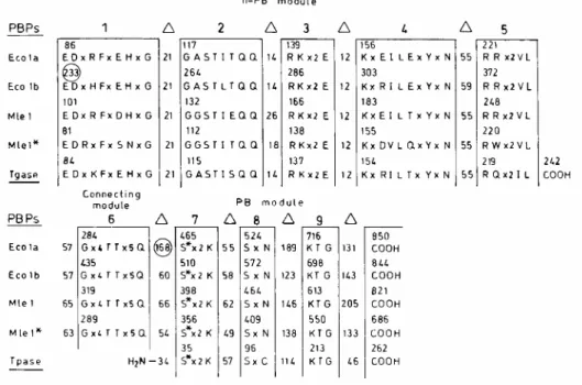
Modular design of M. leprae PBP1*.
ponA of cosmid L222 (nucleotides 7365 to 5305) encodes the 686-amino-acid residue PBP1* (7). This PBP
bears the signature amino acid sequence of the multimodular class A PBPs. The nine motifs shown in Fig. 1 are the consensus derived from the alignment of 11 class A PBPs. PBP1* consists of a V1-to-L225 n-PB module fused to an R302-to-P686 PB module via a G226-to-Q301 connecting module. The n-PB module has a pseudo-signal peptide (the membrane anchor) at the amino end of the polypeptide chain, and the PB module possesses, downstream from motif 9, a 130-amino-acid residue carboxy-terminal extension. In comparison with PBP1*, M.
leprae PBP1 has a much longer tail, E. coli PBP1a has an ~100-amino-acid residue insert between motifs 6 and
7, and E. coli PBP1b has an ~150-amino-acid residue insert upstream from motif 1.
In spite of the highly conserved molecular organization adopted by the M. leprae and E. coli class A PBPs, similarity in the amino acid sequences (after elimination of the inserts and extensions) is low or even statistically insignificant. At the level of the n-PB modules, the members of the pairs M. leprae PBP1*-M. leprae PBP1, M.
PB modules, the members of the pair M. leprae PBP1*-M. leprae PBP1 have 25% identity, and the members of the pairs M. leprae PBP1*-E. coli PBP1a and M. leprae PBP1*-E. coli PBP1b have only 17% identity. For comparative purposes, the n-PB modules of E. coli PBP1a and E. coli PBP1b have 31% identity, and the corresponding PB modules have 28% identity.
FIG. 1. Modular design of the multimodular class A PBP1a and PBP1b of E. coli and PBP1 and PBP1* of M.
leprae and occurrence of the conserved motifs along the amino acid sequences. Intervals (∆) between conserved motifs are expressed in numbers of amino acid residues. Circled numbers indicate the presence of inserts. E. coli possesses a 34,000-Mr
monofunctional transglycosylase (10), and the genes whose products have the signature amino acid sequence of the class A PBPs are present in several bacterial species (15). The transglycosylase (Tgase) motifs shown are those derived from the E. coli gene sequence. The transpeptidase (Tpase) motifs shown are those of the Streptomyces strain K15 PBP (13), which has been biochemically characterized as a DD-transpeptidase (9, 12). The sequences of E. coli PBP1a and PBP1b (Eco1a and Eco1b, respectively) are from reference 2, and the sequences of M. leprae PBP1 and PBP1* (Mle1 and Mt1*, respectively) are from references 1 and 7.
Production of M. leprae (His tag)(R2-P686) PBP1* in E. coli.
M. leprae PBP1* with its membrane anchor (lacking residue V1) was produced in E. coli and labelled with a
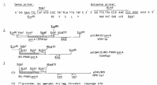
polyhistidine sequence fused to residue R2. Plasmid pDML909, which encodes PBP1* with a polyhistidine tag fused to R2 residue P686 [(His tag)(R2-P686) PBP1*], was constructed (Fig. 2) as follows. (Step 1) The sense and antisense primers were used to synthesize by PCR a 167-bp DNA segment encoding the R2-to-L49 sequence of PBP1*, flanked by an EcoRI site fused to an NdeI site at the 5' end and by an XcmI site fused to an ApaI site at the 3' end. (Step 2) The ponA-containing XbaI-EcoRV 3,800-bp segment of cosmid L222 was inserted into pUCBM20, the resulting plasmid was digested with EcoRI and XcmI (partially), and the excised 1,159-bp fragment was replaced by the EcoRI-XcmI PCR product. (Step 3) The latter plasmid was digested with NdeI and
HindIII, and the excised 2,832-bp fragment carrying the R2-P686 PBP1*-encoding sequence was cloned into
pET28a(+) between NdeI and HindIII, yielding pDML909.
The expression of the modified ponA in E. coli BL21(DE3)/ pDML909 was IPTG
(isopropyl-β-D-thiogalactopyranoside) inducible and under the control of the T7 promoter and lac operator. Transformants were grown at 37°C in Luria-Bertani medium containing 50 µg of kanamycin per ml. When an optical density at 600 nm of 0.6 was reached, various concentrations of IPTG, from 1 µM to 2 mM, were added and the cultures were allowed to grow at various temperatures, from 20 to 37°C, for 1 to 20 h. The plasma membranes of the E. coli transformants were prepared by transforming the cells into spheroplasts at 4°C in the presence of lysozyme and EDTA as described previously (5). The membranes suspended in 40 mM sodium phosphate, pH 7.0, were labelled with 10-4 M [3H]benzylpenicillin (5 µCi/mmol) for 15 min at 37°C and analyzed by sodium dodecyl sulfate-polyacrylamide gel electrophoresis (SDS-PAGE) and Coomassie blue staining and fluorography of the
gels.
The result of these analyses was that the produced protein lacked PB activity when ponA was expressed at 37°C. The optimal conditions for the production of an active (His tag)(R2-P686) PBP1* were to grow the E. coli transformant at 20°C for 7 h after induction with 100 µM IPTG. Under these conditions, the only PBPs detected in membrane samples of the E. coli transformant (containing 80 µg of total proteins) were M. leprae (His tag)(R2-P686) PBP1* and low levels of E. coli PBPs 5 and 6 (lane 1 in Fig. 3).
FIG. 2. Construction of pDML909, which encodes M. leprae (His tag)(R2-P686) PBP1*. PCR using the sense and antisense primers was carried out in 100 µl of 10 mM Tris-HCl, pH 9.0, containing 50 mM KCl, 0.1% Triton, 10% dimethyl sulfoxide, 1.5 mM MgCl2, 0.2 mM deoxynucleoside triphosphate, and 2 U of Taq polymerase (Promega Corporation Benelux, Leiden, The Netherlands).
The mixture was incubated successively at 94°C for 1 min, 35°C for 30 s, and 72°C for 30 s. The cycle was repeated six times. The EcoRI-ApaI PCR product was cloned into pUCBM20 and sequenced.
FIG. 3. SDS-PAGE (10% polyacrylamide) of the membranes of IPTG-induced E. coli BL21(DE3)/pDML909
(lane 1) and the Ni2+ -nitrilotriacetic acid-agarose-purified (His tag)(R2-P686) PBP1* (lanes 2 and 3). Samples were labelled with [3H]benzylpenicillin before analysis. For lanes 1 and 2, fluorography was performed; for lane 3, Coomassie blue staining
was performed. Amounts of proteins were as follows: 80 µg (lanel), 150 ng (lane 2), and 2.5 µg (lane 3). The radioactive, ~40-kDa band in lane 1 is due to E. coli PBPs 5 and 6.
Properties of membrane-bound M. leprae (His tag)(R2-P686) PBP1*.
A study of the membranes of the E. coli transformant led to the conclusion that M. leprae (His tag)(R2-P686) PBP1* differs from M. leprae (His tag) PBP1, E. coli PBP1a, and E. coli PBP1b in several respects.
PBP1* is thermolabile, having half-lives in 10 mM HEPES, pH 7.0, of 60 min at 25°C and less than 5 min at 37°C. M. leprae PBP1, E. coli PBP1a, and E. coli PBP1b have half-lives of 10 min at 60, 45, and 60°C, respectively.
PBP1* is very sensitive to inactivation by β-lactam antibiotics. The values of the rate of enzyme acylation and enzyme deacylation were determined as described previously (6). At 25°C and in 10 mM HEPES, pH 7.0, containing 0.5 M NaCl (half-life, 120 min), the values of the second-order rate constant of acylation by benzylpenicillin, ampicillin, cefotaxime, and cefuroxime are >50,000, 8,800, 8,700, and 1,200 M-1 s-1
respectively, and the corresponding acyl-PBP1*s decay spontaneously with first-order rate constant values of 1.7 × 10⁻⁴ s-1 for cefotaxime and <1 × 10⁻⁴ s-1 for benzylpenicillin, ampicillin, and cefuroxime. For comparative
purposes, the values of the rate of acylation of M. leprae PBP1, E. coli PBP1a, and E. coli PBP1b by benzylpenicillin are ~5 to 10, 800, and 150 M-1 s-1 respectively.
PBP1* can be solubilized by incubating E. coli membrane suspensions containing 1.25 mg of total proteins per ml in 50 mM sodium phosphate, pH 7.0, supplemented with 1 M NaCl and 1% 3-[(3-cholamidopropyl)-dimethylammonio]-1-propanesulfonate (CHAPS) for 40 min at 4°C. The yield, 50%, is independent of the temperature, from 4 to 25°C, and the incubation time, up to 1 h. In contrast, M. leprae PBP1 defies attempts at solubilization with all the detergents tested except cetyltrimethylammonium bromide (1), and E. coli PBP1b is stable and active in Sarkosyl, a denaturing detergent of E. coli PBP1a.
Properties of solubilized M. leprae (His tag) (R2-P686) PBP1*.
The CHAPS-NaCl-solubilized PBP1* was adsorbed on a Ni2+-nitrilotriacetic acid-agarose column, and the column was washed stepwise with 50 to 250 mM imidazole in 50 mM sodium phosphate (pH 7.4)-1% CHAPS-0.5 M NaCl. PBP1*, eluted at 250 mM imidazole, was dialyzed against the phosphate-CHAPS-NaCl buffer, concentrated on polyethylene glycol 4000 and dialyzed against the same phosphate-CHAPS-NaCl solution. All the steps were carried out at 4°C. SDS-PAGE revealed the presence of a single protein which bound penicillin and had the correct molecular mass (lanes 2 and 3 of Fig. 3).
The purified soluble PBP1* has a half-life of 10 min at 25°C in 50 mM sodium phosphate, pH 7.4, containing 0.5 M NaCl, 5% glycerol, 5% ethylene glycol, and 1 % CHAPS. It is acylated by benzylpenicillin and cefuroxime with the same second-order rate constant values as the membrane-bound form. It catalyzes acyl transfer reactions on C6H5-CONH-CHR2-COS-CHR3-COOH thiolesters (the asymmetric carbon atoms of which have the D configuration). Hydrolysis with release of the HS-CHR3-COOH leaving group proceeds until completion, with catalytic rate constant/Km ratios of 4,500 M-1 s-1 when R2 is CH3 and R3 is H and 3,300 M-1 s-1 when R2 is H and R3 is CH3. These values were determined from initial rate measurements as described
previously (11). PBP1* lacks detectable hydrolytic activity when R2 and R3 are both H. In contrast to PBP1*, M.
leprae PBP1 is inert on the three thiolesters tested. One may note that the catalytic rate constant/Km ratio is
equivalent to the second-order rate constant of acylation of the protein by the thiolester.
Concluding remarks.
The nine motifs characteristic of the class A PBPs are present in M. leprae PBP1, M. leprae PBP1*, E. coli PBP1a, and E. coli PBP1b protein sequences in the same order and with the same spacing. In spite of this close similarity in modular design and molecular organization, M. leprae PBP1 and M. leprae PBP1* differ markedly from each other and from E. coli PBP1a and E. coli PBP1b with respect to penicillin sensitivity, thermostability, and reactions to detergents. These differences can be related to the low levels of similarity in the corresponding amino acid sequences. The question of whether the mycobacterial PBPs are bienzymatic (transglycosylase-transpeptidase) polypeptides is left open. To resolve the issue, the peptidoglycan-synthesizing activity of the PBPs should be probed on the mycobacterial lipid II intermediate (or analog).
The lack of efficacy of classical lactam antibiotics against mycobacteria may be due to a combination of β-lactamase production and poor access to PBPs (3) rather than the inertness of the drugs towards the
peptidoglycan cross-linking machinery. M. leprae PBP1* is a high-affinity PBP and is unstable at temperatures above 25°C, and the latter property may be related to the fact that M. leprae characteristically multiplies in the cooler tissues in humans (its optimum in vivo temperature has been shown to be 27 to 30°C in mice) (14). The question of whether the class A PBP1 and PBP1* are functionally redundant is also left open. M. leprae counterparts of the E. coli life cycle PBP2 and PBP3 are other potential targets that deserve to be investigated.
Acknowledgement
This work was carried out in the frame of CEE contract CI1*-CT92-001 between the Department of Chemistry, Bose Institute, Calcutta, India, and the Centre d'Ingénierie des Protéines, University of Liège, Belgium. The work done in Liège was also supported by the Belgian Program on Interuniversity Poles of Attraction initiated by the Belgian State, Prime Minister's Office, Services fédéraux des affaires scientifiques, techniques et culturelles (PAI no. 19 and P4/03), and the Fonds de la Recherche Scientifique Médicale (contract 3.4531.92). The work done in Calcutta was also supported by the Department of Science and Technology, Government of India. The work done in Paris was supported by the Association Française Raoul Follereau.
References
1. Basu, J.r S. Mahapatra, M. Kundu, S. Mukhopadhyay, M. Nguyen-Distèche, P. Dubois, B. Joris, J. Van Beeumen, S. T. Cole, P. Chakrabarti, and J.-M. Ghuysen. 1996. Identification and overexpression in Escherichia coli of a Mycobacterium leprae gene, pon1, encoding a high-molecular-mass class A penicillin-binding protein, PBP1. J. Bacteriol. 178:1707-1711.
2. Broome-Smith, J. K., A. Edelman, S. Yousif, and B. G. Spratt. 1985. The nucleotide sequences of the ponA and ponB genes encoding penicillin-binding proteins 1A and 1B of Escherichia coli K12. Eur. J. Biochem. 147:437-446.
3. Chambers, H. F., D. Moreau, D. Yajko, C. Miick, C. Wagner, C. Hackbarth, S. Kocagbz, E. Rosenberg, W. K. Hadley, and H. Nikaido. 1995. Can penicillins and other β-lactam antibiotics be used to treat tuberculosis? Antimi-crob. Agents Chemother. 39:2620-2624. 4. Eiglmeier, K., N. Honoré, S. A. Woods, B. Caudron, and S. T. Cole. 1993. Use of an ordered cosmid library to deduce the genomic organisation of Mycobacterium leprae. Mol. Microbiol. 7:197-206.
5. Fraipont, C., M. Adam, M. Nguyen-Distèche, W. Keck, J. Van Beeumen, J. Ayala, B. Granier, H. Hara, and J. M. Ghuysen. 1994. Engineering and overexpression of periplasmic forms of the penicillin-binding protein 3 of Escherichia coli. Biochem. J. 298:189-195. 6. Frère, J. M., M. Nguyen-Distèche, J. Coyette, and B. Joris. 1992. Mode of action: interaction with the penicillin-binding proteins, p. 148-197. In M. I. Page (ed.). The chemistry of β-lactams. Blackie Academic and Professional, Glasgow, United Kingdom.
7. Fsihi, H., E. De Rossi, L. Salazar, R. Cantoni, M. Labo, G. Riccardi, H. E. Takiff, K. Eiglmeier, S. Bergh, and S. T. Cole. 1996. Gene arrangement and organization in a ≈76 kb fragment encompassing the oriC region of the chromosome of Mycobacterium leprae. Microbiology 142:3147-3161.
8. Ghuysen, J. M., P. Charlier, J. Coyette, C. Duez, E. Fonzé, C. Fraipont, C. Goffin, B. Joris, and M. Nguyen-Distèche. 1996. Penicillin and beyond: evolution, protein fold, multimodular polypeptides, and multiprotein complexes. Microb. Drug Resist. 2:163-175. 9. Grandchamps, J., M. Nguyen-Distèche, C. Damblon, J. M. Frère, and J. M. Ghuysen. 1995. Streptomyces K15 active-site serine DD-transpeptidase: specificity profile for peptide, thiol ester and ester carbonyl donors and pathways of the transfer reactions. Biochem. J. 307:335-339.
10. Hara, H., and H. Suzuki. 1984. A novel glycan polymerase that synthesizes uncross-linked peptidoglycan in Escherichia coli. FEBS Lett. 168:155-160.
11. Jamin, M., C. Damblon, S. Millier, R. Hakenbeck, and J. M. Frère. 1993. Penicillin-binding protein 2x of Streptococcus pneumoniae: enzymic activities and interactions with β-lactams. Biochem. J. 292:735-741.
12. Nguyen-Distèche, M., M. Leyh-Bouille, S. Pirlot, J. M. Frère, and J. M. Ghuysen. 1986. Streptomyces K15 DD-peptidase-catalysed reactions with ester and amide carbonyl donors. Biochem. J. 235:167-176.
13. Palomeque-Messia, P., S. Englebert, M. Leyh-Bouille, M. Nguyen-Distèche, C. Duez, S. Houba, O. Dideberg, J. Van Beeumen, and J. M. Ghuysen. 1991. Amino acid sequence of the penicillin-binding protein/DD-peptidase of Strep tomyces K15. Biochem. J. 279:223-230.
14. Rees, R. J. W. 1985. The microbiology of leprosy, p. 31-52. In R. C. Hastings (ed.), Churchill Livingstone, Edinburgh, United Kingdom.
15. Spratt, B. G., J. Zhou, M. Taylor, and M. J. Merrick. 1996. Monofunctional biosynthetic peptidoglycan transglycosylases. Mol. Microbiol. 19:639-647.