Differential usage of NF-
j
B activating signals by IL-1b and TNF-
a
in pancreatic
beta cells
F. Ortis
a,b,⇑, M. Miani
a, M.L. Colli
a, D.A. Cunha
a, E.N. Gurzov
a, F. Allagnat
a, A. Chariot
c, D.L. Eizirik
aa
Laboratory of Experimental Medicine, Université Libre de Bruxelles, Brussels, Belgium
b
Department of Anatomy, Cellular Biology and Physiology and Biophysics, Institute of Biology, State University of Campinas (UNICAMP), Campinas, SP, Brazil
c
Laboratory of Medical Chemistry and Unit of Signal Transduction, GIGA Research, CHU Sart-Tilman 4000, Liège, Belgium
a r t i c l e
i n f o
Article history:
Received 22 December 2011 Revised 13 February 2012 Accepted 14 February 2012 Available online 22 February 2012 Edited by Masayuki Miyasaka Keywords: Pancreatic b-cell Cytokine NF-jB IKK complex Interleukin-1b
a b s t r a c t
The cytokines interleukin (IL)-1b and tumor necrosis factor (TNF)-ainduce b-cell death in type 1 dia-betes via NF-jB activation. IL-1b induces a more marked NF-jB activation than TNF-a, with higher expression of genes involved in b-cell dysfunction and death. We show here a differential usage of the IKK complex by IL-1b and TNF-ain b-cells. While TNF-auses IKK complexes containing both IKKaand IKKb, IL-1b induces complexes with IKKaonly; this effect is achieved by induction of IKKb degradation via the proteasome. Both IKKcand activation of the TRAF6-TAK1-JNK pathway are involved in IL-1b-induced IKKb degradation.
Ó 2012 Federation of European Biochemical Societies. Published by Elsevier B.V. All rights reserved.
1. Introduction
Type 1 Diabetes mellitus (T1D) is an autoimmune disease characterized by a selective destruction of the insulin producing b-cells [1]. During the inflammatory process known as insulitis, pro-inflammatory cytokines such as interleukin (IL)-1b, tumor necrosis factor (TNF)-
a
and interferon (IFN)-c
are secreted by im-mune cells invading the islets and contribute for b-cell dysfunction and apoptosis[1]. Activation of the transcription factor NF-j
B is necessary for cytokine-induced b-cell dead[2–6], which is surpris-ing, since in other cell types NF-j
B activation has mostly an anti-apoptotic role[7,8]. This key transcription factor controls, directly or indirectly (mainly via NO production), the expression of several genes and transcription factors in b-cells. These genes are involved in the regulation of survival/apoptosis, function and immune system cells attraction[9–11].NF-
j
B is composed by a family of five members that can form homo or heterodimers, depending on the cell type and stimulus[12–14]. These dimers are normally kept inactive in the cytoplasm by their binding to the inhibitory
j
B (Ij
B) proteins[14]. Binding of IL-1b or TNF-a
to their specific receptors recruits scaffold proteins and activates a cascade of kinases and ligases leading to the Ij
B kinase (IKK) complex activation. This complex induces Ij
B phos-phorylation leading to its degradation, allowing NF-j
B transloca-tion to the nucleus [14]. NF-j
B transcriptional activity can be also regulated by post-translational mechanisms such as phos-phorylation of the p65 subunit by the mitogen-activated protein kinases (MAPKs), IKK complex, AKT (or protein kinase B) and oth-ers[14,15]. Many NF-j
B-activating signalling cascades converge to the IKK complex whose activation therefore has a key role in cell biology[16,17]. This complex is formed by three proteins: IKKa
, IKKb and IKKc
(NEMO)[14,17]. The kinase subunits IKKa
and IKKb share a similar structural identity, including a N-terminal catalytic domain, a central dimerization leucine zipper and a C-terminal helice-loop-helice domain[14,17]. They are associated as homo-or heterodimer to the regulathomo-ory IKKc
subunit[14,17]. In spite of their high similarity, IKKa
and IKKb differ on their function and activation depending on the type of stimulus[18–21].Due to its pro-apoptotic and inflammatory effects, it is impor-tant to better understand the specific characteristics of NF-
j
B acti-vation in b-cells. We have previously shown that cytokine-induced 0014-5793/$36.00 Ó 2012 Federation of European Biochemical Societies. Published by Elsevier B.V. All rights reserved.doi:10.1016/j.febslet.2012.02.021
Abbreviations: KD, knock down; IKK, IjB kinase; IL-1b, interleukin-1b; TNF-a, tumor necrosis factor-a; JNK, jun N-terminal kinase
⇑Corresponding author at: Department of Anatomy, Cellular Biology and Physiology and Biophysics, Institute of Biology, State University of Campinas (UNICAMP), P.O. Box 6109, Campinas, SP, CEP: 13083-865, Brazil. Fax: + 55 19 37886185.
E-mail address:fernandaortis@hotmail.com(F. Ortis).
NF-
j
B activation in b-cells differ from other cell types by its inten-sity and duration[7]. Furthermore, IL-1b induced NF-j
B activation has a more important pro-apoptotic effect than TNF-a
[5,7]. This seems to be related to the stronger intensity of NF-j
B induction by IL-1b and of preferential activation of kinases that modulate NF-j
B and other genes, including the IKK complex and ERK[5,7]. In order to unravel the signal transduction involved in these differ-ences, we presently studied the specific characteristics of IKK activation by IL-1b or TNF-a
in b-cells.2. Materials and methods 2.1. Cell culture and treatment
Insulin-producing INS-1E cells [22], a kind gift from Prof. C. Wolheim (Centre Médical Universitaire, Geneva, Switzerland), and rat fibroblasts 208F cells [European Collection Cell Cultures (ECACC), Salisbury, UK] were cultured as previously described
[7]. Key findings were confirmed in primary FACS-purified rat b-cells, isolated as previously described[23,24]. Adult Wistar rats (Charles River Laboratories Belgium, Brussels, Belgium) were housed and used according to the guidelines of the Belgian Regula-tions for Animal Care; the Ethical Committee for Animal Experi-ments of the ULB approved the experimental protocol. Cells were exposed to recombinant human IL-1b (100 U/ml, a kind gift from Dr. C.W. Reinolds, National Cancer Institute, Bethesda, MD-USA) or recombinant murine TNF-
a
(1000U/ml, Innogenetics, Gent-Bel-gium); these concentrations were selected based on previous dose–response experiments [11,25]. The IKK inhibitors BMS-345541 (Sigma) (15–100l
M), inhibitor IV (Calbiochem) (25l
M) and JNK inhibitor SP600125 (Sigma) (10l
M) were used prior (2–3 h) to cytokine treatment.2.2. Immunofluorescence
Cells were plated in poly-lysine coated cover-slips, and after treatment fixed with 4% paraformaldehyde and permeabilized with 70% acetone +30% methanol. Incubation with anti-p65 (Santa Cruz Biotechnology, Santa Cruz, CA-USA) or anti-hemaglutinin (HA) (Roche Diagnostics, Mannheim-Germany) and secondary antibody anti-rabbit IgG conjugated with FITC (Jackson Immuno-Research, Westgrove, PA-USA) done as previously described[5]. 2.3. RNA interference
Small interfering RNA (siRNA) against IKK
a
, IKKb, IKKc
, TRAF6 and TAK1 SilencerÒSelect Pre-designed siRNA (Ambion, Austin –TX, USA) and Stealth RNAi™ siRNA (Invitrogen, Carlsbad – CA, USA) were used to knock down expression of respective genes. Allstars Negative Control siRNA (Qiagen, Venlo, Netherlands) was used as a negative control. siRNA transfection was done as previ-ously described[26].
2.4. Western blot assay
Total or immunoprecipitated protein extracts were obtained from cells after exposure to treatment and/or transfection with specific siRNA[5,7]. Proteins were fractioned in a 10% SDS–PAGE and transferred to a nitrocellulose membrane. Immunoblot analy-sis was performed with anti-IKK
a
, IKK-b, IKKc
, P-Ij
Ba
(Santa Cruz, Biotechnology, Santa Cruz – CA, USA), TRAF6, TAK1, total JNK, P-JNK, anti-polyubiquitin and anti-a
-tubulin (Cell Signaling, Beverly, MA) antibodies, followed by incubation with a secondary horseradish peroxidase-labeled anti-IgG (Santa Cruz Biotechnology or Cell Signaling). The quantification of the specific bands was doneby the Scion Image (Scion Corporation, Frederick, MD). Values were corrected by values obtained for
a
-tubulin (housekeeping protein). 2.5. IKK kinase assayProtein extracts were obtained as described [5]. An equal amount of protein was incubated for 2 h at 4 °C with either anti-IKK
c
(Santa Cruz Biotechnology) or anti-HA (Roche Diagnostics, Mannheim-Germany; negative control), followed by 2 h incubation with protein A-sepharose™CL-4B beads (GE Healthcare, Uppsala; Sweden). Beads were washed and incubated at 30 °C in kinase buffer with GST-Ij
Ba
recombinant protein and 5 mM ATP [5]. Western blot was performed as described above.3. Results
3.1. Differential usage of IKK complex subunits by IL-1b and TNF-
a
in pancreatic b-cellsTo prevent NF-
j
B activation in b-cells we used an IKK inhibitor (BMS-345541 – BMS) with specificity for IKKb containing IKK com-plexes[27]. Pre-treatment with BMS inhibited TNF-a
-inducedNF-j
B activation (’80%) as measured by p65 (NF-j
B subunit) migration to the nucleus in INS-1E cells (Fig. 1A andSupplementary Fig. 1S A) and primary rat b-cells (Fig. 1B). BMS, however, did not prevent IL-1b-induced NF-j
B activation (Fig. 1A, B andSupplementary Fig. 1S A). An IKK activation assay confirmed that BMS inhibits TNF-a
- but not IL-1b-induced NF-j
B activation (Fig. 1C). Subsequent experi-ments indicated that BMS failure to prevent IL-1b-induced NF-j
B activation was observed at different IL-1b concentrations (from 5 to 100 U/ml) (Supplementary Fig. 2S A), BMS concentrations (40– 80l
M) or time of pre-treatment (from 3 to 16 h) (Supplementary Fig. 2S B). The use of another IKK inhibitor targeting IKKb containing IKK complexes (inhibitor IV), showed again a higher efficiency against TNF-a
than IL-1b (Supplementary Fig. 1S B and C).3.2. IL-1b induces IKKb degradation in pancreatic b-cells
We next analyzed the composition of the IKK complex in INS-1E cells after exposure to IL-1b or TNF-
a
for 10–90 min, since NF-j
B activation by these cytokines starts between 10–30 min [5,7]. Analysis of the IKK complex showed that IL-1b, but not TNF-a
in-duced IKKb disappearance without changing IKKa
expression (Fig. 2A). Neither IL-1b nor TNF-a
modified IKKa
or IKKb levels in rat fibroblast cells (208F) (Supplementary Fig. 3S A), suggesting a preferential effect on b-cells.IL-1b-induced IKKb degradation was already observed after 10 min and lasted up to 2 h (Fig. 2B and Supplementary Fig. 3S B). On the other hand, TNF-
a
did not trigger IKKb degradation at any of the time points studied (Supplementary Fig. 3S B). IL-1b-in-duced IKKb degradation was confirmed in FACS-purified b-cells (Supplementary Fig. 3S C). IKKb degradation is dependent on pro-teasome activity, since MG132 (a propro-teasome activity blocker) pre-vented it (Fig. 2C).3.3. IL-1b induced IKKb degradation relies on IKK
c
The use of specific siRNAs against the components of the IKK complex (
a
, b andc
) indicated that IL-1b does not induce IKKb deg-radation following knock down (KD) of IKKc
(Fig. 3). On the other hand, IKKa
KD did not prevent IL-1b induced IKKb degradation (Fig. 3). The specificity of each siRNA was confirmed by its ability to KD only the specific IKK targeted (Fig. 3A andSupplementary Fig. 4S). Use of a second set of siRNAs against these three subunits confirmed the observations described above (data not shown).3.4. IL-1b induced IKKb degradation uses the TRAF6-, TAK1- and JNK-dependent signalling pathway
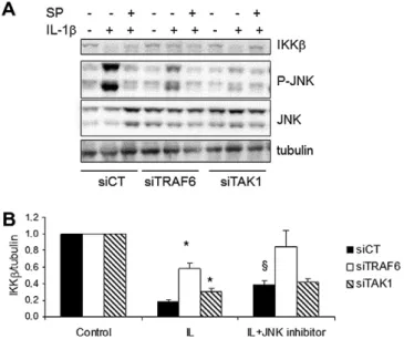
We next investigated the role of TNF receptor associated factor (TRAF)-6, an adaptor protein involved in IL-1b- but not TNF-
a
-in-duced NF-j
B activation[14,28]in IKKb degradation. A specific siR-NA against TRAF6 decreased by >60% its expression and partially prevented IKKb degradation induced by IL-1b (Fig. 4).Knocking-down transforming growth factor (TGF)-b-activated kinase 1 (TAK1), the kinase recruited by TRAF6 for the activation of IKK complex[14,29], was not as efficient as observed for TRAF6 (less than 30%), but this was sufficient to partially prevent IKKb degra-dation induced by IL-1b (Fig. 4). Beside IKK activation, TRAF6 also induces Jun-N-terminal Kinase (JNK) via TAK1[29,30]; in line with this, TRAF6 and TAK1 KD decreased JNK activation (Fig. 4). Of note, IL-1b induced higher activation of JNK than TNF-
a
in b-cells Fig. 1. BMS-345541 inhibits TNF-a- but not IL-1b-induced IKK activation. (A–B): analysis of IL-1b or TNF-a-induced NF-jB (p65 subunit) nuclear localization in INS-1E cells (A) and rat primary b-cells (B) in the presence (grey bars) or absence (white bars) of 3 h pre-treatment with IKK inhibitor BMS-245541 (BMS, 50lM). Cells were exposed to IL-1b (IL), TNF-a(TNF) or left untreated (control) for 30 min. Results are the mean ± SEM of 4 independent experiments.⁄P < 0.05 BMS treated cells vs. respective controls; paired t-test. (C): IKK activity assay; INS-1E cells were pre-treated as described in A–B with 25 or 50lM of BMS and then exposed to IL-1b, TNFaor left untreated (control) for 30 min. Cells were lysed and the IKK complex was immunoprecipitated (IP) with anti-IKKcantibody. IKK activation was measured by its capacity to phosphorylate the substrate glutathione-S-transferase (GST)-IjBa. Western blot for IKKashowed similar IP of the IKK complex. Anti-haemagglutinin (HA) antibody was used as a negative control for IP. The figure is representative of three similar experiments.
Fig. 2. IL-1b, but not TNF-a, induces IKKb degradation in b-cells. (A–B): INS-1E cells exposed to IL-1b (IL) or TNF-a(TNF) or left untreated (control) for the time indicated in the figure. (A): cells were lysed and the IKK complex was immunoprecipitated (IP) with anti-IKKcantibody and Western blot for IKKaand IKKb performed as described in Methods. Anti-haemagglutinin (HA) antibody was used as a negative control for IP. (B): the protein cell lysate was directly used in Western blot assays for IKKaand IKKb; tubulin was analyzed to confirm similar protein loading. (C): INS-1E cells were pre-treated or not with the proteasome inhibitor MG132 (2lM) for 2 h, and then exposed to IL-1b (IL) or TNF-a(TNF) or left untreated (control) for 15 min. Cells were lysed and used in Western blot assay for IKKaand IKKb; tubulin was used to confirm similar protein loading. Figures shown are representative of three to four experiments.
(Supplementary Fig. 5S A); this stimulatory effect of IL-1b on JNK started already at 5 min of treatment, with further increase by 10 min (Supplementary Fig. 5S B and C). To determine if JNK
acti-vation plays a role in IKKb degradation, we used the JNK inhibitor SP600125 (SP). SP inhibited JNK activation by P80%, as measured by its phosphorylation. JNK inhibition partially prevented IL-1b-in-duced IKKb degradation (Fig. 4), an effect similar to the one ob-served with a combination of TAK1 KD and JNK inhibition. This suggests that these two proteins are part of the same signalling pathway involved in IKKb degradation. Use of a second set of siR-NAs confirmed the observations described above (data not shown). 4. Discussion
The main findings of the present study, taken together with our previous observations[5,7], are summarized inFig. 5. We have pre-viously shown that IL-1b induces an earlier and stronger activation of ERK and of the IKK complex, as compared to TNF-
a
, leading to a more intense and protracted NF-j
B activation and consequent expression of downstream genes involved in b-cell dysfunction and death. Furthermore, IL-1b, but not TNF-a
, alone induce INS-1E cell dead [7]. We presently show that there is a differential usage of IKK complexes following b-cell exposure to IL-1b or TNF-a
. IL-1b induces degradation of IKKb via the proteasome, lead-ing to a preferential use of IKK complexes containlead-ing only the IKKa
subunit. This degradation is dependent, at least in part, on the presence of the IKK
c
subunit and activation of the TRAF6 signalling pathway (Fig. 5). Of note, TRAF6 is involved in the activation of NF-j
B by IL-1b but not by TNF-a
[14,17], which may contribute to the different effects of the two cytokines on IKK. Activation of JNK via TAK1 is also important for IL-1b-induced IKKb degradation, suggesting a new effect of this kinase, previously shown to contrib-ute for cytokine-induced b-cell apoptosis[10,31].The IKKb subunit was described as indispensable for NF-
j
B acti-vation by different pro-inflammatory cytokines[32,33]. Due to the importance of NF-j
B activation in auto-immune diseases [33], such as T1D, many ‘‘specific’’ inhibitors for IKK complexes possess-ing the IKKb subunit were developed[27,33]. Recently, however, it was shown that while TNF-a
-induced NF-j
B activation is indeed Fig. 3. IKKb degradation following inhibition of different components of the IKKcomplex. INS-1E cells were transfected with siRNAs specific for IKKa, IKKb or IKKc subunits, or a control siRNA (siCT). Three days after transfection cells were treated either with IL-1b or TNF-a(as indicated in the figure) for 30 min. Western blots for IKKa, IKKb, IKKcand tubulin were then performed. (A): representative figure of 5–8 independent experiments. (B): quantification of the results.⁄
P 6 0.05 cells trans-fected with siIKK vs. siCT under the same treatment conditions.
Fig. 4. Involvement of TRAF6, TAK1 and JNK activation in IL-1b-induced IKKb degradation in b-cells. INS-1E cells were transfected with siRNA specific for TRAF6 (siTRAF6) and TAK1 (siTAK1), or a control siRNA (siCT). Three days after transfection the cells were pre-treated or not with the JNK inhibitor (SP600125) and then left untreated (control) or treated with IL-1b for 30 min, as indicated in the figure. Western blots for IKKb, P-JNK and tubulin were then performed. (A): representative figure of 4–8 independent experiments is shown. (B): quantification of the results. Data are means ± SEM of 4–8 experiments.⁄P 6 0.05 cells transfected with siCT vs.
siTRAF6 or TAK1 with similar treatment.§P 6 0.005 IL-1b treated cells vs IL-1b + JNK
inhibitor transfected with the same siRNA.
Fig. 5. Proposed model for the differential IKK activation by IL-1b and TNF-ain b-cells. In red are the proteins putatively involved in IKKb degradation induced by IL-1b.
dependent of IKKb, IL-1b is able to induce NF-
j
B via IKKb or IKKa
[20]. The heterodimers of IKK
a
and IKKb, in association with IKKc
, are the most common IKK complexes observed in cells containing the three isoforms [14,17,34]. We presently describe, however, that b-cells treated with IL-1b have preferentially IKKa
homodi-mers in association with IKKc
(Fig. 2A). This is probably secondary to IL-1b-induced IKKb degradation. This effect seems to be specific for b-cells, in which NF-j
B activation is pro-apoptotic, since it is not observed in rat fibroblasts (Supplementary Fig. 3S A). Of note, NF-j
B activation in rat fibroblasts is anti-apoptotic, and TNF-a
induces stronger activation of this transcription factor as compared to IL-1b[5], emphasizing the context- and cell-dependent regula-tion of NF-
j
B and downstream genes.IKK
a
and IKKb share high structural similarity, but they play different roles in the regulation of NF-j
B translocation to the nu-cleus[17]and its post-translational modulation[16]. IKKa
regu-lates both NF-j
B transcriptional activity and the regulation of transcriptional co-activators, co-repressors and histones[35–40]. On the other hand, IKKb contains an ubiquitin-like domain which seems to be important for its activation and degradation[41]. In fact, IKKb degradation may be an important mechanism to attenu-ate NF-j
B activation in other cell types, and disruption of this mechanism favors neoplasias [42]. Thus, the presently observed preferential usage of IKK complexes containing only the IKKa
sub-unit following IL-1b exposure might contribute to the more intense NF-j
B activation and expression of downstream genes observed in b-cells[5,7]. Of note, we have previously shown that neither IL-1b nor TNF-a
induce the non-canonical (IKKa
-dependent) NF-j
B pathway[7].Our data suggest that IKKb degradation requires the presence of IKK
c
. IKKc
has no kinase activity, but it allows association between the IKK complex and regulatory proteins that modulate IKK activa-tion/repression [14,28]. Additional experiments are required to clarify how IKKc
contributes for the presently observed IL-1b-in-duced IKKb degradation.Different pro-inflammatory cytokines may contribute to insuli-tis and b-cell apoptosis during development of T1D [1,43]. We presently show that IL-1b and TNF-
a
utilize different ‘‘strategies’’ for NF-j
B activation in pancreatic b-cells, including usage of differ-ent members of the IKK complex. Since NF-j
B is a key transcription factor for insulitis and apoptosis [1,44], this novel information open interesting possibilities for a context-dependent modulation of the transcription factor to protect b-cells in early T1D.Acknowledgements
We thank the personnel from the Laboratory of Experimental Medicine, A. Musuaya, R. Makhnas, M. Pangerl and S. Mertens for excellent technical support. This work was supported by grants from the Fonds National de la Recherche Scientifique (FNRS) Belgium, the Communauté Française de Belgique – Actions de Recherche Concertées (ARC), the European Union (projects Savebeta and Naimit in the Framework Programme 7 of the European Community), Expert Center Grant 2008.40.001 from the Dutch Diabetes Research Foundation, the Belgium Program on Interuniversity Poles of Attraction initiated by the Belgium State (IUAP P6/40), Project WBI-CAPES (Coordenação de Aperfeiçoamen-to de Pessoal de Nível Superior) and the Fundação para a Pesquisa do Estado de São Paulo (FAPESP). AC is a Senior Research at the Belgium National Funds for Research (FNRS).
Appendix A. Supplementary data
Supplementary data associated with this article can be found, in the online version, atdoi:10.1016/j.febslet.2012.02.021.
References
[1] Eizirik, D.L., Colli, M.L. and Ortis, F. (2009) The role of inflammation in insulitis and beta-cell loss in type 1 diabetes. Nat. Rev. Endocrinol. 5, 219–226. [2] Heimberg, H., Heremans, Y., Jobin, C., Leemans, R., Cardozo, A.K., Darville, M.
and Eizirik, D.L. (2001) Inhibition of cytokine-induced NF-jB activation by adenovirus-mediated expression of a NF-jB super-repressor prevents b-cell apoptosis. Diabetes 50, 2219–2224.
[3] Giannoukakis, N., Rudert, W.A., Trucco, M. and Robbins, P.D. (2000) Protection of human islets from the effects of interleukin-1b by adenoviral gene transfer of an IjB repressor. J. Biol. Chem. 275, 36509–36513.
[4] Baker, M.S., Chen, X., Cao, X.C. and Kaufman, D.B. (2001) Expression of a dominant negative inhibitor of NF-jB protects MIN6 b-cells from cytokine-induced apoptosis. J. Surg. Res. 97, 117–122.
[5] Ortis, F. et al. (2008) Induction of nuclear factor-jB and its downstream genes by TNF-aand IL-1b has a pro-apoptotic role in pancreatic b cells. Diabetologia 51, 1213–1225.
[6] Eldor, R. et al. (2006) Conditional and specific NF-jB blockade protects pancreatic b cells from diabetogenic agents. Proc. Natl. Acad. Sci. USA 103, 5072–5077.
[7] Ortis, F., Cardozo, A.K., Crispim, D., Storling, J., Mandrup-Poulsen, T. and Eizirik, D.L. (2006) Cytokine-induced proapoptotic gene expression in insulin-producing cells is related to rapid, sustained, and non-oscillatory nuclear factor-jB activation. Mol. Endocrinol. 20, 1867–1879.
[8] Dutta, J., Fan, Y., Gupta, N., Fan, G. and Gelinas, C. (2006) Current insights into the regulation of programmed cell death by NF-jB. Oncogene 25, 6800–6816. [9] Cardozo, A.K., Heimberg, H., Heremans, Y., Leeman, R., Kutlu, B., Kruhoffer, M., Orntoft, T. and Eizirik, D.L. (2001) A comprehensive analysis of cytokine-induced and nuclear factor-jB-dependent genes in primary rat pancreatic b-cells. J. Biol. Chem. 276, 48879–48886.
[10] Gurzov, E.N., Ortis, F., Bakiri, L., Wagner, E.F. and Eizirik, D.L. (2008) JunB inhibits ER stress and apoptosis in pancreatic b cells. PLoS One 3, e3030. [11] Kutlu, B., Cardozo, A.K., Darville, M.I., Kruhoffer, M., Magnusson, N., Orntoft, T.
and Eizirik, D.L. (2003) Discovery of gene networks regulating cytokine-induced dysfunction and apoptosis in insulin-producing INS-1 cells. Diabetes 52, 2701–2719.
[12] Hoffmann, A., Leung, T.H. and Baltimore, D. (2003) Genetic analysis of NF-jB/ Rel transcription factors defines functional specificities. EMBO J. 22 sb:pages>5530–5539.
[13] Saccani, S., Pantano, S. and Natoli, G. (2003) Modulation of NF-jB activity by exchange of dimers. Mol. Cell 11, 1563–1574.
[14] Hayden, M.S. and Ghosh, S. (2008) Shared principles in NF-jB signaling. Cell 132, 344–362.
[15] Vermeulen, L., De Wilde, G., Notebaert, S., Vanden Berghe, W. and Haegeman, G. (2002) Regulation of the transcriptional activity of the nuclear factor-jB p65 subunit. Biochem. Pharmacol. 64, 963–970.
[16] Chariot, A. (2009) The NF-jB-independent functions of IKK subunits in immunity and cancer. Trends Cell Biol. 19, 404–413.
[17] Hacker, H. and Karin, M. (2006). Regulation and function of IKK and IKK-related kinases. Sci STKE 2006, re13.
[18] Wegener, E. and Krappmann, D. (2008) Dynamic protein complexes regulate NF-jB signaling. Handbook Exp. Pharmacol. 12, 237–259.
[19] Krappmann, D., Wegener, E., Sunami, Y., Esen, M., Thiel, A., Mordmuller, B. and Scheidereit, C. (2004) The IkappaB kinase complex and NF-jB act as master regulators of lipopolysaccharide-induced gene expression and control subordinate activation of AP-1. Mol. Cell Biol. 24, 6488–6500.
[20] Solt, L.A., Madge, L.A., Orange, J.S. and May, M.J. (2007) Interleukin-1-induced NF-jB activation is NEMO-dependent but does not require IKKb. J. Biol. Chem. 282, 8724–8733.
[21] Solt, L.A. and May, M.J. (2008) The IjB kinase complex: master regulator of NF-jB signaling. Immunol. Res. 42, 3–18.
[22] Janjic, D., Maechler, P., Sekine, N., Bartley, C., Annen, A.S. and Wolheim, C.B. (1999) Free radical modulation of insulin release in INS-1 cells exposed to alloxan. Biochem. Pharmacol. 57, 639–648.
[23] Pipeleers, D.G., in’t Veld, P.A., Van de Winkel, M., Maes, E., Schuit, F.C. and Gepts, W. (1985) A new in vitro model for the study of pancreaticaand b cells. Endocrinology 117, 806–816.
[24] Rasschaert, J. et al. (2005) Toll-like receptor 3 and STAT-1 contribute to double-stranded RNA+ interferon-c-induced apoptosis in primary pancreatic b-cells. J. Biol. Chem. 280, 33984–33991.
[25] Cardozo, A.K., Kruhoffer, M., Leeman, R., Orntoft, T. and Eizirik, D.L. (2001) Identification of novel cytokine-induced genes in pancreatic b-cells by high-density oligonucleotide arrays. Diabetes 50, 909–920.
[26] Moore, F. et al. (2009) PTPN2, a candidate gene for type 1 diabetes, modulates interferon-c-induced pancreatic b-cell apoptosis. Diabetes 58, 1283–1291. [27] Burke, J.R. et al. (2003) BMS-345541 is a highly selective inhibitor of IjB
kinase that binds at an allosteric site of the enzyme and blocks NF-j B-dependent transcription in mice. J. Biol. Chem. 278, 1450–1456.
[28] Ghosh, S. and Karin, M. (2002) Missing pieces in the NF-jB puzzle. Cell 109 (Suppl), S81–S96.
[29] Chen, Z.J. (2005) Ubiquitin signalling in the NF-jB pathway. Nat. Cell Biol. 7, 758–765.
[30] Li, X., Commane, M., Burns, C., Vithalani, K., Cao, Z. and Stark, G.R. (1999) Mutant cells that do not respond to interleukin-1 (IL-1) reveal a novel role for IL-1 receptor-associated kinase. Mol. Cell Biol. 19, 4643–4652.
[31] Gurzov, E.N., Ortis, F., Cunha, D.A., Gosset, G., Li, M., Cardozo, A.K. and Eizirik, D.L. (2009) Signaling by IL-1b+IFN-cand ER stress converge on DP5/Hrk activation: a novel mechanism for pancreatic b-cell apoptosis. Cell Death Differ. 16, 1539–1550.
[32] Hayden, M.S. and Ghosh, S. (2004) Signaling to NF-kB. Genes Dev. 18, 2195–2224.
[33] Strnad, J. and Burke, J.R. (2007) IjB kinase inhibitors for treating autoimmune and inflammatory disorders: potential and challenges. Trends Pharmacol. Sci. 28, 142–148.
[34] Scheidereit, C. (2006) IkappaB kinase complexes: gateways to NF-jB activation and transcription. Oncogene 25, 6685–6705.
[35] Anest, V., Hanson, J.L., Cogswell, P.C., Steinbrecher, K.A., Strahl, B.D. and Baldwin, A.S. (2003) A nucleosomal function for IjB kinase-a in NF-j B-dependent gene expression. Nature 423, 659–663.
[36] Hoberg, J.E., Popko, A.E., Ramsey, C.S. and Mayo, M.W. (2006) IjB kinasea -mediated derepression of SMRT potentiates acetylation of RelA/p65 by p300. Mol. Cell Biol. 26, 457–471.
[37] Hoberg, J.E., Yeung, F. and Mayo, M.W. (2004) SMRT derepression by the IjB kinasea: a prerequisite to NF-jB transcription and survival. Mol. Cell 16, 245–255.
[38] Huang, W.C., Ju, T.K., Hung, M.C. and Chen, C.C. (2007) Phosphorylation of CBP by IKKapromotes cell growth by switching the binding preference of CBP from p53 to NF-jB. Mol. Cell 26, 75–87.
[39] Lawrence, T., Bebien, M., Liu, G.Y., Nizet, V. and Karin, M. (2005) IKKalimits macrophage NF-jB activation and contributes to the resolution of inflammation. Nature 434, 1138–1143.
[40] Luo, J.L., Tan, W., Ricono, J.M., Korchynskyi, O., Zhang, M., Gonias, S.L., Cheresh, D.A. and Karin, M. (2007) Nuclear cytokine-activated IKKacontrols prostate cancer metastasis by repressing Maspin. Nature 446, 690–694.
[41] May, M.J., Larsen, S.E., Shim, J.H., Madge, L.A. and Ghosh, S. (2004) A novel ubiquitin-like domain in IjB kinase b is required for functional activity of the kinase. J. Biol. Chem. 279, 45528–45539.
[42] Lee, D.F. et al. (2009) KEAP1 E3 ligase-mediated downregulation of NF-jB signaling by targeting IKKb. Mol. Cell 36, 131–140.
[43] von Herrath, M. and Nepom, G.T. (2009) Animal models of human type 1 diabetes. Nat. Immunol. 10, 129–132.
[44] Cnop, M., Welsh, N., Jonas, J.C., Jorns, A., Lenzen, S. and Eizirik, D.L. (2005) Mechanisms of pancreatic b-cell death in type 1 and type 2 diabetes: many differences. Few Similarities Diabetes 54 (Suppl 2), S97–S107.