Copyright © 2003, American Society for Microbiology. All Rights Reserved.
Egr Family Members Regulate Nonlymphoid Expression of Fas
Ligand, TRAIL, and Tumor Necrosis Factor during
Immune Responses†
Nathalie M. Droin, Michael J. Pinkoski, Emmanuel Dejardin, and Douglas R. Green*
Division of Cellular Immunology, La Jolla Institute for Allergy and Immunology,San Diego, California 92121
Received 3 April 2003/Returned for modification 25 April 2003/Accepted 9 July 2003
The Fas ligand (FasL)/Fas pathway is crucial for homeostasis of the immune system and peripheral tolerance. Peripheral lymphocyte deletion involves FasL/Fas in at least two ways: coexpression of both Fas and its ligand on T cells, leading to activation-induced cell death, and expression of FasL by nonlymphoid cells, such as intestinal epithelial cells (IEC), that kill Fas-positive T cells. We demonstrate here that superantigen Staphylococcus enterotoxin B (SEB) induced a dramatic upregulation of FasL, TRAIL, and TNF mRNA expression and function in IEC from BALB/c and C57BL/6 mice. Using adoptive transfer in which CD4!T cells
from OT-2 T-cell receptor transgenic mice were transferred into recipients, we observed an induction in IEC of FasL, TRAIL, and TNF mRNA after administration of antigen. Specific Egr-binding sites have been identified in the 5! promoter region of the FasL gene, and Egr-1, Egr-2, and Egr-3 mRNA in IEC from mice treated with SEB and from transgenic OT-2 mice after administration of antigen was upregulated. Overex-pression of Egr-2 and Egr-3 induced endogenous ligand upregulation that was inhibited by overexOverex-pression of Egr-specific inhibitor Nab1. These results support a role for Egr family members in nonlymphoid expression of FasL, TRAIL, and TNF.
Apoptosis-inducing members of the tumor necrosis factor (TNF) family, the so-called death ligands, include TNF, FasL, and TNF-related apoptosis-inducing ligand (TRAIL). These ligands and their receptors play pivotal roles in the regulation of the immune system, especially via induction of apoptosis in activated lymphocytes. In particular, they have been implicated in the process of activation-induced cell death (AICD) in T lymphocytes, in which repeated antigen stimulation of T cells induces apoptosis through the induction of death ligand ex-pression and sensitization to death receptor-induced apoptosis (3, 6, 13, 44). Death ligand-mediated AICD is at least one mechanism for peripheral deletion, the process whereby ex-panding clones of activated T cells subsequently decrease in number (31).
The roles for death ligands in immune regulation include expression of death ligands in nonlymphoid tissues. Immune privilege in the anterior chamber of the eye requires functional FasL expression in this organ, which induces apoptosis in in-filtrating lymphocytes (9). Similarly, functional FasL expres-sion on the cornea contributes to graft acceptance in an animal model of cornea transplant (42). More recent studies have shown that epithelial cells of the eye express, in addition to FasL, TRAIL (21) and TNF (8), which may also contribute to immune privilege.
Other nonlymphoid tissues express FasL only during im-mune responses involving extensive T-cell activation, and this is also likely to contribute to peripheral deletion (36). Such
tissues include the lung, liver, and small intestine. More re-cently, we demonstrated that expression of FasL on intestinal epithelial cells (IEC) is induced by TNF in response to antigen (35). TNF-induced FasL expression in IEC was found to depend on activation of NF-"B and its effect on the FasL promoter.
Cyclosporine has been shown to interfere with AICD (41), suggesting that cyclosporine-sensitive transcription factors may be involved. Indeed, several studies have demonstrated roles for the cyclosporine-sensitive nuclear factor of activated T cells (NFAT) transcription factor and for early-growth-response transcription factors, mainly Egr-2 and Egr-3, in T-cell expres-sion of the FasL gene following T-cell receptor (TCR) ligation (7, 38). NFAT is a family of transcription factors that are present in the cytoplasm in a phosphorylated form. After de-phosphorylation mediated by the phosphatase calcineurin, these factors are rapidly translocated into the nucleus, where they activate different genes, including those encoding cell surface receptors, signaling molecules, and cytokines. Double knockout mice lacking both NFATp and NFAT4 exhibit im-paired induction of FasL and a lymphoproliferative disorder, suggesting a key role for these transcription factors in FasL transcription (37).
While several studies postulate that NFAT may be a direct regulator of FasL (19, 20, 23), it has also been demonstrated that the cyclosporine-inhibitable Egr-2 and Egr-3 transcription factors are potent regulators of FasL expression (29, 30). This family is composed of four members: Egr-1 (NGFI-A, krox24, zif268), Egr-2 (krox20), Egr-3 (PILOT), and Egr-4 (NGFI-C). All of these members share a highly conserved DNA-binding domain composed of three zinc finger motifs that bind and transactivate transcription from the consensus sequence GCG GGGGCG (33). Similar to some other transcription factors, Egr members associate with corepressor proteins Nab1 and * Corresponding author. Mailing address: La Jolla Institute for
Al-lergy and Immunology, 10355 Science Center Dr., San Diego, CA 92121. Phone: (858) 558-4675. Fax: (858) 558-3526. E-mail: Dgreen5240 @aol.com.
† La Jolla Institute for Allergy and Immunology article no. 559. 7638
by on October 23, 2007
mcb.asm.org
Nab2, which can modulate transcription of Egr-dependent genes. These factors bind to Egr-1, Egr-2, and Egr-3 by direct proteprotein interactions with a conserved R1 domain, in-hibiting the transactivating potential of these transcription fac-tors (32).
Here, we extend our observations on FasL expression in IEC to other death ligands and transcription factors. We report that Egr-1, Egr-2, and Egr-3 mRNA is upregulated in IEC from BALB/c and C57BL/6 mice challenged with superantigen Staphylococcus enterotoxin B (SEB) and from transgenic OT-2 mice after administration of antigen. Overexpression of Egr-2, Egr-3, or TNF in IEC induces endogenous FasL, TRAIL, and TNF upregulation, whereas Nab1 coexpression completely abolishes these effects. These results support a role for Egr family members in nonlymphoid expression of FasL, TRAIL, and TNF.
MATERIALS AND METHODS
Cell lines and animals.All animals were bred in house or purchased from Jackson Laboratories (Bar Harbor, Maine) and housed under pathogen-free conditions at the La Jolla Institute for Allergy and Immunology. All cell lines were obtained from the American Type Culture Collection (Manassas, Va.), including L1210 and HT29 cells, and grown in suspension culture medium RPMI 1640. Primary IEC were isolated from small intestines according to the proce-dure previously described (22) and cultured in Dulbecco’s modified Eagle’s medium. Each medium was supplemented with 10% (vol/vol) heat-inactivated fetal bovine serum, 2 mML-glutamine, 100 U of penicillin/ml, and 100 #g of streptomycin/ml, and the cells were grown in 5% CO2at 37°C.
Adoptive transfer and antigenic stimulation.CD4!lymphocyte subsets were enriched as previously described (36), and lymphocytes (5 $ 106to 10 $ 106) were introduced into the tail veins of recipient animals. Superantigen SEB (75 to 100 #g/mouse; Sigma) was injected intraperitoneally (i.p.) into BALB/c mice. OT-2 TCR transgenic animals and recipients of OT-2 lymphocytes were treated subcutaneously (s.c.) with the ovalbumin (OVA) peptide consisting of amino acids 323 to 339 (OVA 323-339 peptide) at 300 #g per mouse. The OVA 323-329 peptide introduced s.c. was delivered in complete Freund’s adjuvant (15).
Functional assay for FasL and TRAIL expression.FasL and TRAIL activities were detected by the ability of IEC to induce DNA fragmentation in Fas-positive and TRAIL receptor 2 (TRAIL-R2)-positive target cells (3, 21). Briefly, parental L1210 and L1210-Fas, both containing TRAIL-R2, were labeled with 5 #Ci of [3H]thymidine/ml for 4 h (39). After incubation, cells were rinsed twice with Hanks balanced salt solution (HBSS) to remove unincorporated thymidine. Target cells were then incubated in complete medium with dissociated epithelial cells from the small intestines of control or treated mice in 96-well plates in the presence or absence of Fas-Fc and/or TRAIL-R2–Fc (R&D Systems, Minneap-olis, Minn.). After 18 h, cells were harvested with a multiautomated system harvester, and DNA was counted in a beta counter. DNA fragmentation was determined as previously described (2).
Real-time reverse transcription-PCR (RT-PCR). Total RNA was isolated from primary IEC with Trizol (Gibco BRL, Rockville, Md.) according to the manufacturer’s instructions. RNA was reverse transcribed by Moloney murine leukemia virus reverse transcriptase (Gibco BRL) with random hexamers (Gibco BRL). For FasL gene expression, primers were located in separate exons in genomic DNA in order to eliminate the possibility of genomic DNA contami-nation. Sequence-specific primers for murine FasL, TNF, %-actin, 18S, and lu-ciferase were previously described (35). Specific forward and reverse primers were, respectively, as follows: murine TRAIL, 5&-CCTCTCGGAAAGGGCAT TC-3& and 5&-TCCTGCTCGATGACCAGCT-3&; Egr-1, 5&-GGCGATGGTGG AGACGAGT-3& and 5&-CGGCCAGTATAGGTGATGGG-3&; Egr-2, 5&-TCTC CGTGCCAGAGAGATCC-3& and 5&-GAGGACAGGGAAACGGCTTT-3&; Egr-3, 5&-CACTGGAGACCATCAAGGCC-3& and 5&-GGGCAGGCTGCCGA A-3&; TRAIL-R2, 5&-TGCTGCTCAAGTGGCGC-3& and 5&-GGCATCCAGCA GATGGTTG-3&; human FasL, 5&-CAGTCCACCCCCTGAAAAAAAA-3& and 5&-CCAGAGGCATGGACCTTGAG-3&; 18S, 5&-ACATCCAAGGAAGGCAG-CAG-3& and 5&-TCGTCACTACCTCCCCGG-3&; LacZ, 5&-AATGGCTTTCGC TACCTGGA-3& and 5&-CCATCGCGTGGGCGTA-3&; green fluorescent pro-tein (GFP), 5&-TGGCCCTGTCCTTTTACCAG-3& and 5&-TTTTCGTTGGGA TCTTTCGAA-3&; Renilla, 5&-AATTTGCAGCATATCTTGAACCATT-3& and
5&-GGATTTCACGAGGCCATGAT-3&. Real-time PCR was performed with AmpliTaq Gold polymerase in a PE Biosystems 5700 thermocycler using the SyBr Green detection protocol as outlined by the manufacturer. Briefly, 12 ng of total cDNA, 50 nM (each) primer, and 1$ SyBr Green mixture were used in a total volume of 25 #l.
Plasmid constructs and retrovirus gene transfer.Murine Egr-2, Egr-3, and Nab1 cDNAs were amplified by RT-PCR from total RNA using the Egr-2 forward primer (5&-ATGAACGGAGTGGCGGGAGATGGC-3&), Egr-2 re-verse primer (5&-TCACGGTGTCCTGGTTCGAGAGGT-3&), Egr-3 forward primer (5&-ATGACCGGCAAACTCGCCGAG-3&), Egr-3 reverse primer (5&-T CAGGCGCAGGTGGTGACCAC-3&), Nab1 forward primer (5&-ATGGCCAC AGCCTTACCTAGGACCCTG-3&), and Nab1 reverse primer (5&-CTATCTTG AGTCTTCAGGCTCTG-3&) at the respective ATG start and stop codons. All amplified fragments were cloned directly into the pcDNA3.1V5-TOPO vector (Invitrogen, Carlsbad, Calif.) and sequenced according to the manufacturer’s instructions.
The hemagglutinin-tagged Egr-3 product was generated by PCR using the Egr-3 forward primer containing an EcoRI restriction site (5&-TATAGAATTC ATGACCGGCAAACTCGCCG-3&) and the Egr-3 reverse primer containing a NotI restriction site (5&-TATAGCGGCCGCTTATGCATAATCTGGTACATC GTATGGATAGGCGCAGGTGGTGACCACAGGGGC-3&). The PCR prod-uct was digested with EcoRI and NotI and cloned into the retroviral pMX-IRES-GFP vector, digested with EcoRI and NotI. Retrovirus production, HT29 cell infection, and sorting were conducted as previously described (5).
Human FasL promoter constructs were generated by PCR. The bp '300 and '240 regions were cloned with the 5&-CCCAAGCTTCGGCTGTTATCAGAA AATTGTGGG-3& and the 5&-CCCAAGCTTCTTCTCAGCTTCAGCTGCAA AGCG-3& sense primers together with the 5&-ACGCGTCGACCATGGCAGC TGGTGAGTCAGGCCAGC-3& antisense primer. Egr and NFAT with mutations in the bp '300 region were generated by PCR using the 5&-CCCAAGCTTCG GCTGTTATCAGAAAATTTTAAACGGAAACTTCC-3& and the 5&-CCCAA GCTTCGGCTGTTATCAGAAAATTGTGGGCGTCCCGATCCAGG-3& sense primers, respectively, and Egr with mutations in the bp '240 region was generated by PCR using the 5&-CCCAAGCTTCTTCTCAGCTTCAGCTGCAAAGCTCTA AATGTTTCT-3& sense primer. All amplified fragments were cloned directly into the pGLOW vector (Invitrogen) and sequenced according to the manufacturer’s instructions.
Transient-transfection and reporter assays.In transient-expression experi-ments, all transfections were performed with Superfect (Qiagen, Valencia, Calif.) according to the manufacturer’s instructions and 0.5 #g of each vector. Trans-fected IEC, HT29, and Egr3-HT29 cells were incubated for 18 h at 37°C before analysis by real-time PCR. Due to the low efficiency of transient transfection, TNF, Egr2, Egr3, and Nab1 overexpression was checked by real-time PCR. The antagonist anti-TNF receptor type 1 (TNFR1) antibody (clone 55R-170) was used at a concentration of 2 #g/ml. GFP mRNA was analyzed by real-time RT-PCR, and transfection efficiencies were measured by analysis of Renilla or luciferase reporter mRNA expression.
Protein extraction and Western blot analysis.The antibodies used in this study included anti-TRAIL, anti-FasL (Santa Cruz Biotechnology, Santa Cruz, Calif.), anti-TNF (a gift from Carl F. Ware), and antiactin (ICN, Aurora, Ohio). For protein extraction, cells were washed twice in phosphate-buffered saline (PBS), lysed in buffer containing 150 mM NaCl, 50 mM pH 8.0 Tris-HCl, 0.1% Na-sodium dodecyl sulfate (SDS), and 0.5% Na deoxycholate in the presence of protease inhibitors for 30 min at 4°C and then centrifuged (20 min; 15,000 $ g) and supernatants were collected. For Western blots, after SDS-polyacrylamide gel electrophoresis and electrotransfer to nitrocellulose membranes (Amersham Biosciences), the membranes were incubated for at least 1 h with 5% nonfat milk in TPBS (0.1% Tween 20, 1$ PBS) and for 2 h at room temperature with primary antibodies. Horseradish peroxidase-conjugated goat rabbit, anti-hamster, or anti-mouse secondary antibodies were then added for 30 to 60 min, and proteins were revealed by enhanced chemiluminescence reagent (Pierce, Rockford, Ill.) and autoradiography.
RESULTS
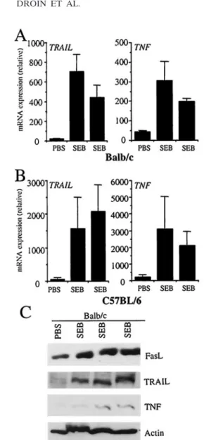
FasL, TRAIL, and TNF mRNA are upregulated in IEC after SEB injection.We have previously established that the super-antigen SEB induces a dramatic upregulation of FasL expres-sion and function in nonlymphoid cells of the liver and small intestine (2). Using real-time RT-PCR, we studied the expres-sion of other death ligands of the TNF family in IEC from
VOL. 23, 2003 TNF LIGAND FAMILY EXPRESSION BY Egr MEMBERS 7639
by on October 23, 2007
mcb.asm.org
BALB/c and C57BL/6 mice injected with PBS or SEB. Inter-estingly, we observed a dramatic upregulation of TRAIL and TNF mRNA in these cells 3 days after SEB injection (Fig. 1A and B). In keeping with our previous observations, an increase in FasL protein levels in response to superantigen-induced activation of peripheral lymphocytes and an increase in TRAIL and TNF protein levels were also observed (Fig. 1C). OT-2 transfer induces FasL, TRAIL, and TNF mRNA up-regulation. hFlp-lacZ transgenic mice express a LacZ con-struct under the control of the proximal promoter of the hu-man FasL gene (36), and antigenic challenge of these mice resulted in increased expression of nonlymphoid FasL as well as LacZ in the epithelium of the small intestine (36). To study
the expression of the different death ligands in IEC, we em-ployed an adoptive-transfer approach in which CD4!T cells from OT-2 TCR (OVA-plus-I-Ab-responsive) transgenic mice were transferred into transgenic hFlp-lacZ recipient mice (OT-23hFlp-lacZ mice). OT-(OT-23hFlp-lacZ recipients were chal-lenged with the OVA peptide; 3 days later IEC were isolated and RNA was assessed by real-time RT-PCR for endogenous FasL, LacZ, TRAIL, and TNF expression. In keeping with our previous findings, we observed that induction of expression of LacZ mRNA correlated with expression of FasL mRNA after administration of antigen (Fig. 2A). We also observed a sig-nificant induction of TRAIL and TNF mRNAs, demonstrating that activated lymphocytes induced accumulation of these dif-ferent ligands at the mRNA level. A similar effect was ob-served when hFlp-lacZ $ OT-2 double-transgenic animals were immunized directly with OVA (Fig. 2B).
FasL, TRAIL, and TNF induction in IEC is TNF dependent. We recently reported that FasL expression in IEC is induced in activated T cells through a TNF-dependent mechanism (35). To determine if TRAIL and TNF mRNA expression in the IEC is also induced by TNF production, we injected TNF into mice and examined ligand expression in the epithelium of the small FIG. 1. IEC FasL, TRAIL, and TNF are induced in response to
superantigen. BALB/c (A) and C57BL/6 (B) mice were injected i.p. with superantigen SEB (75 to 100 #g), and after 3 days IEC total mRNA was isolated and assessed by real-time RT-PCR for TRAIL and TNF mRNA expression. Independent data from two SEB-treated mice are shown for each strain. Absolute mRNA values were determined, normalized to %-actin, and reported as arbitrary units. (C) FasL, TRAIL, and TNF protein levels were also assessed by Western blot-ting. Independent results from three SEB-treated mice are shown.
FIG. 2. OT-2 transfer into hFlp-lacZ transgenic mice and antigen stimulation in OT-2 $ hFlp-lacZ double-transgenic mice simulta-neously induce FasL, TRAIL, and TNF upregulation. CD4! T cells
from OT-2 TCR transgenic mice were transferred into C57BL/6 re-cipients. OT-23hFlp-lacZ recipients (A) and OT-2 $ hFlp-lacZ dou-ble-transgenic (B) mice were challenged with OVA 323-339, and IEC were isolated. The OVA peptide introduced s.c. was delivered in com-plete Freund’s adjuvant. Total mRNA was isolated and assessed by real-time RT-PCR for FasL, LacZ, TRAIL, and TNF mRNA expres-sion. Absolute mRNA values were determined, normalized to %-actin, and reported as arbitrary units.
by on October 23, 2007
mcb.asm.org
intestine. Injection of TNF (2 #g/mouse) resulted in a signif-icant increase in TRAIL and TNF expression in IEC in vivo (Fig. 3A). We then asked if TNF is necessary for upregulation of TRAIL in IEC as it is for upregulation of FasL. To address this question, wild-type and TNF'/'mice were injected with SEB and 3 days later IEC mRNA was isolated and assayed by real-time RT-PCR for TRAIL mRNA expression. We ob-served a significant increase of TRAIL mRNA expression in wild-type mice after SEB injection, whereas little TRAIL mRNA was seen in TNF'/'mice (Fig. 3B). We have previ-ously shown that IEC constitutively express TNFR1 (p55) (35) and not TNFR2 (data not shown). To confirm that the ob-served effect is dependent on TNFR1, wild-type and TNFR1'/' mice were injected with SEB. Three days later total IEC
mRNA was isolated, and TRAIL and TNF mRNA expression was assessed by real-time RT-PCR (Fig. 3C). Levels of TRAIL and TNF mRNA expression in wild-type IEC were significantly increased after SEB injection, whereas no upregulation was seen for TRAIL and TNF in IEC from TNFR1'/'mice (Fig. 3C). Taken together, these results suggest that TNF is involved in TRAIL expression following exposure to antigen.
IEC express functional FasL and TRAIL.FasL present in nonlymphoid tissue has been shown to delete reactive lympho-cytes during viral infection and after exposure to SEB or an OVA peptide (2, 9, 36). To evaluate the function of nonlym-phoid FasL and TRAIL, we isolated IEC from SEB-treated and untreated animals and tested their ability to kill Fas! target cells. As shown in Fig. 4, IEC from SEB-treated mice killed targets bearing the Fas receptor (L1210-Fas), confirming our previous observation (Fig. 4A). Because L1210-Fas also expresses the TRAIL-R2 (Fig. 4B, inset), we used these cells to examine the function of TRAIL by culturing the IEC and target cells in the presence of Fas-Fc or TRAIL-R2–Fc. The apoptosis induced by IEC in L1210-Fas cells was partially in-hibited when Fas-Fc or TRAIL-R2–Fc was included in the culture (Fig. 4B). We also observed a greater protection when both Fas-Fc and TRAIL-R2–Fc were added together to the culture in which IEC and targets were at a higher ratio (10:1), suggesting a possible cooperation between these two ligands in killing. However, the addition of both Fas-Fc and TRAIL-R2–Fc did not completely inhibit cell death, and therefore it is possible that other factors are also involved.
To further evaluate the function of nonlymphoid TRAIL, we isolated IEC from SEB-treated C57BL/6-gld animals and tested their ability to kill TRAIL-R2!target cells (L1210). IEC from SEB-treated C57BL/6 animals induced apoptosis in L1210 cells, and this apoptosis was partially inhibited when TRAIL-R2–Fc was included in the culture (Fig. 4C). IEC from SEB-treated C57BL/6-gld animals induced less apoptosis in L1210 cells, presumably due to nonfunctional FasL, but the observed death was almost completely inhibited when TRAIL-R2–Fc was included in the culture (Fig. 4C). Again, the addi-tion of TRAIL-R2–Fc did not completely inhibit cell death in the higher-ratio (10:1) culture, suggesting that other factors could be involved. Thus, the FasL and the TRAIL induced on IEC by in vivo administration of SEB are biologically active.
Egr transcription factors are upregulated in IEC after SEB and OVA injection. The Egr-1 transcription factor has been shown to be involved in TNF expression, while Egr-2 or Egr-3 expression can induce FasL transcription in nonlymphoid cells (17, 30). These observations suggest a possible role for Egr transcription factors in FasL and TNF expression and perhaps TRAIL expression. Egr family members are zinc finger pro-teins that are rapidly induced in diverse cell types following cell activation by different stimuli. Egr-1 mRNA is known to be constitutively expressed in many tissues, whereas Egr-2 and Egr-3 mRNA is not found in unactivated tissues (34), arguing in favor of de novo synthesis mechanisms. Using real-time RT-PCR, we studied endogenous Egr-1, Egr-2, and Egr-3 gene expression in IEC from BALB/c, C57BL/6, B6129SF2 wild-type, TNF'/', and TNFR1'/'mice injected with PBS or SEB. We observed that Egr-1 mRNA is the major Egr mRNA pressed in these cells, while Egr-2 and Egr-3 mRNA is ex-pressed at lower levels (Fig. 5). Constitutive Egr-2 and Egr-3 FIG. 3. Induction of nonlymphoid TRAIL and TNF mRNA is
de-pendent on TNF. (A) BALB/c mice were injected with TNF (2 #g), and IEC were harvested 12 h later and analyzed for TRAIL and TNF mRNA expression. Absolute mRNA values were determined, normal-ized to 18S RNA, and reported as arbitrary units. Data from three TNF-treated mice are shown. (B) B6129SF2 and TNF'/'mice were
injected with SEB. IEC were isolated 3 days after SEB administration and analyzed for TRAIL mRNA expression. Absolute mRNA values were determined, normalized to %-actin, and reported as arbitrary units. (C) C57BL/6 and TNFR1'/'mice were injected with SEB. IEC
were isolated 3 days after SEB administration and analyzed for TRAIL and TNF mRNA expression. Absolute mRNA values were deter-mined, normalized to %-actin, and reported as arbitrary units.
VOL. 23, 2003 TNF LIGAND FAMILY EXPRESSION BY Egr MEMBERS 7641
by on October 23, 2007
mcb.asm.org
mRNA expression in some tissues is not consistent with pre-vious observations (34) but can be explained by the fact that real-time PCR is more sensitive than Northern analysis or conventional RT-PCR.
In addition, we observed a dramatic upregulation of Egr-1, Egr-2, and Egr-3 mRNA in wild-type IEC 3 days after SEB injection (Fig. 5A). The kinetics of appearance of FasL, TRAIL, and TNF mRNA in these cells after SEB injection correlated with Egr expression (Fig. 1A and B). Egr-1, Egr-2, and Egr-3 mRNA was also induced in IEC from hFlp-lacZ $ OT-2 double-transgenic mice after direct immunization with
OVA (Fig. 5B). To determine if Egr mRNA expression in IEC was dependent on TNF, wild-type and TNF'/' mice were injected with SEB and 3 days later IEC mRNA was isolated and Egr expression was assessed by real-time RT-PCR (Fig. 6A). Egr-1 and Egr-3 mRNA expression in wild-type IEC was significantly increased after SEB injection, whereas no upregu-lation was seen for Egr-1, Egr-2, or Egr-3 in IEC from TNF'/' mice (Fig. 6A).
We have shown that IEC express TNFR1 (35). To confirm that the observed effect is dependent on TNFR1, wild-type and TNFR1'/'mice were injected with SEB and 3 days later IEC FIG. 4. Epithelial cells from the small intestines of SEB-treated mice have functional FasL and TRAIL. BALB/c mice were challenged in vivo with PBS or SEB, and IEC were prepared. (A) IEC were cocultured with [3H]thymidine (TdR)-labeled L1210 or L1210-Fas target cells for 18 h
at the ratios indicated on the x axes. Induction of apoptosis in the targets was assessed as the percentage of specific DNA fragmentation. (B) The contribution of FasL and TRAIL to the killing of L1210-Fas targets expressing TRAIL-R2, as assessed by real-time RT-PCR (inset), was studied by inclusion of 5 mg of Fas-Fc and/or TRAIL-R2–Fc/ml. Induction of apoptosis in the L1210-Fas targets at the indicated ratios was assessed as the percentage of specific DNA fragmentation. (C) C57BL/6 and C57BL/6-gld mice were challenged in vivo with SEB, and IEC were prepared. The IEC were cocultured with [3H]TdR-labeled L1210 target cells for 18 h at the indicated ratios. The contribution of TRAIL to the killing of
L1210 targets was studied by inclusion of 5 mg of TRAIL-R2–Fc/ml. Induction of apoptosis in the L1210 targets at the indicated ratios was assessed as the percentage of specific DNA fragmentation.
by on October 23, 2007
mcb.asm.org
mRNA was isolated and Egr expression was assessed by real-time RT-PCR (Fig. 6B). Egr-2 and Egr-3 mRNA expression in wild-type IEC was significantly increased after SEB injection, whereas no upregulation was seen for Egr-1, Egr-2, or Egr-3 in IEC from TNFR1'/'mice (Fig. 6B).
Egr-2 and Egr-3 transcription factors induce endogenous FasL, TRAIL, and TNF upregulation in IEC. Egr family tran-scription factors contain no known dimerization domains and are known to bind to DNA and activate transcription as mono-mers (32). Since Egr-1 did not appear to have a major role in activation-induced upregulation of FasL transcription (30), we studied endogenous ligand expression after Egr-2 or Egr-3 overexpression in primary IEC. Egr-2 or Egr-3 overexpression was sufficient to induce endogenous FasL mRNA accumula-tion in both cell types (Fig. 7). Interestingly, we also observed an increase in endogenous TRAIL and TNF mRNA after Egr-2 or Egr-3 overexpression in primary IEC (Fig. 7).
Recently, a novel family of corepressors, named Nab, that
inhibits the transcriptional activity of the Egr family of zinc finger transcription factors was identified (43). To determine if endogenous FasL, TRAIL, and TNF mRNA expression is Egr dependent, murine Nab1 was cloned and coexpressed with 2 or 3. As expected, Nab1 expression inhibited Egr-induced FasL, TRAIL, and TNF upregulation in primary IEC (Fig. 7). To further analyze the effects of TNF and Egr mem-FIG. 5. IEC Egr-1, -2, and -3 mRNA is induced in response to
superantigen and after antigen stimulation in OT-2 $ hFlp-lacZ dou-ble-transgenic mice. BALB/c and C57BL/6 (A) and OT-2 $ hFlp-lacZ double-transgenic (B) mice were injected i.p. with superantigen SEB (75 to 100 #g) (A) or challenged with OVA (B), and after 3 days IEC total mRNA was isolated and assessed by real-time RT-PCR for Egr-1, Egr-2, and Egr-3 mRNA expression. Absolute mRNA values were determined, normalized to %-actin, and reported as arbitrary units.
FIG. 6. IEC Egr-1, -2, and -3 mRNA expression is dependent on TNF. B6129SF2 and TNF'/' (A) and C57BL/6 and TNFR1'/'
(B) mice were injected i.p. with superantigen SEB (75 to 100 #g), and after 3 days IEC total mRNA was isolated and assessed by real-time RT-PCR for Egr-1, Egr-2, and Egr-3 mRNA expression. Absolute mRNA values were determined, normalized to %-actin, and reported as arbitrary units.
FIG. 7. Transient overexpression of Egr-2 and Egr-3 induces en-dogenous FasL, TRAIL, and TNF mRNA upregulation in primary IEC. Primary IEC prepared from BALB/c mice were transfected with different constructs expressing murine Egr-2, Egr-3, and/or Nab1. Twenty hours later total mRNA isolated from transfected IEC was analyzed for endogenous FasL, TRAIL, and TNF mRNA expression. Absolute mRNA values were determined, normalized to 18S RNA, and reported as arbitrary units.
VOL. 23, 2003 TNF LIGAND FAMILY EXPRESSION BY Egr MEMBERS 7643
by on October 23, 2007
mcb.asm.org
bers on IEC, we expressed TNF in primary IEC by transient transfection and assayed the effects on FasL and TRAIL mRNA levels (Fig. 8). Primary IEC were transfected with TNF-pcDNA in the presence or absence of an anti-TNFR1 antagonistic antibody, and 20 h later RNA was analyzed by real-time RT-PCR. As we have previously shown (35), there was a significant increase in FasL mRNA levels in TNF-trans-fected IEC, and we also observed an increase in TRAIL mRNA (Fig. 8). The TNF-dependent effects on FasL and TRAIL ex-pression were reduced to background levels when an anti-TNFR1 antagonistic antibody was added to the medium (Fig. 8). We then examined the effect of Nab1 expression in the cells expressing TNF (note that the expression of TNF allowed us to examine the effects of Nab1 coexpression on endogenous li-gand expression in the same cells). As shown in Fig. 8, the expression of the Egr-specific inhibitor Nab1 inhibited TNF-induced FasL and TRAIL upregulation. These results suggest that TNF production induces Egr-dependent expression of FasL and TRAIL in IEC.
To identify DNA elements that control the regulation of FasL transcription, known regions of the human FasL gene 5& of the translation start site containing Egr-binding sites were used to drive the expression of a GFP reporter in primary IEC (Fig. 9). Due to the difficulty in transfecting primary cells with high efficiency to study reporter gene expression by conven-tional assays, we developed an assay to study reporter expres-sion at the mRNA level by real-time RT-PCR. In transient transfections with the indicated GFP reporter constructs, ex-pression of Egr-2 led to a weak induction of the bp '300 human FasL promoter reporter while Egr-3 induced strong transcription (Fig. 9A and B). Surprisingly, truncation of the FasL promoter to nucleotide '240, deleting putative overlap-ping NFAT and Egr sites, did not inhibit the transcriptional activity induced by Egr-2 but eliminated the response to Egr-3, indicating that Egr-3 binds only the putative overlapping
NFAT and Egr sites. To determine if these sites are required for FasL induction in IEC, we tested FasL promoter activity using promoter-GFP constructs with specific mutations in the binding sites for these transcription factors. Mutations within the Egr or NFAT sites completely abolished the activity of the bp '300 construct upon Egr-3 overexpression, suggesting that mutation in the NFAT site affects Egr-3 binding to its own site (Fig. 9). The role of Egr-3 at the putative overlapping NFAT and Egr sites in the FasL promoter was confirmed by using HT29 cells overexpressing Egr-3 (Fig. 9C). In this experiment, FIG. 8. Endogenous FasL and TRAIL mRNA upregulation
in-duced by TNF is inhibited by Nab1 overexpression in IEC. Primary IEC prepared from BALB/c mice were transfected with different con-structs expressing murine Egr-2 or Egr-3 either alone, in the presence of a neutralizing TNFR1 antibody (Ab), or with Nab1. Twenty hours later total mRNA was isolated from transfected IEC and analyzed for FasL (A) and TRAIL (B) mRNA by real-time RT-PCR. Absolute mRNA values were determined, normalized to 18S RNA, and re-ported as arbitrary units.
FIG. 9. Expression of exogenous Egr-3 and Egr-2 protein is suffi-cient to induce FasL-responsive-element-dependent transcription in nonlymphoid cells. Primary IEC prepared from BALB/c mouse cells were cotransfected with different constructs expressing murine Egr-2 (A) or Egr-3 (B) with the indicated GFP reporter plasmids with sche-matic Egr (blue boxes) and NFAT (purple boxes) sites. X, mutated Egr and NFAT sites. GFP mRNA was analyzed by real-time RT-PCR and normalized to luciferase mRNA expression as an efficiency control. (C) HT29 cells were infected with either an internal ribosomal entry site (IRES)-GFP control retrovirus or an IRES-GFP retrovirus ex-pressing wild-type murine Egr-3. Stably infected cells were sorted by fluorescence-activated cell sorter to (95% GFP-expressing cells. Egr-3 mRNA expression was assessed by real-time RT-PCR. (Inset) HT29 and Egr3-HT29 cells were cotransfected with the indicated GFP re-porter plasmids and a Renilla vector as a control. GFP mRNA was analyzed by real-time RT-PCR and normalized to Renilla mRNA.
by on October 23, 2007
mcb.asm.org
this site was again required for Egr-3-induced FasL promoter activity.
DISCUSSION
In this paper, we have shown that IEC express not only FasL but also TRAIL and TNF in response to superantigen- or peptide-induced activation of peripheral lymphocytes. In keep-ing with our previous observations, we found that the expres-sion of these different ligands in IEC depends on TNF (35). Our results suggest a model in which TNF, produced by lym-phoid cells after activation by superantigen or peptide (16), induces Egr-dependent expression of FasL, TRAIL, and TNF in IEC through TNFR1. Biologically active FasL and TRAIL expressed in the small intestine may then kill infiltrating Fas! and TRAIL-R2!T cells (Fig. 10).
One function of the TNF ligand family is the induction of apoptosis. Constitutive expression of TRAIL mRNA has been reported in most normal tissues and cell types (10). TRAIL is not expressed on freshly isolated T cells; however, both TRAIL
and the TRAIL receptor are upregulated upon T-cell activa-tion (12, 25, 40). For example, CD4!V)24NKT cells express TRAIL only when the cells are activated by )-galactosylcer-amide-pulsed dendritic cells and can kill malignant target cells (32). However, the function of this cytokine in normal tissues is still not clear. Recently, a study using mice deficient in TRAIL found an essential role for TRAIL in thymocyte apo-ptosis and inhibition of autoimmunity (18). Our results show that functional FasL and TRAIL are expressed on normal IEC during immune responses, suggesting a possible cooperation of these ligands in peripheral lymphocyte deletion. Our data are in line with recent publications demonstrating cross-activation of different death systems, enabling autoamplification of apo-ptosis following ligation of FasL, TRAIL, and TNF (11). Fol-lowing the same concept, Lee et al. recently reported the expression of a functional TRAIL protein in an immune-priv-ileged site, the anterior chamber of the eye (21). In addition, the pattern of expression of TRAIL in the eye is remarkably similar to that of FasL, suggesting a possible cooperation be-tween these different ligands in the elimination of activated T cells, thereby limiting the damage to the tissues into which the T cells infiltrate.
While the idea that T-cell-induced nonlymphoid expression of molecules such as FasL, TRAIL, and TNF contributes to peripheral deletion is attractive, a possible problem concerns the effect of cyclosporine in these systems. Egr is inhibited by cyclosporine (29), and this accounts in part for the inhibition of FasL expression in activated T cells treated with cyclosporine (4). In contrast, peripheral deletion is not inhibited by cyclo-sporine (2). At present, we envision at least two possibilities. First, it is possible that Egr in IEC and other nonlymphoid cells is not inhibitable by cyclosporine. Alternatively, some nonlym-phoid cells may express FasL (and other related molecules) that is not dependent on Egr. For example, constitutive ex-pression of FasL in the testes is not inhibited by cyclosporine (19).
Studies with the FasL promoter have established the pres-ence of several sites important in its transcription, such as binding sites for NF-"B (14, 26, 27), NFAT (19, 20), activating protein 1 (AP-1) (28), and Egr-2 and -3 (29, 30), although the relative functional importance of these sites might differ among species. We found that injection of the superantigen SEB into wild-type mice or of OVA into OT-2 mice induced Egr-1, Egr-2, and Egr-3 expression in IEC and that this was not observed with mice deficient in TNF expression. Egr-2 and Egr-3 have recently emerged as important transcription factors for FasL expression in lymphoid and nonlymphoid cells (29, 30). Because induction of both Egr-2 and Egr-3 was shown to be cyclosporine sensitive in T cells (29), the expression of the Egr-2 and -3 genes might be dependent on NFAT. In fact, a transcriptionally active NFAT site was identified in the Egr-3 promoter, confirming this hypothesis (24). In addition, it was recently shown that NFAT-mediated induction of Egr-2 and/or Egr-3 is essential for optimal FasL synthesis in vivo (38). We have provided evidence that mutation within distal Egr- or NFAT-overlapping sites completely abolished the transcrip-tional activity of the bp '300 construct in primary IEC and Egr3-HT29 cells, demonstrating that mutation in the NFAT site affects Egr-3-induced FasL expression. However, this ob-FIG. 10. Model of nonlymphoid ligand induction in response to the
presence of activated lymphocytes. Activated lymphocytes produce TNF that binds its receptor, TNFR1, constitutively expressed on the surfaces of IEC (35). TNFR1 activation induces several transcription factors, such as Egr and NF-"B, that in turn induce TNF, FasL, and TRAIL expression in IEC. IEC expressing functional FasL and TRAIL then induce apoptosis of infiltrating Fas- and TRAIL-R-bearing lym-phocytes.
VOL. 23, 2003 TNF LIGAND FAMILY EXPRESSION BY Egr MEMBERS 7645
by on October 23, 2007
mcb.asm.org
servation does not exclude a possible interaction between Egr and endogenous NFAT. We also demonstrated that Egr-3 has no effect on a second putative Egr-binding site using the bp '240 construct in primary IEC and Egr3-HT29 cells, suggest-ing that Egr-2 may act exclusively at this site. In primary IEC, Egr-2 drove reporter expression of constructs containing either the distal or NFAT-overlapping site or the proximal Egr-binding site of the human FasL promoter.
We also found that overexpression of Egr-2 and Egr-3 in IEC was sufficient to induce expression of FasL, TRAIL, and to a lesser extent TNF mRNA. This role for Egr members in death ligand expression is relevant, because expression of the Egr corepressor Nab1 inhibited TNF-induced expression of these ligands in IEC. Therefore, the Egr transcription factor family is likely to be involved in the transcriptional regulation of several members of the TNF ligand family in IEC.
We have also recently reported that expression of FasL by TNF is dependent on NF-"B, one component of the TNFR signaling pathway (35). These results are not contradictory since mutations within both the Egr- and the "B1-binding sites silence the murine FasL promoter (28). Taken together, these results confirm that NF-"B and Egr-2 and -3 are important transcription factors for the expression of murine FasL.
Sequences proximal to bp '182 on the TNF promoter con-tain motifs for several transcription factors, including AP-1, Egr-1, NF-"B, and SP1. For example, lipopolysaccharide acti-vation of macrophages induces the binding of c-Jun, Egr-1, and NF-"B to binding sites within the proximal bp '182 region of the TNF promoter (45). We also observed an Egr-2- and -3-dependent induction of endogenous TNF in A1.1 T cells (N. M. Droin and D. R. Green, data not shown), suggesting a redundancy between the different members of this family.
Studies with the TRAIL promoter have recently established the importance of several NF-"B binding sites for TRAIL tran-scription in T cells (1). Sequence analysis of the TRAIL pro-moter also revealed several potential NFAT and AP-1 sites and one potential Egr site (N. M. Droin and D. R. Green, data not shown), suggesting that the involvement of the Egr family members in TRAIL regulation may be direct. Thus, regulation of TRAIL may parallel in many respects that of FasL and TNF. Further functional studies of cooperative interactions between transcription factors (Egr members and NF-"B) bound to these different regulatory elements will be essential to our understanding of the transcriptional regulation of the FasL, TRAIL, and TNF genes in epithelia after activation of periph-eral lymphocytes.
Taken together, these data suggest a model of ligand tran-scriptional regulation converging on the recruitment of com-plexes composed by shared components such as the Egr mem-bers and NF-"B. This hypothesis suggests a very subtle regulation of these common complexes by different combina-tions of transcription factors in the context of different en-hancer sequences. We must now determine which transcription factors are required physiologically for expression of each li-gand in nonlymphoid tissues and activated T cells. Such studies will shed light on the specificity of these complexes and help us to understand the expression of FasL, TRAIL, and TNF in different cell types during immune responses.
ACKNOWLEDGMENTS
This work was supported by National Institutes of Health grant AI44828 and the Fondation pour la Recherche Medicale (to N.M.D.).
We thank Carl F. Ware for the TNF antibody.
REFERENCES
1. Baetu, T. M., H. Kwon, S. Sharma, N. Grandvaux, and J. Hiscott. 2001. Disruption of NF-"B signaling reveals a novel role for NF-"B in the regu-lation of TNF-related apoptosis-inducing ligand expression. J. Immunol.
167:3164–3173.
2. Bonfoco, E., P. M. Stuart, T. Brunner, T. Lin, T. S. Griffith, Y. Gao, H.
Nakajima, P. A. Henkart, T. A. Ferguson, and D. R. Green.1998. Inducible nonlymphoid expression of Fas ligand is responsible for superantigen-in-duced peripheral deletion of T cells. Immunity 9:711–720.
3. Brunner, T., R. J. Mogil, D. LaFace, N. J. Yoo, A. Mahboubi, F. Echeverri,
S. J. Martin, W. R. Force, D. H. Lynch, C. F. Ware, et al.1995. Cell-autonomous Fas (CD95)/Fas-ligand interaction mediates activation-induced apoptosis in T-cell hybridomas. Nature 373:441–444.
4. Brunner, T., N. J. Yoo, D. LaFace, C. F. Ware, and D. R. Green. 1996. Activation-induced cell death in murine T cell hybridomas. Differential reg-ulation of Fas (CD95) versus Fas ligand expression by cyclosporin A and FK506. Int. Immunol. 8:1017–1026.
5. Dejardin, E., N. M. Droin, M. Delhase, E. Haas, Y. Cao, C. Makris, Z. W. Li,
M. Karin, C. F. Ware, and D. R. Green.2002. The lymphotoxin-beta receptor induces different patterns of gene expression via two NF-"B pathways. Im-munity 17:525–535.
6. Dhein, J., H. Walczak, C. Baumler, K. M. Debatin, and P. H. Krammer. 1995. Autocrine T-cell suicide mediated by APO-1/(Fas/CD95). Nature 373: 438–441.
7. Dzialo-Hatton, R., J. Milbrandt, R. D. Hockett, Jr., and C. T. Weaver. 2001. Differential expression of Fas ligand in Th1 and Th2 cells is regulated by early growth response gene and NF-AT family members. J. Immunol. 166: 4534–4542.
8. Elzey, B. D., T. S. Griffith, J. M. Herndon, R. Barreiro, J. Tschopp, and T. A.
Ferguson.2001. Regulation of fas ligand-induced apoptosis by tnf. J. Immu-nol. 167:3049–3056.
9. Griffith, T. S., T. Brunner, S. M. Fletcher, D. R. Green, and T. A. Ferguson. 1995. Fas ligand-induced apoptosis as a mechanism of immune privilege. Science 270:1189–1192.
10. Griffith, T. S., and D. H. Lynch. 1998. TRAIL: a molecule with multiple receptors and control mechanisms. Curr. Opin. Immunol. 10:559–563. 11. Herr, I., C. Posovszky, L. D. Di Marzio, M. G. Cifone, T. Boehler, and K. M.
Debatin.2000. Autoamplification of apoptosis following ligation of CD95-L, TRAIL and TNF-alpha. Oncogene 19:4255–4262.
12. Jeremias, I., I. Herr, T. Boehler, and K. M. Debatin. 1998. TRAIL/Apo-2-ligand-induced apoptosis in human T cells. Eur. J. Immunol. 28:143–152. 13. Ju, S. T., D. J. Panka, H. Cui, R. Ettinger, M. el-Khatib, D. H. Sherr, B. Z.
Stanger, and A. Marshak-Rothstein.1995. Fas(CD95)/FasL interactions re-quired for programmed cell death after T-cell activation. Nature 373:444– 448.
14. Kasibhatla, S., L. Genestier, and D. R. Green. 1999. Regulation of fas-ligand expression during activation-induced cell death in T lymphocytes via nuclear factor "B. J. Biol. Chem. 274:987–992.
15. Kearney, E. R., K. A. Pape, D. Y. Loh, and M. K. Jenkins. 1994. Visualization of peptide-specific T cell immunity and peripheral tolerance induction in vivo. Immunity 1:327–339.
16. Kramer, B., T. Machleidt, K. Wiegmann, and M. Kronke. 1995. Superanti-gen-induced transcriptional activation of the human TNF gene promoter in T cells. J. Inflamm. 45:183–192.
17. Kramer, B., A. Meichle, G. Hensel, P. Charnay, and M. Kronke. 1994. Characterization of a Krox-24/Egr-1-responsive element in the human tumor necrosis factor promoter. Biochim. Biophys. Acta 18:413–421.
18. Lamhamedi-Cherradi, S. E., S. J. Zheng, K. A. Maguschak, J. Peschon, and
Y. H. Chen.2003. Defective thymocyte apoptosis and accelerated autoim-mune diseases in TRAIL'/'mice. Nat. Immunol. 4:255–260.
19. Latinis, K. M., L. L. Carr, E. J. Peterson, L. A. Norian, S. L. Eliason, and
G. A. Koretzky.1997. Regulation of CD95 (Fas) ligand expression by TCR-mediated signaling events. J. Immunol. 158:4602–4611.
20. Latinis, K. M., L. A. Norian, S. L. Eliason, and G. A. Koretzky. 1997. Two NFAT transcription factor binding sites participate in the regulation of CD95 (Fas) ligand expression in activated human T cells. J. Biol. Chem.
272:31427–31434.
21. Lee, H. O., J. M. Herndon, R. Barreiro, T. S. Griffith, and T. A. Ferguson. 2002. TRAIL: a mechanism of tumor surveillance in an immune privileged site. J. Immunol. 169:4739–4744.
22. Lin, T., T. Brunner, B. Tietz, J. Madsen, E. Bonfoco, M. Reaves, M. Huflejt,
and D. R. Green.1998. Fas ligand-mediated killing by intestinal intraepithe-lial lymphocytes. Participation in intestinal graft-versus-host disease. J. Clin. Investig. 101:570–577.
23. Macian, F., C. Garcia-Rodriguez, and A. Rao. 2000. Gene expression elicited
by on October 23, 2007
mcb.asm.org
by NFAT in the presence or absence of cooperative recruitment of Fos and Jun. EMBO J. 19:4783–4795.
24. Mages, H. W., R. Baag, B. Steiner, and R. A. Kroczek. 1998. Utilization of an NF-ATp binding promoter element for EGR3 expression in T cells but not fibroblasts provides a molecular model for the lymphoid cell-specific effect of cyclosporin A. Mol. Cell. Biol. 18:7157–7165.
25. Mariani, S. M., and P. H. Krammer. 1998. Differential regulation of TRAIL and CD95 ligand in transformed cells of the T and B lymphocyte lineage. Eur. J. Immunol. 28:973–982.
26. Matsui, K., A. Fine, B. Zhu, A. Marshak-Rothstein, and S. T. Ju. 1998. Identification of two NF-"B sites in mouse CD95 ligand (Fas ligand) pro-moter: functional analysis in T cell hybridoma. J. Immunol. 161:3469–3473. 27. Matsui, K., S. Omura, H. Cui, S. L. Schauer, G. E. Sonenshein, and S. T. Ju. 1997. Proteasome regulation of Fas ligand cytotoxicity. Eur. J. Immunol.
27:2269–2278.
28. Matsui, K., S. Xiao, A. Fine, and S. T. Ju. 2000. Role of activator protein-1 in TCR-mediated regulation of the murine fasl promoter. J. Immunol. 164: 3002–3008.
29. Mittelstadt, P. R., and J. D. Ashwell. 1998. Cyclosporin A-sensitive tran-scription factor Egr-3 regulates Fas ligand expression. Mol. Cell. Biol. 18: 3744–3751.
30. Mittelstadt, P. R., and J. D. Ashwell. 1999. Role of Egr-2 in up-regulation of Fas ligand in normal T cells and aberrant double-negative lpr and gld T cells. J. Biol. Chem. 274:3222–3227.
31. Mogil, R. J., L. Radvanyi, R. Gonzalez-Quintial, R. Miller, G. Mills, A. N.
Theofilopoulos, and D. R. Green.1995. Fas (CD95) participates in periph-eral T cell deletion and associated apoptosis in vivo. Int. Immunol. 7:1451– 1458.
32. Nieda, M., A. Nicol, Y. Koezuka, A. Kikuchi, N. Lapteva, Y. Tanaka, K.
Tokunaga, K. Suzuki, N. Kayagaki, H. Yagita, H. Hirai, and T. Juji.2001. TRAIL expression by activated human CD4!V alpha 24NKT cells induces in vitro and in vivo apoptosis of human acute myeloid leukemia cells. Blood
97:2067–2074.
33. O’Donovan, K. J., W. G. Tourtellotte, J. Millbrandt, and J. M. Baraban. 1999. The EGR family of transcription-regulatory factors: progress at the interface of molecular and systems neuroscience. Trends Neurosci. 22:167– 173.
34. Patwardhan, S., A. Gashler, M. G. Siegel, L. C. Chang, L. J. Joseph, T. B.
Shows, M. M. Le Beau, and V. P. Sukhatme.1991. EGR3, a novel member
of the Egr family of genes encoding immediate-early transcription factors. Oncogene 6:917–928.
35. Pinkoski, M. J., N. M. Droin, and D. R. Green. 2002. Tumor necrosis factor alpha up-regulates non-lymphoid Fas-ligand following superantigen-induced peripheral lymphocyte activation. J. Biol. Chem. 277:42380–42385. 36. Pinkoski, M. J., N. M. Droin, T. Lin, L. Genestier, T. A. Ferguson, and D. R.
Green.2002. Nonlymphoid Fas ligand in peptide-induced peripheral lym-phocyte deletion. Proc. Natl. Acad. Sci. USA 99:16174–16179.
37. Ranger, A. M., M. Oukka, J. Rengarajan, and L. H. Glimcher. 1998. Inhib-itory function of two NFAT family members in lymphoid homeostasis and Th2 development. Immunity 9:627–635.
38. Rengarajan, J., P. R. Mittelstadt, H. W. Mages, A. J. Gerth, R. A. Kroczek,
J. D. Ashwell, and L. H. Glimcher.2000. Sequential involvement of NFAT and Egr transcription factors in FasL regulation. Immunity 12:293–300. 39. Rouvier, E., M. F. Luciani, and P. Golstein. 1993. Fas involvement in Ca2!
-independent T cell-mediated cytotoxicity. J. Exp. Med. 177:195–200. 40. Sheikh, M. S., T. F. Burns, Y. Huang, G. S. Wu, S. Amundson, K. S. Brooks,
A. J. Fornace, Jr., and W. S. el-Deiry.1998. p53-dependent and -independent regulation of the death receptor KILLER/DR5 gene expression in response to genotoxic stress and tumor necrosis factor alpha. Cancer Res. 58:1593– 1598.
41. Shi, Y. F., B. M. Sahai, and D. R. Green. 1989. Cyclosporin A inhibits activation-induced cell death in T-cell hybridomas and thymocytes. Nature
339:625–626.
42. Stuart, P. M., T. S. Griffith, N. Usui, J. Pepose, X. Yu, and T. A. Ferguson. 1997. CD95 ligand (FasL)-induced apoptosis is necessary for corneal allo-graft survival. J. Clin. Investig. 99:396–402.
43. Swirnoff, A. H., E. D. Apel, J. Svaren, B. R. Sevetson, D. B. Zimonjic, N. C.
Popescu, and J. Milbrandt.1998. Nab1, a corepressor of NGFI-A (Egr-1), contains an active transcriptional repression domain. Mol. Cell. Biol. 18:512– 524.
44. Yang, Y., M. Mercep, C. F. Ware, and J. D. Ashwell. 1995. Fas and activation-induced Fas ligand mediate apoptosis of T cell hybridomas: inhibition of Fas ligand expression by retinoic acid and glucocorticoids. J. Exp. Med. 181: 1673–1682.
45. Yao, J., N. Mackman, T. S. Edgington, and S. T. Fan. 1997. Lipopolysac-charide induction of the tumor necrosis factor-alpha promoter in human monocytic cells. Regulation by Egr-1, c-Jun, and NF-"B transcription fac-tors. J. Biol. Chem. 272:17795–17801.
VOL. 23, 2003 TNF LIGAND FAMILY EXPRESSION BY Egr MEMBERS 7647
by on October 23, 2007
mcb.asm.org