doi:10.1136/hrt.2007.122796
2009;95;164-170
Heart
Luc A Piérard and Patrizio Lancellotti
Non-invasive imaging
Echocardiography in the emergency room:
http://heart.bmj.com/cgi/content/full/95/2/164
Updated information and services can be found at:
These include:
References
http://heart.bmj.com/cgi/content/full/95/2/164#BIBL
This article cites 30 articles, 18 of which can be accessed free at:
Rapid responses
http://heart.bmj.com/cgi/eletter-submit/95/2/164
You can respond to this article at:
service
Email alerting
the top right corner of the article
Receive free email alerts when new articles cite this article - sign up in the box at
Topic collections
(9110 articles)
Clinical diagnostic tests(1276 articles)
Acute coronary syndromes(1470 articles)
Echocardiography(9992 articles)
Drugs: cardiovascular systemArticles on similar topics can be found in the following collections
Notes
http://journals.bmj.com/cgi/reprintform
To order reprints of this article go to:
http://journals.bmj.com/subscriptions/
go to:
Heart
To subscribe to
NON-INVASIVE IMAGING
Echocardiography in the
emergency room
Luc A Pie´rard, Patrizio Lancellotti
University Hospital, Departmentof Cardiology, Liege, Belgium Correspondence to: Professor Luc Pierard, Department of Cardiology, University Hospital, B-4000 Liege, Belgium; lpierard@chu. ulg.ac.be
The evaluation and management of patients who arrive at the emergency department with acute symptoms and/or worrying clinical condi-tion is a daily challenge. Careful history taking— when it is possible—and clinical examination remain the mandatory first steps. It is of the utmost importance to obtain a timely accurate diagnosis and to immediately stratify the risk to the patient’s life. Therefore, choice of the correct investigation is essential. Among the possible tools, imaging modalities are frequently required. Echocardiography is the most versatile method. It is non-invasive, can be performed at the bedside, provides rapid results, and avoids con-trast injection and exposure of the patient to radiation. Echocardiography is now available in most emergency rooms, allowing immediate, standard transthoracic examination by an emer-gency department physician. For some indica-tions and some ultrasonic modalities, specialised and trained physicians or sonographers are necessary for performance and interpretation.
This article focuses on the clinical information that can be obtained from echocardiography according to the most frequent presentations suggesting a cardiovascular emergency.
ACUTE CHEST PAIN: SUSPICION OF ACUTE CORONARY SYNDROME
Patients with acute chest pain account for a notable proportion (20–30%) of medical admis-sions to the emergency department.1 Acute
cor-onary syndromes can be easily identified in the presence of typical chest pain and a diagnostic ECG, and confirmed by serial changes in cardiac enzymes. However, a vast majority of patients have atypical chest pain and/or normal or non-diagnostic ECG; early determination of serum troponin frequently is negative. Marginal eleva-tions of troponin in this clinical setting result in uncertainty. To avoid unnecessary prolonged hospital observation (which is costly) and mis-taken discharge (with the associated potential for litigation), rapid cost effective and accurate risk stratification is important. Numerous alternative strategies are possible (table 1).
Echocardiography can be performed only at rest, conventionally or with implementation of an ultrasound contrast agent. Echocardiography can also be coupled with exercise or pharmacological stress testing.
Rest echocardiography
A brief echocardiographic examination can be performed in ,10 min. The primary role of rest echocardiography when performed in the emer-gency room is to assess the presence and extent of regional wall motion abnormalities. A dyssynergic ventricular region can be ischaemic, stunned, hibernating or necrotic. In particular, the distinc-tion between ongoing ischaemia and previous myocardial infarction may be difficult; in addition, acute ischaemia can occur in segments adjacent to necrotic tissue. On the other hand, the absence of segmental dyssynergy cannot definitely exclude a recent episode of ischaemia in the absence of stunning. Regional wall motion abnormalities can also be present in other conditions, such as myocarditis, right ventricular (RV) pressure or volume overload, pre-excitation, left bundle branch block or the presence of a pacemaker.
The main limitation is that recording of reliable images can be technically difficult if a skilled echocardiographer is not available. The interpreta-tion of subtle wall mointerpreta-tion abnormalities requires training and experience. The current development of telemedicine may be helpful; an expert can guide the recording and interpret images accurately.
A major advantage is the versatility of the echocardiogram. Cardiac causes of acute chest pain other than coronary artery disease can be detected (box 1).
Contrast echocardiography
Slow bolus intravenous injections or continuous infusion of an ultrasonic contrast agent containing small microbubbles results in the opacification of the left ventricle, improving endocardial border detection, and permits the observation of myocar-dial perfusion. The assessment of both regional function and perfusion at the bedside represents a significant advantage. A multicentre study com-pared contrast echocardiography and gated single photon emission computed tomography (SPECT) in more than 200 patients presenting to the emergency room with chest pain and non-diag-nostic ECG. Both methods provided more incre-mental information for early cardiac events than did the demographic, clinical and ECG assessment.2
Another study in the same clinical setting used contrast echocardiography in 957 patients. This technique provided accurate short, intermediate, and long term prognostic information. Normal regional function was most efficient for predicting
lowest risk. For a given regional function, normal perfusion was indicative of a very low risk whereas abnormal perfusion identified a high risk for acute coronary syndrome.3
However, the choice of appropriate technical settings and correct inter-pretation of images require experience which is not easily obtained by emergency department physi-cians.
Stress echocardiography
Current guidelines recommend pre-discharge exer-cise testing in patients with acute chest pain, normal or non-diagnostic ECG, and normal serum troponin values. The diagnostic accuracy of ST segment changes is limited, however.
Exercise echocardiography requires trained phy-sicians, and patients who are capable of exercising. Echocardiograms are performed at rest and either immediately after treadmill or bicycle exercise, or preferably at peak stress during supine bicycle exercise.4
Exercise echocardiography and exercise SPECT have been shown to provide comparable short term prognostic information in the triage of chest pain patients, allowing safe early discharge; the negative
predictive value was 97% in both methods, but exercise echocardiography is preferable because of a higher positive predictive value.5
Pharmacological stress echocardiography can be used in patients unsuitable for exercise testing. Graded dobutamine infusion with addition of atropine if necessary, or high dose dipyridamole and atropine, can be used as stressors. Early dobutamine stress after admission to the emer-gency room has been shown to be feasible and safe. Pre-discharge dobutamine stress echocardiography has important and independent prognostic value in low risk, troponin negative, chest pain patients.6
As compared to exercise ECG, dobutamine stress echocardiography was found to be more cost effective: the mean length of stay in the hospital was lower; and no event occurred in a 2 month follow-up in patients with a normal dobutamine test, whereas the event rate was 11% in patients with normal exercise ECG.7 Several studies
com-bining patients submitted to exercise echocardio-graphy or dobutamine stress echocardioechocardio-graphy have also demonstrated good risk stratification, especially through a high negative predictive value.8 9
In summary, echocardiography has an important role in the evaluation of chest pain in the emergency room when the ECG is non-diagnostic. Rest echocardiography alone has diagnostic limita-tions. Contrast or stress echocardiography have substantial value for the accurate detection of acute coronary syndromes and for prediction or risk. The strength of these methods is their high negative predictive value. The positive predictive value depends on the patient’s characteristics, in particular his probability of suffering from signifi-cant coronary artery disease.
ACUTE CHEST PAIN: OTHER CAUSES Pericarditis
Individuals with pericarditis usually present with chest pain, which is classically increased in the lying position and during inspiration. The pain can be transient. The ECG may show ST segment elevation but it is usually widespread and concave upwards. A pericardial friction rub is audible in only one third of patients and frequently transi-ent.10
Elevated cardiac troponin can be detected in half of the patients.11Echocardiography is helpful if
pericardial effusion is detected; this occurs in two thirds of cases.12 However, regional wall motion
abnormalities may be present if pericarditis is associated with myocarditis, and small pericardial effusion is a frequent complication of acute myocardial infarction.
In the absence of pericardial effusion, increased pericardial thickness is detectable, more easily from the subcostal window.
Aortic dissection
Dissection of the aorta is a life threatening condition for which rapid diagnosis and correct management are essential.13Chest pain is typically
Table 1 Resting imaging modalities for triage of patients with acute chest pain and non-diagnostic ECG
Methods Advantages Limitations
Coronary angiography High accuracy Invasive
Low uncertainty Limited immediate availability
Resting echocardiography
Widely available
Diagnosis of causes other than CAD
Difficult interpretation of regional dyssynergy
Normal if no ischaemia at rest
Contrast echocardiography
Improved endocardial detection Regional function and perfusion Rapid examination
Challenging interpretation of perfusion images
Multislice CT Rapid image acquisition Requires apnoea and contrast injection Diagnosis of causes other than CAD Radiation
Coronary anatomy
Cardiac MRI Detection of function, perfusion and necrosis
No isotopes
Limited immediate availability Quantitative information
SPECT High negative predictive value Not available ‘‘after hours’’ Wide validation
CAD, coronary artery disease; CT, computed tomography; MRI, magnetic resonance imaging; SPECT, single photon emission computed tomography.
Box 1 Acute chest pain—cardiac causes detectable by rest echocardiography
c Acute coronary syndrome
c Pericarditis
c Aortic dissection (low sensitivity of transthoracic echocardiography) c Hypertrophic cardiomyopathy
c Aortic stenosis c Mitral valve prolapse
severe, and immediately of maximum intensity. Clinical suspicion is easier if the pain is migrating and in the presence of specific malperfusion syndrome. Rapid and non-invasive diagnostic imaging is required; the choice of the modality depends on the relative immediate availability.14
Transthoracic echocardiography can be performed without delay to visualise the aortic root and arch, but the distal ascending aorta and descending aorta are more difficult to evaluate. The specificity is high (90%). The sensitivity has been shown to vary from 55–75%; the proportion of false negative studies decreases when all ultrasonic windows are used, including right parasternal, subcostal, supr-asternal, abdominal or even dorsal views. Clearly, a normal transthoracic echocardiographic examina-tion cannot rule out aortic dissecexamina-tion. A major pitfall is the possibility of reverberation artefacts. The presence of a mobile intimal flap separating the true and false lumen can be observed in the proximal ascending aorta. Severe complications can also be identified from the transthoracic approach: acute aortic regurgitation, pericardial effusion, or regional wall motion abnormalities suggestive of dissection involving a coronary artery.
Transoesophageal echocardiography is the pre-ferred ultrasonic modality (fig 1).15
The blind region is limited to the most distal part of the ascending aorta and the proximal part of the arch. An intimal flap, a large false lumen and a true lumen are diagnostic. The assessment of entry is essential to distinguish between type A and type B, as their management strategies are strikingly different.16Several features may be able to
differ-entiate the true and false lumen. The false lumen is usually larger; its diameter increases in diastole; intense spontaneous contrast is frequent in the
presence of blood stasis; and thrombus can be observed. Accuracy of transoesophageal echocar-diography requires skilled hands. This method of imaging is the preferred first choice in the emergency room in centres where an experienced cardiologist is readily available.17 The positive
predictive value is of course dependent on the disease prevalence and is lower than magnetic resonance imaging (MRI) in the low prevalence populations.18
Other causes of acute aortic syndrome include intramural haematoma and penetrating athero-sclerotic ulcers. Although their management remains controversial, proper identification is of clinical importance. Transthoracic echocardiogra-phy is of limited value and the transoesophageal approach is more efficient. The semiology of intramural haematoma is well known: .7 mm crescent or circumferential heterogenous thicken-ing of the aortic wall. Longitudinal extension is usually important. Sometimes, an echo-free region may be observed, suggesting haemorrhage or liquefaction of the haematoma. If the diagnosis is questionable, other imaging modalities such as MRI may be necessary.19
A penetrating athero-sclerotic ulcer most frequently occurs in the descending aorta; the transoesophageal window is thus mandatory, but computed tomography and MRI perform better.
Pulmonary embolism
Chest pain is frequent (65%) in patients admitted to the emergency room with a pulmonary embo-lism, but is usually associated with dyspnoea. Both symptoms and clinical signs are not specific and the differential diagnosis is frequently difficult with acute coronary syndromes or other condi-tions.20
If echocardiography is immediately avail-able, important information can be rapidly obtained. The visualisation of a large mobile, serpentine thrombus trapped in the right heart chambers (embolism in transit) is rare but makes the diagnosis evident.21Transoesophageal
echocar-diography is a useful method to visualise pulmon-ary embolism, especially if it is central. In this case, the sensitivity is 90–95% and the specificity is 100%.22
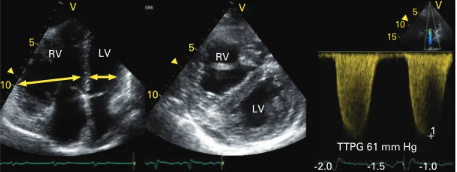
In practice, echocardiography provides excellent assessment of the acute haemodynamic consequences. The main findings, although non-specific, are dilatation of the right heart chambers and of the inferior vena cava, abnormal motion of the interventricular septum, and an abnormal ratio of right ventricular (RV) diameter or area to left ventricular (LV) diameter or area. A ratio between end diastolic right to LV diameter .0.6 and a ratio of end diastolic right to LV area .1.0 are consistent with massive pulmonary embolism associated with RV dysfunction (fig 2).23
Another interesting feature is hypokinesis of the mid RV free wall and preserved contraction of the RV apex: the so-called McConnell sign.24
RV systolic pressure, and thus systolic pulmonary arterial (PA) pressure in the absence of pulmonary valve stenosis, can be estimated from the peak Figure 1 Transoesophageal echocardiographic examination obtained in a patient with
acute aortic dissection. EO, entry orifice; FL, false lumen; H, intense spontaneous contrast + thrombus; TL, true lumen.
velocity of the tricuspid regurgitant jet and application of the simplified Bernoulli equation: PA systolic pressure = right atrial pressure + 4V2.
Right atrial pressure is estimated by clinical examination of the jugular veins or by the diameter of the inferior vena cava and its respiratory changes. In the presence of a first episode of pulmonary embolism, systolic PA pressure cannot increase to more than 40–50 mm Hg. A chronic process—repetitive episodes of pulmonary embo-lism or chronic cor pulmonale—is probable if the PA pressure is .60 mm Hg (peak velocity of tricuspid regurgitant jet .3.5 m/s). New ultrasonic modalities such as tissue Doppler imaging, permit-ting the measurement of RV free wall velocities and strain, can add significant information on RV function.
The parameters cited above not only help in the diagnosis of pulmonary embolism but can also predict prognosis. They could be used and inte-grated with troponin and B type natriuretic peptide (BNP) elevation to decide upon the therapeutic option—for example, anticoagulation versus thrombolytic therapy.25 26
HYPOTENSION AND JUGULAR VEIN DISTENSION
The association of hypotension and elevated jugular venous pressure may be present in different
clinical conditions, such as acute heart failure, cardiac tamponade, RV infarction, severe pulmon-ary embolism, and exacerbation of chronic obstruc-tive pulmonary disease (COPD). Echocardiography is well suited to potentially provide rapid diagnosis in this clinical setting.
Cardiac tamponade
Cardiac tamponade corresponds to acute or sub-acute compression of the heart. It results from important and/or rapid accumulation of fluid in the pericardial space that increases intrapericardiac pressure above intracavitary pressure. Diastolic filling is reduced and ventricular ejection is compromised. Compensatory tachycardia and vasoconstriction initially maintain a normal car-diac output. At a critical level of intrapericardial pressure, cardiac output and arterial pressure fall. Pulsus paradoxus is present: LV stroke volume decreases during inspiration because of reduced LV preload and paradoxal motion of the interventri-cular septum resulting from RV volume overload. These clinical features correspond to the echocar-diographic findings. The echocarechocar-diographic diag-nosis of this life threatening condition is usually rapid and simple (fig 3).
Two dimensional echocardiography shows the presence, location and volume of pericardial effu-sion. The presence of fibrin, thrombus, sponta-neous contrast or separated effusion usually implies surgical drainage. The most important aspect in the emergency room is the assessment of haemodynamic consequences.27 Indeed, the
repercussions mainly depend on the rapidity of fluid accumulation rather than on effusion size. The earlier sign indicating a risk of tamponade is right atrial diastolic collapse, but its specificity is moderate. Diastolic RV collapse occurs later, is more specific, and is best appreciated in the parasternal short axis view. Dilatation of the inferior vena cava without change during deep inspiration has a good sensitivity but a moderate specificity for the diagnosis of tamponade. Pulsed wave Doppler is essential. Changes in velocities during inspiration should be recorded at low speed. The most interesting characteristics include inspiratory increase of peak pulmonary ejection velocity (.40%) and of RV inflow velocity (.85%), as well as a parallel reduction during inspiration of LV inflow and aortic ejection velocities.
If the patient’s condition requires urgent peri-cardiocentesis, the procedure can be echocardio-graphically guided.27
The subcostal view is important to determine the distance between the patient’s skin and the effusion. If necessary, the correct position of the pericardial needle can be visualised and, in the case of doubt, agitated saline contrast can be injected to confirm the intraper-icardial position of the needle. Echocardiography can of course be used to verify whether the effusion has been completely drained. Transoesophageal echocardiography is rarely indi-cated in this setting.
Figure 2 Echocardiographic examination of a patient admitted for a recurrent episode of pulmonary embolism. The right ventricle (RV) was enlarged compared with the left ventricle (LV) and the ratio between end diastolic RV to LV diameter was .0.6. The trans-tricuspid pressure gradient (TTPG) was notably increased, indicating severe pulmonary hypertension.
Figure 3 Echocardiographic examination of a patient admitted for cardiac tamponade. The diagnosis was confirmed by the combination of a pericardial effusion (PE), a complete right ventricular collapse, and the significant respiratory changes in mitral E wave velocity.
Acute decompensated heart failure
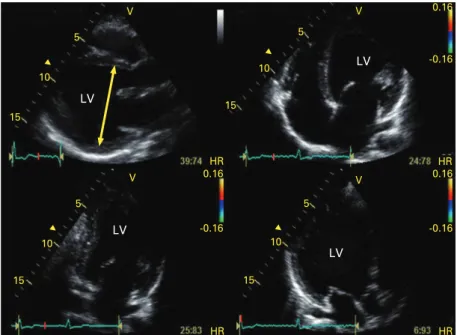
Numerous patients, especially the elderly, are admitted to the emergency room for acute decom-pensated heart failure (figs 4 and 5). In addition to hypotension and jugular vein distension, these patients present with dyspnoea and orthopnoea. Pulmonary rales, peripheral oedema and hepato-jugular reflux provide evidence of elevated filling pressures. Rapid measures of BNP or NT-proBNP enhance the accuracy of diagnosis. Rapid distinc-tion between heart failure due to systolic versus diastolic dysfunction should be obtained since there are significant differences in treatment. An
immediate echocardiogram at the bedside in the emergency room makes it possible to visualise dilatation of the heart chambers, diffuse or regional abnormalities in contraction, functional mitral and/or tricuspid regurgitation or, in contrast, normal LV volume, enlarged left atrium, LV hypertrophy and preserved ejection fraction. Numerous additional measures and detailed eva-luation of systolic and diastolic function can be obtained later during the hospital stay.
Severe pulmonary embolism
The role of echocardiography in this clinical setting has been already described.
ACUTE PULMONARY OEDEMA
Acute cardiogenic pulmonary oedema may result from acute events or from acute deterioration of a chronic disease. The main mechanisms include reduced outflow, reduced inflow or backward flow. Some conditions are obvious causes of pulmonary oedema, such as a large acute myocardial infarction or severe tachyarrhythmias. The patients should first be treated with oxygen, nitrates and loop diuretics. Physical examination is insensitive in detecting a cardiac murmur. Thus, Doppler echo-cardiography can be useful and performed when treatment has been initiated.
Hypertensive crises induce exacerbation of dia-stolic dysfunction. In this setting, sydia-stolic dysfunc-tion is not observed, even transiently; the LV ejection fraction can be normal and similar to that found later after treatment.28
Doppler echocardiography can rapidly reveal valvular heart disease, such as severe aortic stenosis, severe acute aortic regurgitation due to endocarditis or rupture of a sinus of Valsalva in the LV, huge mitral regurgitation resulting from papillary muscle or primary chordae rupture, or severe dysfunction of a prosthetic valve. Dynamic exacerbation of chronic ischaemic mitral regurgita-tion is another condiregurgita-tion, probably more difficult to ascertain, because early treatment, especially glyceryl trinitrate, substantially decreases mitral regurgitant volume. Ischaemic mitral regurgitation may appear to be mild or moderate, although it could have been severe at the onset of the syndrome. If this condition is suspected, exercise Doppler echocardiography could reveal it a few days later, showing a dynamic increase in mitral regurgitation resulting in an increase in pulmonary vascular pressure.29 30
CONCLUSIONS
Echocardiography should be available in the emergency room for immediate or rapid observa-tion at the bedside of many possible abnormalities that help diagnose cardiovascular syndromes or diseases. This quite versatile imaging method can find a place in the diagnostic algorithm of most clinical acute presentations. This article has only focused on the most frequent or important applications.
Figure 4 Echocardiographic examination showing depressed left ventricular (LV) systolic function in a patient admitted for acute dyspnoea.
Figure 5 Echocardiographic examination showing preserved left ventricular (LV) systolic function in a patient admitted for acute dyspnoea. The diagnosis of hypertrophic cardiomyopathy was made. The spectral tissue Doppler derived E/Ea ratio revealed an increased LV filling pressure confirming the diagnosis of diastolic heart failure. E, early mitral inflow velocity; Ea, diastolic mitral annular velocity.
Competing interests: In compliance with EBAC/EACCME guide-lines, all authors participating in Education in Heart have disclosed potential conflicts of interest that might cause a bias in the article. The authors have no competing interests.
REFERENCES
1. Blatchford O, Capewell S. Emergency medical admissions in Glasgow: general practices vary despite adjustments for age, sex and deprivation. Br J Gen Pract 1999;49:551–4.
2. Kaul S, Senior R, Firschke C, et al. Incremental value of cardiac imaging in patients presenting to the emergency department with chest pain and without ST-segment elevation: a multicenter study. Am Heart J 2004;148:129–36.
3. Tong KL, Wang XQ, Rinkevich, et al. Myocardial contrast echocardiography versus thrombolysis in myocardial infarction score in patients presenting to the emergency department with chest pain and a nondiagnostic electrocardiogram. J Am Coll Cardiol 2008;46:920–7.
4. Pie´rard LA. Echocardiographic monitoring throughout exercise: better than the post-treadmill approach? J Am Coll Cardiol 2007;50:1864–66.
c Editorial on the comparison of post- and peri-exercise stress echocardiography.
5. Conti A, Sammicheli L, Gallini C, et al. Assessment of patients with low-risk chest pain in the emergency department: head-to-head comparison of exercise stress echocardiography and exercise myocardial SPECT. Am Heart J 2005;149:894–901.
6. Bholasing R, Cornel JH, Kamp O, et al. Prognostic value of predischarge dobutamine stress echocardiography in chest pain patients with a negative cardiac troponin T. J Am Coll Cardiol 2003;41:596–602.
7. Nucifora G, Badano LP, Sarraf-Zadegan N, et al. Comparison of early dobutamine stress echocardiography and exercise electrocardiographic testing for management of patients presenting to the emergency department with chest pain. Am J Cardiol 2007;100:1068–73.
c Study reporting the superiority of early dobutamine stress echocardiography in patients presenting with chest pain. 8. Colon PJ, Cheirif J. Long-term value of stress echocardiography in
the triage of patients with atypical chest pain presenting to the emergency department. Echocardiography 1999;16:171–7.
c Study showing the prognostic value of stress echo-cardiography in patients presenting with acute chest pain. 9. Jeetley P, Burden L, Senior R. Stress echocardiography is superior to exercise ECG in the risk stratification of patients presenting with acute chest pain with negative troponin. Eur J Echocardiogr 2006;7:155–64.
c Important study reporting the superiority of stress echocardiography in patients presenting with acute chest pain.
10. Imazio M, Demichelis B, Parrini I, et al. Day-hospital treatment of acute pericarditis: a management program for outpatient therapy. J Am Coll Cardiol 2004;43:1042–6.
11. Bonnefoy E, Godon P, Kirkorian G, et al. Serum cardiac troponin I and ST-segment elevation in patients with acute pericarditis. Eur Heart J 2000;21:832.
12. Imazio M, Bobbio MD, Cecchi E, et al. Colchicine in addition to conventional therapy for acute pericarditis. Circulation 2005;27:2012–16.
13. Hagan PG, Nienaber Ca, Isselbacher EM, et al. The International Registry of Acute Aortic Dissection (IRAD): new insights into an old disease. JAMA 2000;283:897–903.
14. Cigarroa JE, Isselbacher EM, DeSanctis RW, et al. Diagnostic imaging in the evaluation of suspected aortic dissection. Old standards and new directions. N Engl J Med 1993;328:35–43. 15. Nienaber CA, von Kodolitsch Y, Nicolas V, et al. The diagnosis of
thoracic aortic dissection by noninvasive imaging procedures. N Engl J Med 1993;328:1–9.
16. Nienaber CA, Spielmann RP, van Kodolitsch Y, et al. Diagnosis of thoracic aortic dissection. Magnetic resonance imaging versus transesophageal echocardiography. Circulation 1992;85:434–47.
c Important study comparing the accuracy of magnetic resonance imaging versus transoesophageal
echocardiography for the diagnosis of thoracic aortic dissection.
17. Erbel R, Oelert H, Meyer J, et al. Effect of medical and surgical therapy on aortic dissection evaluated by transesophageal echocardiography. Implications for prognosis and therapy. The European Cooperative Study Group on Echocardiography. Circulation 1993;87:1604–15.
18. Moore AG, Eagle KA, Bruckman D, et al. Choice of computed tomography, transesophageal echocardiography, magnetic resonance imaging, and aortography in acute aortic dissection: International Registry of Acute Aortic Dissection (IRAD). Am J Cardiol 2002;89:1235–8.
19. Nienaber CA, Sievers HH. Intramural haematoma in acute aortic syndrome: more than one variant of dissection? Circulation 2002;106:284–5.
20. Stein PD, Henry JW. Clinical characteristics of patients with acute pulmonary embolism stratified according to their presenting syndromes. Chest 1997;112:974–9.
21. Casazza F, Bongarzoni A, Centonze F, et al. Prevalence and prognostic significance of right-sided cardiac mobile thrombi in acute massive pulmonary embolism. Am J Cardiol 1997;79:1433–5.
Echocardiography in the emergency room: key points
Acute coronary syndrome
c Echocardiography is helpful in patients with non-diagnostic ECG and negative
troponin.
c The absence of segmental dyssynergy on resting echocardiography cannot
exclude recent ischaemia.
c Rest echocardiography alone has diagnostic limitations.
c Normal perfusion on contrast echocardiography is indicative of very low risk. c Stress echocardiography is recommended in patients with non-diagnostic ECG
and negative troponins.
c The positive predictive value of stress echocardiography is mainly based upon
the likelihood of significant coronary artery disease. Acute heart failure
c Rapid diagnosis in patients presenting with hypotension and jugular vein
distension.
c Immediate risk stratification and provides help in clinical decision making. c Distinction between heart failure due to systolic versus diastolic dysfunction. c Cardiac tamponade: association of pericardial effusion, right atrial diastolic
collapse and respiratory changes (.40% of decrease) in mitral E velocity.
c Severe pulmonary embolism: association of right ventricular dilatation,
hypokinesis of the mid right ventricular free wall, significant pulmonary hypertension and elevated BNP.
You can get CPD/CME credits for Education in Heart
Education in Heart articles are accredited by both the UK Royal College of Physicians (London) and the European Board for Accreditation in Cardiology— you need to answer the accompanying multiple choice questions (MCQs). To access the questions, click on BMJ Learning: Take this module on BMJ Learning from the content box at the top right and bottom left of the online article. For more information please go to: http://heart.bmj.com/misc/education. dtl
c RCP credits: Log your activity in your CPD diary online (http://www.
rcplondon.ac.uk/members/CPDdiary/index.asp)—pass mark is 80%.
c EBAC credits: Print out and retain the BMJ Learning certificate once you have
completed the MCQs—pass mark is 60%. EBAC/ EACCME Credits can now be converted to AMA PRA Category 1 CME Credits and are recognised by all National Accreditation Authorities in Europe (http://www.ebac-cme.org/ newsite/?hit = men02).
Please note: The MCQs are hosted on BMJ Learning—the best available learning website for medical professionals from the BMJ Group. If prompted, subscribers must sign into Heart with their journal’s username and password. All users must also complete a one-time registration on BMJ Learning and subsequently log in (with a BMJ Learning username and password) on every visit.
22. Leibowitz D. Role of echocardiography in the diagnosis and treatment of acute pulmonary thromboembolism. J Am Soc Echo 2001;14:921–6.
c An excellent review on the role of echocardiography in the diagnosis of pulmonary embolism.
23. Goldhaber SZ. Thrombolysis for pulmonary embolism. N Engl J Med 2002;347:1131–2.
24. McConnell MV, Solomon SD, Rayan ME, et al. Regional RV dysfunction detected by echocardiography in acute pulmonary embolism. Am J Cardiol 1996;78:469–73.
25. Douketis JD, Crowther MA, Stanton EB, et al. Elevated cardiac troponin levels in patients with submassive pulmonary embolism. Arch Intern Med 2002;162:79–81.
26. ten Wolde M, Tulevski II, Mulder JW, et al. Brain natriuretic peptide as a predictor of adverse outcome in patients with pulmonary embolism. Circulation 2003;107:2082–4.
27. Maisch B, Seferovic PM, Ristic AD, et al. Guidelines on the diagnosis and management of pericardial disease. Eur Heart J 2004;25:587–610.
28. Gandhi SK, Powers JC, Nomeir AM, et al. The pathogenesis of acute pulmonary edema associated with hypertension. N Engl J Med 2001;344:17–22.
c Important study showing that acute pulmonary oedema
results from acute diastolic dysfunction and not from acute systolic dysfunction in hypertensive patients.
29. Pie´rard LA, Lancellotti P. The role of ischemic mitral regurgitation in the pathogenesis of acute pulmonary edema. N Engl J Med 2004;351:1627–34.
c First clinical paper showing that dynamic mitral regurgitation could represent a mechanism of acute pulmonary oedema.
30. Levine RA. Dynamic mitral regurgitation – more than meets the eye. N Engl J Med 2004;351:1681–4.
Multiple choice questions
Education in Heart Interactive
(heart.bmj.com/misc/education.dtl)Education in Heart (EiH) articles each have an accompanying series of six multiple choice questions. These are hosted on BMJ Learning—the best available learning website for medical professionals from the BMJ Group. Each article is submitted to EBAC (European Board for Accreditation in Cardiology;
ebac-cme.org) for 1 or 2 hours of external CPD credit.
Free access for subscribers: For full details of the resources available to subscribers please see:
heart.bmj.com/misc/education.dtl#access
How to access the questions: Click onBMJ Learning:Take this module on BMJ Learningfrom the online article content box, table of contents or EiH collection (heart.bmj.com/cgi/collection/ heart_education).
If prompted, subscribers must sign into Heart with their journal’s username and password.
Please note:All users must also complete a one-time registration on BMJ Learning. Users will then subsequently log in (with a BMJ Learning username and password) on every visit in order to log activity and provide appropriate access.
Activating your subscription to Heart: If you have not yet activated your online subscription to Heart, please visitjournals.bmj.com/cgi/activate/basicand enter your six digit (all numeric) customer number (found above your address label with your print copy). If you have any queries, please contact
Case based learning: You may also be interested in the cardiology interactive case histories published in association with Heart, for more information please see:heart.bmj.com/misc/education.dtl#ichs