ABSTRACT
A silicone tissue-expander prosthesis (STEP) connected with a subcutaneously located self-sealing valve system was introduced surgically to displace small bowel outside the treatment volume in order to decrease radiation-induced small bowel injury in 42 patients with a gynecological malignancy before radiation therapy. According to the FIGO classification, there were 13 stage IB, 19 stage IIB, 6 stage IIIB, and 4 stage IVA patients. All patients received external pelvic (n=40) or pelvic and paraaortic (n=2) radiotherapy with a median total dose of 59.4 Gy (range: 45-70.4). Intracavitary brachytherapy was given in 38 patients with a median dose of 30 Gy (range: 10-45). Overall and disease-free survival were 46% and 44%, respectively at 5 years. Acute and late toxicity were graded according to the WHO and RTOG/EORTC classification system, respectively. During external radiotherapy there were 28 patients with G0, 9 with G1 and 5 with G2 gastrointestinal toxicity. During brachytherapy, the same toxicity was G0 in 35 patients, and G1 in 6. At the end of the treatment only 5 patients had G1 gastrointestinal toxicity. No gastrointestinal toxicity was recorded at 3 and 6 months following treatment. Only three patients developed major complications requiring surgery: 2 (one small bowel obstruction and one ileus with abscess) related to STEP and one related to radiation therapy at 32 Gy (mechanical ileus) resulting with surgical correction and
application of a STEP to complete her treatment. We conclude that STEP is correlated with very low rates of gastrointestinal toxicity due to major reductions of small bowel quantity within the radiation volume without any major surgical toxicity related to its placement. [Turk J Cancer 2004;34(1):11-18]
KEY WORDS:
Silicone tissue-expander prosthesis, radiotherapy, small bowel toxicity, gynecologic cancer
INTRODUCTION
Radiation therapy (RT) is widely used as a curative treatment in several gynecologic malignancies either as a primary treatment or combined to surgery (1-5). The small bowel is a major dose limiting structure during the course of pelvic radiation, especially when the doses exceed 40-45 Gy. Diarrhea is a common acute side effect encountered after intestinal irradiation in more than 70% of the patients (6,7). Late intestinal radiation toxicity (chronic malabsorp-tion, intestinal obstrucmalabsorp-tion, perforamalabsorp-tion, fistula) is rare, and
A pilot study of silicone tissue
expander prosthesis to protect the
small bowel during radiation therapy
for uterine malignancies
ABDERRAH‹M ZOUHAIR1, JEAN-FRANCOIS DELALOYE2, MAHMUT ÖZfiAH‹N1, DAVID AZRIA1, JEAN-FRANCOIS CUTTAT3, ALESSANDRO LEVORATO1, HUU-PHUOC DO1, PHILIPPE COUCKE1 Centre Hospitalier Universitaire Vaudois (CHUV), Departments of 1Radiation Oncology, 2Gynecology and Obstetrics and 3Surgery, Lausanne-Switzerland
Two patients with local relapse and 40 with newly diagnosed tumors were treated. According to the Interna-tional Federation of Gynecology and Obstetrics (FIGO) staging system, there were 13 stage IB, 19 stage IIB, 6 stage IllB, and 4 stage IVA patients. Surgical procedure consisted of staging laparatomy in 19 patients, Wertheim operation in 5, total abdominal hysterectomy (TAH) and bilateral salpingo-oopherectomy (BSO) in 4, BSO in 11, and TAH in 4 (Table 1). Previous surgical history included appendectomy in 8 patients, appendectomy and cholecys-tectomy in one, hysterectomy in 5, colectomy for benign disease in one, caesarian in one, inguinal hernia in one, and tubal ligation in four patients.
All patients received external pelvic (n=40), or pelvic and paraaortic (n=2) RT with a median total dose of 59.4 (45-70.4) Gy using the standard four-field technique. The median dose per fraction of 2.0 (1.6-2.0) Gy was prescribed at the isocenter according to the ICRU recommendations may necessitate surgical correction associated with higher
morbidity and mortality (8,9). A recent randomized study of radical surgery versus radiotherapy for early stage cervical cancer showed 5% more ileal obstruction in patients treated with surgery and postoperative radiotherapy versus 1% if radiotherapy is used as the sole treatment modality (10). The aim of the radiation oncologist is to maintain the best therapeutic index in malignant disease and, therefore, avoid serious complications resulting from combined modalities. In collaboration with the Department of Gynecology and Obstetrics, we used a silicone tissue expander prosthesis (STEP) at the time of surgery removing as much as possible small bowel outside the irradiation volume in order to decrease early and late toxicity as well.
This study prospectively assesses the efficacy of tissue expanders implanted at the time of surgery for gynecologic malignancies, and appraises its efficacy in decreasing chronic intestinal toxicity with long time follow-up.
PATIENTS AND METHODS
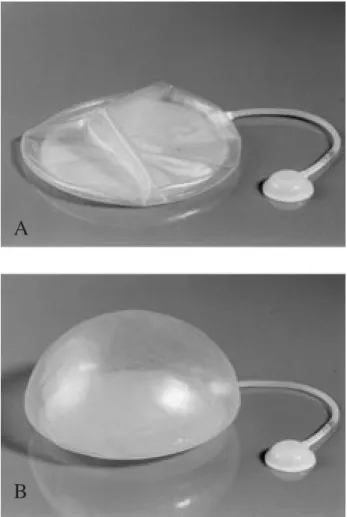
Between 1990 and 1995, 42 patients with cervical (n=36) or endometrial (n=6) cancer including squamous-cell carcinoma (n=36), adenocarcinoma (n=4) or sarcoma (n=2) were treated. Median age was 48 years (range: 25-70). Forty-one patients underwent surgical placement of a temporary STEP connected with a subcutaneously located self-sealing valve system before RT (it was placed in one patient after mechanical ileus with inflammatory reaction at 32 Gy). This technique allows elimination of the small bowel outside the RT volume, thus, reducing the risks of acute and late small bowel toxicity. The expander is fixed within the pelvis using a vicryl mesh, and filled with 400 ml isotonic saline solution (Figure 1A and 1B). No other surgical procedure was necessary to secure the device in position. Radiation therapy (RT) was administered either exclusively (n=30) or in postoperative (n=12) setting. All patients underwent treatment-planning simulation using oral contrast medium to highlight the amount of small bowel within the radiation fields. Figure 2A, 2B, 3A, and 3B illustrate the simulation radiographies with or without STEP, respectively.
Fig 1 (A,B). Silicone tissue expander. (A): empty, (B): filled with isotonic saline solution
A
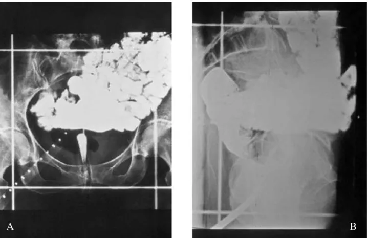
Fig 2 (A,B). Simulation films illustrating the importance of small bowel volume within the irradiation volume. (A): AP/PA fields; (B): lateral fields
A
B
Fig 3 (A,B). Simulation films after introduction of STEP; there is an upward displacement of the small bowel outside the treated volume. (A): AP/PA fields, (B): lateral fields
A
(11). All patients were treated with at least 6-MV photons from a linear accelerator. STEP does not alter the isodose distribution because its density is similar to the density of human tissues. Six patients included in this study were treated according to a local protocol combining hyperfrac-tionated RT (1.6 Gy/fraction) and cisplatin-based chemo-therapy.
lntracavitary brachytherapy boost using a cesium source was given in all but 2 patients with a median dose of 30 (10-45) Gy according to the Manchester system. Brachy-therapy was started at the end of external pelvic RT in 33 patients, during external RT in 6, and before external RT in one. Median AP/PA field surface was 270 (164-879) cm2,
and median lateral opposed field surface 204 (159-318)
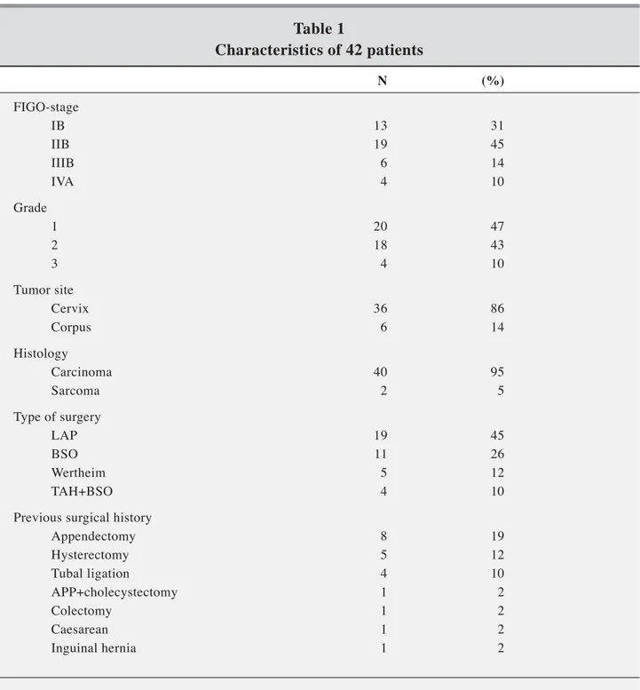
Table 1
Characteristics of 42 patients
N (%) FIGO-stage IB 13 31 IIB 19 45 IIIB 6 14 IVA 4 10 Grade 1 20 47 2 18 43 3 4 10 Tumor site Cervix 36 86 Corpus 6 14 Histology Carcinoma 40 95 Sarcoma 2 5 Type of surgery LAP 19 45 BSO 11 26 Wertheim 5 12 TAH+BSO 4 10Previous surgical history
Appendectomy 8 19 Hysterectomy 5 12 Tubal ligation 4 10 APP+cholecystectomy 1 2 Colectomy 1 2 Caesarean 1 2 Inguinal hernia 1 2
LAP: laparotomy; BSO: bilateral salpingo-oopherectomy; TAH: total abdominal hysterectomy; FIGO: International Federation of Gynecology and Obstetrics; APP: appendectomy
cm2. Median measured small bowel surface was 6 (0-107)
cm2 in the AP/PA fields, and 0 (0-20) cm2 in the lateral
fields (Table 2). The median follow up was 75 months (range: 2-9 years).
RESULTS
As of January 1999, the 5-year overall, and disease-free survivals were 46% and 44%. Acute and late toxicities were graded according to World Health Organization (WHO) and European Organization of Research and Treat-ment of Cancer/Radiation Therapy and Oncology Group (EORTC/RTOG) criteria, respectively (12). During external RT, there were 28 patients with G0, 9 with G1, and 5 with G2 gastrointestinal system (GIS) toxicity (Table 3). During brachytherapy, GIS toxicity was G0 in 35 patients, and G1
in 6. At the end of the treatment, only 5 patients had G1 GIS toxicity. No GIS toxicity was recorded either at 3 and 6 months following treatment, or until the end of the whole follow-up period. Only three patients developed major complications requiring surgery (none of them had previous surgery): two (one small bowel obstruction and one ileus with abscess) were related to STEP, and one was related to previous surgical interventions and RT (mechanical ileus with inflammatory reaction at 32 Gy) resulting with surgical correction and application of a STEP to complete her treatment.
STEP caused discomfort in two patients necessitating volume reduction of the saline solution.
The surgical removal of the tissue expander under local anesthesia was done immediately following the end of treatment without complications in all patients.
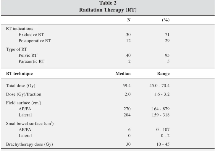
Table 2
Radiation Therapy (RT)
N (%) RT indications Exclusive RT 30 71 Postoperative RT 12 29 Type of RT Pelvic RT 40 95 Paraaortic RT 2 5RT technique Median Range
Total dose (Gy) 59.4 45.0 - 70.4
Dose (Gy)/fraction 2.0 1.6 - 3.2
Field surface (cm2)
AP/PA 270 164 - 879
Lateral 204 159 - 318
Smal bowel surface (cm2
)
AP/PA 6 0 - 107
Lateral 0 0 - 2
Brachytherapy dose (Gy) 30 10 - 45
Table 3
Acute and late radiation-related toxicities according to the WHO
and RTOG/EORTC scoring systems, respectively
G0 (%) G1 (%) G2 (%) G3 (%) G4 (%) Acute Diarrhea 28 (67) 9 (21) 5 (12) - -Skin - 40 (95) 2 (5) - -Late Small bowel - - - - 2 (5)
WHO classification: grade 0, absence of diarrhea; grade 1, transient diarrhea <2 liquid stools/day; grade 2, tolerable diarrhea >2 liquid stools/day; grade 3, intolerable diarrhea necessitating a specific treatment or cessation of irradiation; grade 4, hemorrhagic diarrhea or dehydratation
EORTC/RTOG: European Organization of Research and Treatment of Cancer/Radiation Therapy and Oncology Group, Grade 4: surgical intervention
DISCUSSION
Radiation therapy is largely used in gynecologic malig-nancies as a single treatment modality, or combined to surgery with or without chemotherapy (10, 13-20). Follow-ing hysterectomy, the small intestine moves into the pelvis. Small intestine being a highly radiosensitive organ in the abdomen and the pelvis and major side effects are observed when RT doses exceed 40-45 Gy (21-23). Acute side effects during RT cause patient discomfort but can also alter therapeutic results by increasing overall treatment time leading to late complications such as chronic radiation enteropathy (24-26). Late intestinal radiation injury is rare, and can result with chronic malabsorption syndrome (partial or complete obstruction, perforation, or fistula). Important acute small bowel toxicity can lead to severe late toxicity, therefore, affecting patient’s quality of life. Management of patients with such sequel generally requires surgical correction by experimental hands because of high postop-erative morbidity (27).
A number of approaches have been used in order to displace the small intestine outside of the irradiation volume using small bowel contrast during simulation, manoeuvres in displacing small bowel, thiol compounds as radioprotector
(30), prostaglandin inhibitors or sucralfat, non surgical methods as patient positioning (prone, supine, decubitus, Trendelenburg), external compression and bladder distension during treatment, and conformal treatment planning using individualized normal-tissue blocks (6, 28-35). Surgical techniques include omental sling, uterine retroversion, use of an absorbable mesh sling or the placement of a removable pelvic spacer (36-38).
The insertion of a STEP into the abdominal cavity to displace small bowel from the pelvic irradiation field has been utilized first by Sugarbaker in the management of advanced or recurrent rectal carcinoma (39). Our technique and initial experience using the same device is reported elsewhere and, to our knowledge, it is the largest prospective series in the gynecologic cancer setting (40,41).
Herbert et al. (42), in their series of 14 patients using STEP, reported acute G1 or G2 GIS toxicity according to the RTOG criteria in 5 patients, and G2 GIS toxicity requiring medication in 4 patients. Another study from the Fox Chase Center using STEP in 34 patients was started in 1989, and published in 1994 (43). In their updated results including 57 patients with gastrointestinal and gynecologic malignancies, they reported overall 16 complications
References
1. Horiot JC, Pigneux J, Pourquier H, et al. Radiotherapy alone in carcinoma of the intact uterine cervix according to G. H. Fletcher guidelines: A French cooperative study of 1383 cases. Int J Radiat Oncol Biol Phys 1988;14:605-11. 2. Eiffel PJ, Moughan J, Owen J, et al. Patterns of radiotherapy
practice for patients with squamous carcinoma of the uterine cervix: Patterns of care study. Int J Radiat Oncol Biol Phys 1999;43:351-8.
3. Lanciano R, Thomas G, Eiffel PJ. Over 20 years of progress in radiation oncology: cervical cancer. Semin Radiat Oncol 1997;7:121-6.
4. Kinney WK, Alvarex RD, Reid GC, et al. Value of adjuvant whole-pelvis irradiation after Wertheim hysterectomy for early-stage squamous carcinoma of the cervix with pelvic nodal metastasis: a matched-control study. Gynecol Oncol 1989;34: 258-62.
5. Perez CA, Grigsby PW, Camel HM, et al. Radiation alone or combined with surgery in stage IB, IIA, and IIB carcinoma of uterine cervix: Update of a nonrandomized comparison. Int J Radiat Oncol Biol Phys 1995;31:703-16.
6. Gallagher MJ, Brereton HD, Rostock RA, et al. A prospective study of treatment techniques to minimize the volume of pelvic small bowel with reduction of acute and late effects associated with pelvic irradiation. Int J Radiat Oncol Biol Phys 1986;12:1565-73.
7. Pedersen D, Bentzen SM, Overgaard J. Early and late radio-therapy morbidity in 442 consecutive patients with locally advanced carcinoma of the uterine cervix. Int J Radiat Oncol Biol Phys 1994;29:941-52.
8. Letschert JGJ. The prevention of radiation-induced small bowel complications. Eur J Cancer 1995;31A:1361-5. 9. Letschert JGJ, Lebesque JV, Aleman BMP, et al. The volume
effect in radiation-related late small bowel complications: results of a clinical study of the EORTC Radiotherapy
Cooperative Group in patients treated for rectal carcinoma. Radiat Oncol 1994;32:116-23.
10. Landoni F, Maneo A, Colombo A, et al. Randomised study of radical surgery versus radiotherapy for stage Ib-IIa cervical cancer. Lancet 1997;350:535-40.
11. International Commission on Radiation Units and Measure-ments (ICRU). Prescribing, Recording, and Reporting Photon Beam Therapy, ICRU Report 50. Bethesda. ICRU Publications Office; 1993.
12. Cox JD, Stetz J, Pajak TF. Toxicity criteria of the Radiation Therapy Oncology Group (RTOG) and the European Orga-nization for Research and Treatment of Cancer (EORTC). Int J Radiat Oncol Biol Phys 1995;31:1341-6.
12. Hopkins MP, Morley GW. Stage IB squamous cell cancer of the cervix: Clinicopathologic features related to survival. Am J Obstet Gynecol 1991;164:1520-7.
13. Hopkins MP, Morley GW. Radical hysterectomy versus radiation therapy for stage IB squamous cancer of the cervix. Cancer 1991;68:272-7.
14. Logue JP, Hale RJ, Wilcox FL, et al. Carcinoma of the cervix: An analysis of prognostic factors, treatment and patterns of failure following Wertheim's hysterectomy. Int J Gynecol Cancer 1992;2:323-7.
15. Morris M, Eiffel PJ, Lu J, et al. Pelvic radiation with con-current chemotherapy compared with pelvic and paraaortic radiation for high risk cervical cancer. N Engl J Med 1999;340:1137-43.
16. Thomas G, Dembo A, Ackerman I, et al. A randomized trial of standard versus partially hyperfractionated radiation with or without concurrent 5-fluorouracil in locally advanced cervical cancer. Gynecol Oncol 1998;69:137-45.
17. Rose PG, Bundy BN, Watkins EB, et al. Concurrent cisplatin-based radiotherapy and chemotherapy for locally advanced cervical cancer. N Engl J Med 1999;340:1144-53.
associated with STEP (abscess in 4 patients, abscess and fistula in 1, fistula after removal of STEP in 4, early STEP withdrawal in 5, fill-port tubing erosion in 1, and capsule mistaken for bladder in 1) (44).
In series without STEP, symptoms of acute GIS toxicity including G2 or G3 diarrhea occur in more than 70% of patients undergoing pelvic irradiation (45,46). In our series, only 5 patients (12%) had G2 acute GIS toxicity confirming the efficiency of STEP in excluding the small intestine outside the treatment volume. Only 2 patients (5%) devel-oped major late toxicity requiring surgery (one small bowel
obstruction and one ileus with abscess). The reason for this low rate of late GIS toxicity is probably related to the homogeneity of our series including only gynecologic tumors without major abdominal surgery for GIS tumors compared to other series.
This prospective study demonstrated that the placement of STEP before RT in patients with gynecologic malignan-cies resulted with decreased irradiated small bowel volume and, therefore, very low rates of acute and late gastrointes-tinal toxicity.
18. Keys HM, Bundy BN, Stehman FB, et al. Cisplatin, radiation, and adjuvant hysterectomy compared with radiation and adjuvant hysterectomy for bulky stage IB cervical carcinoma. N Engl J Med 1999;340:1154-61.
19. Whitney CW, Sause W, Bundy BN, et al. A randomized comparison of fluorouracil plus cisplatin versus hydroxyurea as an adjunct to radiation therapy in stages IIB-IVA carcinoma of the cervix with negative para-aortic lymph nodes. J Clin Oncol 1999;17:1339-48.
20. Withers RH, Taylor JMG, Maciejewski B. Treatment volume and tissue tolerance. Int J Radiat Oncol Biol Phys 1988;14:751-9.
21. Emami B, Lyman JT, Brown A, et al. Tolerance of normal tissue to therapeutic irradiation. Int J Radiat Oncol Biol Phys 1991;21:109-22.
22. Yeoh E, Horowitz M, Russo A, et al. Retrospective study of the effects of pelvic irradiation for carcinoma of the cervix on gastrointestinal function. Int J Radiat Oncol Biol Phys 1993;26:229-37.
23. Gyrinski TH, Rey A, Roche B, et al. Overall treatment time in advanced cervical carcinomas: a critical parameter in treatment outcome. Int J Radiat Oncol Biol Phys 1993;27:1051-6.
24. Hauer-Jensen M. Late radiation injury of the small intestine: Clinical, pathophysiologic and radiobiologic aspects. A review. Acta Oncol 1990;29:401-15.
25. Letschert JGJ, Lebesque JV, Aleman BMP, et al. The volume effect in radiation-related late small bowel complications results of a clinical study of the EORTC Radiotherapy Cooperative Group in patients treated for rectal carcinoma. Radiother Oncol 1994;32:116-23.
26. Schellhammer PF, Jordan GH, EL-Mahdi AM. Pelvic com-plications after interstitial and external beam irradiation of urologic and gynecologic malignancy. World J Surg 1986;10:259-68.
27. Herbert SH, Curran WJ, Solin LJ, et al. Decreasing gas-trointestinal morbidity with the use of small bowel contrast during treatment planning for pelvic irradiation. Int J Radiat Oncol Biol Phys 1991;20:835-42.
28. Green N, Iba G, Russel Smith W. Measures to minimize small intestine injury in the irradiated pelvis. Cancer 1975;35:1640-75.
29. Mitsuhashi N, Takahashi I, Takahashi M, et al. Clinical study of radioprotective effects of amifostine (YM-08310, WR 2721) on long-term outcome for patients with cervical cancer. Int J Radiat Oncol Biol Phys 1993;26:407-11.
30. Rose PG, Haltyer SA, Su CM. The effect of indomethacin on acute radiation-induced gastrointestinal injury: a morpho-logic study. J Surg Oncol 1992;49:231-8.
31. Kochhar R, Patel DMF, Dhar A, et al. Radiation-induced proctosigmoiditis: prospective, randomized, double-blind
controlled Trial of oral sulfasalazine plus rectal steroids versus rectal sucralfate. Digest Dis Sci 1991;36:103-7. 32. Caspers RJL, Robert JL, Hop WCJ, et al. Irradiation of true
pelvis for bladder and prostatic carcinoma in suppine, prone or trendelenburg position. Int J Radiat Oncol Biol Phys 1983;9:589-93.
33. Green N. The avoidance of small intestine injury in gyneco-logic cancer. Int J Radiat Oncol Biol Phys 1983;9:1385-90. 34. Gunderson LL, Russell AH, Llewellyn HJ, et al. Treatment planning for colorectal cancer: radiation and surgical tech-niques and value of small-bowel films. Int J Radiat Oncol Biol Phys 1985;11:1379-93.
35. Cohen AM, Gunderson LL, Welch CE. Selective use of adjuvant radiation therapy in resectable colorectal adenocar-cinoma. Dis Colon Rectum 1981;24:247-51.
36. Snijders-Keilholz A, Trimbos JB. A preliminary report on new efforts to decrease radiotherapy related small bowel toxicity. Radiother Oncol 1991;22:206-8.
37. Durig M, Steenblock U, Heberer M, et al. Prevention of radiation injuries to the small intestine. Surg Gynecol Obstet 1984;159:162-4.
38. Sugarbaker PH. Intrapelvic prosthesis to prevent injury of the small intestine with high dosage pelvic irradiation. Surg Gynecol Obstet 1983;157:269-71.
39. Coucke PA, Cuttat JF, Mirimanoff RO. Small bowel protection with STEP. Strahlenther Onkol 1992;168:226-9.
40. Delaloye JF, Cuttat JF, Coucke PA, et al. Protection of the small bowel with a silicone tissue expander prosthesis and a polyglycolic acid mesh during radiation therapy for cervical carcinoma. Br J Obstet Gynaecol 1994;101:541-2. 41. Herbert SH, Solin LJ, Hoffman JP, et al. Volumetric analysis
of small bowel displacement from radiation portals with the use of a pelvic tissue expander. Int J Radiat Oncol Biol Phys 1993;25:885-93.
42. Hoffman JP, Lanciano R, Carp NZ. Morbidity after intrap-eritoneal insertion of saline-filled tissue expanders for small bowel exclusion from radiotherapy treatment field. Am Surg 1994;60:473-83.
43. Hoffman JP, Sigurdson ER, Eisenberg BL. Use of saline-filled tissue expanders to protect the small bowel from radiation. Oncology 1998;12:51-4.
44. Henriksson R, Franzen L, Littbrand B. Prevention and therapy of radiation-induced bowel discomfort. Scand J Gastroenterol 1992;27:7-11.
45. Resbeut M, Marteau PH, Cowen D, et al. A randomized double blind placebo controlled multicenter study of mesala-zine for the prevention of acute radiation enteritis. Radiother Oncol 1997;44:59-63.