Universite de Sherbrooke
SENSITIZATION OF PLASMID DNA TO IONIZING
RADIATION BY PLATINUM CHEMOTHERAPEUTIC
DRUGS
by
MOHAMMAD REZAEE
Department o f Nuclear M edicine and Radiobiology
Thesis submitted to the Faculty o f Medicine and Health Sciences for the
degree o f Doctorate o f Philosophy (PhD) in Radiation Sciences and
Biomedical Imaging
Sherbrooke, Quebec, Canada
August 2013
Jury Committee
Professor Leon Sanche
Professor Darel Hunting
Professor Benoit Paquette
Professor James Chow
Professor Patrick Ayotte
Professor Brigitte Guerin
Thesis Supervisor, Radiation Sciences and Biomedical Imaging
Thesis Supervisor, Radiation Sciences and Biomedical Im aging
President, Radiation Sciences and
Biomedical Im aging
Examiner, D epartm ent o f Radiation
Oncology, University o f Toronto
Examiner, D epartm ent o f Chemistry, University o f Sherbrooke
Examiner, Radiation Sciences and
Biomedical Imaging
1+1
Published Heritage Branch Direction du Patrimoine de I'edition 395 Wellington Street Ottawa ON K 1A0N 4 Canada 395, rue Wellington Ottawa ON K1A 0N4 CanadaYour file Votre reference ISBN: 978-0-499-00411-6 Our file Notre reference ISBN: 978-0-499-00411-6
NOTICE:
The author has granted a non
exclusive license allowing Library and Archives Canada to reproduce, publish, archive, preserve, conserve, communicate to the public by
telecomm unication or on the Internet, loan, distrbute and sell theses
worldwide, for commercial or non commercial purposes, in microform, paper, electronic and/or any other formats.
AVIS:
L'auteur a accorde une licence non exclusive permettant a la Bibliotheque et Archives Canada de reproduire, publier, archiver, sauvegarder, conserver, transmettre au public par telecomm unication ou par I'lnternet, preter, distribuer et vendre des theses partout dans le monde, a des fins com merciales ou autres, sur support microforme, papier, electronique et/ou autres formats.
The author retains copyright ownership and moral rights in this thesis. Neither the thesis nor substantial extracts from it may be printed or otherwise reproduced without the author's permission.
L'auteur conserve la propriete du droit d'auteur et des droits moraux qui protege cette these. Ni la these ni des extraits substantiels de celle-ci ne doivent etre imprimes ou autrement
reproduits sans son autorisation.
In compliance with the Canadian Privacy A ct some supporting forms may have been removed from this thesis.
W hile these forms may be included in the document page count, their removal does not represent any loss of content from the thesis.
Conform em ent a la loi canadienne sur la protection de la vie privee, quelques
form ulaires secondaires ont ete enleves de cette these.
Bien que ces form ulaires aient inclus dans la pagination, il n'y aura aucun contenu manquant.
A B ST R A C T
Sensitization o f Plasmid DNA to Ionizing Radiation by Platinum C hem otherapeutic Drugs by M ohamm ad Rezaee
Department o f N uclear M edicine and Radiobiology
Thesis submitted to the Faculty o f M edicine and Health Sciences for the degree o f Doctorate o f Philosophy (PhD) in Radiation Sciences and Biomedical Imaging, Faculty o f
M edicine and Health Sciences, University o f Sherbrooke, Sherbrooke, Q uebec, Canada
Concomitant chemoradiation therapy based on platinum chem otherapeutic drugs (Pt- drugs) is a common treatment m odality for several types o f cancers and has dramatically improved patient survival. The radiosensitization capacity o f Pt-drugs results essentially from their binding to nuclear DNA. Although several mechanisms such as increase in the radiation damage to DNA and inhibition o f their repair have been proposed, the contribution and efficiency o f the underlying molecular mechanisms o f the radiosensitization rem ain unknown. This PhD thesis determines the relative efficiency o f Pt-drugs, in term s o f the type o f drug and the quantity o f Pt-DNA adducts, in the sensitization o f DNA to the direct and indirect effects o f ionizing radiations, and elucidates the m ajor mechanism responsible for this radiosensitization. In particular, it addresses the role o f low-energy electrons (LEEs), hydroxyl radicals and hydrated electrons in the radiosensitization o f D N A m odified by Pt- drugs. This thesis includes a review o f the literature on the m olecular basis o f radiotherapy, Pt-based chemotherapy, and their com bination in cancer treatment. Five articles, on which I am first author, are presented, and followed by a com prehensive discussion that integrates all results and their implications in the clinic and future research.
With respect to the direct effect o f radiation, LEEs are found to be the m ain species responsible for the enhancement in DNA dam age, particularly cluster dam age including DSB and interduplex cross-links. Irradiation o f a 3199-bp plasm id DNA m odified by an average o f 2 Pt-drug adducts with 10-eV electrons results in significant increases in DSB formation by factors o f 3.1, 2.5 and 2.4, respectively, for carboplatin, cisplatin and oxaliplatin relative to unmodified DNA. Irradiation o f these samples with subexcitation-energy electrons (i.e., 0.5 eV) generates substantial number o f DSB in the m odified DNA, while no DSB is observed in the unmodified DNA. Since 0.5 eV is well below that energy required for the electronic excitation o f organic molecules, dissociative electron attachm ent m ust be the main mechanism responsible for the formation o f strand breaks in the presence o f Pt-adducts. For indirect effects o f radiation, our results show that both hydroxyl radicals and hydrated electrons are responsible for the enhanced formation o f dam age in m odified DNA. In the presence o f Pt-adducts, hydroxyl radicals mainly contribute to the SSB formation, while hydrated electrons are the main species responsible for the DSB formation.
Our results indicate that carboplatin and oxaliplatin have higher efficiency than cisplatin in the enhancement o f radiation dam age to DNA. At low frequencies o f Pt-D NA adducts (i.e., less than 3. lx l O'4 adducts per nucleotide), radiosensitization o f DNA, in term s o f the damage per adduct, increases by an order o f magnitude com pared with that at large frequencies o f adducts. In conclusion, Pt-drug modification is an extrem ely efficient m eans o f enhancing the formation o f DNA DSBs by both LEEs and hydrated electrons created by ionizing radiation. Key-words: Cisplatin, Carboplatin, Oxaliplatin, Electron, DNA damage, Radiosensitization
medicaments chim iotherapeutiques platines Par M oham m ad Rezaee
Departement de medecine nucleaire et de radiobiologie
These presente a la Faculte de medecine et des sciences de la sante en vue de l ’obtention du diplome de philosophiae doctor (Ph.D .) en science des radiations et imagerie biomedicale, Universite de Sherbrooke, Sherbrooke, Quebec, Canada, J1H 5N4
La radiochimiotherapie concomitante, basee sur les m edicam ents antineoplasiques platines (Pt-antineoplasiquesm), est une modalite de traitem ent utilise contre plusieurs types de cancers et a considerablement ameliore la survie des patients. Parmi ces medicam ents anticancereux, les analogues de platine sont les plus couram m ent utilises. Leur capacite a radiosensibiliser resulte essentiellement de leur liaison a l'ADN nucleaire. Bien que plusieurs mecanismes aient ete proposes telles que l'augmentation des dom m ages induits a l'ADN et l'inhibition de leur reparation, la contribution et l’efficacite des m ecanism es m oleculaires sous-jacents a la radiosensibilisation restent inconnus. La presente etude exam ine l'efficacite Pt-antineoplasiques a sensibiliser l'ADN aux rayonnements ionisants et determ ine le role des electrons secondaires, des radicaux d'hydroxyles et des electrons hydrates dans ce processus. Cette these com prend un revue de des donnees scientifiques concem ant la base m oleculaire de la radiotherapie, de la chimiotherapie Pt-antineoplasiques et de leur com binaison dans le traitem ent de cancer. Cinq articles, done je suis premier auteur, sont presentes suivis d'une discussion qui integre mes resultats et leurs implications dans la clinique et la recherche future.
En ce qui conceme l'effet direct des radiations, les electrons de faible energie s ’averent etre la principale espece responsable de l’augmentation des dom m ages a l’ADN, en particulier les dommages multiples localises, les CDBs et les pontages inter-brin. L'irradiation de plasmides de 3199 paires de bases, contenant en moyenne deux adduits Pt-ADN, avec des electrons de 10 eV conduit a une augmentation significative des CDBs par des facteurs de 3.1, 2.5 et 2.4, respectivement, pour le carboplatine, le cisplatine et l'oxaliplatine par rapport a l ’irradiation des plasmides non modifies. L'irradiation avec des electrons de 0.5 eV genere un nombre substantiel de CDBs dans les plasmides modifies, alors qu'aucune CDB n ’est observee dans les plasmides non modifies. Puisque 0.5 eV est une energie bien inferieure a celle necessaire a l'excitation electronique des molecules organiques, l'attachem ent dissociatif de 1’electron doit etre le principal mecanisme responsable de la formation de cassures en presence de Pt- antineoplasiques. Pour les effets indirects des rayonnem ents, nos resultats m ontrent que les radicaux hydroxyles et les electrons hydrates sont, tous les deux, responsables de la formation accrue des dommages dans l'ADN modifie. En presence d ’adduits Pt-A DN , les radicaux hydroxyles contribuent principalement a la formation de cassures simple brin, tandis que les electrons hydrates sont les principales especes responsables de la formation de CDBs.
Nos resultats indiquent que le carboplatine et l'oxaliplatine sont plus efficaces que le cisplatine pour augmenter les dommages a l'ADN. A faible concentration de Pt-ADN (soit moins de 3.1x10 adduit par nucleotide), la radiosensibilisation de l'ADN, en term es de dommages par adduit, est d ’un ordre de grandeur superieure a celle aux concentrations elevees. En conclusion, l’ajout de Pt-antineoplasiques est un m oyen extrem em ent efficace d'augmenter la formation de CDBs dans l’ADN par 1’interm ediaire des electrons de faible energie et des electrons hydrates produits par les rayonnem ents ionisants.
TABLE OF CONTENTS
LIST O F TABLES i
LIST O F FIGURES iii
LIST OF ABBREVIATIONS ix
LIST OF SYM BOLS xii
I. INTRODUCTION 1
1.1. Concomitant Chemoradiation Therapy 1
1.2. Platinum Chemotherapeutic Drugs 3
1.3. M olecular Basis o f Radiation Therapy 7
1.3.1. Biological Effects o f Radiation 7
1.3.2. Interaction o f Therapeutic Radiation with Biological M atter 10
1.3.3. Interaction o f LEEs with Condensed M atter 13
1.3.3.1. Resonant Processes 14
1.3.3.2. Non-resonant Processes 16
1.3.4. Interaction o f LEEs with DNA 17
1.4. Radiosensitization Effects o f Platinum Chem otherapeutic Drugs 19
1.5. Research Project 22
II. RESULTS - ARTICLES 25
11.1. First Article 25
DNA-platinum thin films for use in chem oradiation therapy studies
11.2. Second Article 49
Absolute cross section for low-energy-electron damage to condensed macromolecules: A case study o f DNA
11.3. Third Article 74
N ew insights into the mechanism underlying the synergistic action o f ionizing radiation with platinum chem otherapeutic drugs: The role o f low energy electrons
11.4. Fourth Article 102
A single subexcitation-energy electron can induce a double strand break in DNA modified by platinum chem otherapeutic drugs
11.5. Fifth Article 131
III. D ISC U SSIO N 156
III. 1. Pt-drugs enhance the formation o f DNA dam age by both the 156
direct and indirect effects o f radiation
111.2. Radiosensitization o f modified DNA depends on the type o f Pt- 160 drugs
111.3. Radiosensitization o f Pt-drugs depends on the num ber o f Pt-D NA 161 adducts
IV. C O N C L U S IO N 166
A C K N O W L E D G E M E N T 168
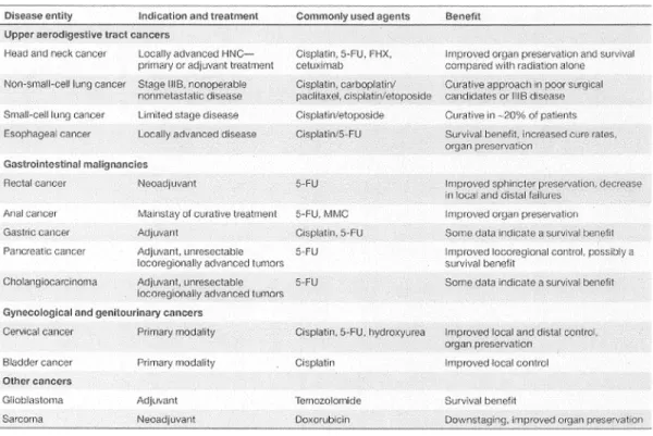
LIST OF TABLES Chapter I
Table 1.1 Overview o f cancers and indications in w hich concurrent 2
chemoradiotherapy is used.
Chapter II: First Article Chapter II: Second Article
Table 1 Values for the ratios R 1.2(0) o f the initial slope o f the exposure- 63
response curves between all possible thickness pairs hi (nm ) and h2 (nm) am ong the 10, 15 and 20 nm films at 10 eV, as well as the 10, 20 and 30 nm films at 100 eV. Corresponding values o f the AL in the plasmid DNA, X (nm), deduced from Eq. (13) along with the calculated average. SE is standard error.
Table 2 Cross Section <x(10' 14 cm 2) to induce a SB in plasm id DNA by 64 10- and 100-eV electron impact on different film thicknesses h
(nm). SE is standard error.
Table 3 Present and previous CS data to induce SB in DNA by LEE 67
impact. The / / value is the penetration factor used to correct the measured CS obtained from thick films, a' ( 1 014 cm 2)and a (10‘ 14 cm2) present the m easured CS per plasm id for loss o f SC before and after a p p ly in g //, respectively. a„ (1 0 17 cm2) stands for the CS per nucleotide to induce DNA SB.
Chapter II: Third Article
Table 1 Yields o f DNA dam age (xlO' 8 dam age/Gy/bp) for 10-keV (a) 80 and 10-eV (b) electron irradiation o f DNA with and w ithout
modification by Pt-drugs.
Table 2 Enhancement Factors for the induction o f SSB, DSB and 88
interduplex CL as a function o f the num ber o f Pt-adducts per nucleotide. Si and S2 denote the slopes o f the fitted lines to the Enhancement Factor curves for the DNA dam ages presented in Fig. 3 at ratios less and more that 3 .lx lO'4 Pt-adducts per nucleotide, respectively. R i,2 is the ratio o f Si to S2.
Chapter II: Fourth Article
Table 1 Yields o f DNA damage (x 10' 8 dam age/Gy/bp) for soft X-rays, 109 10- and 0.5-eV electron irradiations o f DNA with and w ithout
modification by platinum anticancer drugs.
Table SI Total number o f DSB (x 10'6) per plasm id induced by 0-2 Gy o f 127 0.5-eV electrons in unm odified DNA and DNA m odified by
cisplatin, carboplatin or oxaliplatin via single- and double-hit
i
film thickness h (nm), mass o f the D NA mDNA (ng), film area A (cm 2). These films were deposited on the either tantalum (Ta) or glass substrates by lyophilisation and irradiated by either electrons or X-rays.
Chapter II: Fifth Article
Table 1 Yields o f SSB and DSB (Strand Break/Gy/base pair) induced by 140 the ^ C o y-rays in DNA and cisplatin/DNA samples containing 5
mM tris and saturated with either N2 or N2O. Enhancement factors present increase in the yields due to the presence o f cisplatin.
Table 2 Yields o f SSB and DSB (Strand Break/Gy/base pair) induced by 143 the ^ C o y-rays in DNA and cisplatin/D NA sam ples containing
200 mM tris and saturated with either N2 or N2O. Enhancement factors present increase in the yields due to the presence o f cisplatin.
Table 3 The yields o f SSB and DSB induced by *OH, 'H and e~q for the 145 DNA and cisplatin/DNA samples in 5 mM tris in the presence o f
N 2.
Chapter III
Table III. 1 Enhancement Factors for the induction o f SSB, DSB and interduplex CL by either 10-eV electrons or gam m a-rays as a function o f the number o f cisplatin-DNA adducts per nucleotide. Si and S2 denote the slopes o f the fitted lines to the Enhancem ent Factor curves at ratios less and more that 3. lx lO'4 Pt-adducts per nucleotide, respectively. R.1,2 is the ratio o f Si to S2.
163
LIST OF FIGURES C h a p te r I
Figure 1.1 Chemical structure o f cisplatin, carboplatin and oxaliplatin. 4
Figure 1.2 Schematic for the formation o f various cisplatin-DNA adducts, 5 including intrastrand cross-links, interstrand cross-link, DNA-
Protein cross-link and mono-functional binding to guanine. Carboplatin and oxaliplatin also form the sam e type o f DNA adducts as cisplatin.
Figure 1.3 Packaging DNA into the chromatin structure. 10
Figure 1.4 (a) The calculated probability o f energy loss in the soft collision o f 13 a charge particle with DNA, liquid water, gaseous w ater and
gaseous hexane molecules, with no mom entum transfer. The calculation is based on the dipole oscillator strength distribution. (b) The energy distribution o f secondary electrons generated by primary ions at different energies in water.
Figure 1.5 The yields for the formation o f SSB and DSB by electrons o f 4-100 18 eV (a) and 0-4 eV (b).
C h a p te r II: F irs t A rticle
Figure 1 Comparison o f the percentages o f DNA supercoiled (a), DNA 33
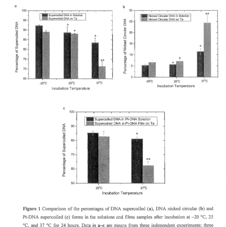
nicked circular (b) and Pt-DNA supercoiled (c) forms in the solutions and films sam ples after incubation at - 2 0 °C, 25 °C, and 37 °C for 24 hours. Data in a - c are means from three independent experiments; three samples at each tem perature are analyzed in each experiment; error bars show standard deviations.
Figure 2 Kinetics o f binding o f Pt-com pounds to plasm id DNA. The Pt- 36 compounds are: (a) cisplatin with the initial ratios in the solution
o f 20:1, (b) 200:1, and (c) carboplatin with the initial ratios o f 40:1 and (d) 200:1. The curves show the quantity o f bound Pt- compounds per DNA molecule at different incubation tim es at 25 °C. Data in a - d are means from three m easurements; error bars show standard deviations. The continuous black lines are exponential fits to the data.
Figure 3 Impact o f tris on the reaction o f DNA platination. Pt-DNA ratios 37 in the cisplatin-DNA solutions incubated during 45, 90, and 180
minutes at 25 °C are com pared in the presence and absence o f tris. Data are means from three m easurements; error bars show standard deviations.
solution, and (b) on tantalum substrate, after incubation for 2, 4, and 8 hours at 25 °C. Data are means from three m easurem ents; error bars show standard deviations.
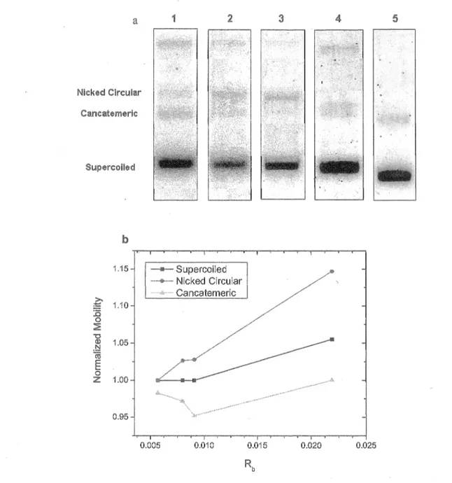
Figure 5 Mobility o f Cispatin-DNA molecules in agarose gel. (a) M igration 41 o f the different configurations o f cisplatin-DNA m olecules
separated by electrophoresis. Lane 1 is for a DNA sam ple and lanes 2-5 are for cisplatin-DNA sam ples with the num ber o f bound cisplatin molecules per nucleotide, Rb, o f 0.0057, 0.008, 0.0091, and 0.0219, respectively, (b) N orm alized m obility o f the nicked circular, supercoiled, and concatem eric forms o f Pt-DNA sam ples at different Rb in gel electrophoresis.
Chapter II: Second Article
Figure 1 Simulation o f exposure-response curves for loss o f the SC D N A 57 based on the Eq. (5) for three different DNA film thicknesses o f
10, 20 and 40 nm and for a fixed AL in absence o f film charging.
Figure 2 Simulation o f the norm alized num ber o f SC DNA in various 59 depths o f a DNA film at different irradiation tim es (t) in absence
o f charging (a) and in presence o f charging (b).
Figure 3 Simulation o f the exposure-response curves for loss o f SC DNA in 60 a 40 nm thick film for different charging time constants (x).
Figure 4 Exposure response curve for lyophilized plasm id DNA films o f 61 10, 15 and 20 nm average thickness, irradiated with 10-eV
electrons (a) and 10, 20 and 30 nm average thickness, irradiated with 100-eV electrons (b). The dash-point lines are guides for eye. Each data point corresponds to the m ean value o f three samples with the relevant standard deviation.
Figure 5 Calculation o f the initial slope o f the exposure-response curve 65 based on the theoretical model and fitting to the m easured data sets
for 10-eV (a) and 100-eV (b) electrons.
Chapter II: Third Article
Figure 1 Enhancement factors in the yields o f SSB,DSB and interduplex 81 CL induced by 10-keV (a) and 10-eV (b) electrons in the presence
o f cisplatin, carboplatin and oxaliplatin.
* indicates a P-value < 0.05, when the different Pt-drug-DNA complexes are compared to unmodified DNA.
f denotes a P-value < 0.05, when the different Pt-drug-DNA complexes are compared to each other.
Figure 2 Figure 3 Figure el Figure e2 Figure e3 Figure e4
Exposure-response curve for the formation o f DSB (a,b) and 85 interduplex CL (c,d) by either 10-eV or 10-keV electrons in DNA
modified by cisplatin, carboplatin and oxaliplatin. Data are means ± standard deviation from five measurements. They have been fitted by employing a least-squares regression analysis.
Enhancement factors in the yields o f SSB, DSB and interduplex 87 CL as a function o f the num ber o f Pt-adducts per nucleotide for
DNA modified by cisplatin (a), carboplatin (b) and oxaliplatin (c) irradiated with 10-eV electrons. The fitted lines are based on a least-square regression analysis.
Kinetics o f binding cisplatin to plasm id DNA at three different 97 initial ratios o f cisplatin to DNA in the solution: (a) 200:1, (b)
80:1, and (c) 20:1. The curves show the quantity o f bound cisplatin per DNA molecule at different incubation times at room temperature. Data in a - c are means ± SD (standard deviation) from three measurements. The dash lines are exponential fits to the data.
Kinetics o f binding oxaliplatin to plasm id D N A at three different 98 initial ratios o f oxaliplatin to DNA in the solution: (a) 200:1, (b)
80:1, and (c) 20:1. The curves show the quantity o f bound oxaliplatin per DNA m olecule at different incubation tim es at room temperature. Data in a - c are m eans ± SD (standard deviation) from three measurements. The dash lines are exponential fits to the data.
Kinetics o f binding carboplatin to plasm id DNA at three different 99 initial ratios o f carboplatin to DNA in the solution: (a) 200:1, (b)
100:1, and (c) 30:1. The curves show the quantity o f bound carboplatin per DNA m olecule at different incubation tim es at room temperature. Data in a - c are m eans ± SD (standard deviation) from three measurements. The dash lines are exponential fits to the data.
Schematic view o f UHV electron irradiator cham ber and the 100 principal components. The assembly consists essentially o f a
rotatable circular platform (F) connected to a rotary drive (E) and two types o f electron guns, i.e., a low-energy electron gun (C) mounted on a linear drive (G ) and a fixed high energy electron gun (H) (Kimball Physics Inc.). The former produces a beam adjustable in energy between 0.5 and 1000 eV, with the spot size o f the beam varying between 2 and 50 mm at w orking distances o f 10 and 50 mm. The latter generates an electron beam adjustable within the 0.5 - 20 keV energy range and in spot sizes at the fixed working distance o f 20 cm. The spot size is controlled by an electrostatic lens system using a triode configured electron source with a control grid aperture, i.e., a sym m etric einzel focus lens,
irradiate an area o f about 0.9 cm2 which was 7 times larger than the DNA film. A 0.3 mm wide slit followed by a Faraday cup detector and a phosphorescent screen were used to calibrate the electron current and its spatial distribution. For irradiation by low- and high- energy electrons, the DNA films were directly transferred to the UHV cham ber, which can be opened by a quick access port (D) from the inside o f a glove box (B), kept under a dry N2 atmosphere during lyophilization o f a DNA solution on the subtracted and holder. The UHV cham ber was then evacuated for 24 hours by a hydrocarbon-free turbom olecular pum p to a pressure o f 5 x 10'9 Torr m easured by an ion gauge (A) at room temperature.
Chapter II: Fourth Article
Figure 1 Exposure-response curve for the formation o f nicked circular (left) 107 and linear (right) DNA configurations corresponding to the SSB
and DSB, respectively, by 0.5-eV electrons in DNA unm odified or m odified by cisplatin, carboplatin and oxaliplatin. The sample films were deposited on DNA. Data are means ± SD (standard deviation) from five measurements. They have been fitted by a least squares regression analysis.
Figure 2 Exposure-response curve for the formation o f nicked circular (a) 111 and linear (b) DNA configurations corresponding to the SSB and
DSB, respectively, by 1486-eV X-ray photons in unm odified DNA or DNA modified by cisplatin, carboplatin and oxaliplatin deposited on glass substrates. Data are means ± SD (standard deviation) from five measurements. They have been fitted by a least squares regression analysis.
Figure 3 (a) A double helix DNA (I) and the formation o f a TN I on the 115 phosphate group in the absence (II) and presence (III) o f Pt-drugs.
Formation o f Pt-adducts such as interstrand crosslinks between two guanines, as shown here, causes to distort DNA conform ation by unwinding and bending its double helix. Such distortion modifies physical and chemical stability o f the DNA leading to the weakening o f the chemical bonds, w hich could enhance the formation o f a TNI and its decay into DEA. X corresponds to N H3, N H3 and C6Hio(NH2)2 for cisplatin, carboplatin and oxaliplatin, respectively. dR denotes the deoxyribose moiety, (b) Schematic diagram showing potential energy curves for negatively-charged singly (curve I) and doubly charged phosphate groups (i.e., formation o f a TNI) in the absence and presence o f Pt-drugs (curves II and III, respectively). Re is the equilibrium intem uclear
distances o f the singly charged phosphate group, Rc and R e are the crossing points in unmodifed and m odified DNA, respectively. The presence o f Pt-adducts is expected to shift the crossing point towards Re (i.e., R e < Rc) resulting in enhanced DEA and subsequently formation o f DNA strand breaks by subexcitation- energy electrons. F-C region and AD denote transition in Frank- Condon region and autodetachment, respectively.
Figure SI Schematic view o f the apparatus used to irradiate DNA sam ples 129 with 1.486 keV A1 Ka X-ray photons. The apparatus com prises a
chamber evacuated to pressure below 5 m Torr, connected to a pressure gauge (A) and an adjustable leak valve (B) connected to a nitrogen gas source. This valve stabilizes the nitrogen pressure at about 20 mTorr in the main cham ber to control the plasm a current. A negative potential o f 3.4 kV is applied to a concave alum inum cathode (C) through a high-voltage electrical feedthrough (D) fixed in a glass-ceramic (M acor) support (E) and placed as a cap on a long quartz tube (F). A nitrogen plasm a discharge with 5.5 mA current is formed between the cathode and an aluminum foil target (G). Aluminum atoms are ionized by electrons incident on the thin foil and characteristic Ka X-rays with energy 1.486 keV are emitted outside the cham ber through a flight tube (H) continuously flushed with helium gas at atmospheric pressure. X- ray traverse the helium gas and then a thin foil o f M ylar (I) to enter a small chamber, where the plasm id DNA films deposited on the different substrates have been inserted on six aluminum plates o f 44.5 mm diameter (J). These plates are fixed at different positions around a brass rotating disc (K) to allow irradiation o f samples directly by X-rays, for different periods o f tim e (i.e., various radiation doses) in the presence o f specific am ounts o f gases or vapours introduced by valves (L). In the present experiments, the distance o f 1.7 ± 0.05 mm betw een the M ylar foil and the surface o f the plates is occupied by dry N2 at atmospheric pressure. Lyophilized samples o f plasm id DNA are placed very close to the M ylar foil to avoid too m uch photon absorption by the surrounding atmosphere. Furtherm ore, GAFCHROMIC® HD-810 radiochromatic dosimetry film (A dvanced M aterials G roup o f International Specialty Products Technologies Inc., Wayne, NJ, USA) were used to measure the incident photon fluence for each irradiation period.
C h a p te r II: Fifth A rticle
Figure 1 Dose-response curves for the formation o f circular and linear DNA 139 by 60Co y-rays for the DNA and cisplatin DNA samples in 5 m M
tris. Panels A and B indicate the curves for the circular DNA in
samples saturated with N2 and N2O, respectively. Data in A - D are means ± SD from three experiments. They have been fitted by employing a least squares regression analysis.
Figure 2 Dose-response curves for the formation o f circular and linear DNA by 60Co y-rays for the DNA and cisplatin DNA samples in 200 mM tris. Panels A and B indicate the curves for the circular DNA o f the samples saturated with N2 and N2O, respectively. The curves for the linear DNA are shown in the panels C and D for the samples saturated with N2 and N2O, respectively. Data in A - D are means ± SD from three experiments. They have been fitted by employing a least squares regression analysis.
Chapter III
Figure III. 1 Enhancement factors in the yields o f SSB, DSB and interduplex CL induced by 10-eV electrons in solid films o f DNA and 60Co □ - rays in the aqueous solution o f DNA in the presence o f cisplatin. Figure III.2 Enhancement factors in the yields o f DSB (a), interduplex CL (b)
and SSB (c) as a function o f the num ber o f Pt-DNA adducts per nucleotide for DNA modified by cisplatin and irradiated w ith 10- eV electrons and y-rays. The fitted lines are based on a least- square regression analysis.
Figure III.3 Dose-response curves for the lose o f circular (a) and the formation o f linear DNA (b) by 60Co y-rays in the presence o f cisplatin interstrand (square) and intrastrand (circle) CLs.
LIST OF ABBREVIATIONS
5-FU 5-flourouracil
A Adenine
AD Autodetachment
AE radionuclide Auger-electron em itting radionuclide
AEA Adiabatic Electron Affinity
AL Attenuation Length
amu Atomic mass unit
bp Base pair
C 4’ Carbon atom at position 4 o f deoxyribose m olecule
cal calorie
Carboplatin cis-diammine( 1,1 -cyclobutane-dicarboxylato)platinum II
C-C Carbon-Carbon
CCRT Concom itant Chem oratiation Therapy
CHO Chinese H amster Ovary
Cisplatin cis-diam m inedichloroplatinum II
CL Cross-link C-N Carbon-Nitrogen C -0 Carbon-Oxygen CRT Chemoradiation Therapy CS Cross Section DACH Diaminocyclohexane
ddH2 0 Distilled deionized w ater
DEA Dissociative Electron A ttachm ent
DMSO Dimethyl Sulfoxide
DNA Deoxyribonucleic Acid
DNA-PKcs DNA-dependent protein kinase, catalytic subunit
DSB Double-Strand Break
E. coli Escherichia coli
EDTA Ethylenediaminetetraacetic acid
eV Electron-volt
F-C region Frank-Condon region
FHX 5-flourouracil, hydroxyurea and radiation
G Guanine
G2-M phase Gap2-M itosis phase
HNC Head and neck cancer
ICL Interstrand cross-link
ICP-MS Inductively Coupled Plasma - M ass Spectroscopy
LEE (LEEs) Low-Energy Electrons
LEET Low-Energy Electron Transmission
LEPET Low-Energy Photoelectron Transmission
LET Linear Energy Transfer
MFP Mean Free Path
ML M onolayer
MMC M itomycin C
MSA M olecular Self-Assembly
N Nucleotide
NbP Number o f base pair
N7 Nitrogen at position 7 o f purin bases
NA Not Applicable
N ER Nucleotide Excision Repair
NHEJ N on-Hom ologous End Joining
N M R N uclear M agnetic Resonance
Oxaliplatin Trans-R,R-l,2-diam inocyclohexane oxalate platinum II
p Phosphate
PLDR Potentially Llthal dam age repair
Pt Platinum
Pt-DNA adduct DNA adduct form ed by Platinum -based chem otherapeutic drugs
Pt-drugs Platinum-based chem otherapeutic drugs
PtTC Chloroterpyridine platinum
P-value Probability value
RNA Ribonucleic Acid
ROS Reactive Oxygen Species
S Phase Synthesis Phase
SAM Self-assembly M onolayer
SB Strand Break
SC Supercoiled
SD Standard Deviation
SE Standard Error
SLDR Sunlethal damage repair
SSB Single strand break
Ta Tantalum
TAE Tris-Acetic acid-EDTA
TE Tris-EDTA
TNI Transient negative ion
Tris tris(hydroxym ethyl)aminom ethane
UHV Ultrahigh vacuum
UV Ultraviolet
VBE Vertical binding energy
A area
£ Exposure
Fluence
Y Yield o f damage per absorbed dose
Y ’ Yield o f damage per incident electron
{dT j pdx)c Collision stopping power
CTC Cross section o f electron capture
CTDEA Cross section o f dissociative electron attachm ent
INTRODUCTION 1
I. INTRODUCTION
1.1. Concom itant Chemoradiation Therapy:
Concomitant chemoradiation therapy (CCRT) is the concurrent com bination o f chemotherapeutic drugs with ionizing radiation. CCRT is now applied to cancer patients as a primary treatment modality. In addition, this is a standard method for adjuvant and neoadjuvant therapy, when surgery is the prim ary treatm ent (Seiwert, Salama and Vokes, 2007). Table 1.1 shows a brief overview o f m alignancies that are frequently treated by CCRT. This treatment modality has been reported to increase the killing o f tum our cells, to improve the locoregional control o f tum ours, to preserve the organ affected by tum our cells, and to enhance patients’ survival (Boscolo-Rizzo, Gava, M archiori, Baggio and da Mosto, 2011; Candelaria, Garcia-Arias, Cetina and D uenas-Gonzalez, 2006; Peters III et al., 2000; Salama, Seiwert et Vokes, 2007; Samant et al., 1999). The m ost significant clinical rationale supporting the adm inistration o f CCRT is the role o f chem otherapeutic drugs as radiosensitizers that enhance local therapy w hile also providing systemic therapy (Seiwert, Salama and Vokes, 2007).
The interaction o f radiation with anticancer drugs occurs at m olecular, cellular and tissue levels via various proposed m echanism s (H ennequin and Favaudon, 2002; Spalding and Lawrence, 2006). At the m olecular level, CCRT can result in an increase or modification in the radiation-induced dam age to DNA. Concom itant exposure o f cultured cells to radiation and etoposide (i.e., a Topoisom erase II inhibitor), for example, has been shown to enhance the formation o f double-strand break (D SB) due to the conformation changes in chromatin and DNA (Foray, Arlett and M alaise, 1997; Yu, Giocanti, Averbeck, M egnin-Chanet and Favaudon, 2000). Chem otherapeutic agents can also inhibit or alter DNA repair processes leading to the conversion o f sublethal D N A damage induced by radiation to lethal damage. A ntim etabolite drugs such as 5-fluorouracil and gemcitabine, for instance, radiosensitize tum our cell by inhibition o f DNA synthesis and repair through depletion o f nucleotide triphosphate pool and thym idylate synthase inhibition (Lawrence, Tepper and Blackstock, 1997; M cGinn and Lawrence, 2001).
Table 1.1. Overview of cancers and indications of drugs used in different concurrent chemoradiotherapy. (Reprint with permission from Macmillan Publishers Ltd: Nature Clinical Practice Oncology (Seiwert, Salama and Vokes, 2007), Copyright 2007).
Gastrointestlnal malignancies
At the cellular level, CCRT can interfere with cell cycle and promote apoptosis. The cooperative effects of chemotherapy and radiotherapy on cytokinesis have been reported to increase cellular toxicity leading to cell cycle arrest and apoptosis (Rennequin and Favaudon, 2002). The anticancer drugs inducing DNA damage in the synthesis (S) phase of the cell cycle such as Topoisomerase 1 inhibitors (e.g., camptothecin) has been
'
shown to sensitize cells in S phase to ionizing radiation resulting in cell death (Rennequin, Giocanti, Balosso and Favaudon, 1994). Sorne anticancer drugs such as paclitaxel and docetaxel are able to block the cell cycle at the G2-M phase, leading to synchronization of the cell cycle at the most radiosensitive phase, and increase the efficacy of the subsequent radiotherapy (Hei, Piao, Geard and Hall, 1994).
At the tissue level, radiation can improve tumour retention of chemotherapeutic drugs and can increase vascular permeability, resulting in an increase in the concentration of chemotherapeutic drugs in the tumour tissue (Spalding and Lawrence, 2006). In
INTRODUCTION 3
addition, reduction o f tumour volume due to treatm ent with one m odality can result in reoxygenetation and thus enhancement o f the tum our cell sensitivity to radiotherapy or chemotherapy (Mason et al., 1999; M ilas, H unter, M ilross, Saito and Peters, 1995).
Over the last thirty years, many studies have been devoted to finding an optimum protocol for a synergistic combination between chem otherapy and radiotherapy (i.e., timing, choice and dosage o f the drugs and radiation) by attempting to understand the basic mechanisms responsible for the com bination, how ever these m echanism s and their contribution to the synergistic action still remain the subject o f active investigation.
1.2. Platinum Chemotherapeutic Drugs:
Cisplatin, carboplatin and oxaliplatin (Fig. 1.1) are platinum -based
chemotherapeutic drugs (Pt-drugs) that are w idely used in cancer treatm ent. Cisplatin (cis-diamminedichloroplatinum II) has significant activity against several forms o f cancer, including ovarian, cervical, esophagus, non-sm all-cell lung, bladder, head and neck cancer (Boulikas, Pantos, Beilis and Christofis, 2007). However, severe side effects including nephrotoxicity, neurotoxicity, em etogenesis and ototoxicity as w ell as tum our resistance to the drug limit its clinical applications (Kelland, 2007). The latter mainly arises from reduced uptake, increased efflux, inactivation by sulphur-containing molecules such as glutathione and m etallothionein, increased capability to repair Pt- drugs-DNA adducts (Pt-DNA adducts), and tolerance to these adducts (W ang and Lippard, 2005).
Carboplatin (cis-diammine (1,1-cyclobutane-dicarboxylato) platinum II), a second generation o f the Pt-drug, is less toxic to kidney, gastrointestinal tract and nervous system compared to cisplatin (Boulikas, Pantos, Beilis and Christofis, 2007). The spectra o f cancers that can be treated by carboplatin are sim ilar to those o f cisplatin and it has thus often replaced cisplatin (Go and Adjei, 1999). The third generation o f Pt-drugs, oxaliplatin (trans-R ,R-l,2-diam inocyclohexane oxalate platinum II), has a different pattern o f sensitivity and activity against cisplatin-resistant cancers (Boulikas, Pantos, Beilis and Christofis, 2007). Such distinctive characteristics are believed to result from different recognition and repair processes for oxaliplatin-D NA adducts com pared to
H3N ^ C l H3N Cl h3n h3n \ / " / \
o
o
o
o
o
Cisplatin Carboplatin Oxaliplatin
Fig. 1.1. Chemical structures of cisplatin, carboplatin and oxaliplatin.
cisplatin and carboplatin (Di Francesco, Ruggiero and Riccardi, 2002). O xaliplatin has lower nephrotoxicity and ototoxicity than cisplatin; how ever neurotoxicity is its most severe adverse effect (Lersch et al., 2002). It is a standard chem otherapeutic drug for the treatment o f colorectal cancer, and is usually com bined w ith other chem otherapeutic drugs such as 5-fluorouracil and leucovorin (De V ita et al., 2005).
Pt-drug molecules enter cells mainly via passive diffusion, but their uptake is slower than other small chem otherapeutic molecules due to their high polarity (Gately and Howell, 1993). They are also transported actively into cells through both copper transporter-1 and organic cation transporters (Kelland, 2007). Inside the cell, a Pt-drug molecule converts to chemically reactive forms via its hydrolysis, in w hich one or two ligands are substituted by w ater molecules (Ciarimboli et al., 2005; W ang and Lippard, 2005). The activated Pt-drug reacts with cellular com ponents through ligand exchange at the platinum atom (Hannon, 2007; Schwietert and M cCue, 1999). O wing to the slow kinetics o f the hydrolysis reaction (e.g., Ua ~ 4 h for cisplatin), the drug can diffuse through the cytoplasm, enter the nucleus, and react with DNA (Jung and Lippard, 2007). Carboplatin has a less labile leaving group (i.e., the cyclobutane di-carboxylate ligand) than the chloride and oxalate ligands in cisplatin and oxaliplatin, respectively; hence, it shows a lower reactivity to hydrolysis and to reaction with other biom olecules, thus permitting the administration o f larger doses and subsequently a greater accumulation inside the cell nucleus relative to both cisplatin and oxaliplatin (Go and Adjei, 1999). Owing to the lower reactivity o f carboplatin, on the other hand, higher concentrations are required to achieve the same cytotoxic effects as for cisplatin (Kelland, 2007).
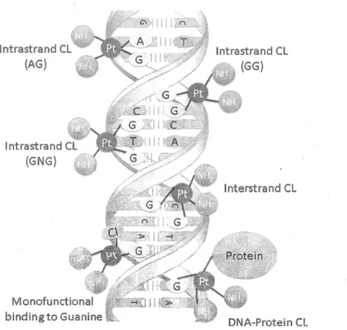
INTRODUCTION lntrastrand CL (AG) lntrastrand CL (GNG) bindingto Guanine 5 lnterstrand CL DNA-Protein CL
Fig. 1.2. Schematic for the formation of various cisplatin-DNA adducts, including intrastrand cross-links, interstrand cross-link, DNA-Protein cross-link and mono-functional binding to guanine. Carboplatin and oxaliplatin also form the same type ofDNA adducts as cisplatin.
The pnmary target of Pt-drugs is nuclear DNA, although other cellular components such as RNA, proteins and membrane phospholipids interact with the drugs (Jung and Lippard, 2007; Reedijk, 2009). Pt-drugs specifically bind to the nitrogen atom at position seven (N7) of the imidazole ring of the purine bases (i.e., the most accessible moiety with nucleophilic characteristics in a double-stranded DNA), mainly guanine (G) and lesser amount to Adenine (A). There are two main reasons for the preferential binding of Pt-drugs to G: (1) Under physiological conditions, N7 of G has higher nucleophilic tendency than that of A since it is more basic than A, and (2) the reaction rate for Pt-drug binding at N7 of 6-oxopurines ( e.g., G) increases, by a factor of 1.6, compared to N7 of 6-aminopurines such as A, owing to the sterically hinderance of the respective amine group (Martin, 1999). Binding of Pt-drugs to DNA forms various DNA
adducts including intrastrand cross-links (CLs) [l,2-d(G pG ), l,2-d(A pG ), and 1,3- d(GpNpG)], interstrand CL, and m onofunctional binding to guanine (Fig. 1.2) (Jamieson and Lippard, 1999; Kelland, 2007). D espite the similarity in the types o f DNA adducts, each Pt-drug produces different proportions o f the specific adducts. Cisplatin and oxaliplatin, for instance, mostly form l,2-d(G pG ) intrastrand CL, w hereas the major carboplatin adduct is l,3-d(G pN pG ) intrastrand CL (Todd and Lippard, 2009). The adducts distort the conformation o f DNA by unwinding and bending, causing the destabilization o f the double helix (Jung and Lippard, 2007; Kelland, 2007; Poklar, Pilch, Lippard, Redding, Dunham and Breslauer, 1996). Structural analysis o f these Pt-DNA adducts by X-ray crystallography and nuclear m agnetic resonance (N M R) spectroscopy has shown that each adduct distorts DNA structure in a distinctive m anner. For example, intrastrand CLs bend DNA duplex tow ard the m ajor groove resulting in a w ider and shallower m inor groove (Jung and Lippard, 2007), whereas interstrand CL bends DNA toward the minor groove leading to a w idening o f the m ajor groove (Coste et al., 1999). The presence o f DACH ligand in oxaliplatin m olecule also results in several conformational differences between DNA adducts formed by oxaliplatin and cisplatin, predominantly due to the interaction between DACH ligand and DNA constituents (Sharma, Gong, Temple, Bhattacharyya, D okholyan and Chaney, 2007).
Such distorted DNA is recognized by several cellular proteins that activate various signal transduction pathways, some o f which lead to D NA-dam age recognition and repair (e.g., high-mobility group proteins) but others mediate the cytotoxicity o f Pt- drugs resulting in cell cycle arrest and apoptosis (Jung and Lippard, 2007) . Nucleotide excision repair (NER) is the m ajor pathway known to remove Pt-drugs from DNA (Ferry, Hamilton and Johnson, 2000). It has been shown that the progression o f both DNA and RNA polymerases along the DNA is blocked at the site o f platination, which results in the inhibition o f replication and transcription processes (Todd and Lippard, 2009). If Pt- DNA adducts are not repaired, it will lead to cell death via an apoptotic pathway (Jung and Lippard, 2007).
Ionizing radiation modulates cancer cell response to the chem otherapeutic effect o f Pt-drugs in several ways. Both Low-dose fractionated and hyper-fractionated radiation modify DNA repair capacity o f a cancer cell that has been reported to enhance the
INTRODUCTION 7
cytotoxicity o f Pt-drugs, particularly in lung, head and neck cancers (G upta et al., 2011; Jeremic et al., 2000). Radiation can also increase vascular perm eability in the blood-brain barrier, leading to a greater accumulation o f the drugs in central nervous system tum ors (Cao et al., 2005). Moreover, radiation induces dam age to cellular m em brane, resulting in the enhancement o f cellular uptake o f the drugs (Yang, D ouple and W ang, 1995). Both the increased vascular permeability and dam age to cellular m em brane prolong tum or retention o f Pt-drugs and their effective concentration inside the cell (Spalding and Lawrence, 2006).
1.3. M olecular Basis of Radiation Therapy:
Radiotherapy is an effective and widespread m ethod for treating cancer with curative, palliative and conservative purposes. A steady rise in the num ber o f cancer patients results in increasing dem and for radiotherapy services in Europe and N orth America. Roughly 45-55% o f cancer patients require radiotherapy at some point and about 20-25% will have more than one course o f radiation treatm ent (Connell and Heilman, 2009; Rosenblatt et al., 2013).
The main goal o f physics-based technology in radiotherapy is to improve the ratio between an optimal radiation dose in the tum our tissue and the possible low est dose in the healthy organs. Such an energy deposition o f radiation in tum our and healthy tissues has serious biological consequences for the irradiated tissues and their cells, including lethal, sublethal, and potentially lethal damages that result in either local control or treatment o f cancer and toxicity o f health tissues as well. In addition to the technological improvements, the clinical practice o f radiotherapy has been therefore influenced by biology underlying the responses o f tum our and healthy cells to ionizing radiation (Connell and Heilman, 2009).
1.3.1. Biological Effects o f Ionizing Radiation:
Ionizing radiation eradicates a m alignant tissue via interaction w ith its cellular components. With respect to DNA as a main cellular target o f radiation, the biological impact o f ionizing radiation results predominantly from the formation o f a variety o f
lesions in DNA via energy deposition into the DNA itself (i.e., direct effect) and its surrounding molecular environment, particularly water molecules (i.e., indirect effect) (Goodhead, Thacker and Cox, 1993; O N eill and Fielden, 1993; O 'Neill and W ardman, 2009). The energy deposition generates interm ediate species including ions, radicals, excited molecules and free electrons in a nanom eter-scale volume. These species subsequently interact with DNA to induce cluster lesions (Goodhead, 2006; W ard,J.F., Webb C.F., Limoli C.L., Milligan J.R., 1990; W ard, 1994).
Owing to the considerable amount o f w ater in a cell (about 70% o f cellular mass), highly reactive species toward DNA arise from w ater radiolysis; the m ost importantly
hydroxyl radicals (*OH), hydrated electrons (e~q ) and hydrogen atom ( H) (Clem ens von
Sonntag, 1987). It has been suggested that the indirect effect o f radiation resulting from the w ater radiolysis has substantial contribution (i.e., 30 - 70 %) to the formation o f DNA damage (DeLara, Jenner, Townsend, M arsden and O'Neill, 1995; N ikjoo et al., 2002). The reactive species induce a variety o f D N A lesions such as base modifications, sugar damage leading to a base release and strand breaks (SSB and D SB), alkali-labile sites, and DNA-protein crosslink (Clemens von Sonntag, 2006). Among the various DNA lesions, strand breaks (particularly DSB) are believed to have considerable biological impact including lethality, mutagenesis and carcinogenesis (Negrini, Gorgoulis and Halazonetis, 2010; O'Driscoll and Jeggo, 2006; Obe, Johannes and Schulte-Frohlinde, 1992).
’OH is the most reactive radical generated from radiolysis o f w ater that interacts with DNA constituents at close to diffusion-controlled rate (Sevilla and Bernhard, 2008). Two main reactions o f *OH with nucleobases and sugar m oiety are addition to C=C and C=N double bonds (e.g., formation o f 8-oxoguanine) and hydrogen abstraction. 'OH has also been known as the main radical to induce DNA strand break via interaction with sugar moieties (e.g., hydrogen abstraction from C 4 ’ o f sugar) (Clemens von Sonntag,
2006). In contrast, *H and e~ seems to be less potent at producing DNA damage,
particularly strand breaks, despite the fact that they are highly reactive reducing species (Clemens von Sonntag, 1987; Li, Sevilla and Sanche, 2003). The observation o f different
INTRODUCTION 9
ionization potentials o f eaq due to the localization o f electron in the surface and inside
(bulk) o f water, however, suggest that the previous notion o f e~q as a single equilibrated
species in water seems to be too simple (Alizadeh and Sanche, 2012; Donald, Leib, O'Brien, Holm and Williams, 2008; Siefermann et al., 2010; Verlet, Bragg, Kammrath, Cheshnovsky and Neumark, 2005). Accordingly, experimental and theoretical studies
report that e~aq, in particular at the surface o f w ater can induce DNA dam age, even strand
breaks in the solvated DNA via electron transfer to a DNA subunits (e.g., phosphate group) and subsequent dissociation o f a chemical bond (Nguyena, M aa, Luoa, Bristowb, Jafffayc and Lu, 2011; Siefermann and Abel, 2011; W ang, N guyen and Lu, 2009).
Although w ater represents a considerable fraction o f cellular mass and the species arising from water radiolysis are highly reactive with DNA to induce a variety o f lesions, DNA helix in a cell nucleus is known to be highly packed by forming a complex with histon protein (i.e., nucleosome) (Luger, M ader, Richmond, Sargent and Richmond, 1997) , which is further organized into a higher order chromatin structure in eukaryotic cells (Fig. 1.3) (Felsenfeld and G roudine, 2003). In this configuration, nuclear DNA, which includes a closely bound hydration shell with less than ~ 13 H2O per nucleotide, is thus surrounded by a minimal am ount o f free water. This configuration reduces the indirect effects o f radiation by protecting DNA from the diffusible radicals (Sevilla and Bernhard, 2008). M oreover, since hydroxyl radicals highly react with other biom olecules and its reaction rate with cellular com ponents is close to diffusion-controlled rates, only those created in the immediate vicinity o f DNA can induce damage (Clemens von Sonntag, 2006). Therefore, it should be recognized that intermediate species arising from the direct effect o f radiation, including DNA subunit radical cations, free electrons and electronically excited DNA subunits, play a considerable role in the induction o f DNA damage, particularly for irradiation with high linear energy transfer (LET) particles (Hirayama et al., 2009).
1.3.2. Interaction of Therapeutic Radiation with Biological Matter:
Despite various modalities in radiotherapy such as intensity modulated radiotherapy, tomotherapy, stereotactic radiotherapy, and brachytherapy, the types of radiation beam are essentially limited to photons including gamma- and X-rays, and charged particles including electrons and to much smaller extend protons and carbon ions (Thariat, Hannoun-Levi, Sun Myint, Vuong and Gérard, 2013). The dominant processes involved in the interaction of these ionizing radiations with biomolecules are ionization and excitation with the contribution of the energy deposited of 80% and 20%, respectively (Inokuti, 1995).
Short region of DNA double helix
"Beads on a string" form of chromatin 30-nm chromatin libre of packed nucleosomes Section of chromosome in an extended torm Condensed section ot chromosome Entire mitotic chromosome
Fig. 1.3. Packaging DNA into the chromatin structure. (Reprint with permission from Macmillan
INTRODUCTION 11 Photons interact with m atter through five processes including Com pton scattering, pair production, photoelectric effect, Rayleigh scattering and photonuclear interactions (Attix, 2004). Therapeutic photon beams o f 0.2 - 20 M eV ionize biom olecules mainly through two types o f interactions including Compton scattering and pair production. In the former, a photon interacts with a weakly bound (i.e., outer shell) electron o f the scattering atom and transfers a fraction o f its energy resulting in the ejection o f the electron and scattering o f the photon with lower energy. Such ejected electrons have a wide range o f energy distribution, with a maximum energy less than the energy o f interacting photons. Depending on the transferred energy, the scattered photon subsequently interacts with another molecule via one o f the interaction processes. At energies less than 100 keV, while the interaction cross section for Com pton scattering substantially decreases in biological matter, the photoelectric effect is the dominant interaction process. In this effect, the photon interacts w ith a tightly bound (i.e., inner shell) electron o f the scattering atom and transfers its entire energy to the atomic electron. This photon vanishes and a photoelectron is ejected from one o f the atomic inner shells. Similar to photoelectric effect, pair production is an absorption process in w hich the incident photon disappears and creates an electron and a positron, via the interaction with a strong Coulomb force field near an atomic nucleus. Obviously, the threshold energy required for the pair production occurring near a nuclear field is 1.02 M eV (i.e., sum o f the rest mass energy o f both electron and positron). In biological m atter, however, its interaction cross section is only substantial for energies higher than 10 MeV. For photon energies in the range o f 100 to 200 keV, Compton scattering is the predom inant ineraction process in biological media (Attix, 2004).
Charged particles lose their energy in a m anner that is distinctly different from that o f photons. Since a charged particle is surrounded by its Coulom b electric force field, it interacts with the atoms via two types o f interactions: soft and hard collisions (Attix, 2004). When a charged particle passes an atom at a considerable distance, the impact o f the particle’s Coulomb force field, which is term ed soft collision, distorts the atom resulting in its excitation to a higher energy level, or ionization by ejecting a valence electron. When the distance between incident charged particle and an atom is o f
the order o f the atomic dimensions, hard collision occurs. In this case, the incident particle interacts primarily with a single atomic electron, w hich is then ejected from the atom with considerable kinetic energy. A lthough the probability o f hard collisions is much less than that o f soft collisions, the fraction o f the energy transferred by incident particles is generally comparable for these tw o processes.
The energetic electrons, produced by Compton scattering, pair production and hard collision, have a wide energy distribution that interact with m atter and they prim arily lose their kinetic energy in a process sim ilar to that o f soft collisions. In this coulombic interaction, an incident charged particle transfers a very small am ount o f its kinetic energy and momentum to biological matter. Figure 1.4(a) shows that the distribution o f energy loss for energetic electrons interacting with DNA is m ostly between a few eV and
100 eV with the mean energy loss at 23 eV (LaV em e and Pimblott, 1995). Owing to the ionization threshold o f organic molecules, this am ount o f energy is capable o f ionizing the molecules to produce cations and secondary electrons (SEs). Calculation o f the energy distribution o f SEs indicates that the vast m ajority o f the electrons have energies below 30 eV and the most probable electron energy is betw een 9 and 10 eV. Fig 1.4(b) shows the results o f such a calculation for protons and He cations o f different energies (Pimblott and LaVeme, 2007). Such a distribution is also independent o f the mass o f the fast charged particle, such that high-energy electrom agnetic and any o f charged-particle ionizing radiation produce such a distribution o f SEs. Therefore, electrons o f 0-30 eV, termed low-energy electrons (LEEs), are generated in copious num ber (i.e., on the order o f 3 x l0 4/M eV o f deposited energy) by any type o f electrom agnetic or charged-particle ionizing radiation and carry a substantial fraction o f the energy o f the prim ary ionizing radiation.
Low-energy secondary electrons can also be created via the A uger effect. When an electron is ejected from an inner atomic shell by the photoelectric effect or a hard collision, the formed excited state rapidly returns to the ground state via filling the electron vacancy by another electron from a less tightly bound shell. This process leads to
INTRODUCTION 13 0 0 5 005 b a 'H o n 0 1 M eV - - 1 M eV 1 0 M eV 1 0 0 M eV Hexane-yas ^JWater-bqwd Vv. Water-gas ' v \ , DNA 0 0 4 ‘H e io n • 4 M eV 0.00 0 0 0 0 40 60 80 100 Energy, c (eV) 10 100 1000 E le c tr o n e n e r g y (eV )
Fig. 1.4. (a) The calculated probability of energy loss in the soft collision o f a charge particle with DNA, liquid water, gaseous water and gaseous hexane molecules, with no momentum transfer. The calculation is based on the dipole oscillator strength distribution [with permission from the Author, (LaVeme et Pimblott, 1995)]. (b) The energy distribution o f secondary electrons generated by primary ions at different energies in water [with permission from the Author, (Pimblott et LaVeme, 2007)].
a cascade o f electron transitions by w hich the atom exchanges one energetically deep inner shell vacancy for a num ber o f relatively shallow outer shell vacancies. Conduction- band electrons finally neutralize these vacancies. Each electronic transition is accompanied by the emission o f either a fluorescence X -ray photon or an A uger electron. Therefore, such an inner shell-ionized atom returns to its stable configuration by emitting a number o f Auger electrons with a range o f energies between a few eV and a few keV (Humm, JL. Howell, RW. Rao, DV, 1995; Nikjoo, Em fietzoglou and Charlton, 2008).
1.3.3. Interaction o f LEEs with Condensed Matter:
When the electron wavelength is shorter than the atomic dim ension (e.g., for moderate- and high-energy electrons), the electron can be considered to interact with the medium through individual localized processes, separated by mean free path much larger than the molecular dimension (Nikjoo, Emfietzoglou, W atanabe and Uehara, 2008). In contrast, LEEs have wavelengths com parable to interm olecular distances; hence they interact with solid and liquid media through delocalized processes predom inantly including static and correlation interactions with neighbouring molecules (i.e., distortion o f the bound electron density o f neighbouring molecules in the presence o f a field o f the
incident electron, resulting in electronic, vibrational, and/or rotational excitations), excitation transfer (i.e., energy transfer o f an excited state o f a molecule to a neighboring molecule, leading to the excitation o f that molecule), and coherent scattering. These processes mainly result from two types o f force between an incoming electron and a target molecule: (1) the induced polarization attraction w hich distorts the target orbitals, and (2) the exchange force which describes the electron-target system w ave function (Carsky and Curik, 2012). At these low energies, particularly less than 15 eV, there is a strong probability that one or m ore partial waves o f the electron w ave function undergo constructive interference (i.e., resonance) within the scattering molecule (Sanche, 1991). Therefore, LEE-molecule interaction can be described by resonant and nonresonant (direct) phenomena.
I.3.3.1 Resonant Process:
Electron resonances occur when an incoming electron is tem porarily captured into an unfilled orbital o f a molecule for a time longer than the usual scattering time. Therefore, the resonant processes involve the formation o f a transient negative ion (TNI) with a survival time between 10"15 and 10' 3 s. There are two major types o f TNIs or electron resonances: shape and core-excited resonances (Schulz, 1973). W hen the captured electron in a TNI occupies a previously unfilled orbital in its ground state, the transitory state is referred to as a single-particle or shape resonance. The energy level o f this type o f resonances lies above their parents. In this case, the electron-m olecule potential forms a penetrable barrier due to the angular momentum from the nonzero momentum partial wave content o f the incoming electron, that is responsible for the temporary trapping o f the electron near the molecule. The term “shape” represents the shape o f the potential that is responsible for the electron trapping. If a TNI is formed by two electrons occupying previously unfilled orbitals, the transitory state is called a “core excited” or two-particle, one-hole resonance. Such resonances can lie slightly below or above the energy o f the electronically excited state o f its neutral parent. In the latter case, a momentum barrier (i.e., a penetrable barrier resulted from the angular mom entum ) in the electron-molecule potential, sim ilar to the shape resonance, contributes to retain the electron in the molecule; hence it is referred as core-excited shape resonance (Sanche,
INTRODUCTION 15
2009a). In the former case, the incoming electron is essentially captured by the positive electron affinity o f an electronically excited state o f the molecule. Since such a TNI falls in the category o f Feshbach resonances, they usually have a relatively long lifetime o f the order o f 1 012 to 10' 14 s. Such a long lifetime entails a substantial increase in the interaction time between an incoming electron and the scattering molecule, which results in an enhancement o f inelastic cross sections by orders o f magnitude (Caron and Sanche, 2012).
Decay channels o f a m olecular TNI are usually m ore than its atomic counterpart since it has more degrees o f freedom. Dissociative electron attachm ent (DEA), electron autodetachment, autoionization and resonance stabilization are the four possible decay channels o f a TNI (Sanche, 1991) . DEA will result in the dissociation o f a TNI, if the TNI has three conditions: 1) dissociative state accessible in the Frank-Condon region, 2) resonance lifetime longer than about h a lf a vibration period o f the anion (Sanche, 2012), and 3) positive electron affinity for one o f the possible fragments. Under these conditions, one or several bonds o f the TNI dissociate via a DEA decay channel to produce a stable anion and a neutral atomic or m olecular species in the ground (reaction
1) or an excited state (reaction 2):
RH + e" -» (RE)" - > / ? + / / “ (or R" + h ), Reaction 1
R H + e" -» (RH)*~ -> R * + H " (or R" + H * ). Reaction 2
Electron autodetachment or autoionization (o f the anion) results in a departing electron with lower kinetic energy and a neutral molecule in a rotationally, vibrationally and/or electronically excited state:
RH + e" -> (RH)*~ —> (RH )* + e" . Reaction 3
Depending on the electronically excited state (i.e., dissociative or non-dissociative) and its energy (i.e., transferred from the previously attached electron), the excited state may autodissociate into neutral or ionic fragments:
(RH )*-+ R + H (or R - + H + ) . Reaction 4
When the parent molecule o f a TNI has a positive electron affinity, if the TNI in an excited state can transfer its energy to another m olecule or molecules, it can become a permanent stable anion. This phenomenon is called charge stabilization:
Autoionization can also lead to cation formation when tw o electrons autodetach from the
TNI. In this case, we have:
I.3.3.2. Non-resonant Process:
The non-resonant interactions o f an incoming electron and a scattering molecule mainly result in the ionization and excitation (i.e., rotational, vibrational and/or electronic excitation states) processes. Interaction cross sections for this type o f scattering, in contrast to the resonances, change slowly with incident electron energy. The curves indicating the yield o f molecular fragments resulting from non-resonant electron impact (i.e., yield functions) are usually distinguished by a smooth and m onotonically rising signal above a threshold and a broad peak at higher energies (Fig. 1.5.a, the curves shown by dashed lines) (Huels, Boudai'ffa, Cloutier, Hunting and Sanche, 2003). The threshold is the energy required for the dissociation o f a neutral excited interm ediate state formed via non-resonant interaction:
RH + e" ->• (RH)*~ -» (RH)~ + AE . Reaction 5
RH +e~ -> ( R H y -> (R H Y + 2e Reaction 6
RH+e~(E
0)->(RH)*+e~(E)
(E < E„).
Reaction 7Such threshold energy corresponds to the lowest dissociation energy o f (RH) * w hich may produce neutral or ionized fragments: