2017 ESC/EACTS Guidelines for the management of
valvular heart disease
The Task Force for the Management of Valvular Heart Disease of the European Society of
Cardiology (ESC) and the European Association for Cardio-Thoracic Surgery (EACTS)
Authors/Task Force Members: Volkmar Falk
*1(EACTS Chairperson) (Germany), Helmut Baumgartner
*(ESC
Chairperson) (Germany), Jeroen J. Bax (The Netherlands), Michele De Bonis
1(Italy), Christian Hamm (Germany),
Per Johan Holm (Sweden), Bernard Iung (France), Patrizio Lancellotti (Belgium), Emmanuel Lansac
1(France),
Daniel Rodriguez Mu~
noz (Spain), Raphael Rosenhek (Austria), Johan Sjo¨gren
1(Sweden),
Pilar Tornos Mas (Spain), Alec Vahanian (France), Thomas Walther
1(Germany), Olaf Wendler
1(UK),
Stephan Windecker (Switzerland), Jose Luis Zamorano (Spain)
Document Reviewers: Marco Roffi (CPG Review Coordinator) (Switzerland), Ottavio Alfieri
1(EACTS Review
Coordinator) (Italy), Stefan Agewall (Norway), Anders Ahlsson
1(Sweden), Emanuele Barbato (Italy),
He´ctor Bueno (Spain), Jean-Philippe Collet (France), Ioan Mircea Coman (Romania), Martin Czerny (Germany),
Victoria Delgado (The Netherlands), Donna Fitzsimons (UK), Thierry Folliguet
1(France), Oliver Gaemperli
(Switzerland), Gilbert Habib (France), Wolfgang Harringer
1(Germany), Michael Haude (Germany),
Gerhard Hindricks (Germany), Hugo A. Katus (Germany), Juhani Knuuti (Finland), Philippe Kolh (Belgium),
Christophe Leclercq (France), Theresa A. McDonagh (UK), Massimo Francesco Piepoli (Italy), Luc A. Pierard
(Belgium), Piotr Ponikowski (Poland), Giuseppe M.C. Rosano (UK/Italy), Frank Ruschitzka (Switzerland),
Evgeny Shlyakhto (Russian Federation), Iain A. Simpson (UK), Miguel Sousa-Uva
1(Portugal), Janina Stepinska
(Poland), Giuseppe Tarantini (Italy), Didier Tche´tche´ (France), Victor Aboyans (CPG Supervisor) (France)
The disclosure forms of all experts involved in the development of these guidelines are available on the ESC website http://www.escardio.org/guidelines.
*Corresponding authors. Volkmar Falk, Department of Cardiothoracic and Vascular Surgery, German Heart Center, Augustenburger Platz 1, 13353 Berlin, Germany and Department of Cardiovascular Surgery, Charite Berlin, Charite Platz 1, 10117 Berlin, Germany. Tel: +49 30 4593 2000, Fax: +49 30 4593 2100,
E-mail: [email protected]. Helmut Baumgartner, Division of Adult Congenital and Valvular Heart Disease, Department of Cardiovascular Medicine, University Hospital Muenster, Albert Schweitzer Campus 1, Building A1, 48149 Muenster, Germany. Tel: +49 251 834 6110, Fax: +49 251 834 6109, E-mail: helmut.baum [email protected].
ESC Committee for Practice Guidelines (CPG) and National Cardiac Societies document reviewers listed in the Appendix.
1
Representing the European Association for Cardio-Thoracic Surgery (EACTS). ESC entities having participated in the development of this document:
Associations:Acute Cardiovascular Care Association (ACCA), European Association of Cardiovascular Imaging (EACVI), European Association of Percutaneous Cardiovascular Interventions (EAPCI), Heart Failure Association (HFA).
Working Groups:Cardiovascular Pharmacotherapy, Cardiovascular Surgery, Grown-up Congenital Heart Disease, Valvular Heart Disease.
The content of these European Society of Cardiology (ESC) Guidelines has been published for personal and educational use only. No commercial use is authorized. No part of the ESC Guidelines may be translated or reproduced in any form without written permission from the ESC. Permission can be obtained upon submission of a written request to Oxford University Press, the publisher of the European Heart Journal and the party authorized to handle such permissions on behalf of the ESC ([email protected]).
Disclaimer. The ESC/EACTS Guidelines represent the views of the ESC and the EACTS and were produced after careful consideration of the scientific and medical knowledge and the evidence available at the time of their publication. The ESC and the EACTS are not responsible in the event of any contradiction, discrepancy and/or ambiguity be-tween the ESC/EACTS Guidelines and any other official recommendations or guidelines issued by the relevant public health authorities, in particular in relation to good use of healthcare or therapeutic strategies. Health professionals are encouraged to take the ESC/EACTS Guidelines fully into account when exercising their clinical judgment, as well as in the determination and the implementation of preventive, diagnostic or therapeutic medical strategies; however, the ESC/EACTS Guidelines do not override, in any way whatsoever, the individual responsibility of health professionals to make appropriate and accurate decisions in consideration of each patient’s health condition and in consultation with that patient and, where appropriate and/or necessary, the patient’s caregiver. Nor do the ESC/EACTS Guidelines exempt health professionals from taking into full and careful consideration the relevant official updated recommendations or guidelines issued by the competent public health authorities in order to manage each patient’s case in light of the scientifically accepted data pursuant to their respective ethical and professional obligations. It is also the health professional’s responsibility to ver-ify the applicable rules and regulations relating to drugs and medical devices at the time of prescription.
VC2017 The European Society of Cardiology. For permissions please email: [email protected].
The article has been co-published with permission in the European Heart Journal [10.1093/eurheartj/ehx391] on behalf of the European Society of Cardiology and the European Journal of Cardio-Thoracic Surgery [10.1093/ejcts/ezx324] on behalf of the European Association for Cardio-Thoracic Surgery. All rights reserved in
For the Web Addenda which include background information and detailed discussion of the data that have provided the basis for the recommendations see https://academic.oup.com/ejcts/article-lookup/doi/10.1093/ejcts/ezx324#supplementary-data. Keywords:Guidelines • Valve disease • Valve surgery • Percutaneous valve intervention • Aortic regurgitation • Aortic stenosis • Mitral regurgitation • Mitral stenosis • Tricuspid regurgitation • Tricuspid stenosis • Prosthetic heart valves
TABLE OF CONTENTS
Abbreviations and acronyms . . . 618
1. Preamble . . . 618
2. Introduction. . . 619
2.1 Why do we need new guidelines on valvular heart disease? . . 619
2.2 Content of these guidelines. . . 619
2.3 New format of the guidelines . . . 619
2.4 How to use these guidelines . . . 620
3. General comments . . . 620
3.1 Patient evaluation . . . 620
3.1.1 Echocardiography . . . 620
3.1.2 Other non-invasive investigations . . . 620
3.1.2.1 Stress testing. . . 620
3.1.2.2 Cardiac magnetic resonance . . . 620
3.1.2.3 Computed tomography . . . 621 3.1.2.4 Cinefluoroscopy. . . 621 3.1.2.5 Biomarkers. . . 621 3.1.3 Invasive investigations. . . 621 3.1.3.1 Coronary angiography . . . 621 3.1.3.2 Cardiac catheterization. . . 622 3.1.4 Assessment of comorbidity . . . 622 3.2 Risk stratification. . . 622
3.3 Special considerations in elderly patients . . . 622
3.4 Endocarditis prophylaxis . . . 623
3.5 Prophylaxis for rheumatic fever . . . 623
3.6 Concept of the Heart Team and heart valve centres . . . 623
3.7 Management of associated conditions . . . 623
3.7.1 Coronary artery disease . . . 623
3.7.2 Atrial fibrillation. . . 624
4. Aortic regurgitation. . . 625
4.1 Evaluation . . . 625
4.1.1 Echocardiography . . . 625
4.1.2 Computed tomography and cardiac magnetic resonance 625 4.2 Indications for intervention . . . 625
4.3 Medical therapy . . . 627
4.4 Serial testing . . . 627
4.5 Special patient populations . . . 627
5. Aortic stenosis . . . 628
5.1 Evaluation . . . 628
5.1.1 Echocardiography . . . 628
5.1.2 Additional diagnostic aspects, including assessment of prognostic parameters. . . 629
5.1.3 Diagnostic workup before transcatheter aortic valve implantation. . 630
5.2 Indications for intervention . . . 630
5.2.1 Indications for intervention in symptomatic aortic stenosis . .630 5.2.2 Choice of intervention mode in symptomatic aortic stenosis . . . 633
5.2.3 Asymptomatic aortic stenosis . . . 633
5.3 Medical therapy . . . 633
5.4 Serial testing . . . 633
5.5 Special patient populations . . . 633
6. Mitral regurgitation. . . 634
6.1 Primary mitral regurgitation . . . 634
6.1.1 Evaluation. . . 634
6.1.2 Indications for intervention . . . 635
6.1.3 Medical therapy . . . 636
6.1.4 Serial testing. . . 636
6.2 Secondary mitral regurgitation . . . 636
6.2.1 Evaluation. . . 636
6.2.2 Indications for intervention . . . 637
6.2.3 Medical therapy . . . 638
7. Mitral stenosis . . . 638
7.1 Evaluation . . . 638
7.2 Indications for intervention . . . 638
7.3 Medical therapy . . . 639
7.4 Serial testing . . . 639
7.5 Special patient populations . . . 639
8. Tricuspid regurgitation . . . 640
8.1 Evaluation . . . 641
8.2 Indications for intervention . . . 642
9. Tricuspid stenosis . . . 643
9.1 Evaluation . . . 643
9.2 Indications for intervention . . . 643
9.3 Medical therapy . . . 643
10. Combined and multiple valve diseases . . . 644
11. Prosthetic valves . . . 644
11.1 Choice of prosthetic valve. . . 644
11.2 Management after valve intervention . . . 644
11.2.1 Baseline assessment and modalities of follow-up . . . 645
11.2.2 Antithrombotic management . . . 645
11.2.2.1 General management. . . 645
11.2.2.2 Target international normalized ratio . . . 646
11.2.2.3 Management of vitamin K antagonist overdose and bleeding . . . 646
11.2.2.4 Combination of oral anticoagulants with antiplatelet drugs . . . 646
11.2.2.5 Interruption of anticoagulant therapy for planned invasive procedures . . . 646
11.2.3 Management of valve thrombosis . . . 648
11.2.4 Management of thromboembolism. . . 650
11.2.5 Management of haemolysis and paravalvular leak. . . 650
11.2.6 Management of bioprosthetic valve failure . . . 650
11.2.7 Heart failure . . . 650
12. Management during non-cardiac surgery . . . 652
12.1 Preoperative evaluation. . . 652
12.2 Specific valve lesions . . . 652
12.2.1 Aortic stenosis . . . 652
12.2.2 Mitral stenosis . . . 652
12.2.3 Aortic and mitral regurgitation . . . 652
12.3 Perioperative monitoring . . . 652
13. Management during pregnancy . . . 652
13.1 Native valve disease . . . 652
13.2 Prosthetic valves . . . 653
14. To do and not to do messages from the Guidelines . . . 654
15 What is new in the 2017 Valvular Heart Disease Guidelines? . . . . 656
16. Appendix . . . 658 17. References . . . 659 ES C/EA CTS GUIDELIN ES
ABBREVIATIONS AND ACRONYMS
DPm
Mean transvalvular pressure gradient
2D
Two-dimensional
3D
Three-dimensional
ABC
Age, biomarkers, clinical history
ACE
Angiotensin-converting enzyme
ACS
Acute coronary syndrome
ARB
Angiotensin receptor blocker
AVA
Aortic valve area
BAV
Balloon aortic valvuloplasty
BNP
B-type natriuretic peptide
BSA
Body surface area
CABG
Coronary artery bypass grafting
CAD
Coronary artery disease
CI
Contra-indication(s)
CMR
Cardiovascular magnetic resonance
CPG
Committee for Practice Guidelines cardiac
resynchronization therapy
CT
Computed tomography
EACTS
European Association for Cardio-Thoracic
Surgery
ECG
Electrocardiogram
EDV
End-diastolic velocity
EROA
Effective regurgitant orifice area
ESC
European Society of Cardiology
EuroSCORE
European System for Cardiac Operative Risk
Evaluation
INR
International normalized ratio
IV
Intravenous
LA
Left atrium/left atrial
LMWH
Low-molecular-weight heparin
LV
Left ventricle/left ventricular
LVEDD
Left ventricular end-diastolic diameter
LVEF
Left ventricular ejection fraction
LVESD
Left ventricular end-systolic diameter
LVOT
Left ventricular outflow tract
MSCT
Multislice computed tomography
NOAC
Non-vitamin K antagonist oral anticoagulant
NYHA
New York Heart Association
PCI
Percutaneous coronary intervention
PISA
Proximal isovelocity surface area
PMC
Percutaneous mitral commissurotomy
RV
Right ventricle/right ventricular
SAVR
Surgical aortic valve replacement
SPAP
Systolic pulmonary arterial pressure
STS
Society of Thoracic Surgeons
SVi
Stroke volume index
TAVI
Transcatheter aortic valve implantation
TOE
Transoesophageal echocardiography
TTE
Transthoracic echocardiography
TVI
Time–velocity interval
UFH
Unfractionated heparin
VHD
Valvular heart disease
VKA
Vitamin K antagonist
V
maxPeak transvalvular velocity
1. PREAMBLE
Guidelines summarize and evaluate available evidence with the aim of assisting health professionals in selecting the best
management strategies for an individual patient with a given condition. Guidelines and their recommendations should facili-tate decision making of health professionals in their daily prac-tice. However, the final decisions concerning an individual patient must be made by the responsible health professional(s) in consultation with the patient and caregiver as appropriate.
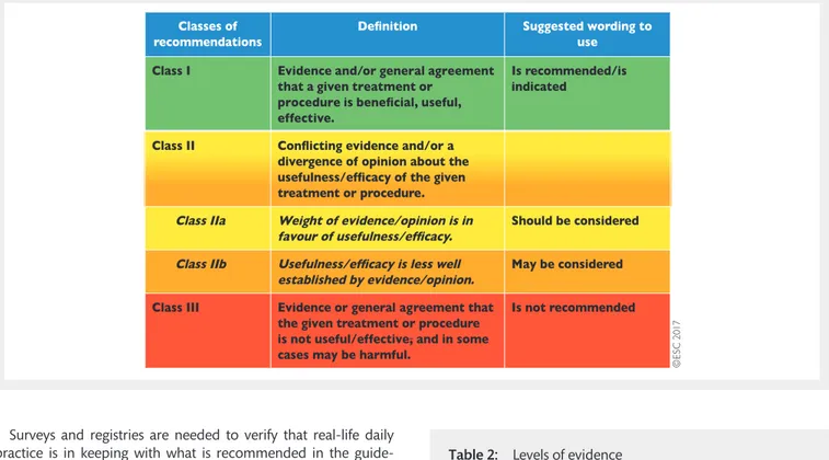
A great number of guidelines have been issued in recent years by the European Society of Cardiology (ESC) and by the European Association for Cardio-Thoracic Surgery (EACTS) as well as by other societies and organisations. Because of the impact on clinical practice, quality criteria for the development of guidelines have been established in order to make all decisions transparent to the user. The recommendations for formulating and issuing ESC Guidelines can be found on the ESC website (https://www.escar dio.org/Guidelines/Clinical-Practice-Guidelines/Guidelines-develop ment/Writing-ESC-Guidelines). ESC Guidelines represent the offi-cial position of the ESC on a given topic and are regularly updated. Members of this Task Force were selected by the ESC and EACTS to represent professionals involved with the medical care of patients with this pathology. Selected experts in the field undertook a comprehensive review of the published evidence for management of a given condition according to ESC Committee for Practice Guidelines (CPG) policy and approved by the EACTS. A critical evaluation of diagnostic and therapeutic procedures was performed, including assessment of the risk–benefit ratio. The level of evidence and the strength of the recommendation of particular management options were weighed and graded ac-cording to predefined scales, as outlined in Tables1and2.
The experts of the writing and reviewing panels provided dec-laration of interest forms for all relationships that might be per-ceived as real or potential sources of conflicts of interest. These forms were compiled into one file and can be found on the ESC website (http://www.escardio.org/guidelines). Any changes in declarations of interest that arise during the writing period were notified to the ESC and EACTS and updated. The Task Force received its entire financial support from the ESC and EACTS without any involvement from the healthcare industry.
The ESC CPG supervises and coordinates the preparation of new Guidelines. The Committee is also responsible for the en-dorsement process of these Guidelines. The ESC Guidelines undergo extensive review by the CPG and external experts, and in this case by EACTS-appointed experts. After appropriate revi-sions the Guidelines are approved by all the experts involved in the Task Force. The finalized document is approved by the CPG and EACTS for publication in the European Heart Journal and in the European Journal of Cardio-Thoracic Surgery. The Guidelines were developed after careful consideration of the scientific and medical knowledge and the evidence available at the time of their dating.
The task of developing ESC/EACTS Guidelines also includes the creation of educational tools and implementation programmes for the recommendations including condensed pocket guideline versions, summary slides, booklets with essential messages, sum-mary cards for non-specialists and an electronic version for digi-tal applications (smartphones, etc.). These versions are abridged and thus, if needed, one should always refer to the full text ver-sion, which is freely available via the ESC website and hosted on the EHJ website. The National Societies of the ESC are encour-aged to endorse, translate and implement all ESC Guidelines. Implementation programmes are needed because it has been shown that the outcome of disease may be favourably influenced by the thorough application of clinical recommendations.
Surveys and registries are needed to verify that real-life daily practice is in keeping with what is recommended in the guide-lines, thus completing the loop between clinical research, writing of guidelines, disseminating them and implementing them into clinical practice.
Health professionals are encouraged to take the ESC/EACTS Guidelines fully into account when exercising their clinical judg-ment, as well as in the determination and the implementation of preventive, diagnostic or therapeutic medical strategies. However, the ESC/EACTS Guidelines do not override in any way whatsoever the individual responsibility of health professionals to make appropriate and accurate decisions in consideration of each patient’s health condition and in consultation with that pa-tient or the papa-tient’s caregiver where appropriate and/or neces-sary. It is also the health professional’s responsibility to verify the rules and regulations applicable to drugs and devices at the time of prescription.
2. INTRODUCTION
2.1. Why do we need new guidelines on valvular
heart disease?
Since the previous version of the guidelines on the manage-ment of VHD was published in 2012, new evidence has accumulated, particularly on percutaneous interventional techniques and on risk stratification with regard to timing of intervention in VHD. This made a revision of the recommen-dations necessary.
2.2. Content of these guidelines
Decision making in VHD involves accurate diagnosis, timing of intervention, risk assessment and, based on these, selection of
the most suitable type of intervention. These guidelines focus on acquired VHD, are oriented towards management and do not deal with endocarditis or congenital valve disease, including pul-monary valve disease, as separate guidelines have been published by the ESC on these topics.
2.3. New format of the guidelines
The new guidelines have been adapted to facilitate their use in clinical practice and to meet readers’ demands by focusing on condensed, clearly represented recommendations. At the end of each section,Key points summarize the essentials. Gaps in evidence are listed to propose topics for future research. The guideline document is harmonized with the simultaneously published chap-ter on VHD of the ESC Textbook of Cardiology, which is freely available by Internet access (https://academic.oup.com/eurheartj/ article-lookup/doi/10.1093/eurheartj/ezx324#supplementary-data).
Table 2: Levels of evidence
Level of evidence A
Data derived from multiple randomized clinical trials or meta-analyses.
Level of evidence B
Data derived from a single randomized clinical trial or large non-randomized studies.
Level of evidence C
Consensus of opinion of the experts and/ or small studies, retrospective studies, registries. ©E S C 2 0 1 7
Table 1: Classes of recommendations
ES
C/EA
CTS
GUIDELIN
The guidelines and the textbook are complementary. Background information and detailed discussion of the data that have provided the basis for the recommendations can be found in the relevant book chapter.
2.4 How to use these guidelines
The Committee emphasizes that many factors ultimately deter-mine the most appropriate treatment in individual patients within a given community. These factors include the availability of diagnostic equipment, the expertise of cardiologists and sur-geons, especially in the field of valve repair and percutaneous intervention and, notably, the wishes of well-informed patients. Furthermore, owing to the lack of evidence-based data in the field of VHD, most recommendations are largely the result of ex-pert consensus opinion. Therefore, deviations from these guide-lines may be appropriate in certain clinical circumstances.
3. GENERAL COMMENTS
The aims of the evaluation of patients with VHD are to diagnose, quantify and assess the mechanism of VHD as well as its conse-quences. Decision making for intervention should be made by a ‘Heart Team’ with a particular expertise in VHD, comprising car-diologists, cardiac surgeons, imaging specialists, anaesthetists and, if needed, general practitioners, geriatricians and heart fail-ure, electrophysiology or intensive care specialists. The ‘Heart Team’ approach is particularly advisable in the management of high-risk patients and is also important for other subsets, such as asymptomatic patients where the evaluation of valve reparability is a key component in decision making. The essential questions in the evaluation of a patient for valvular intervention are sum-marized in Table3.
3.1 Patient evaluation
Precise evaluation of the patient’s history and symptomatic status as well as proper physical examination, in particular auscultation and search for heart failure signs, are crucial for the diagnosis and management of VHD. In addition, assessment of the extrac-ardiac condition—comorbidities and general condition—require particular attention.
3.1.1 Echocardiography.
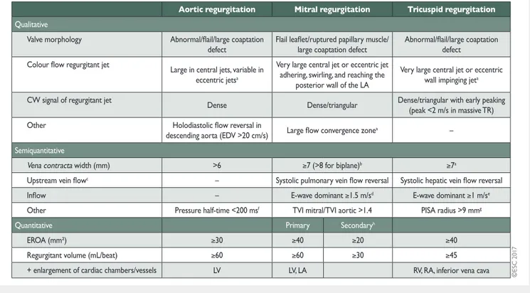
Following adequate clinical evalu-ation, echocardiography is the key technique used to confirm the diagnosis of VHD as well as to assess its severity and prognosis. It should be performed and interpreted by properly trained per-sonnel [1].Echocardiographic criteria for the definition of severe valve stenosis and regurgitation are addressed in specific documents [2–4]. Recommendations for stenotic lesions are indicated in the corresponding sections and quantification of regurgitant lesions is summarized in Table4. An integrated approach including vari-ous criteria is strongly recommended instead of referring to sin-gle measurements. Echocardiography is also key to assess valve morphology and function as well as to evaluate the feasibility and indications of a specific intervention.
Indices of left ventricular (LV) enlargement and function are strong prognostic factors. Pulmonary artery pressure should be
estimated as well as right ventricular (RV) function [5]. Transoesophageal echocardiography (TOE) should be considered when transthoracic echocardiography (TTE) is of suboptimal quality or when thrombosis, prosthetic valve dysfunction or endocarditis is suspected. Intraprocedural TOE is used to guide percutaneous mitral and aortic valve interventions and to moni-tor the results of all surgical valve operations and percutaneous valve implantation or repair.
3.1.2 Other non-invasive investigations.
3.1.2.1 Stress testing. The primary purpose of exercise testing is to unmask the objective occurrence of symptoms in patients who claim to be asymptomatic or have non-specific symptoms, and is especially useful for risk stratification in aortic stenosis [8]. Exercise testing will also determine the level of recommended physical activity, including participation in sports.
Exercise echocardiography may identify the cardiac origin of dyspnoea. The prognostic impact has been shown mainly for aortic stenosis and mitral regurgitation [9].
The search for flow reserve (also called ‘contractile reserve’) using low-dose dobutamine stress echocardiography is useful for assessing aortic stenosis severity and for operative risk stratifica-tion in low-gradient aortic stenosis with impaired LV funcstratifica-tion as well as to assess the potential of reverse remodelling in patients with heart failure and functional mitral regurgitation after a mitral valve procedure [10,11].
3.1.2.2 Cardiac magnetic resonance. In patients with inadequate echocardiographic quality or discrepant results, cardiac magnetic resonance (CMR) should be used to assess the severity of valvular Table 3: Essential questions in the evaluation of patients for valvular intervention
VHD: valvular heart disease.
aLife expectancy should be estimated according to age, sex,
lesions, particularly regurgitant lesions, and to assess ventricular volumes, systolic function, abnormalities of the ascending aorta and myocardial fibrosis. CMR is the reference method for the evaluation of RV volumes and function and is therefore particu-larly useful to evaluate the consequences of tricuspid regurgita-tion [12].
3.1.2.3 Computed tomography. Multislice computed tomography (MSCT) may contribute to evaluation of the severity of valve dis-ease, particularly in aortic stenosis [13, 14] and of the thoracic aorta. MSCT plays an important role in the workup of patients with VHD considered for transcatheter intervention, in particular transcatheter aortic valve implantation (TAVI), and provides valu-able information for pre-procedural planning. Owing to its high negative predictive value, MSCT may be useful to rule out coron-ary artery disease (CAD) in patients who are at low risk of atherosclerosis.
3.1.2.4 Cinefluoroscopy. Cinefluoroscopy is particularly useful for assessing the kinetics of the occluders of a mechanical prosthesis. 3.1.2.5 Biomarkers. B-type natriuretic peptide (BNP) serum levels are related to New York Heart Association (NYHA) functional class and prognosis, particularly in aortic stenosis and mitral re-gurgitation [15]. Natriuretic peptides may be of value for risk stratification and timing of intervention, particularly in asymp-tomatic patients.
3.1.3 Invasive investigations.
3.1.3.1 Coronary angiography. Coronary angiography is indicated for the assessment of CAD when surgery or an intervention is planned, to determine if concomitant coronary revascularization is indicated (see following table of recommendations) [16]. Alternatively, coronary computed tomography (CT) can be used to rule out CAD in patients at low risk for the condition.
Table 4: Echocardiographic criteria for the definition of severe valve regurgitation: an integrative approach (adapted from Lancellottiet al. [2,6,7])
CW: continuous wave; EDV: end-diastolic velocity; EROA: effective regurgitant orifice area; LA: left atrium/atrial; LV: left ventricle/ventricular; PISA: proximal isovelocity surface area; RA: right atrium/right atrial; RV: right ventricle; TR: tricuspid regurgitation; TVI: time–velocity integral.
a
At a Nyquist limit of 50–60 cm/s.
b
For average between apical four- and two-chamber views.
c
Unless other reasons for systolic blunting (atrial fibrillation, elevated atrial pressure).
d
In the absence of other causes of elevated LA pressure and of mitral stenosis.
e
In the absence of other causes of elevated RA pressure.
f
Pressure half-time is shortened with increasing LV diastolic pressure, vasodilator therapy, and in patients with a dilated compliant aorta, or lengthened in chronic aortic regurgitation.
gBaseline Nyquist limit shift of 28 cm/s.
hDifferent thresholds are used in secondary mitral regurgitation where an EROA >20 mm2and regurgitant volume >30 ml identify a subset of patients at
increased risk of cardiac events.
ES
C/EA
CTS
GUIDELIN
3.1.3.2 Cardiac catheterization. The measurement of pressures and cardiac output or the assessment of ventricular performance and valvular regurgitation by ventricular angiography or aortography is restricted to situations where non-invasive evaluation is incon-clusive or discordant with clinical findings. When elevated
pulmonary pressure is the only criterion to support the indication for surgery, confirmation of echo data by invasive measurement is recommended.
3.1.4 Assessment of comorbidity.
The choice of specific examinations to assess comorbidity is directed by the clinical evaluation.3.2 Risk stratification
Risk stratification applies to any sort of intervention and is required for weighing the risk of intervention against the ex-pected natural history of VHD as a basis for decision making. Most experience relates to surgery and TAVI. The EuroSCORE I (http://www.euroscore.org/calc.html) overestimates operative mortality and its calibration of risk is poor. Consequently, it should no longer be used to guide decision making. The EuroSCORE II and the Society of Thoracic Surgeons (STS) score (http://riskcalc.sts.org/stswebriskcalc/#/) more accurately discrim-inate high- and low-risk surgical patients and show better cali-bration to predict postoperative outcome after valvular surgery [17,18]. Scores have major limitations for practical use by insuffi-ciently considering disease severity and not including major risk factors such as frailty, porcelain aorta, chest radiation etc. While EuroSCORE I markedly overestimates 30-day mortality and should therefore be replaced by the better performing EuroSCORE II in this regard, it is nevertheless provided in this document for comparison, as it has been used in many TAVI studies/registries and may still be useful to identify the subgroups of patients for decision between intervention modalities and to predict 1-year mortality. Both scores have shown variable results in predicting the outcomes of intervention in TAVI but are useful for identifying low-risk patients for surgery. New scores have been developed to estimate the risk of 30-day mortality in pa-tients undergoing TAVI, with better accuracy and discrimination, albeit with numerous limitations [19,20].
Experience with risk stratification is being accumulated for other interventional procedures, such as mitral edge-to-edge re-pair. It remains essential not to rely on a single risk score figure when assessing patients or to determine unconditionally the indi-cation and type of intervention. Patient’s life expectancy, ex-pected quality of life and patient preference should be considered, as well as local resources. The futility of interventions in patients unlikely to benefit from the treatment has to be taken into consideration, particularly for TAVI and mitral edge-to-edge repair [21]. The role of the Heart Team is essential to take all of these data into account and adopt a final decision on the best treatment strategy. Finally, the patient and family should be thor-oughly informed and assisted in their decision on the best treat-ment option [22].
3.3 Special considerations in elderly patients
Poor mobility, as assessed by the 6-minute walk test, and oxygen dependency are the main factors associated with increased mor-tality after TAVI and other VHD treatments [23,24]. The combin-ation of severe lung disease, postoperative pain from sternotomy or thoracotomy and prolonged time under anaesthesia in pa-tients undergoing traditional surgical aortic valve replacement (SAVR) may contribute to pulmonary complications. There is a Management of CAD in patients with VHD (adapted
from Windeckeret al.[16])
Recommendations Classa Levelb
Diagnosis of CAD
Coronary angiographycis recommended
before valve surgery in patients with severe VHD and any of the following:
• history of cardiovascular disease
• suspected myocardial ischaemiad
• LV systolic dysfunction
• in men >40 years of age and postmeno-pausal women
• one or more cardiovascular risk factors.
I C
Coronary angiography is recommended in the evaluation of moderate to severe sec-ondary mitral regurgitation.
I C
CT angiography should be considered as an alternative to coronary angiography before valve surgery in patients with severe VHD and low probability of CAD or in whom conventional coronary angiography is technically not feasible or associated with a high risk.
IIa C
Indications for myocardial revascularization CABG is recommended in patients with a primary indication for aortic/mitral valve surgery and coronary artery diameter stenosis >_70%.e
I C
CABG should be considered in patients with a primary indication for aortic/mitral valve surgery and coronary artery diameter stenosis >_50–70%.
IIa C
PCI should be considered in patients with a primary indication to undergo TAVI and coronary artery diameter stenosis >70% in proximal segments.
IIa C
PCI should be considered in patients with a primary indication to undergo transcath-eter mitral valve interventions and coro-nary artery diameter stenosis >70% in proximal segments.
IIa C
CABG: coronary artery bypass grafting; CAD: coronary artery disease; CT: computed tomography; LV: left ventricular; MSCT: multislice computed tomography; PCI: percutaneous coronary intervention; TAVI: transcatheter aortic valve implantation; VHD: valvular heart disease. a Class of recommendation. b Level of evidence. c
MSCT may be used to exclude CAD in patients who are at low risk of atherosclerosis.
dChest pain, abnormal non-invasive testing. e>_50% can be considered for left main stenosis.
gradual relationship between the impairment of renal function and increased mortality after valvular surgery, TAVI and trans-catheter mitral edge-to-edge repair [25], especially when glom-erular filtration rate is < 30 ml/min. Coronary, cerebrovascular and peripheral artery disease have a negative impact on early and late survival after surgery and TAVI [22].
Besides specific organ comorbidities, there is growing interest in the assessment of frailty, an overall marker of impairment of functional, cognitive and nutritional status. Frailty is associated with increased morbidity and mortality after surgery and TAVI [26]. The assessment of frailty should not rely on a subjective ap-proach, such as the ‘eyeball test’, but rather on a combination of different objective estimates. Several tools are available for as-sessing frailty [23,26,27].
3.4 Endocarditis prophylaxis
Antibiotic prophylaxis should be considered for high-risk proced-ures in patients with prosthetic valves, including transcatheter valves, or with repairs using prosthetic material and those with previous episodes of infective endocarditis [28]. Recommendations regarding dental and cutaneous hygiene and strict aseptic meas-ures during any invasive procedmeas-ures are advised in this population. Antibiotic prophylaxis should be considered in dental procedures involving manipulation of the gingival or periapical region of the teeth or manipulation of the oral mucosa [28].
3.5 Prophylaxis for rheumatic fever
Prevention of rheumatic heart disease should preferably be ori-ented towards preventing the first attack of acute rheumatic fever. Antibiotic treatment of group AStreptococcus sore throat is key in primary prevention. In patients with rheumatic heart dis-ease, secondary long-term prophylaxis against rheumatic fever is recommended. Lifelong prophylaxis should be considered in high-risk patients according to the severity of VHD and exposure to group AStreptococcus [29–31].
3.6 Concept of the Heart Team and heart valve
centres
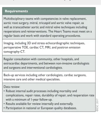
The main purpose of heart valve centres as centres of excellence in the treatment of VHD is to deliver better quality of care. This is achieved through greater volumes associated with specialization of training, continuing education and clinical interest. Specialization will also result in timely referral of patients before irreversible ad-verse effects occur and evaluation of complex VHD conditions. Techniques with a steep learning curve may be performed with better results in hospitals with high volumes and more experience [32]. These main aspects are presented in Table5.
A heart valve centre should have structured training pro-grammes [32]. Surgeons and cardiologists performing any valve intervention should undergo focused training as part of their basic local board certification training. Learning new techniques should take place through mentoring to minimize the effects of the ‘learning curve’.
The relationship between case volume and outcomes for sur-gery and transcatheter interventions is complex but should not be denied [33–35]. However, the precise numbers of procedures per individual operator or hospital required to provide
high-quality care remain controversial and more scientific data are required before solid recommendations can be provided. Nevertheless, standards for provision of cardiac surgery that con-stitute the minimal core requirements have been released [36]. Experience in the full spectrum of surgical procedures—including valve replacement; aortic root surgery; mitral, tricuspid and aortic valve repair; repair of complicated valve endocarditis such as root abscess; treatment of atrial fibrillation as well as surgical myocardial revascularization—must be available. The spectrum of interventional procedures in addition to TAVI should include mi-tral valvuloplasty, mimi-tral valve repair (edge-to-edge), closure of atrial septal defects, closure of paravalvular leaks and left atrial (LA) appendage closure as well as percutaneous coronary inter-vention (PCI). Expertise in interinter-ventional and surgical manage-ment of vascular diseases and complications must be available. Comprehensive recording of performance and patient outcome data at the level of the given heart valve centre is essential, as well as participation in national or ESC/EACTS registries.
3.7 Management of associated conditions
3.7.1 Coronary artery disease.
The use of stress tests to detect CAD associated with severe valvular disease is discouraged be-cause of their low diagnostic value and potential risks. A sum-mary of the management of associated CAD is given in section 3.1.3.1 (see table of recommendations on the management of CAD in patients with VHD) and is detailed in specific guide-lines [16].Table 5: Recommended requirements of a heart valve centre (modified from Chamberset al. [32])
3D: three-dimensional; CT: computed tomography; MRI: magnetic resonance imaging; TOE: transoesophageal echocardiography.
ES
C/EA
CTS
GUIDELIN
3.7.2 Atrial fibrillation.
Non-vitamin K antagonist oral anti-coagulants (NOACs) are approved only for non-valvular atrial fib-rillation, but there is no uniform definition of this term [37]. Recent subgroup analyses of randomized trials on atrial fibrilla-tion support the use of rivaroxaban, apixaban, dabigatran and edoxaban in patients with aortic stenosis, aortic regurgitation or mitral regurgitation presenting with atrial fibrillation [38–41]. The use of NOACs is discouraged in patients who have atrial fibrilla-tion associated with moderate to severe mitral stenosis, given the lack of data and the particularly high thromboembolic risk. Despite the absence of data, NOACs may be used in patients who have atrial fibrillation associated with an aortic bioprosthesis >3 months after implantation but are strictly contraindicated in patients with any mechanical prostheses [42,43].Surgical ablation of atrial fibrillation combined with mitral valve surgery is effective in reducing the incidence of atrial fibril-lation, but at the expense of more frequent pacemaker implant-ation, and has no impact on short-term survival [44]. Surgical ablation should be considered in patients with symptomatic atrial fibrillation and may be considered in patients with asymptomatic atrial fibrillation if feasible with minimal risk. The decision should factor in other important variables, such as age, the duration of atrial fibrillation and LA size. Surgical excision or external clipping of the LA appendage may be considered combined with valvular surgery, although there is no evidence that it decreases thrombo-embolic risk. For patients with atrial fibrillation and risk factors for stroke, long-term oral anticoagulation is currently recom-mended, although surgical ablation of atrial fibrillation and/or surgical LA appendage excision or exclusion may have been per-formed [37]. Recommendations for the management of atrial fib-rillation in VHD are summarized in the following table.
Key points
• Precise evaluation of the patient’s history and symptomatic status as well as proper physical examination are crucial for the diagnosis and management of VHD.
• Echocardiography is the key technique to diagnose VHD and assess its severity and prognosis. Other non-invasive investiga-tions such as stress testing, CMR, CT, fluoroscopy and bio-markers are complementary, and invasive investigation beyond preoperative coronary angiography is restricted to situations where non-invasive evaluation is inconclusive. • Risk stratification is essential for decision making to weigh the
risk of intervention against the expected natural history of VHD. • Decision making in elderly patients requires special consider-ations, including life expectancy and expected quality of life, with regards to comorbidities and general condition (frailty). • Heart valve centres with highly specialized multidisciplinary
teams, comprehensive equipment and sufficient volumes of procedures are required to deliver high-quality care and pro-vide adequate training.
• NOACs may be used in patients with atrial fibrillation and aortic stenosis, aortic regurgitation, mitral regurgitation or aortic bioprostheses >3 months after implantation but are contraindicated in mitral stenosis and mechanical valves. Gaps in evidence
• Better tools for risk stratification need to be developed, par-ticularly for the decision between surgery and catheter inter-vention and for the avoidance of futile interinter-ventions.
Management of atrial fibrillation in patients with VHD
Recommendations Classa Levelb
Anticoagulation
NOACs should be considered as an alternative to VKAs in patients with aortic stenosis, aortic regurgitation and mitral
regurgitation presenting with atrial fibrillation [38–41]. IIa B
NOACs should be considered as an alternative to VKAs after the third month of implantation in patients who have
atrial fibrillation associated with a surgical or transcatheter aortic valve bioprosthesis. IIa C
The use of NOACs is not recommended in patients with atrial fibrillation and moderate to severe mitral stenosis. III C
NOACS are contraindicated in patients with a mechanical valve [45]. III B
Surgical interventions
Surgical ablation of atrial fibrillation should be considered in patients with symptomatic atrial fibrillation who undergo
valve surgery [37]. IIa A
Surgical ablation of atrial fibrillation may be considered in patients with asymptomatic atrial fibrillation who undergo
valve surgery, if feasible, with minimal risk. IIb C
Surgical excision or external clipping of the LA appendage may be considered in patients undergoing valve surgery
[46]. IIb B
LA: left atrial; NOAC: non-vitamin K antagonist oral anticoagulant; VHD: valvular heart disease; VKA: vitamin K antagonist.
aClass of recommendation. bLevel of evidence.
• Minimum volumes of procedures per operator and per hos-pital that are required to achieve optimal treatment results need to be defined.
• The safety and efficacy of NOACs in patients with surgical or transcatheter bioprostheses in the first 3 months after im-plantation should be studied.
4. AORTIC REGURGITATION
Aortic regurgitation can be caused by primary disease of the aor-tic valve cusps and/or abnormalities of the aoraor-tic root and as-cending aortic geometry. Degenerative tricuspid and bicuspid aortic regurgitation are the most common aetiologies in Western countries, accounting for approximately two-thirds of the under-lying aetiology of aortic regurgitation in the Euro Heart Survey on VHD [47]. Other causes include infective and rheumatic endo-carditis. Acute severe aortic regurgitation is mostly caused by in-fective endocarditis and less frequently by aortic dissection.
4.1 Evaluation
4.1.1 Echocardiography.
Echocardiography (TTE/TOE) is the key examination to describe valve anatomy, quantify aortic re-gurgitation, evaluate its mechanisms, define the morphology of the aorta and determine the feasibility of valve-sparing aortic surgery or valve repair [48,49].Essential aspects of this evaluation include:
• Assessment of valve morphology: tricuspid, bicuspid, unicus-pid or quadricusunicus-pid valve.
• Determination of the direction of the aortic regurgitation jet in the long-axis view (central or eccentric) and its origin in the short-axis view (central or commissural).
• Identification of the mechanism, following the same principle as for mitral regurgitation: normal cusps but insufficient coaptation due to dilatation of the aortic root with central jet (type 1), cusp prolapse with eccentric jet (type 2) or retraction with poor cusp tissue quality and large central or eccentric jet (type 3) [48]. • Quantification of aortic regurgitation should follow an
inte-grated approach considering all qualitative, semi-quantitative and quantitative parameters [2,6] (Table4).
• Measurement of LV function and dimensions. Indexing LV diameters for body surface area (BSA) is recommended in pa-tients with small body size (BSA <1.68 m2) [50]. New param-eters obtained by three-dimensional (3D) echocardiography, tissue Doppler and strain rate imaging may be useful, particu-larly in patients with borderline left ventricular ejection fraction (LVEF), where they may help in the decision for surgery [51]. • Measurement of the aortic root and ascending aorta in the
2-dimensional (2D) mode at four levels: annulus, sinuses of Valsalva, sinotubular junction and tubular ascending aorta [52]. Measurements are taken in the parasternal long-axis view from leading edge to leading edge at end diastole, except for the aortic annulus, which is measured in mid systole. As it will have surgical consequences, it is important to differentiate three phenotypes of the ascending aorta: aortic root aneurysms (sinuses of Valsalva >45 mm), tubular ascending aneurysm
(sinuses of Valsalva <40–45 mm) and isolated aortic regurgita-tion (all diameters <40 mm). The calcularegurgita-tion of indexed values has been recommended to account for body size [53].
• Definition of the anatomy of the aortic valve cusps and as-sessment of valve reparability should be provided by pre-operative TOE if aortic valve repair or a valve-sparing surgery of the aortic root is considered.
• Intraoperative evaluation of the surgical result by TOE is man-datory in patients in whom the aortic valve is preserved or re-paired in the procedure.
4.1.2 Computed tomography and cardiac magnetic
reso-nance.
CMR should be used to quantify the regurgitant fraction when echocardiographic measurements are equivocal. In patients with aortic dilatation, gated MSCT is recommended to assess the maximum diameter. CMR can be used for follow-up, but indica-tion for surgery should preferably be based on CT measurements. Different methods of aortic measurements have been reported and this may result in diameter discrepancies of 2–3 mm that could influence therapeutic management. To improve reproduci-bility, it is recommended to measure diameters using the inner-inner edge technique at end diastole on the strictly transverse plane by double oblique reconstruction perpendicular to the axis of blood flow of the corresponding segment. Diameters at the an-nulus, sinus of Valsalva, sinotubular junction, tubular ascending aorta and aortic arch level should be reported. Maximum root diameter should be taken from sinus to sinus rather than sinus to commissure diameter, as it correlates more closely to long-axis leading edge to leading edge echo maximum diameters [54,55].4.2 Indications for intervention
Acute aortic regurgitation may require urgent surgery. It is primar-ily caused by infective endocarditis and aortic dissections. Specific guidelines deal with these entities [28, 56]. The indications for intervention in chronic aortic regurgitation are summarized on the next page (recommendations on indications for surgery in severe aortic regurgitation and aortic root disease) and in Figure1 and may be related to symptoms, status of the LV or dilatation of the aorta.
In symptomatic patients, surgery is recommended irrespective of the LVEF value, except for extreme cases, as long as aortic re-gurgitation is severe and the operative risk is not prohibitive [57]. In asymptomatic patients with severe aortic regurgitation, im-pairment of LV function (ejection fraction <_50%) and LV enlarge-ment with an LV end-diastolic diameter (LVEDD) >70 mm or left ventricular end-systolic diameter (LVESD) >50 mm are associated with worse outcome and surgery should therefore be pursued when these cut-offs are reached [58]. In patients with small body size, LVESD should be related to BSA and a cut-off of 25 mm/m2 BSA appears to be more appropriate [50]. In patients not reach-ing the thresholds for surgery, close follow-up is needed and ex-ercise testing should be performed to identify borderline symptomatic patients. In truly asymptomatic patients, regular as-sessment of LV function and physical condition are crucial to identify the optimal time for surgery. A rapid progression of ven-tricular dimensions or decline in venven-tricular function on serial testing is a reason to consider surgery.
In patients with a dilated aorta, the rationale for surgery has been best defined in patients with Marfan syndrome and root
ES
C/EA
CTS
GUIDELIN
dilation [59]. Root aneurysms need to have root replacement, with or without preservation of the native aortic valve, but defin-itely with coronary reimplantation. In contrast, tubular ascending aortic aneurysms require only a supracommissural tube graft re-placement without coronary reimplantation. In patients with aortic diameters borderline for aortic surgery, the family history, age and anticipated risk of the procedure should be taken into consider-ation. In individuals with a bicuspid aortic valve and no significant valve regurgitation, prophylactic surgery should be considered with aortic diameters >_55 mm or >_50 mm when additional risk fac-tors or coarctation are present (see table of recommendations on indications for surgery in severe aortic regurgitation and aortic root disease). Surgery is indicated in all patients with Marfan syn-drome and a maximal aortic diameter >_50 mm. In patients with Marfan syndrome and additional risk factors and in patients with a TGFBR1 or TGFBR2 mutation (including Loeys–Dietz syndrome), Figure 1: Management of aortic regurgitation. AR: aortic regurgitation; BSA: body surface area; LVEDD: left ventricle end-diastolic diameter; LVEF: left ven-tricular ejection fraction; LVESD: left ventricle end-systolic diameter.
a
See table of recommendations on indications for surgery in severe aortic regurgitation and aortic root disease for definition.
b
Surgery should also be considered if significant changes in LV or aortic size occur during follow-up (see table of recommendations on indications for surgery in severe aortic regurgitation and aortic root disease in section 4.2).
Indications for surgery in (A) severe aortic regurgitation and (B) aortic root disease (irrespective of the severity of aortic regurgitation)
Indications for surgery Classa Levelb
A. Severe aortic regurgitation
Surgery is indicated in symptomatic patients [57,58,66,67]. I B
Surgery is indicated in asymptomatic patients with resting
LVEF <_50% [57,58]. I B
Surgery is indicated in patients undergoing CABG or
sur-gery of the ascending aorta or of another valve. I C
Heart Team discussion is recommended in selected patientscin whom aortic valve repair may be a feasible
alternative to valve replacement.
I C
Surgery should be considered in asymptomatic patients with resting ejection fraction >50% with severe LV dilata-tion: LVEDD >70 mm or LVESD >50 mm (or LVESD >25 mm/m2BSA in patients with small body size) [58,66].
IIa B
B. Aortic root or tubular ascending aortic aneurysmd(irrespective of the
severity of aortic regurgitation)
Aortic valve repair, using the reimplantation or remodel-ling with aortic annuloplasty technique, is recommended in young patients with aortic root dilation and tricuspid aortic valves, when performed by experienced surgeons.
I C
Surgery is indicated in patients with Marfan syndrome who have aortic root disease with a maximal ascending aortic diameter >_50 mm.
I C
Surgery should be considered in patients who have aortic root disease with maximal ascending aortic diameter: • >_45 mm in the presence of Marfan syndrome and
additional risk factorseor patients with a
TGFBR1 orTGFBR2 mutation (including Loeys–Dietz syndrome).f
• >_50 mm in the presence of a bicuspid valve with additional risk factorseor coarctation.
• >_55 mm for all other patients.
IIa C
When surgery is primarily indicated for the aortic valve, replacement of the aortic root or tubular ascending aorta should be considered when >_45 mm, particularly in the presence of a bicuspid valve.g
IIa C
BSA: body surface area; CABG: coronary artery bypass grafting; CT: computed tomography; ECG: electrocardiogram; LV: left ventricular; LVEDD: left ventricular end-diastolic diameter; LVEF: left ventricular ejection fraction; LVESD: left ventricular end-systolic diameter.
aClass of recommendation. bLevel of evidence.
cPatients with pliable non-calcified tricuspid or bicuspid valves who
have a type I (enlargement of the aortic root with normal cusp motion) or type II (cusp prolapse) mechanism of aortic regurgitation [6,48,49].
dFor clinical decision making, dimensions of the aorta should be
con-firmed by ECG-gated CT measurement.
eFamily history of aortic dissection (or personal history of spontaneous
vascular dissection), severe aortic regurgitation or mitral regurgitation, desire for pregnancy, systemic hypertension and/or aortic size increase >3 mm/year (on repeated measurements using the same ECG-gated imaging technique measured at the same level of the aorta with side-by-side comparison and confirmed by another technique).
fA lower threshold of 40 mm may be considered in women with low
BSA, in patients with aTGFBR2 mutation or in patients with severe extra-aortic features [60].
gConsidering age, BSA, aetiology of the valvular disease, presence of a
bicuspid aortic valve and intraoperative shape and thickness of the ascending aorta.
surgery should be considered at a maximal aortic diameter >_45 mm [60]. In the latter group, women with low BSA, patients with aTGFBR2 mutation or patients with severe extra-aortic fea-tures appear to be at particularly high risk and surgery may be considered already at a lower threshold of 40 mm [60]. In aortic roots >_55 mm, surgery should be considered irrespective of the degree of aortic regurgitation and type of valve pathology [61]. For patients who have an indication for aortic valve surgery, an aortic diameter >_45 mm is considered to indicate concomitant surgery of the aortic root or tubular ascending aorta. The patient’s stature, the aetiology of the valvular disease (bicuspid valve) and the intra-operative shape and wall thickness of the ascending aorta should be taken into account for individual decisions.
Although valve replacement is the standard procedure in the ma-jority of patients with aortic regurgitation, valve repair or valve-sparing surgery should be considered in patients with pliable non-calcified tricuspid or bicuspid valves who have a type I (enlargement of the aortic root with normal cusp motion) or type II (cusp pro-lapse) mechanism of aortic regurgitation [6,48,49]. In experienced centres, valve-sparing root replacement and valve repair, when feas-ible, yield good long-term results with low rates of valve-related events as well as better quality of life [62–65]. The choice of the sur-gical procedure should be adapted to the experience of the team, the presence of an aortic root aneurysm, characteristics of the cusps, life expectancy and desired anticoagulation status. Patients in whom the Heart Team identifies the aortic valve to be repairable should be referred to appropriate surgical teams for the procedure.
4.3 Medical therapy
Medical therapy can provide symptomatic improvement in indi-viduals with chronic severe aortic regurgitation in whom surgery is not feasible. In patients who undergo surgery but continue to suffer from heart failure or hypertension, angiotensin-converting enzyme (ACE) inhibitors, angiotensin receptor blockers (ARBs) and beta-blockers are useful [68,69].
In patients with Marfan syndrome, beta-blockers and/or losar-tan may slow aortic root dilatation and reduce the risk of aortic complications and should be considered before and after surgery [70–72]. By analogy, while there are no studies that provide evi-dence, it is common clinical practice to advise beta-blocker or losartan therapy in patients with bicuspid aortic valve if the aortic root and/or ascending aorta is dilated.
Women with Marfan syndrome and an aortic diameter >45 mm are strongly discouraged from becoming pregnant without prior repair because of the high risk of dissection. Although an aortic diameter <40 mm is rarely associated with aortic dissection, a completely safe diameter does not exist. With an aorta between 40 and 45 mm, previous aortic growth and family history are im-portant for advising pregnancy with or without aortic repair [73]. Although the actual risk of dissection is not well-documented in the setting of bicuspid valves, counselling against pregnancy is rec-ommended in the setting of aortic diameters >50 mm [74].
The level of physical and sports activity in the presence of a dilated aorta remains a matter of clinical judgement in the absence of evidence. Current guidelines are very restrictive, particularly re-garding isometric exercise, to avoid a catastrophic event [75]. This at-titude is clearly justified in the presence of connective tissue disease.
Given the family risk of thoracic aortic aneurysms, screening and referral for genetic testing of the patient’s first-degree relatives with appropriate imaging studies is indicated in patients with connective
tissue disease. For patients with bicuspid valves it is appropriate to have an echocardiographic screening of first-degree relatives.
4.4 Serial testing
All asymptomatic patients with severe aortic regurgitation and normal LV function should be seen for follow-up at least every year. In patients with a first diagnosis, or if LV diameter and/or ejection fraction show significant changes or come close to thresholds for surgery, follow-up should be continued at 3–6-month intervals. In inconclusive cases, BNP may be helpful, as its elevation during follow-up has been related to deterioration of LV function [76]. Patients with mild to moderate aortic regurgita-tion can be reviewed on a yearly basis and echocardiography performed every 2 years.
If the ascending aorta is dilated (>40 mm) it is recommended to perform CT or CMR. Follow-up assessment of the aortic di-mension should be performed using echocardiography and/or CMR. Any increase >3 mm should be validated by CT angiog-raphy/CMR and compared to baseline data.
4.5 Special patient populations
If aortic regurgitation requiring surgery is associated with severe mitral regurgitation, both should be addressed during the same operation.
In patients with moderate aortic regurgitation who undergo coronary artery bypass grafting (CABG) or mitral valve surgery, the decision to treat the aortic valve is controversial, as data show that progression of moderate aortic regurgitation is very slow in patients without aortic dilatation [77]. The Heart Team should decide based on the aetiology of aortic regurgitation, other clinical factors, the life expectancy of the patient and the patient’s operative risk.
Key points
• The evaluation of aortic regurgitation requires consideration of valve morphology and the mechanism and severity of re-gurgitation, including careful assessment of aortic dilatation. • In asymptomatic patients with severe aortic regurgitation,
careful follow-up of symptomatic status and LV size and func-tion is mandatory.
• The strongest indication for valve surgery is the presence of symptoms (spontaneous or on exercise testing) and/or the documentation of LVEF <50% and/or end-systolic diameter >50 mm.
• In patients with a dilated aorta, definition of the aortic path-ology and accurate measurements of aortic diameters are cru-cial to guide the timing and type of surgery.
• Aortic valve repair and valve-sparing aortic surgery instead of aortic valve replacement should be considered in selected cases in experienced centres.
Gaps in evidence
• The impact of earlier markers of LV dysfunction on postoper-ative outcome requires further research.
• Criteria for the decision between valve replacement and valve repair must still be refined.
ES
C/EA
CTS
GUIDELIN
• Potential differences in the risk of aortic complications de-pending on subtypes of aortic aneurysms (site and morph-ology) should be studied.
• The effect of medical treatment on aortic enlargement in pa-tients with bicuspid aortic valve needs to be studied.
5. AORTIC STENOSIS
Aortic stenosis is the most common primary valve disease leading to surgery or catheter intervention in Europe and North America, with a growing prevalence due to the ageing population.
5.1 Evaluation
5.1.1 Echocardiography.
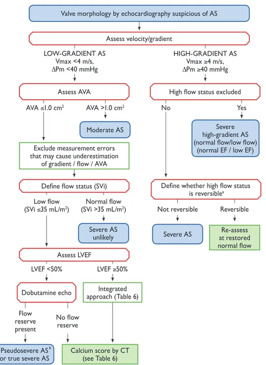
Echocardiography is the key diagnostic tool. It confirms the presence of aortic stenosis; assesses the degree of valve calcification, LV function and wall thickness; detects the presence of other associated valve disease or aortic pathology and provides prognostic information. Doppler echocardiography is the preferred technique for assessing the severity of aortic stenosis [4].Figure2and Table6provide a practical stepwise approach for the assessment of aortic stenosis severity. Details can be found in a recent position paper from the European Association of Cardiovascular Imaging [4].
Figure 2: Stepwise integrated approach for the assessment of aortic stenosis severity (modified from Baumgartneret al. [4]).aHigh flow may be reversible in settings
such as anaemia, hyperthyroidism, arteriovenous shunts.bPseudosevere AS is defined by an increase to an AVA >1.0 cm2with flow normalization.
DPm: mean transvalvular pressure gradient; AS: aortic stenosis; AVA: aortic valve area; CT: computed tomography; EF: ejection fraction; LVEF: left ventricular ejection fraction; SVi: stroke volume index; Vmax: peak transvalvular velocity.
Although valve area represents, from a theoretical perspec-tive, the ideal measurement for assessing the severity of aor-tic stenosis, it has technical limitations in clinical pracaor-tice. It must, for clinical decision making, always be considered together with flow rate, mean pressure gradient (the most ro-bust measurement), ventricular function, size and wall thickness, degree of valve calcification, blood pressure and functional status. Hypertensive patients should be reassessed when normotensive [4]. Four categories of aortic stenosis can be defined:
• High-gradient aortic stenosis (valve area <1 cm2, mean gradi-ent >40 mmHg). Severe aortic stenosis can be assumed irre-spective of whether LVEF and flow are normal or reduced. • Low-flow, low-gradient aortic stenosis with reduced ejection
fraction [valve area <1 cm2, mean gradient <40 mmHg, ejec-tion fracejec-tion <50%, stroke volume index (SVi) <_35 ml/m2]. Low-dose dobutamine echocardiography is recommended in this setting to distinguish truly severe aortic stenosis from pseudosevere aortic stenosis, which is defined by an increase to an aortic valve area (AVA) of >1.0 cm2with flow normaliza-tion. In addition, the presence of flow reserve (also termed contractile reserve; increase of stroke volume >20%) has prog-nostic implications because it is associated with better out-come [10,78].
• Low-flow, low-gradient aortic stenosis with preserved ejection fraction (valve area <1 cm2, mean gradient <40 mmHg, ejec-tion fracejec-tion >_50%, SVi <_35 ml/m2). This is typically encoun-tered in the elderly and is associated with small ventricular size, marked LV hypertrophy and frequently a history of hypertension [79,80]. The diagnosis of severe aortic stenosis in this setting remains challenging and requires careful exclusion of measurement errors and other reasons for such
echocardiographic findings (Table6). The degree of valve cal-cification by MSCT is related to aortic stenosis severity and outcome [13,14,81]. Its assessment has therefore gained increasing importance in this setting.
• Normal-flow, low-gradient aortic stenosis with preserved ejec-tion fracejec-tion (valve area <1 cm2, mean gradient <40 mmHg, ejection fraction >_50%, SVi >35 m2). These patients will in gen-eral have only moderate aortic stenosis [14,82–84].
5.1.2 Additional diagnostic aspects, including assessment
of prognostic parameters.
Exercise testing is recommended in physically active patients for unmasking symptoms and for risk stratification of asymptomatic patients with severe aortic stenosis [85].Exercise stress echocardiography may provide prognostic in-formation in asymptomatic severe aortic stenosis by assessing the increase in mean pressure gradient and change in LV func-tion during exercise [86].
TOE provides additional evaluation of concomitant mitral valve abnormalities. It has gained importance in the assessment before TAVI and after TAVI or surgical procedures [87].
MSCT and CMR provide additional information on the dimen-sions and geometry of the aortic root and ascending aorta and the extent of calcification. It has become particularly important for the quantification of valve calcification when assessing aortic stenosis severity in low-gradient aortic stenosis [13,14,81]. CMR may be useful for the detection and quantification of myocardial fibrosis, providing additional prognostic information regardless of the presence of CAD [88].
Natriuretic peptides have been shown to predict symptom-free survival and outcome in normal and low-flow severe aortic stenosis [89,90] and may be useful in asymptomatic patients to determine optimal timing of intervention.
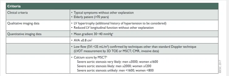
Table 6: Criteria that increase the likelihood of severe aortic stenosis in patients with AVA <1.0 cm2and mean gradient <40 mmHg
in the presence of preserved ejection fraction (modified from Baumgartneret al. [4])
3D: three-dimensional; AVA: aortic valve area; CMR: cardiovascular magnetic resonance; LV: left ventricular; LVOT: left ventricular outflow tract; MSCT: multi-slice computed tomography; SVi: stroke volume index; TOE: transoesophageal echocardiography.
aHaemodynamics measured when the patient is normotensive.
bValues are given in arbitrary units using Agatston method for quantification of valve calcification.
ES
C/EA
CTS
GUIDELIN
Retrograde LV catheterization to assess the severity of aortic stenosis is no longer routinely performed. Its use is restricted to patients with inconclusive non-invasive investigations.
5.1.3 Diagnostic workup before transcatheter aortic valve
implantation.
MSCT is the preferred imaging tool to assess the anatomy and dimensions of the aortic root, size and shape of the aortic valve annulus, its distance to the coronary ostia, the distribu-tion of calcificadistribu-tions and the number of aortic valve cusps. It is es-sential to evaluate the feasibility of the various access routes, as this provides information on minimal luminal diameters, athero-sclerotic plaque burden, the presence of aneurysms or thrombi, vessel tortuosity and thoracic and LV apex anatomy. CMR—as an alternative technique—is, in this context, inferior to MSCT with re-gards to assessment of inner vessel dimensions and calcifications. 3D TOE can be used to determine aortic annulus dimensions but remains more operator- and image quality–dependent than MSCT. However, TOE is an important tool for monitoring the pro-cedure and evaluating the results, especially if complications occur.5.2 Indications for intervention
The indications for aortic valve interventions are summarized on the next page (see table of indications for intervention in aortic stenosis and recommendations for the choice of intervention mode) and in Table7and are illustrated in Figure3.
5.2.1 Indications for intervention in symptomatic aortic
stenosis.
Early therapy should be strongly recommended in all symptomatic patients with severe aortic stenosis because of their dismal spontaneous prognosis. The only exceptions are patients with severe comorbidities indicating a survival of < 1 year and pa-tients in whom severe comorbidities or their general condition at an advanced age make it unlikely that the intervention will im-prove quality of life or survival.As long as the mean gradient remains >40 mmHg, there is vir-tually no lower ejection fraction limit for intervention, whether surgery or TAVI. The management of patients with low-gradient aortic stenosis is more challenging:
• In patients with low-flow, low-gradient aortic stenosis and reduced ejection fraction in whom the depressed ejection fraction is predominantly caused by excessive afterload, LV function usually improves after intervention [10,104]. Conversely, improvement in LV function after intervention is uncertain if the primary cause is scarring due to extensive myocardial infarction or cardiomyopathy. Intervention is def-initely advised when severe aortic stenosis is confirmed at increasing flow (true severe aortic stenosis) [10], while pa-tients who are classified as having pseudosevere aortic sten-osis at increasing flow should receive conventional treatment for heart failure [105]. Although the outcome of patients without flow reserve is compromised by a higher operative mortality, SAVR (as well as TAVI) has also been shown to im-prove ejection fraction and clinical status in such patients [10,78,104]. Decision making should take into account the clinical condition (in particular the comorbidities), the degree of valve calcification, the extent of coronary disease and the feasibility of concomitant or staged revascularization. The
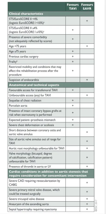
Table 7: Aspects to be considered by the Heart Team for the decision between SAVR and TAVI in patients at increased surgical risk (see Table of Recommendations in section 5.2.)
CABG: coronary artery bypass grafting; CAD: coronary artery disease; EuroSCORE: European System for Cardiac Operative Risk Evaluation; LV: left ventricle; SAVR: surgical aortic valve replacement; STS: Society of Thoracic Surgeons; TAVI: transcatheter aortic valve implantation.
a
STS score (calculator: http://riskcalc.sts.org/stswebriskcalc/#/calculate); EuroSCORE II (calculator: http://www.euroscore.org/calc.html); logistic EuroSCORE I (calculator: http://www.euroscore.org/calcge.html); scores have major limitations for practical use in this setting by insufficiently con-sidering disease severity and not including major risk factors such as frailty, porcelain aorta, chest radiation etc [103]. EuroSCORE I markedly overesti-mates 30-day mortality and should therefore be replaced by the better per-forming EuroSCORE II with this regard; it is nevertheless provided here for comparison as it has been used in many TAVI studies/registries and may still be useful to identify the subgroups of patients for decision between intervention modalities and to predict 1-year mortality.

![Table 9: Echo scores: Wilkins score [145], Cormier score [150], and Echo Score “Revisited” for immediate outcome prediction [146]](https://thumb-eu.123doks.com/thumbv2/123doknet/5437509.127695/26.918.90.845.134.656/table-scores-wilkins-cormier-revisited-immediate-outcome-prediction.webp)

