Eur. J. Biochem. 38,301-306 (1973)
The
Wall Peptidoglycans
of Neisseria perf lava, Moraxella
gl
ucidolytica,
Pseudomonas alculigenes
and
Proteus vulgaris Strain
P
18
Jean-Pierre MARTIN, Jacqueline FLECK, Michele MOCK, and Jean-Marie GHUYSEN Institut de Bactbriologie, Unit6 d'Enseignement et de Recherche de Mkdecine,
Universiti! Louis Pasteur, Strasbourg, and
Service de Microbiologie, Facult6 de Mkdecine, Institut de Botanique, Universit6 de Liege (Received Nay 30, 1973)
The primary structure of the wall peptidoglycans of Neisseria perflava, Moraxella glucidolytim and Pseudomonas alcaligenes is similar to those of the Enterobacteriaceae Proteus vulgaris and
Escherichia coli, suggesting that the peptidoglycans of all gram-negative bacteria contain nzeso-
diaminopimelic acid and belong to the same chemotype I. Differences are observed with regard to the thickness of the peptidoglycan layer in the cell envelopes, the extent of peptide crosslinking and the occurrence of N,O-diacetylmuramic acid residues.
I n contrast to the wall peptidoglycans of the gram-positive bacteria which have been extensively studied, the peptidoglycans of very few gram- negative bacteria have been submitted to thorough structural investigations [1,2]. It is known that, in
Escherichia coli [3,4], the glycan chains are composed of alternating
p-
1,4-linked N-acetylglucosamine and N-acetylmuramic acid residues. The N-acetylmur- amic acid residues are substituted by L-ahnyl-y-D- glutamyl-(L)-meso-diaminopimelyl-(L)-D-alanine pep- tide units and some of these peptides are, in turn, crosslinked through C-terminal D-alanyl-(D)-?neso- diaminopimelic acid linkages (peptidoglycan of chemotype I) [l]. The peptidoglycan of Proteusvulgaris PI8 exhibits the same primary structure [5,6], except that some of the N-acetylmuramic acid residues are 0-acetylated on C6.
I n the course of the present study, the peptido- glycans of three taxonomically different gram-nega- tive bacteria belonging to the genera Neisseria,
Moraxella and Pseudomonas, were isolated and their
structures were compared to that of
P.
vulgaris P18 [61.MATERIALS AND METHODS Strains and Growth Conditions
Neisseria perflava was isolated from a throat
swab, Moraxella glucidolytica from a peritoneal liquid, Pseudomonas alcaligenes from a pus wound and Proteus vulgaris strain P18 from a coproculture 171. The bacteria were grown in a New Brunswick fermentor (72 liters) a t 37 "C with strong aeration.
Moraxella, Neisseria and Pseudomonas were grown
on a Merck Ntihrbouillon Standard I liquid medium for 24 h and Proteus was grown either on the Nahr- bouillon or on a modified Medill liquid medium [8] for 16 h. The cultures of Neisseria, Moraxella and
Pseudomonas were sterilized a t 120 "C for 30 min. Cells were collected with a Sharpless continuous-flow centrifuge and were lyophilized.
Model Compounds
The following compounds were used: the tetra- peptides L-analyl-y-D-glutamyl-(L)-meso-diamino- pimelyl-(~)-~-alanine and L-alanyl-D-isoglutaminyl- (L)-meso-diaminopimelyl-(L)-D-alanine were isolated from walls of Bacillus megaterium [4] and of Clostri- dium perfringens [9], respectively. The disaccharides
,$-1,4-N-acetylglucosaminyl-N-acetylmuramic acid and ,!I - 1,4
-
N-acetylglucosaminyl- N,O-
diacetylmu- ramic acid were isolated from walls of Lactobacillusacidophilus strain 63 AM Gasser [lo].
Detection Reagents
Oligosaccharides from peptidoglycans were de- tected by fluorescence [Ill. Amino acids, free amino groups and peptides were detected with ninhydrin
(0.5O/, in isopropyl alcohol).
Chromatography
The following solvents were used: (I) chloroform- methanol-acetic acid (44:5: 1, v/v/v), (11) methan- ol-pyridine-12 NHC1-water (8: 1 :0.21: 1.79, v/v/- v/v), (111) benzylalcohol- - chloroform-methanol-
302 Wall Peptidoglycans of Gram-Negative Bacteria Eur. J. Biochem. water-ammonia (15 : 15 : 15 : 3 : 1, v/v/v/v/v) and (IV)
1-butanol-acetic acid-water (3: 1 :I, v/v/v).
Analytical Methods
Reducing groups (Park- Johnson procedure), acetamido sugars (Morgan-Elson reaction), hexos- amines (Morgan-Elson reaction after chemical acetyl- ation), amino acids and terminal amino-groups (fluorodinitrobenzene technique) were measured as previously described [ 12,131. Quantitative analysis of amino acids (after 16 h of hydrolysis in 6
N
HC1 a t 105 "C) and of hexosamines (after 4 h of hydrolysis in 3 N HC1 a t 95 "C) were also carried out with the help of an Unichrom (Beckman) amino acid analyzer. The method described in the Beckman technical manual was slightly modified according to Guire [14]. Separation of amino acids and amino sugars was carried out a t 55 "C on one single column (0.9x
50 cm) packed with the Beckman resin type AA-15. Two buffers were used in sequence: 0.2N sodium citrate buffer pH 2.875 and 0.35 N sodium citrate buffer pH 5.25. The buffer change was set a t2 h. Under these conditions, muramic acid, diamino- pimelic acid and glucosamine appeared as well- defined single peaks.
The isomers of diaminopimelic acid were identified as described by Bricas et al. [15]. LL- and meso-di- aminopimelic acid were separated by chromatog- raphy (after hydrolysis in 6
N
HC1 for 16 h a t 120 "C) on Whatman no. 1 paper, in solvent I1 [16]. Didi- nitrophenylated DD- and meso-diaminopimelic acidwere separated and monodinitrophenyl-meso-di- aminopimelic acid was isolated by chromatography on silica-gel (G) thin-layer plates in solvent I11 [I?']. Disaccharides and 0-acetyldisaccharides were separ- ated by chromatography on Whatman no. 1 paper in solvent IV.
Preparation of the Wall Peptidoglycans
Lyophilized cells of Proteus (10 g) were suspended in 1 1 of boiling 4O/, sodium dodecylsulfate solution. The suspension was stirred for 2 h, and kept over- night a t room temperature [19]. The colorless gel obtained after repeated washings and centrifugations (30 min a t 66000
x
g ) was incubated with the Bacillus subtilis protease, in 0.05 M phosphate buffer pH 7.4 [20] for 2-3 h a t 37 "C. Lyophilized cells of Neisseria, Moraxella and Pseudomonas were first disrupted by ultrasonic treatment for 20 min. After removal of intact cells by centrifugation a t 12000x
g for 5 min, the cell envelopes were isolated by different,ial centri- fugation and treated with boiling 4 sodium dode- cylsulfate solution under the same conditions as those described above. All the peptidoglycan materi- als were treated with trypsin (ratio enzyme-sub- strate = 1 :25, w/w) in 0.1 M phosphate buffer pH 8,for 1 to 2 h a t 37 "C, and were washed 5 times with water by centrifugation a t 66 000 x g for 30 min.
Enzymic Degradation of Peptidoglycans
Egg-white lysozyme and Chalaropsis B enzyme
[all are endo-N-acetylmuramidases with hydro- lyse ,& 1,4- N-acetylmuramyl-N-acetylglucosamine linkages. Degradation of the peptidoglycans with lysozyme (grade I Sigma) were performed a t 37 "C in 0.06 M phosphate buffer pH 6.2 (ratio, enzyme- substrate = 1 : 25, w/w). Degradations with Chalarop- sis B enzyme were performed in 0.02 M acetate buffer pH 4.5 (ratio, enzyme-substrate = 1 : 100,
When acting on disaccharide peptide fragments,
Xtreptomyces N-acetylmuramyl-L-alanine amidase hydrolyses the linkages between the N-acetyl- muramic acid residues and the peptide units [22]. Degradations were carried out a t 37 "C in 0.025 M acetate buffer pH 5.4.
w/w).
Gel Filtrations
Fractionations were carried out by filtrations in 0.1 M LiCl, on two columns connected in series, of Sephadex G-50 fine (720 ml) and Sephadex G-25 fine (400 ml). The gel filtration properties of the compounds were expressed in terms of distribution coefficients :
( V e - V,)
K* = Vi
with V , elution volume, V , = Ve of totally excluded
material and Vi = V e - V , where Vi is the value
of V e for NaC1.
Electrophoreses
Electrophoreses were carried out on Whatman no. 3-mm paper using an electrorheophor apparatus (Pherograph-type), a t pH 4 (pyridine-acet,ic acid- water, 2:9: 1000, v/v/v) and a t pH 2 (0.5 N formic acid).
RESULTS
Location of the Wall-Peptidoglycan Layer in the Cell Envelopes
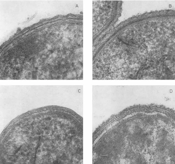
The electron micrographs of thin sections (Fig. 1) of intact cells of Neisseria, Pseudomonas and Moraxel- la showed the multi-layered structure of the cell envelopes. As reported earlier [ 2 3 ] , a similar structure was visible in Proteus only after brief treatment a t high temperature (5 min at 80 "C). According to previous studies, the innermost layer of the cell wall
(i.e. the G, layer according to De Petris' terminology)
[24], directly superimposed upon the three-layered cytoplasmic membrane, is the peptidoglycan. The approximate thicknesses of the peptidoglycan layers
Vo1.38, No.2, 1973 J.-P. MARTIN, J. FLECK, M. MOCK, and J.-M. GHUYSEN 303
Fig. 1. Electron micrographs of thin sections of various gram-negative bacteria (magnification x 240000). (A) Neisseria perjlava, (B) Noraxella glucidolytica, ( C ) Pseudomonas alcaligenes and (D) Proteus vulgaris P18 (treated 5 min at 80 "C)
were 10 nm in Neisseria, 12 nm in Moraxella, 6 nm in Pseudonaonas and 8 nm in Proteus. I n Moraxella,
the thick G, electron-dense layer was surrounded by a wavy layer of homogeneous appearance, whereas in the three other organisms, this outermost layer had a typical membrane structure. The isolated peptidoglycan preparations appeared as homo- geneous electron-dense layers.
Chemical Composition of the Peptidoglycans
The preparations obtained from Moraxella and
Neisseria were pure peptidoglycan materials. They
consisted of muramic acid, glucosamine, alanine, meso-diaminopimelic acid and glutamic acid occur- ring in the molar ratio, 1 : 1 :2 : 1 : I (Table 1). The peptidoglycans of Pseudomonas and Proteus had the same chemical composition but they represented only about 50°/,, dry weight, of the final prepara-
tions. Small amounts of non-peptidoglycan amino acids were also present in these latter preparations. Based 011 these estimations, the poptidoglycan layers
represented about 2.5O/,, dry weight, of the cells of
Moraxella, 0.6O/, of the cells of Neisseria, 0.44O],
of the cells of Pseudomonas and 0.28O/, of the cells of Proteus.
304 Wall Peptidoglycans of Gram-Negative Bacteria Eur. J. Biochem.
Table 1. Chemical comrosition of the peptidoglycans isolated from Neisseria, Moraxella, Pseudomonas and Proteus
N e i s a e T i a Moraxella Pseudomonas Proteus
Constituent
Content Molar ratio Content Molar ratio Content Molar ratio Content Molar ratio
nmol/mg nmol/mg nmol/mg nmolimg
Glutamic acid 980 1 850 0.9 404 1 560 1 acid 990 1 950 1 380 1 530 1 Glucosamine 848 0.8 800 0.8 462 1.1 620 1.1 Muramic acid 720 0.75 820 0.8 430 1.1 590 1.1 AIanine 1900 1.9 1560 1.8 880 2.3 920 1.75 meso- Diaminopimelic
-
c m "2
800 0 E D 700 + a m E 600 & 500.
> ._ a, c-
g 400 0, 2 300 rn c ._ 2 200 T ) B 0 ._ - 0 $ 0 30 60 90 120 150 180 0 30 60 90 120 150 180 T i m e ( m i n ) 0 J ._Fig. 2. Effect of egg-white lysozyme on peptidoglycans of Neisseria, Moraxella, Pseudomonas and Proteus. (A) Liberation of reducing groups (for conditions, see text). (B) Same experiment after prior treatment of the peptidoglycans by 0.02 N
NaOH for 1 h a t 37 O C . Neisseria (o-o), Moraxella (o----o), Pseudomonas (o...~), and Proteus (.-.-.-o)
Degradation of the Peptidoglycans into Disaccharide-Peptide Units
Chalaropsis B endo-N-acetylmuramidase com- pletely solubilized the peptidoglycan preparations. The amounts of disaccharide-peptide units in the degraded peptidoglycan preparations, expressed in nequiv. per mg dry weight were: 950 for Neisseria, 900 for Moraxella, 360 for Pseudomonas and 580 for Proteus. I n contrast to the Charopsis B enzyme, egg-white lysozyme had a very weak lytic effect and exposed reducing groups which representcd only 25O/, to 60°/, of the theoretical amounts (Fig.2). Prior treatment with 0.02 N NaOH for 1 h a t 37 "C (under which conditions 0-acetyl groups, if present, should be hydrolyzed) [IS] considerably increased the lysozyme sensitivity of the peptidoglycan of Mor- axella, but had very little effect on that of the other peptidoglycans (Fig. 2).
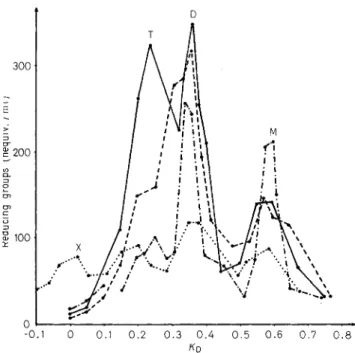
Fractionation of the Degraded Peptidoglycans Filtration of the Chalaropsis-B-degraded peptido- glycans, on two linked SephadexG-50 to 0-25
columns, in 0.1 M LiC1, yielded four fractions (X, T, D and M) from the peptidoglycan of Pseudomonas and three fractions (T, D and M) from the peptido- glycans of Neisseria, Moraxella and Proteus (Fig. 3). All the fractions were separately desalted by filtra- tion in water on Sephadex G-I5 columns.
The various fractions were homogeneous and in- distinguishable from one another by paper electro- phoresis at pH 4. Their chemical compositions were identical to those of the original peptidoglycan prep- arations. Estimation of terminal amino groups revealed that 93 to 98
,Ilo
of the meso-diaminopimelic acid residues in fractions M, 46 to 50°/, in fractions D, 28 to 35O/, in fractions T and 20°/, in fraction X (from Pseudomonas) had one amino group free. These determinations, together with the K D values (Fig. 2 ) allowed to identify the fractions M ( K D = 0.55) asdisaccharide peptide monomers, fractions D ( K D = 0.35) as bisdisaccharide peptide dimers, fractions T ( K D = 0.20) as trisdisaccharide peptide trimers
and fraction
X
(from Pseudomonas; K D = 0 ) prob-ably as a tetramer. Finally, the electrophoretic migrations in 0.2 M formic acid (under which condi-
YO]. 38, NO. 2, 1973 J.-P. MARTIN, J. FLECK, M. MOCK, and J.-M. GRUYSEN 305
tions disaccharide-peptide compounds of various sizes can be separated), confirmed the monomeric and oligomeric structures of the various fractions. The proportions of disaccharide peptide monomer and oligomers present in the various degraded peptido- glygans greatly varied depending upon the bacterial species (Table 2 ) .
Fractions M, D and T showed heterogeneity by chromatography on silica-gel thin-layer plates in
t
0-0.1 0 0.1 0.2 0 . 3 0.4 0 . 5 0 . 6 0.7 0.8
KD
Fig.3. Filtration on Sephadex G-50 to G-25, in 0.1 M LiCl, of
the Chalaropsis-B-degraded peptidoglycans from Neisseria, Moraxella, Pseudomonas and Proteus. Results are expressed in nequiv. reducing groups per ml. Fractions were of 4 ml. For conditions, see text. Neisseria (0-o), Noraxella (0----o), Pseudomonas (o.... . 0 ) , and Proteus (o-.-.-o)
solvent IV (Fig.4). Fractions N gave rise to sub- fractions MI and M, and fractions D to sub-fractions D,, D, and D,. Again, these sub-fractions had the same chemical compositions as the undegraded peptidoglycans. Under these conditions, fractions T and X could not be separated into individual sub- fractions.
Structure of the Disaccharide Units
The disaccharide peptide monomers, M, and M,, were degraded into free disaccharides and free pep- tide units with the help of the Streptomyces N-acetyl- muramyl-L-alanine amidase. The molar absorption coefficient of the free disaccharides (Morgan-Elson reaction; 30 min of heating in l o / , borate) suggested that the glycoside linkages between N-acetyl- glucosamine and N-acetylmuramic acid were
p-
1,4[ill.
The free disaccharides from fractionsMI
and fractions M, had R g values of 0.56 and 0.36, respec-tively (paper chromatography in solvent IV). These
RF
values were those of authenticp-
1,4-N-acetyl- glucosaminyl-N-acetylmuramic acid andp-
1,4-N- acetylglucosaminyl-N,O-diacetylmuramic acid, re- spectively. When treated with 0.02 N NaOH for 1 hTable 2. Proportion of peptide subunits occurring in the form
of monomer, dimer, trimer and tetramer in the Chalaropsis-B- degraded peptidoglycans of Neisseria, Moraxella, Pseudomonas
and Proteus
Form of Peptidoglycans of
subunit Neisseria Moraxella Pseudomonas Proteus
” 0 “lo “ 0 “1. Monomer 21 31 28 22 Dimer 21 53 32 48 Trimer 58 16 24 20 Tetramer 0 0 16 0 X T A B C D E F G H Fractions D Fractions M
Fig.4. Silica-gel thin-layer chromatography in solvent I V (two runs) of the various fractions obtained by filtration on SepharZex
G-50 to G-25 columns (see Fig.3). X = Fraction X from Pseudomonas; T = trimers; A-D = dimers from Neisseria (A),
Noraxella (B), Pseudomonas (C) and Proteus (D); E-H = monomers from Neisssria (E), Moruxella (F), Pseudomonas (G), and Proteus (H)
306 MARTIN, FLICK, MOCK, and GHUYSEN : Wall Peptidoglycans of Gram-Negative Bacteria Ear. J. Biochem.
a t 37 "C, the 0-aceCyldisaccharide from fractions M was converted, a t least partially, into 0-acetyl-free disaccharide.
The bisdisaccharide peptide dimers D,, D, and D, were also treated with the Streptomyces amidase. The two disaccharide residues present in the dimers D, appeared to be 0-acetylated whereas those of dimers D, were free of 0-acetyl substituents. I n dimer D,, one disaccharide residue was 0-acetylated and the other was not.
Identification of the Peptide Monomers The peptide monomers obtained by action of the amidase upon the fractions M were indistinguishable by paper electrophoresis a t pH 4 from the standard peptide L-alanyl-y-D-ghtamy~-(L)-meso-diamino- pimelyl- (L)-D-alanine (Materials and Methods) and were more anionic than the peptide L-alanyl-D-iso- glutamin$- ( L) meso-diaminopimelyl- (L) -D -ahnine, demonstrating that the carboxyl groups of glutamic acid and meso-diaminopimelic acid were not amidated.
COKCLUSI0NS
Insofar as can be judged, the wall peptidoglycans of the four taxonomically different gram-negative bacteria : Moraxella, Neisseria, Pseudomonas and Proteus have identical primary structures (meso-di- aminopimelic-acid-containing peptidoglycans of chemotype I ; see Introduction) [l] and contain 0-acetyl substituents on some of their N-acetyl- muramic acid residues. This structure is also that found in E . coli except that in this latter organism the glycan strands are not substituted by 0-acetyl groups. 0-Acetyl groups occur in the peptidoglycans of some gram-positive bacteria (Staphylococcus aureus and Lactobacillus acidophilus) [ 1,101.
of the dry weight of the cells of Moraxella but less than of the cells of Neisseria, Pseudomonas and Proteus. The thicknesses of the G2 peptidoglycan layers in the cell envelopes also vary according to the bacteria. I n addition, differences were observed with regard to the extent of peptide cross linking. The peptido- glycan of Neisseria was especially highly cross linked, 58 of the peptide units occurring as trimers, 2l0/, as dimers and 2i0/, as uncross-linked mono-
The peptidoglycans represent 2.5
mers. Oligomers higher than dimers had not been detected in the peptidoglycan of E . coli [3,4].
This work was supported in part by grants (to J.M.G.) from the Fonds de la Recherche Fondant entale Collective, Brussels (No 1000), by the Centre National de la Recherche Bcientifique, France, and by the Institut National de la Santd et de la Recherche Mkdicale. France.
REFERENCES
1. Ghuysen, J. M. (1968) Bacteriol. Rev. 32, 435-464. 2. Schleifer, K. H. & Kandler, 0. (1972). Bacteriol. Rev. 36, 3. Weidel, W. & Pelzer, H. (1964) Advan. Enz. 26, 193. 4. Van Heijenoort, J., Elbaz, L., DezBlBe, P., Petit, J. F.,
Bricas, E. & Ghuysen, J. M. (1969) Biochemistry, 8, 207.
407-477.
5. Martin, H. H. (1964) J . Gen. Microbiol. 36, 441. 6. Fleck, J., Mock, M., Minck, R. & Ghuysen, J. N. (1971) 7. Tulasne, R. (1949) C. R. Skances SOC. Biol. Fil. 143, 8. Minck, R., Kirn, A. & Galleron, M. (1957) Ann. Inst. 9. Leyh-Bouille, M., Bonaly, R., Ghuysen, J. M., Tinelli, R. 10. Coyette, J. 8: Ghuysen, J. M. (1970) Biochemistry, 9, 11. Sharon, N. (1964) Proc. Symp. Fleming's Lysozyme 12. Ghuysen, J. M., Tipper, D. J. 85 Strominger, J. L. (1966) 13. Ghuysen, J. M., Bricas, E., Lache, M. & Leyh-Bouille, 14. Guire, P. (1971) Anal. Biochem. 42, 1.
Biochim. Biophys. Acta, 233, 489. 286.
Pasteur (Paris) 92, 138.
& Tipper, D. J. (1970) Biochemistry, 9, 2944. 2935.
( 3 r d ) , Milan, April 3-5, 44/RT. Methods Enzymol. 8, 685. M. (1968) Biochemistry, 7 , 1450.
15. Bricas, E., Ghuysen, J. AT. & DezBNe, P. (1967) Bio- 16. Rhuland, L. E., Work, E., Deunam, R. F. 8: Hoare, 17. Jusic, D., Rov, C.. Schochcr. A. J. & Watson. R. W.
chemistry, 6 , 2598.
D. S. (1955) J . Am. Chem. Soc. 77, 4844. (1963) Can."*J. Biochem. Physiol. 41, 817. Biochem. Biophys. Res. Commun. 42, 4.
18. Arak, Y., Naktani, R., Hayaski, H. & Ito. E. (1971) 19. Braun, V. & Rehn, K. (1969) Eur. J . Biochem. 10, 426. 20. Fleck, J. & Mock, M. (1971) C. R. Rebd. Skances. Ser. D
Sci. Nat. (Paris) Acad. Sci. 272, 1560.
21. Hash, J. N. & Rothlauf, N. V. (1967) Bid. Chem. 242, 853.
22. Ghuysen, J. &I., Dierickx, L., Coyette, J., Leyh-Booille, M., Guinand, M. & Campbell, J. N. (1969) Biochem- istry, 8, 213.
23. Fleck, J. & Mock, M. (1972) Ann. Inst. Pasteur (Paris) 123, 319.
24. De Petris, S. (1967) J . Ultrastruct. Res. 19, 45.
25. Winter, A. J., Katz,W. & Martin, H. H. (1971) Biochim. 26. Heilman, H. Biophys. Acta, 244, D. (1972) 58. Eur. J . Biochem. 31, 456.
J.-P. Martin, J. Fleck, and M. Mock, Institut de Bactbriologie, Virologie et Immunologie GBnbrale, 3 rue KoeberlB, F-67000 Strasbourg, France
J.-M. Ghuysen, Service de Microbiologie, DBpartement de Botanique, Universit6 de Liege au Sart-Tilman, par B-4000 Liege 1, Belgium