1
,
1 1 1 1L
1 1 1)
1
DEFECTIVE EPO PRODUCTION CONTRIBUTES
TO ANEMIA PERSISTENT AFTER ARF
IN NEPHROPATHIA EPIDEMICA (NE)
J. CLEMENT', R. DE BOCK', Y. BEGUIN'
IBelgian Zoonosis Workgroup, Queen Astrid Military Hospital, Bruynstraat, B-1l20 Brussels, Belgium
'Middelheim Hospital Antwerp, Hematology-Oncology, Lindendreef l, B-2020 Anlwerp, Belglum 'Department of Medicine, Hematology, Université du Sart-Tilman, B-4000 Liège, Belgium Key Words: Acute renal failure, anemia, hanta virus, nephropathia epidemica, diffuse intravascular
coagulation, erythropoielin
Running title: DEFECTIVE EPO PRODUCTION CONTRIBUTES TO ANEMIA PERSISTENT AFTER ARF IN NEPHROPATHIA EPIDEMICA (NE)
SUMMARY : NE is an infectious, newly recognized form of acute renal failure (ARF) caused by Hantavirus. In NE cases, anemia is a rare finding of unclear etiology, since NE-induced ARF is most often mild and of short duration, and since hemorrhages are only minor or absent altogether.
We describe here 3 male patients with serologically proven (IFA
IgG
&
ELISA IgM positive) NE, who aIl developed normocyticnormochromic anemia early in theirclinical course, without any evidence of internaI hemorrhage. Analysis of hematological and clotting parameters on admission pointed to a condition
suggestive for microangiopathic hemolytic anemia with or without (subclinical) DIC. Later in the clinical course, measurement of newly established indicators of erythropoiesis disclosed in 2 patients with persistent anemia inappropriately low serum levels of erythropoietin (EPO) and of transferrin receptor (TfR). In contrast, EPO and TfR were within normal range in the patient who showed only a mild and transient degree of anemia.
In conclusion : rapid-onset anemia in the early phase of NE is
consistent with microangiopathic hemolysis. A delayed correction however of this anemia in the convalescent phase of NE seems
linked to defective EPO production, probably as a consequence of (peri)tubular in jury in the kidney, a frequent finding in acute Hantavirus-nephritis.
INTRODUCTION
surprisingly little has been published concerning the
pathophysiology of anemia in acute renal failure (ARF) (1,2). Erythropoiesis is basically regulated by [1] the red cell mass
(trigger), determining in the kidney production of [2]
erythropoietin (stimulator), which in turn stimulates in the bone marrow the [3] erythropoietic activity (effector). These 3
successive steps are assessed by : 1/ hematocrit, 2/ circulating erythropoietin (EPO) levels, 3/ serum levels of transferrine receptor (TfR) , which was recently showed to be a reliable
quantitative assay of total erythropoeisis in animal as weIl as in man (3).
To our knowledge, these 3 parameters were not assessed together so far in cases of anemia linked to ARF.
We were struck by an outspoken and/or prolonged degree of anemia in sorne cases of ARF due to Nephropathia epidemica (NE). This is a. mild European variant of an infectious form of acute interstitial nephritis, caused by a rodent-borne Hemorrhagic fever virus,
called
Hantavirus.
Whereas in the Far East, more severe forms of Hantavirus infections may occur with internaI bleedings,hemorrhages are only minor (epistaxis, petechiae) or absent
altogether in European NE. Thus, anemia in NE is a rare finding of unclear etiology, since NE-induced ARF is most often mild and of short duration. The aim of this paper is to try to elucidate
mechanisms of protracted anemia in 3 recently confirmed NE-cases. SUBJECTS AND METHODS : 1. Subjects
Patients A, Band C are white males aged 24, 26 and 32 years respectively, who each presented with a history of sudden fever, general malaise, outspoken lumbar pain and gastro-intestinal disturbances. A transient ARF developed in each, with spontaneous remission within 2 to 3 weeks. Patient C underwent a percutaneous kidney biopsy, which showed mild interstitial infiltrates and edema without glomerular alterations, i.e. a picture typical for NE. The diagnosis was confirmed in each case by the demonstration in IFA of rising titers of IgG antibodies, and in p-capture ELISA of IgM antibodies against puumala virus, the etiologic agent of NE. Patient A was thoroughly examined on a hematological ward. 2. Methods
*
Erythropoietin (EPO) Assay : Circulating EPO levels were measured by a commercially available RIA (Incstar corp.,Stillwater, Minnesota, USA) using recombinant human Epo (rHu Epo)
*
Transferin Receptor (TfR) Assay : Human placental receptor-transferrin complex was purified and injected repeatedly into rabbits. Rabbit anti-TfR antibodies wpre removed by passingthrough a columm of hum an diferric transferrin coupled to Affigel 15 (Bio-Med, Richmond, CA). These specific anti-TfR Abs were
incorporated in an ELISA assay. (3). RESULTS
1. Assessment of renal
&
hematological evolutionNormocytic normochromic anemia developed in each patient without any evidence of internaI hemorrhage. Renal, hematological and clotting parameters of the 3 patients are summarized on Table l
(only the most abnormal values during the clinical course of each patient are given). Stem cell culture and flow cytometry both of
peripheral blood and of bone marrow were normal in patient A. Bone marrow examination disclosed in each patient an enhanced
megakaryocytosis and a normal (Pat A) or slightly diminished (Pat B) erythropoietic activity. The most severe anemia (Hematocrit 24%, hemoglobin 8.5 g/dl) was noted 25 days after onset of
symptoms (and 10 days after discharge from hospital) in Pat C, at a moment wh en renal function was almost completely restored
(s.Creat down from 12 tot 2 mg
%).
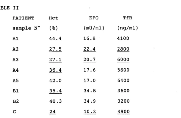
Ambulant gastroscopy, performed to exclude internaI hemorrhage, was found within normal limits. 2. Assessment of erythropoiesis factorsEPO-Ievels and Transferrin receptor (TfR) levels are given on Table II, together with the Hct levels registered the same day.
TABLE l
S.Creat. Ret Plate lets FSP APT'!' Schisto Indir.Bili
mg% % Xl09/l Dg/ml see -cytes % -rubin mg
pat A 3.8 27.1 25 8,000 52.6 3 1.66
pat 8 9.7 30.6 12 2,000 48.6 neg 0.6
pat C 12 24 192 D.d. D.d. D.d. 0.6
Normals <1.2 40-54 >140 "500 24-39 neg <0.8
renal, hematological
&
clotting parameters in Pat A,B,C with NE. n.d. = not doneTABLE II PATIENT Hct EPO TfR sample N° (%) (mU/ml) (ng/ml) Al 44.4 16.8 4100 A2 27.5 22.4 2800 A3 27.1 20.7 6000 A4 36.4 17.6 5600 A5 42.0 17.0 6400 B1 35.4 34.8 3600 B2 40.3 34.9 3200 C 24 10.2 4900
sample numbers denote successive blood samplings during the
clinical course (1 : admission to 4 : discharge). Abnormally low values are underlined.
Lowest "physiological" values
EPO at Hct 37% : 8 and at Hct 27% : 50 mU/ml
TfR at Hct 37 % : 3500 at Hct 27 % :
>
10.000 ng/mlalso lead to micro-angiopathic hemolysis, the signs of which were
found in our patients on admission : thrombocytopenia together with elevated indirect bilirubin and LDH (Table I). A further step in this pathophysiology may ne (subclinical) diffuse intravascular coagulation (DIC), as also found initially in sorne of our patients
(Table I).
However, all these clotting alterations disappeared together with the other inflammatory signs during the clinical course of our patients, whereas low hematocrits persisted, thus excluding micro-angiopathic hemolysis as the only explanation of anemia.
DISCUSSION
With a possible exception for Pat B, the degrees of normocytic
normochromic anemia found in these patients with serologically
proven NE were out of proportion with the degree of ARP and
persisted after restoration to normal or near normal levels of
kidney function. The typical thrombocytopenia, as often found in
the initial phase of
NE,was probably missed in Pat C, who was
hospitalized only 7 days after the onset of symptoms. After
admission however, he developed thrombocytosis up to 553xl0 9/lit,
as often seen as a rebound phenomenon after initial
thrombocytopenia.
The thrombocytopenia is probably the expression of plate let
activation secondary to
endothelial lesions,the most constant
histopathological finding in Hantavirus disease (4,5). This
microvascular damage may be a direct effect of virus replication
in the endothelial cell (5) but immunomediated endotheliai in jury
triggered by viral infection has aiso been proposed (6).
Widespread endotheliai lesions with platelet activation may
clinical course were
inappropriately lowto the degree of anemia
in Pats A
&
C, but only borderline low in Pat B, who showed the
least severe and the most transient degree of anemia. This pattern
of erythropoiesis, consisting of [1] lowHct, [2] low EPO and [3]
low TfR is consistent with the so-called state of
"defective EPOproduction"
as clearly demonstrated so far in chronic renal
failure (3). Low EPO levels, accounting for the slow increase in
hemoglobin following recovery, have been demonstrated in ARF of
varied (septic and non-septic) etiology and are not specifie for
NE.
Peritubular in jury has been proposed as underlying mechanism
CONCLUSION : A transient state of "defective EPO production" as demonstrated in 3 NE patients with ARF can be interpreted as a late consequence of (peri)-tubular injury in the kidney, a frequent finding in acute Hantavirus-nephritis.
REFERENCES
1. Nielsen OJ, Thaysen JH. Erythropoietin deficiency in acute renal failure. Lancet 1989;i:624-5.
2. Lipkin GW, Kendall R, Russon LJ, Turney JH, Norfolk DR, Brownjohn AM. Erythropoietin deficiency in acute renal failure. Nephrol Dial Transplant 1990;5:920-2.
3. Huebers HA, Beguin Y, Pootrakul P, Einspahr D, Finch CA. Intact transferrin receptors in human plasma and their relation to erythropoiesis. Blood 1990;75:102.
4. cosgriff TM. HFRS, four decades research. Ann Intern Med 1989;110(4):313-6.
5. Zhu P, Yang WS. Effects of Hantaan virus on human
endothelial cells and their significance in pathogenesis of hemorrhagic fever with renal syndrome. Chin Med J Engl
1991;104(11):924-9.
6. penseiro MN, Sharefkin JB, Dieffenbach CW, Hay J. Hantaan virus infection of human endothelial cells. J Virol
1992;66(10):5929-36.
Correspondence address
Dr. J. Clement Belgian Zoonosis Workgroup (CTISM) Queen Astrid Military Hospital
Bruynstraat, B-1120 Brussels, Belgium Tel +32/2/268.00.50 Ext 2464 - 2442 Fax +32/2/268.72.49