Université de Montréal
Analyse des mécanismes cellulaires et moléculaires du guidage
axonal sérotoninergique in vitro
par
Bahram Sharif Askari
Département de pathologie et biologie cellulaire
Faculté de médecine
Mémoire présenté à la Faculté des études supérieures
en vue de l1obtention du grade de maîtrise
en pathologie et biologie cellulaires
option système nerveux
Mai 2006
I
J
(J
L
f
n
Université
(III
de Montréal
Direction des bibliothèques
AVIS
L’auteur a autorisé l’Université de Montréal à reproduire et diffuser, en totalité ou en partie, par quelque moyen que ce soit et sur quelque support que ce soit, et exclusivement à des fins non lucratives d’enseignement et de recherche, des copies de ce mémoire ou de cette thèse.
L’auteur et les coauteurs le cas échéant conservent la propriété du droit d’auteur et des droits moraux qui protègent ce document. Ni la thèse ou le mémoire, ni des extraits substantiels de ce document, ne doivent être imprimés ou autrement reproduits sans l’autorisation de l’auteur.
Afin de se conformer à la Loi canadienne sur la protection des renseignements personnels, quelques formulaires secondaires, coordonnées ou signatures intégrées au texte ont pu être enlevés de ce document. Bien que cela ait pu affecter la pagination, il n’y a aucun contenu manquant. NOTICE
The author of this thesis or dissertation has granted a nonexclusive license allowing Université de Montréal to reproduce and publish the document, in
partor in whole, and in any format, solely for noncommercial educational and research purposes.
The author and co-authors if applicable retain copyright ownership and moral rights in this document. Neither the whole thesis or dissertation, nor substantial extracts from it, may be printed or otherwise reproduced without the author’s permission.
In compliance with the Canadian Privacy Act some supporting forms, contact information or signatures may have been removed from the document. While this may affect the document page count, t does flot represent any loss of content from the document.
Université de Montréal
Faculté des études supérieures
Ce mémoire intitulé
Analyse des mécanismes cellulaires et moléculaires du guidage axonal
sérotoninergique in vitro
présenté par:
Bahram Sharif Askari
a été évalué par un
jury
composé des personnes suivantes:
Dr Louis-Éric TRUDEAU
président-rapporteur
Dr Guy DOUCET
directeur de recherche
Dr Timothy KENNEDY
membre du jury
Des travaux antérieurs du laboratoire avaient démontré la capacité des neurones sérotoninergiques (5-HT) à reconnaître des signaux de guidage spécifiques dans des extraits de membranes de diverses régions cibles du cerveau. Les objectifs du présent travail étaient de déterminer la réponse axonale de neurones 5-HT individuels exposés à un choix de substrats appliqués en bandes alternantes d’extraits de membranes du cortex cérébral (Ctx), du striatum (Str) ou du mésencéphale ventral (VM) néonatals et de caractériser ou identifier les molécules contribuant à ce guidage axonal.
Nous avons observé que les axones 5-HT formaient des branchements axonaux de préférence sur des bandes de membranes de la même origine cérébrale que celles où étaient localisés leurs corps cellulaires. Cette observation indique que les axones reconnaissent des
signaux de guidage distincts, selon les régions cérébrales. De plus, il semble qu’il y ait une
induction par contact de la spécificité de reconnaissance axonale, au niveau du corps cellulaire ou de la croissance initiale de l’axone. puisque l’adhérence des corps cellulaires 5-HT sur les membranes extraites de régions cérébrales normalement dénuées de neurones 5-HT, se faisait présumément au hazard. Cette activité a été abolie des membranes du VM par un traitement avec la phospholipase-C spécifique du phosphatidylinositol (PI-PLC), qui défait les ancrages glycosylphosphatidylinositols (GPI), de même que des membranes du Ctx traitées avec une protéine de fusion EphA3-Fc, qui bloque les éphrines-A. Les éphrines A étant les ligands membranaires à ancrage GPI des récepteurs EphA, elle constituent de bons candidats moléculaires pour cette activité de guidage. La poursuite de ces travaux permettra de déterminer si les membranes des autres régions testées perdront aussi leur activité de guidage avec les mêmes traitements et si on pourra modifier la réponse des
11
axones 5.-HT en jouant avec l’expression des récepteurs EphA ou des éphrines-A dans ces neurones.
Mots clés:
Rat, neurobiologie, développement, guidage axonal, raphé dorsal. sérotonine, culture cellulaire neurale, protéines à ancre GPI, éphrines, récepteurs Eph
Résumé anglais:
Previous work in our laboratory bas shown that serotonergic (5-HT) neurones had the capacity to recognize specific guidance signais in cellular membranes extracted from various target brain regions. The objectives of the present work were to determine the axonal response of individual 5-HT neurones challenged wiffi a choice of substrates applied as aitemating stripes made of membrane extracts from the neonatal cerebral cortex (Ctx), striatum (Str), or ventral midbrain, and to characterize or identify ffie molecules involved in this axon guidance activity. We observed that 5-HT axons branched preferentialiy on the membrane stripes of the same cerebral origin as those on which their parent celi bodies were lying. This observation indicates that the axons recognize guidance signais ifiat are brain region-specific. Moreover, it appears that there is a contact induction of the specificity of axonal recognition, at the level of the perikarya or intitial axon growth, since perikaryai adherence to any stripes occurred presumably at random on membranes extracted from target brain regions that are normally devoid of 5-HT neurons.
Ibis axon guidance activity was abolished from VM membranes treated with the phosphatidylinositol-specific phospholipase-C (PI-PLC), winch removes the GPI anchors, as weil as from Ctx membranes treated with the fusion protein EphA3-fc, winch blocks ephrin As. Ephrin-As being the GPI-anchored membrane ligands of EphA receptors, they constitute good candidates for fuis axon guidance activity. Further experiments will teil if the membranes from the other tested brain regions will also lose their axon branching inducing activity foilowing similar treatments and whether one will be able to modify the response of 5-HT axons by changing the expression ofEphAs or ephrin-As in these neurons.
iv
Key words:
Rat, neurobiology, development, axon guid&ice, dorsal raphe, serotonin neurons, fleurai ceÏi culture, GPI-anchored proteins, ephrins, Eph receptors
Table of Contents:
RÉSUMÉFRANÇAIS I
MOTS CLÉS ii
RÉSUMÉ ANGLAIS III
KEYWORDS W
TABLE 0F CONTENTS V
LISI 0F FIGURES t VII
LIST 0F ABBREVIATIONS VIII
DEDICATION IX
ACKNOWLEDGEMENTS X
1. INTRODUCTION I
1.1. FUNCTI0N&i. ROLES 0F THE5-HTSYSTEMS 2
1.2. Organisation ofihe 5-HT systems 5
1.2.1 The raphe nuclei 5
1.2.3 Rostral 5-HTprojections 6
1.2.3.2Projections ofthe caudalraphe nuclei 7
1.3.DEvELOPMENT 0fTHE5-HTSYSTEMS $
1.3.1. Time ofappearance of5-HT neurons 8
1.3.2. Development of5-HT axonat projections 9
1.3.3. Molecutes affecting the growth of5-HT axons 10 1.3.4. Exfracellular mafrix proteins rnd integrins 13
1.3.5. CelÏ adhesion molecutes 13
1.3.6 Trophicfactors 14
1.3.7. Morphogens in axon guidance 15
1.3.8. Netrins and their receptors 17
1.3.9. Siits andRobos 18
1.3.10. Semaphorins and their receplors 19
1.3.11. Ephrins andEph receptors 20
vi
2. MATER1ALS ANDMETHODS .22
2.1. CELLMEMBRANE PREPARATION 22
2.2. ALIERNAnNG MEMBRANE STIUPE PREPARATION 24
2.3.PROEPARATI0N 0f DISSOCIATED CELLS FROM THE FOETAL DORSAL RAPHE 25
2.4. 1MwN0cy’rocf1EvusTRY 26
2.5.IMAGE ANALYSIS AND QUANTIFICATiON 26
3. RESULTS
3.1.EER1MENTAL FRAMEWORK 2$
3.2. SEROT0NERGIC AXONS PREFER MEMBRANE TOPLLSTRWES 28
3.3.AxoN GROWTH ON MEMBRANES FROM DWFERENT BRAINREGIONS 29
3.4.MEMBRANESTREATEDWTTHPI-PLCNO LONGER INDUCE5-HTAXON BRANCH[NG 31 3.5.TRETMENT WflH EPFIA3-Fc ATTENUATES5-HTAXON BRANCHING 32
4. DISCUSSION 45
4.1. SELECTIVE BRM’.JCHII’JG 0F5-HTAXONS IN DISTINCT SUBSTRATE STRIPES 45
4.2. NATURE 0F THE GUIDANCE CUES TNVOLVED IN5-HTAXON BRANCHING 47 4.3.INDUCTION 0F 111E CAPACITY 0F 5-HT AXONS TO RECOGNIZE IRE GU[DANCE CUES 50
4.4.FuluRE EXPERIMENTS 51
5. CONCLUSIONS
RÉFÉRENCES 54
List of
Figures:
FIGURE 1.DissEcTioN 0F Cix, STR ANDVMTISSUE PIECES FROM NEONATAL RAT BRAIN. AFTER PETIT ET AL,
2005(wITH PERMISSION) 23
FIGuRE2.DoRsAL RAPHE NEURONS CULTURED ON STRIPES 0FPLLOR CELL MEMBRANES PREPARED FROM
01f FERENT BRAIN REGIONS 35
FIGuRE3.DoRSAL RAPHE NEURONS ONTO MEMBRANES TREATED WITHPI-PLCOR EPHA3-Fc 36
FIGuRE4.SEROTONERGIC AXON BRANCHING (NUMBER 0F “AXON BRANCHES” AND “AXON LENGTH”), IN STRIPES
0F NEONATAL BRAIN CELL MEMBRANE, ALTERNATING WITH STRIPES 0F POLY-L-LYSFNE(PLL),ACCORDING
TO PARENT CEIL BODY LOCATION IN MEMBRANE ORPLLSTRIPES (“CELL BODY POSITION”, ON THEXAxis).
3$
FIGuRE5. SER0TONERGIC AXON BRANCHING (NuMBER 0F “AXON BRANCHES” AND “AXON LENGTR”), IN
ALTERNAT[NG STRIPES 0F CELL MEMBRANE PREPARED FROM THE NEONATAL CTX, STR ORVM,ACCORDING
TO PARENT CELL BODY LOCATION IN SPECIFIC MEMBRANE STRIPES (“CELL BODY POSITION”, ON THEX
AXIS) 40
FIGURE6.SEROT0NERGIC AXON BRANCHING (NuMBER 0f “AXON BRANCI-IES” AND “AXON LENGTH”), IN
ALTERNATING STRIPES 0F CELL MEMBRANE PREPARED FROM TE-TE NEONATALSTR OR VM,UNTREATED(VM
OR STR) OR TREATED WITH THE ENZYMEPI-PLC (VMPI-PLC) 42
FIGuRE7. SEROT0NERGIcAXON BRANCHING (NuMBER 0F “AXON BRANCHES” AND “AXON LENGTH”), ni
ALTERNATING STRIPES 0F CELL MEMBRANE PREPARED FROM THE NEONATALCTX,UfREATED (CTx) OR
TREATED WITH EITHER TE-lE FUSION PROTEtN EPHA3-FC (CTXEPHA3-FC), OR THE CONTROL FC PROTEIN
VIII
Lïst of Abbreviations:
5-HT: 5-hydroxytryptamine, (sérotonine)
CAM: ceil adhesion molecule (molécule dadhérence cellulaire) CNS : central nervous system (système nerveux central)
Ctx cerebral cortex (cortex cérébral)
ECM : extracellular matrix (matrice extracellulaire) Effi : ephrin (éphrine)
Eph : Eph receptor (récepteur Eph)
IgCAMs : immunoglobulin CAMs (CAM de la superfamille des immunoglobulines) NCAM : neural CAM (CAM neurale)
Str: striatum
VM: ventral midbrain (mésencéphale ventral)
PI-PLC : phosphatidylinositol-specific phospholipase C GPI : glycosylphosphatidylinositol
GDNf : Guai celi line-derived neurotrophic factor TGf-f3 : transforming growth factor-[3
fGf : fibroblast growth factors Wnt $ wingless
Shh : sonic hedgehog Robo : Roundabout
To rny wife, Manzar
X
Acknowledgements
I wish to thank Pr. Guy Doucet for his supervision of this thesis, Audrey Petit for teaching me the ceil culture and sffipe assay techniques, David Bouvier for his help in preparing figures, Moogeh Bahamoori for continuing this project, Miguel Chagnon for statistical analyses, and Michel Lauzon for his help with fluorescence microscopy, confocal microscopy and image analysis. I am also grateful to the Département de pathologie et biologie cellulaire for its financial support and to the Canadian Institutes for Health Research which supported this pro] ect.
The main objective of our iaboratory is to understand the cellular and molecular factors that control the guidance of serotonergic (5-HT, for 5-hydroxytryptamine, serotonin, or serotonergic) axons projecting from the dorsal raphe to the forebrain and midbrain of the developing and aduit rodent.
The laboratory had shown previously that neural grafis from ifie fetal striatum (Str). implanted into the striatum were better innervated by the neonatal or aduit host 5-HT axons than those from the ventral midbrain (VM) (Mounir et al., 1994; Pierret et al., 1998; Doucet & Petit, 2002). furthermore, it was demonstrated that astrocytes present in the grafi, could influence the host-derived 5-HT innervation. Indeed, astrocytes derived from the neonatal cortex (Ctx) or Str could promote the innervation of VM grafis, whereas astrocytes prepared from the VM had no effect on the 5-HT innervation ofVM or Str grafis (Petit et al., 2001).
To fiirther define the molecules involved in ffie guidance of these 5-HT axons, experiments were done in vitro, using fleurai expiant cultures, either in coilagen gel or onto substrates made of cellular membrane from various brain regions (Petit et ai., 2005). This experimentation disciosed several activities associated with ce!! membranes and influencing 5-HT axon growth. first, an axon growth promoting activity was detected in membranes from ail tested CNS regions, including spinal cord, altemating with PLL in the alternating stripe assay. Second, an axon growth inhibitory activity was found by culturing dorsal raphe expiants onto a small carpet of membranes from the Str or VM, adjacent to a second carpet from a different target brain region: 5-HT axons then stopped at the border between the two membrane carpets. Third. it appeared that 5-HT axons are induced to recognize the latter inbibitory factors during their transit on the VM or Str membranes. Indeed, these axons were
2
flot stopped at the border between a Ctx membrane carpet and another, distal carpet from any other brain region. Moreover, treating VM or Str membranes in the proximal carpet, with PI PLC or high KC1, removed this inducing activity in sucli a way that 5-HT axons were no longer inhibited to grow onto a distal carpet of membranes from another brain region. This suggested that the inducing activity was associated with glycosylphosphatydilinositol (GPI) anchored membrane proteins in complex with peripheral proteins bound to the extracellular surface (Petit et al., 2005).
In the present study, we developed another in vitro assay that allowed exarnining the reaction of individual 5-HT neurones, using dissociated neuronal cultures of the foetal dorsal raphe onto altemating stripes of substrates made of membranes from various brain regions, or of poly-L-Iysine.
In the following, I wiIl first describe the context ofthis work by briefly reviewing the functions, organization and development of the 5-HT neuronal systems, I will then review briefly the current knowledge about the major axon guidance molecules.
1.1. Functional roles of the 5-HT systems
Serotonin is found in central nervous system (CNS) neurons as well as in several types of peripheral ceils. In neurons, 5-HT acts mainly as a neurotransmitter, or neuromodulator, but is also known to exert various effects at different developmental stages and in different brain regions, such that it is sometimes difficuit to separate transmitter actions and other types of effect, e.g., neurotrophic. In the CNS, it also influences neural development, as we will see below, including neuronal proliferation, neurite outgrowth and synapse formation. Moreover, 5-HT and 5-HT receptors have been implicated in neuronal
plasticity and memory formation. (Azmitia et al., 1981; Lauder et aL, 1982; Lauder, 1990; Gaspar et aÏ., 2003).
In the mature CNS, 5-HT is involved in several homeostatic functions, such as the central regulation of blood pressure, sodium and glucose balance and body fluid homeostasis (Harding et aï., 2004). It also has an important role in various aspects ofbehaviour including sex, feeding, and affective behaviour, as weYl as depression and anxiety (Harding et al., 2004). Appetite, the sleep-wake cycle and cognition are ail affected by up- or down regulafion of 5-HT secretion (Serrats et al., 2005).
Alterations of the 5-HT systems have been reported in neurodegenerative and psychiatrie disorders, including Mzheimer’ s disease, schizophrenia, autism, depression, as well as in normal aging (Harding et al., 2004). In general, though, the exact contribution of 5-HT, or 5-HT neurons, to pathogenesis remains to be clarified.
The various physiological or pharmacoÏogical effects of the 5-HT systems are mediated by multiple receptor subtypes, classified into 7 groups, named 5-HT1 to -7. This classification is based on thefr amino acid sequence, gene structure (Russo et aï., 2005), pharmacological and functional characteristics (Russo et aÏ., 2005). AIl of the 5-HT receptors belong to the G-protein-coupled receptor family, except 5-HT3 which is unique among 5-HT receptors as a ligand-gated ion channel and ionotropic receptor(Tiemey, 2001; Maxwell et aL, 2003; Russo et al., 2005). These receptors are differentially scattered in the
CNS.
Among the 7 classes of 5-HT receptors, the 5-HT 1 class includes 5 subtypes (lA to
1F) (Lanfumey & Hamon, 2004), concentrated in choroid plexus, dentate gyrus, substantia
4
The 5-HT2 class comprises 3 subtypes: 2A to 2C, with extensive distributions in CNS, except 5-HT2B, which is more restricted in distribution (Leysen, 2004). The 5-HT2B receptors are localized in the olfactory tubercle, layer IV of the neocortex, claustrum, lateral
amygdala, nucleus accumbens and hippocampus (Russo et al., 2005).
The 5-HT3 receptors include two subunits, 3A and 33. They become permeable to Na+ and K+, when activated, and cause membrane depolarisation (Grant, 1995; Tiemey,
2001; Maxwell et aï., 2003).
The 5HT4 class, discovered 15 years ago, only lias one subtype, that may be a target for the treatment of cognitive deficits, feeding disorders and abdominal pain (Baez et al.,
1995; Bockaert et aÏ., 2004).
The 5-HT5 receptors include subtypes SA and 5B. The 5-HT5A receptor was detected in rat, mouse and humari, but 5-HT5B appears to be expressed only in mouse and rat (Nelson, 2004). The expression of the 5-HT5 receptors is essentially restricted to the CNS, with 5-HT5A receptors showing an extensive distribution and 5-HT5B being more restricted (Nelson, 2004). Expression of 5-HT5 in the suprachiasmatic nucleus raises the possibility that they are involved in the regulation of blood circulation (Russo et al., 2005).
The 5-HT6 receptors are newly discovered G-protein-coupled receptors that have a high affinity for a broad spectrum of psychiatric drugs (Woolley et al., 2004; Russo et al., 2005). They are expressed in the CNS and involved in the regulation of glutamatergic and cholinergic neuronal activity, as weIl as in cognition (Woolley et al., 2004).
The 5-HT7 receptors are expressed mainly in thalamus, hypothalamus and hippocampus, with low expression in the amygdala and cerebral cortex (Russo et al., 2005). There are 4 5-HT7 subtypes, 7A to 7D, found in rat tissues (Russo et al., 2005). They have
known functions in peripheral tissue, such as smooth muscle relaxation, alimentary tractus and cardiovascular system (Thomas & Hagan, 2004). They are implicated in reaulation of cicardian pacemaker function, in the suprachiasmatic nucleus, and may play a role in CNS disorders such as cognitive disturbances, anxiety and migraine (Thomas & Hagan, 2004; Russo et al.. 2005).
1.2. Organisation ofthe 5-HT systems 1.2.1 The raphe nuctei
Serotonergic neurons aie located in the raphe nuclei, which constitute a collection of ceil groups along ffie midiine region of the brainstem tegmentum, from the rostral midbrain to the medulla-spinal cord transition, at the level of the pyramidal tract decussation (Steinbusch & Nieuwenhuvs. 1983; Harding et al., 2004). The raphe nuclei comprise the caudal linear nucleus, nucleus raphe dorsalis, median raphe nucleus, nucleus raphe magnus, nucleus raphe obscurus and nucleus raphe pallidus (Steinbusch & Nieuwenhuys, 1983; Harding et al., 2004).
Serotonergic neurons have also been classified into nine cell groups: BI-39, in a caudal to rostral order ($teinbusch & Nieuwenhuys, 1983; Harding et al., 2004). Group BI is located in raphe pallidus, in the caudal medulla; B2, in nucleus raphe obscums; 33, in raphe magnus; 34, in the central grey of the medulia oblongata; 35, in the pontine median raphe; 36 in dorsal raphe; and the 37 and B8 groups in the midbrain dorsal and median raphe nuclei. respectively; 38 also extends into the caudal linear nucleus, while B9 ceils are located in the medial lemrilscus (Steinbusch & Nieuwenhuys, 1983). The caudal linear nucleus is a subnucleus of the ventral mesencephalic tegmentum, iocated dorsal to nucleus
6
interpeduncuaris, on the midline, between the two red nuclei. h is the most rostral raphe nucleus.
Arnong the above nuclei, nucleus raphe dorsalis is the largest one. It is situated in the ventral part of the central grey matter, overlapping the mesencephalon and rostral part of the tegmentum pontis, just ventral to the aquaductus cerebri and rostral part of the fourth ventricle and also dorsal to the fasciculus longitudinalis medialis (Steinbusch & Nieuwenhuys, 1983; Homung, 2003). It includes four areas wiffi particularly high celi density: one in the caudal rhombencephalic part (caudal part) and the other three in the mesencephalic region, designated as dorsomedian, ventromedian and lateral parts. The
dorsomedian part has the highest ceil density (Steinbusch & Nieuwenhuys, 1983). 1.2.3 Rextra! 5-HT projections
Tne raphe nuclei may be roughly divided into rostral (oral) and caudal raphe nuclei, according to their projections. The rostral raphe nuclei have ascending projections to the forebrain, while the caudal raphe nuclei send projections primarily to the lower brainstem and spinal cord (Homung, 2003; Harding et al., 2004).
The rostral groups, B7-B$, include the median raphe, the caudal linear and the dorsal raphe nuclei (Homung, 2003; Harding et al., 2004). These nuclei send their efferent projections essentialiy to the forebrain and upper brainstem (Homung, 2003; Harding et aL. 2004). They grow as a fascicle in the marginal zone. withïn the medial forebrain bundie (Wallace & Lauder, 1992: Rubenstein, 1998). The olfactory bulbs. thalamus, hypothalamus. striatum. septal area, cerebral cortex and hippocampus are the major target structures of ifiese fibers (Harding et ai.. 2004).
In the rat brain, 5-HT neurons are present in large numbers within the dorsal raphe. These neurons give rise to the majority of the ascending 5-HT projections to the forebrain. The caudal extension of the raphe dorsalis, in the pons, constitutes the B6 group of 5-HT neurons, while the larger 37 group is situated in the midbrain periaqueductal gray. Dorsal raphe 5-HT axons project to most regions of forebrain and midbrain (Vertes, 1991; Vertes & Kocsis, 1994). The rostral part of the dorsal raphe nucleus sends significantly denser projections than its caudal part to ail neocortical regions (Vertes, 1991; Vertes & Kocsis, 1994). On the offier hand, the caudal part of the nucleus provides moderately dense projections to the hippocampal formation, which receives no projection from the rostral part (Vertes, 1991; Vertes & Kocsis, 1994). Most midbrain structures, including the ventral midbrain, ventral tegmental area, and the substantia nigra are innervated by the 37 group of 5-HT neurons, winch originate from the dorsal arid median raphe (Vertes, 1991; Harding et al., 2004). The striatum is aiso iimervated by the dorsal and median raphe 5-HT neurons (Vertes, 1991). Many of the 5-HT neurons which project to the striatum send axon collaterals to ifie substantia nigra in the ventral midbrain (van der Kooy & Hattori, 1980).
The dorsal and median raphe nuclei provide dense parallel 5-HT projections to the cerebral cortex (Homung, 2003). Most of these fibers enter the cerebral cortex afler passing through the diagonal bands of Broca and the septal area, while only a fraction of 5-HT fibers enter the telencephalon via the ganglionic eminence (Rubenstein, 1998).
1.2.3.2 Projections ofthe caudal raphe nuctel
Raphe magnus, raphe obscunas and raphe pallidus form the caudal group of 5-HT ceils (Bi-33). These ceils, with a predorninance from group 33, provide the main source of 5-HT innervation to the spinal cord (Harding et al., 2004). AIl the grey matter of the spinal
8
cord contains 5-HT fibres, but higher densities are found in the intermediolateral column, layer 10 (central), layers 1 and 2 ofthe dorsal hom, and motor nuclei (Harding et aL, 2004).
Most parts of the brainstem and cerebellum receive dense 5-HT projections, mainly from group B3, but also from groups B6, B7, 38 and B9 (Harding et al., 2004).
In addition to the caudal brainstem and spinal cord, several hypothalamic nuclei, the central nucleus of amygdala, and the dorsolateral periaqueductal grey are innervated by 5-HT neurons ofthe caudal groups (Harding et al., 2004).
1.3. Development of the 5-HT systems 1.3.1. Time of appearance of 5-HT neurons
The development of the 5-HT system was initialiy examined in 1972 by Oison and Seiger in the rat (Wallace & Lauder, 1992). They provided information regarding the time of appearance (embryonic day, or E; postnatal day, or P) and subsequent development of the dorsal raphe 5-HT neurons and their axona projections.
In rnost species, including rat and mouse, the development of the 5-HT neuronal system begins very early, with 5-HT immunoreactive neurons being visible by E13 in the raphe (Wallace et aL, 1982; Wallace & Lauder, 1983; Wallace, 1992; Gaspar et al., 2003). Serotonin neurons frrst differentiate as two separate clusters: a rostral group oriented just caudal to the mesencephalic flexure and giving risc to most ascending 5-HT fibres, and a caudal group in the medulla oblongata giving rise to most of the descending 5-HT fibres (Wallace et al., 1982; Wallace & Lauder, 1983; Wallace & Lauder, 1992; Gaspar et al., 2003).
1.3.2. Development of 5-HT axonat projections
The ascending projections of the raphe nuclei can be detected as early as E 1 2-E 13 in mouse and E13 in rat, as soon as 5-HT ceil bodies become visible in the rostral rhombencephalon (B4-B9) (Wallace & Lauder, 1983; Lauder. 1990; Wallace, 1992; Gaspar et al., 2003). These fibres branch as they reach the caudal diencephalon, by E14-E15. At tbis time, they spread out in dorsoventral and mediolateral directions and they enter into the interrnediate zone (Lauder, 1990). The number of 5-HT—immunoreactive (5-HT-IR) fibres increases in ifie lateral hypothalamus by fate E15 and early E16. These fibres can be visualised at the border of the diencephalon and telencephalon at E16 and, by E17, this projection has reached the frontal pole of the t&encephalon. A dense plexus of 5-HT-IR fibres exists in flic septal area by E18 and many ofthem pass through this region to reach the neocortex rnedialiy. wbile offier 5-HT fibres traverse the ventral portion of the ganglionic erninence to enter the lateral neocortex (Wallace, 1992).
The first 5-HT fibers appear in developing neocortex by E16-17 in rat and mouse (Janusonis et al., 2004). Such early 5-HT projections to the marginal zone raised the possibility that they may influence cortical development. lie 5-HT innervation of cerebral cortex originates from the dorsal and median raphe (Don et al., 1996; Homung, 2003; Morgane et al., 2005). The dorsal raphe was said to provide fine axons with small varicosities while ifie median raphe would provide beaded axons with large varjcosities. Fine 5-HT terminais abound in various cortical areas, but beaded 5-HT axon terminais reach mainly the outer cortical layers (Morgane et aÏ., 2005). Afler arniving in the cortex, 5-HT axons divide in two tangential sheets, above and below the cortical plate. lien ffiey arborize gradually and send branches to ail cortical layers (Don et al., 1996; Janusonis et al., 2004).
These projections reach the different regions of the neocortex by the end of embryonÏc life, the occipital cortex being reachedjust shortly after birth (Don et al., 1996). The aduit pattem of 5-HT innervation in neocortex is completed by the end ofthe 3rd postnatal week (Don et al., 1996).
1.3.3. Motecutes affecting tlte growth of 5-HT axons
The growth of axons occurs at their tip, involving the growth cone, a structure specialised in sensing the local chemical environment for directed orientation (Tessier Lavigne & Goodman, 1996). In many instances, the axonal growth cone needs to navigate over long distances, along specific pathways to fmd the correct innervation targets. The growth cone is thus a highly motile structure, with a core fihled with microtubules, and peripheral appendices, known as lamellipodia and filopodia. winch are fihled with actin microfflaments. The latter appendices are responsible for sensing and integrating the multiple signais present in its environment. Then, growth cones translate these signais into cytoskeletal changes, winch determine the direction of extension and rate of growth. The growth cone must therefore express receptors for appropniate axon guidance molecules present in its microenvironment.
Growth cone guidance depends on a least 4 different types of extracellular signais: contact attraction (short range, non diffusible molecules), chemo-attraction (long range, diffusible molecuies), contact repulsion (short range, non diffusible) and chemo-repulsion (long range, diffiisible) (Tessier-Lavigne & Goodman, 1996). These mechanisms act in a coordinated manner to direct path±nding. Axons also fasciculate to constitute white matter tracts, Such fasciculation also appears to depend on a balance between attraction and repulsion among axons and wiffi ifie surrounding environrnent.
The above four types of guidance cues have been associated with several families of molecules identified over ifie past 20 years. Interestingly, any given family of mole-cules may be categorised in more than one type of signal (Tessier-Lavigne & Goodman, 1996). Semaphorins, for example may be reieased into the extracellular space and diffuse to form concentration gradients for long range effects. 3ut other members of this family are transmembrane or glycosylphosphatidylinositol- (GPI-) anchored proteins. Neffins are similarly considered as diffiisible proteins, but some GPI-anchored members have been identified recently (Nakashiba et aI., 2000). They must then be considered as short-range cues. Moreover, ifie difffisible semaphorins and netrins have been showu to bind to extraceflular matrix proteins or proteoglycans (Manitt & Kennedy, 2002; Kantor et al., 2004).
ihese mechanisms are mediated by ligand-receptor interactions and depend on the intraceliular signaliing machinery offfie axon (Tessier-Lavigne & Goodman, 1996; Barton et al., 2004). Ihus, the same guidance moiecules may elicit diverse actions, such as attraction, repulsion, STOP signais, or collateral brandi formation (e.g., netrins, ephrins/Eph), depending on ifie sets of receptors that are expressed at the surface of the recipient axon, as well as on the signalIing paffiways that are activated. Furthermore some guidance moiecules, or their receptors, interact wiffi one another to control axonal outgrowth (e.g.: DCC and Robo). For example, the commissural axons of the vertebrate spinal cord are guided by the coordinated action of members of netiin and Shh families (acting as chemoattractants) and members of the sut and semaphorin families (acting as chemorepellents) (Tessier-Lavigne & Goodman, 1996; Barton et ai., 2004).
12
Axon guidance molecules comprise molecules of the extracellular matrix; ceil adhesion molecules (CAM) of the irnmunogÏobulin (1g) or cadherin types; offier types of transmembrane or GPI-anchored proteins. such as epbrins (Effi) and Ephs, semaphorins, or netrins; and diffusible proteins such as netrins, semaphorins, siits, Shh, Wnt, and their respective receptors (integrins, CAM, Eph, Efu, neuropillins/plexins, Robos, etc). Trophic factors, sucli as fibroblast growth factor (FGf), or neurotrophins may also influence axonal branching (Tessier-Lavigne & Goodman, 1996; Barton et al., 2004).
Currently, littie is known about the factors that determine the projections or branching offfie 5-HT axons in any specific part ofthe CNS.
The last decade has seen the discovery of several moiecules affecting axon growth, including secreted proteins such as trophic factors (guai celi line-derived trophic factor or GDNT; neurotrophins, fibrobÏast growth factors, or FGFs: S-1003); morphogens (Shh. wingless/integrated or Wnt), and cytokines (ciliary neurotrophic factor, or CNTF), as well as members of so-called axon guidance molecules, wbich may be secreted (netrins. slits, semaphorins) or membrane anchored (ephrins, semaphorins, celi adhesion molecules, cadherins) (Tessier-Lavigne & Goodman, 1996).
Even 5-HT itself has been dernonstrated to have trophic effects on 5-HT axons, perhaps via an action on glia (Azmitia et al., 1981; Lauder et al., 1983; Lauder, 1990; Gaspar et aL, 2003). The receptor subtypes underlying these effects have not yet been identified, aifflougli some reports have suggested that 5-HT lA receptors might be involved (Whitaker-Azinitia et aI.. 1990).
1.3.4. Extracettutar rnatrixproteins and integrins
Lamiuins, tenascins, coliagens. fibronectin, vitronectin, thrombospondin and several types of proteoglycans are extracellular matrix (ECM) molecules that may promote or inhibit axon growth (Tessier-Lavigne & Goodman, 1996). Their receptors are predominantly members offfie integrin family, but may also include 1g CAIvIs, or proteoglycans.
Integrins belong to a large family of transmembrane glycoproteins, with different heterodimeric Œ/13 subunits, with the f31 integrins representing the largest group, expressed in the nervous tissue by neurones and guai ceils (Tate et al., 2004; Andressen et al., 2005). Most integrins are connected to bundies of actin filaments via molecules such as talin, ti
actinin and filamin. The most important function of integrins is indeed to mediate
cytoskeleton - extracellular mati-ix interactions. There is currently no data about the
contribution of ECM and their receptors in 5-HT axon guidance or growth. 1.3.5. Ceit adhesion motecutes
Cadherin and immunoglobulin CAMs (IgCAMs) are two large familles of ceil adhesion molecules (CAMs) that are involved in axon pathfinding. Both families include over 100 members (Walsh & Doherty, 1997). Most of them serve as ligands as well as receptors (ofien by homophilic interactions), but some of them act as heterophilic ligands or receptors for other ceil-surface or extracellular matrix (ECM) molecules (Tessier-Lavigne & Goodman, 1996). Most of these proteins are transmembrane molecules, but neural CAM (NCAM) exists in 3 forms, including a GPI-anchored version that may also function as a diffusible guidance cue (Walsh & Doherty, 1997).
Several IgCAMs contain a cytoplasmic region with protein tyrosine kinase or protein tyrosine phosphatase domains but most of them do flot. Ceil adhesion molecules not only can
14
mediate celi-celi adhesion in vitro, but aiso have been implicated in axon fasciculation in vivo. Indeed. IgCAMs such as f asciclin II or L1/NgCAM can function to pull axons together. It has been proposed that fasciculation resuits from the relative balance between attractive and inhibitory forces, involving CAMs, among others (Tessier-Lavigne & Goodman, 1996). The expression of polysialyÏated NCAM has been shown to be influenced by 5-HT in aduit rat brain (Brezun & Daszuta, 1999; 2000a b).
1.3.6 Trophicfactors
Guai ceil line-derived neurotrophic factor (GDNF) and neurturin are members of the TGF-13 gene family, with known patterning roles in early stages of development and promotion of neuronal survival (Hynes & Rosenthal, 1999; Zihlmann et al., 2005). These neurotrophic factors act through receptor tyrosine kinase stimulation —GFRŒ and Ret (Mehien. 2005). GDNF is expressed at high levels in the developing rat striaturn and low levels in the ventral midbrain (Zihimann et al., 2005). Several studies have shown that GDNF or neurturin influence dopaminergic and serotonergic neuron survivai and neurite growth in culture (Beck et al., 1996; Schailer et al., 2005; Zihlrnann et ai., 2005; Ducray et aL. 2006). Furthermore, GDNF increases motor neuron suniival during development and following lesion in vitro and in vivo (Beck et al., 1996).
BDNf is a member of the neurotrophin family, which also includes NGF, NT-3 and NT4/5. It interacts with high affmity with the receptor tyrosine kinase, TrkB, which is expressed in 5-HT neurons, notably Rumajogee et al., 2002). Studies have also demonstrated that BDNF can induce sprouting of 5-HT axons in the adult rodent cerebral cortex (Mamounas et al., 1995; Mamounas et al., 2000), or on fetal 5-HT neurons in culture Nishi et al., 2000; Djalali et aL, 2005), Observations in BDNF knockout mice suggest that
BDNF may also influence the development of 5-HT neurones through indirect action onto astrocytes and oligodendrocytes and their respective expression of.S 1 003 and myelin basic protein (Djalali et aL, 2005).
The flbroblast growth factor (FGF) family contairis at least 23 members. found in different multicellular organisms which signal through tyrosine kinase receptors (Cayuso & Marti, 2005; Sanchez-Camacho et al., 2005). The f Gf s are implicated in the induction of posterior neural tissue, and their signais can control 5-HT cell fate in the anterior neurai plate ($anchez-Camacho et al., 2005). fGF8 has midbrain-inducing and polarizing abilities (Sanchez-Camacho et al., 2005). It is expressed in the midbrain-hindbrain boundary and anterior neural ridge, as early as E9 in rat embryo and lias inductive actions for the differentiation of 5-HT neurons by E14 in rat embryo (Ye et aL, 1998; Hynes & RosenthaL 1999). In vitro and in vivo experiments have also revealed that f GF8 influences the guidance oftrochlear axons ont of the neural tube (Sanchez-Camacho et al., 2005).
$100 proteins are members of caicium-binding proteins which play major roles in cellular processes such as inflammation, exocytosis, transcription, proliferation and differentiation (Adami et al.. 2001; Donato, 2001; Marenhoiz et al., 2004). The $100 family includes more than 20 different proteins, which act in tissue-specific or celi-type-specific manmers. Among these, $10013, winch is expressed in astrocytes has been shown to induce axon outgrowtb (Donato, 2001), including in 5-HT neurons (Whitaker-Azmitia et aL, 1990; Nishi et aI., 2000; Nishiyama et al.. 2002; Djalali et ai., 2005).
1.3.7. Morphagens in izxon grddance
Morphogens are secreted extraceflular signalling molecules winch diffuse away from their source, and can induce various cellular responses including important mechanisms
16
underlying the progressive patteming ofthe embryo (Cayuso & Marti, 2005; Mefflen, 2005; Sanchez-Camacho et aI., 2005). The best known morphogens are the hedgehogs (hh), wingless/integrated (Wnt), fibroblast growth factors (FGf s, see Trophic factors) and transforming growth factor-f3s (TGf- [3).
Sonic hedgehog (Shh) is expressed by the notochord and neural floor plate, and is involved in ifie differentiation of the floor plate, motoneurons, and ventral intemeurons (Cayuso & Marti, 2005; Mehlen, 2005; Sanchez-Camacho et al., 2005). It is also involved in the differentiation of dorsal brain structures such as the cerebellum, neocortex, and tectum (Cayuso & Marti, 2005) as well as in the proximo-distal and dorso-ventral patterning of the optic vesicle during eye formation (Sanchez-Camacho et aL, 2005). Serotonergic neurons differentiation is also regulated by $hh (Hendricks et al., 1999; Hynes & Rosenthal, 1999; Ding et al., 2003). Recently, floor plate-derived Shh has been involved in the guidance of spinal cord commissural axons towards the ventral midiine (Charron et al., 2003).
Wnts are a large family of secreted glycoproteins iniplicated in tissue patterning, celi proliferation, and differentiation in several tissues, including nervous tissue (Cayuso & Marti, 2005; Mehien, 2005; Sanchez-Camacho et al., 2005). The Wnts are related to drosophila wingless protein, winch regulates ceil-to-celi interactions. They influence survival of vertebrate neural crest celis and CNS progenitors, during embryogenesis (Cayuso & Marti, 2005; Mefflen, 2005; Sanchez-Camacho et al., 2005). Two different signalling pathways (“Wnt canonical” and Wnt/Ca2± pathway) are activated following binding of Wnts to their frizzled receptors, winch are G-protein-coupled receptors (Sanchez-Camacho et al., 2005). The growth of commissural axons, in fly as well as in rodents also involves Wnt signalling (Sanchez-Camacho et al., 2005). In particular, Wnt-5 has a direct role as a
repellent axon guidance cue for anterior commissural axons, whereas Wnt-4 is a guidance eue required for the anterior growth of commissural axons (Sanchez-Camacho et al., 2005). 1.3.8. Netrins and their receptors
Netrins constitute a small family of 6 members first identified as axon guidance eues in vertebrates. The netrins gene family includes four members in mammals: netrin-1, -3, -Gi and -4 or B-netrin (Nakashiba et al., 2000). They are srnall proteins (—600 amino acids), related to the much larger laminins (Manitt & Keimedy, 2002). Most netrins are secreted proteins, which direct ceil migration and axonal growth cone guidance, during neural development. Recently, however, new members with a GPI-anchor, netrin-Gi, with 6 isofoms (netiin-G la-f) and netrin—G2 wiffi 3 isofonns (netrin-G2a-c), have been identified (Nakashiba et al., 2000; Lin et ai., 2003; Barallobre et al., 2005). These netrin-G proteins, expressed as early as El 2 in several areas of ifie CNS, reach ffieir highest level of expression at the perinatal stage (Barallobre et al., 2005).
Netrin-l and -2 were first identified in chicken as proteins expressed by floor plate celis and having an important role in the guidance of spinal cord commissural axons (Kennedy et ai, 1994). Netrin-3 has different properties, and it expressed in motor, sensory and sympathetic neurons during peripheral nervous system development. It is expressed also in mesenchymal and muscle celis (Barallobre et aL. 2005). Members of this family were later found to be multifunctional in axon guidance, attracting some axons and repelling others (Tessier-Lavigne & Goodman, 1996; Manitt & Keimedy, 2002).
Netiins have a homologue, U}.JC-6, in the nematode C. elegans, for which receptors had already been identified: UNC-40 and UNC-5. UNC-40 tumed out to have an already known homologue in mammals, Deleted in Colorectal Cancer (DCC), and anoffier that was
18
recently identified, neogenin, which are the receptors mediating the allraction elicited by netflns. UNC-5 homologues, UNC5H-1 and -2 have also been found in vertebrates and are mediating the repulsive effects of netrins. However. mediation of this repulsive action depends on the co-expression of UNC-40 (Tessier-Lavigne & Goodman, 1996; Dickson, 2002).
There is currently no available information on the expression of netrins in 5-Fff fleurons. Preliminary work by A. Petit did flot detect DCC in 5-HT neuron (Petit, Kennedy, and Doucet, unpublished).
1.3.9. Stits and Bobos
Roundabout (Robo) was first identified through gefletic screening in drosophila as the receptor for a midiine repellent suspected to be responsible for midline axon guidance defects (Dickson, 2002). The repellent was found to be Sut, a large secreted protein. Several Slit and Robo homologues ($lit-1 to -3; Robo-l to -3) have later been identified in vertebrates (Dickson, 2002). Siits are involved in several functions, such as stimulating sensory axon branching and elongation, but midiine guidance in drosophila and formation of the optic cbiasm in vertebrates are their best known fimctions (Dickson, 2002). Siits are expressed by midiine floor plate celis, whereas Robos are expressed by longitudinal axons, preventing ipsilateral axons from crossing, and commissural axons from re-crossing, the midline.
Inactivation of Siits was shown to affect major 5-HT projections in Slit-1 and Sut-2 mutant animais where a significant percentage of 5-HT fibres abnormally crossed the midiine in the basal telencephalon (Bagri et al., 2002). Up to now, Sut- 1 and Sut-2 are
only axon guidance molecules to have been involved in the guidance of ascending 5-HT axons (Bagri et al., 2002; Mclness & Michaud. 2005).
1.3.10. Semaphorins and their receptors
Semaphorins are a large family of celi surface and secreted guidance molecules which function as axon chemorepellent or growth inhibitors. The family is defined by the presence of a conserved 420 amino acid sema domain at the NH2 terminal. They are divided into 8 classes, including secreted and celi surface —transmembrane or GPI-anchored
- members (Barton et al., 2004). Some of these classes are found in invertebrates (classes 1
& 2) while others are found in vertebrates (classes 3-7), and one (class V) is encoded by viruses (lessier-Lavigne & Goodman. 1996; Dickson. 2002). Approximately 20 members of the semaphorin family have been identified in higher vertebrates, among winch one third are secreted molecules, one is attached to the ceil surface via a OPI-anchor, and the remaining ones are trans-membrane molecules (Tamagnmie & Comoglio, 2004). One of the trans membrane and most of the secreted semaphorins function in repelling specific subset of axons in culture, while two of the secreted ones can act either as axon repellents or attractants (Tessier-Lavigne & Goodman, 1996; Dickson, 2002).
Plexins are ifie primary receptors of semaphorins. Many, and perhaps ail. semaphorin receptor complexes include a plexin. Many transmembrane semaphorins bind dfrectly to plexins, but secreted semaphorins (class 3), in vertebrates, bind to a second class of receptor component named neuropilins ÇDickson, 2002; Tamagnone & Comoglio. 2004). Plexins are a large famïly of trans-membrane proteins, grouped in 4 subfamilies, plexin-A to -D. on the basis of sequence similarit (Dickson. 2002: Swiercz et al., 2002).
20
Ihere is cm-rently no available information on ifie expression of semaphorins or their receptors in 5-HT neurons or on their possible roÏe in the guidance of 5-HT axons during development.
1.3.11. Ephrins and Eph receptors
Ephrins (Effi) were discovered, in 1994 as important regulators of the retinotopic projections onto the vertebrate visual tectum (Cheng & Flanagan, 1994; Drescher et al., 1995). Ibis family of membrane-bound guidance molecules was classified in two major classes according to the type of insertion in ifie ceil membrane, a GPI- anchor for EfnAs and a transmembrane domain for EfnBs (Eph Nomenclature Committee, 1997). Ephrin-As and EfnBs respectiveÏy interact with EphA and EphB tyrosine kinase receptors. Currently, 9 Efiis and 15 Eph receptors have been identified in mammals (Murai & Pasquale, 2003; Martinez & Soriano, 2005). The 9 mammalian EphA receptors bind to 5 EfnAs, with a variable affmity, while the 6 EphBs do similarly with the 3 EfnBs. However, this general mIe has a few exceptions: EphA4 being able to bind both EthAs and EfnBs, and EthA5 recognizing EphB2 in addition to EphAs (Huot, 2004; Surawska et al., 2004).
Ephrins and Ephs are involved in several neural developmental processes, including topographie mapping, brain commissure formation and axon giiidance (Martinez & Soriano, 2005). EphA5 and EfnA5 are expressed in dopaminergic neurons in the ventral midbrain, suggesting their involvement in the regulation of substantia nigra-striatum interactions (Halladay et aL, 2004).
Analyses of dominant negative EphÂ5 transgenic mice suggested ifiat titis receptor might be involved in 5-HT axon growth, since 5-HT levels were decreased in these mice (Halladay et aI, 2004). The latter observations are interesting in the contexi of our study,
wbich suggests that EfnAs influence 5-HT axon branching in vitro, on membranes from the cerebral cortex, striatum or ventral midbrain. At least sorne EphA receptors (EphA5, EphA7) are expressed in the dorsal raphe (Maisonpierre et al., 1993) (seewww.genepaint.org).
1.4. Objectives of the present work
Our hypothesis is that 5-HT axon guidance molecules are associated with celi membranes in 5-HT innervation target fields. Our objectives were to test ffie capacity of individual 5-HT neurons to recognÏze guidance cues present in distinct target brain regions and to identify molecules involved in these processes. As will be described, mir work has fiilfilled ail 3 objectives, at least partially: we have found that 5-HT axons can discriminate membranes extracted from ifie neonatal cerebral cortex (Ctx), striatum (Str) and ventral midbrain (VM). Moreover, we observed that the choice of a membrane substrate by 5-HT axons is detennined by the position of the ceil body on the same type of membranes; strongly suggesting an induction of guidance cue recognition. Lastly, our experiments indicate that ephrin-As are involved in 5-HT axon branchîng, at ieast on membranes extracted from the cerebral cortex.
2. Materials and Methods
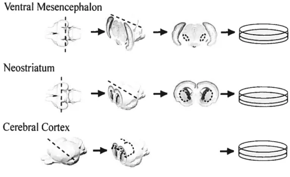
2.1. Celi membrane preparation
Ceil membranes were prepared as described by (Petit et al., 2005), and modified from (Waiter et al., 1987). Newborn (P0) female Sprague-Dawiey rats were quickly decapitated with sharp scissors, and blocks of Ctx, VM or $tr (Figurel) were dissected in Gey’s Balanced Sait Solution (GBSS; Sigma G-9779) supplemented with glucose (6.5mg/ml; Sigma G-7021).
The pia matter was removed and suces were eut with a sharpened tungsten needle (unsheaffied, tungsten-rhenium, Omega Engineering mc, WW 26-020). Tungsten needles were sharpened by immersing the wfre in NaOH (iN) and applying a voltage of 4-5 V). The dissected tissues were transferred into 1.5 ml centrifuge tubes (Coming) containing 1 ml H
buffer which includes 1OmM fris-HCL (Sigma T3253) pH 7.4, 1.5 mM CaCI2 and 1 mM Spermidin plus 40 pi protease inhibitor (Protease inhibitor, complete EDTA-free; Roche no.
Ventral Mesencephalon
:
N eostri atum
a:z
CerebraÏ Cortex
-FIGuRE 1. DISSECTION 0F CTx, STR ANDVMTISSUE PIECES FROM NEONATAL RAT BRAIN. AFTER PETIT ET AL.,2005 (wITH PERMISSION).
Tissues were homogenized by several strokes through P1000 and P200 pipettes and then a 1 ml syringe, until ail tissue debris had disappeared. Homogenization was continued in an ultrasound bath (Branson 2200) during 5-10 min. The homogenate was centrifuged during 20 min at 50 000 g, at 40
C, in a sucrose step gradient (300 jil sucrose 5 % in upper phase and 700 jil sucrose 50 % in iower phase; Sigma no. S-5390, in 5 ml centrifuge tubes; Beckman) using an ultracentrifuge (Sorval Ultra pro 80; rotor AH650). The inter-band, containing the celi membrane fraction was colÏected and washed twice in 1 ml sodium phosphate buffered saline (PBS, iX, pH 7.2) and centrifiiged for 10 min at 16 000 g (Eppendorf Model 4515C centrifuge), at 4°C. The pellet was re-suspended in 1 ml PBS and the concentration of purified membrane was determined by optical density (OD) at 220 nm in a spectrophotometer (Ultraspec 2100 pro). The concentration was then adjusted with PBS
24
glycerol (50 % PBS- 50 % glycerol; F isher no. G33-1) to a standard level yielding an OD of 1.0.
At this point, membranes could be treated with PI-PLC or EphA3-Fc. For PI-PLC, 10 L of PI-PLC (1 U/mL, Sigma-Aldrich) were added to 500 tL of membrane preparation from neonatal VM during 1 h, at 37°C (Petit et al., 2005). The membranes were then rinsed and re-suspended at the same OD. Neurons were cultured on stripes of PI-PLC-treated VM membranes altemating with stripes of untreated VM or Str membranes.
for EphA3-fc treatment (Janis etal., 1999), 200 ng ofthe protein (R&D) were added to 500 .tL of membrane suspension from neonatal Ctx. As controls, other Ctx membranes were treated with fc alone (Jackson ImmunoResearch Laboratories; 420 ng in 500 tI of membrane suspension). The membranes were rinsed and re-suspended, as above. Neurons were then cultured onto stripes of EpbA3-Fc, or fc, altemating with stripes of untreated membranes from Ctx.
2.2. Alternatmg membrane stripe preparation
Altemating stripes of membrane substrates were prepared as described (Canson et al., 1987; Petit et al., 2005). A 100 t1 drop ofpurified membrane was first mixed wiffi 3 pi of a suspension of blue fluorescent polymer micro spheres (1:825; B500; Duke Scientific, Palo Alto, CA). The labelled membrane suspension was applied to a polycarbonate nucleopore filter (piore size 0.1 jlm; Whatman), deposited on top of a silicone matrix with 90 jim-wide parallel channels (Jurgen Jung, Max-?lank Institute, Tuebingen, Germany). The labelled membrane was aspirated across the filter by applying a negative pressure (-0.8 mbar) in the underlying channels with a vacuum pump (model DAA- V175-EB; GA$T, Benton Harbor, MI), until the filter stripes above the channels were saturated.
for preparation of membrane stripes altemating with poly-L-lysine (PLL), these
stripes were printed onto coverslips pre-coated with poly-L-lysine (PLL; 20ig/ml, Sigma; P.2636). for altemating stripes of different membranes preparations, the filter with saturated stripes was transferred onto a nylon grid matrix (fine mesh, provided by J. Jung) and then a 100 pi droplet of a secondtype of membrane suspension was applied and aspirated as above, to fil the non-saturated stripes of the filter. These altemating membrane stripes were printed onto PLL pre-coated coverslips. These coverslips were kept at 37 oc until used, on the same day.
2.3. Preparation of dissociated ceils from the foetal dorsal raphe
Rat foetuses (E14-E15) were dissected in HBS$ iX (Gibco: 14180-053 or 14170-112) supplemented with 1% HEPES solution (Gibco; 156030-80 or 156030-106). Explants of the dorsal raphe were transferred onto a 15 ml centrifuge tube containing 4.5 ml HBS$/HEPES; (1.0 ml HEPES in 100 ml Hank’s solution), supplemented with 500 pi
trypsin (2.5 %) and incubated at 37°c for 15-20 min. The tissue was re-suspended every 5 min. with a Pasteur pipette (under the culture hood).
Afler dissociation, the suspension was supplemented with 5 ml Neurobasal complete Medium and 1.0 ml Heat-Inactivated Horse Senim (HIHS), and was centrifliged during 10 min at 2 000 g (centra cL2, 15 ml tube). The supematant was discarded and the pellet re suspended 500 pi of Neurobasal Complete medium. Celis were counted in a 10 jil sample using a hmocytometer. The dissociated dorsal raphe neurons were plated (‘—1 8 000/cm2)
ontothemembrane stipe substrates and incubated in Neurobasal Complete medium for 72 h
26
2.4. Immunocytochemistry
Cultures were fixed in 4 % paraformaldehyde in 0.1 M sodium phosphate buffer (PB, pH 7.4) for 1 h at room temperature. They were pre-incubated in PBS containing 10 % bovine serum albumin (Sigma), 1 % normal goat serum (Jackson), 0,05 % -1 % Triton X100, for 3-4 h, at room temperature. They were incubated during 16 h with a polyclonal antïserum against 5-HT (1:5 000, Diasorin, Stiliwater, IVIN) and a monoclonal antibody against ‘3m-tubulin (1:200; Sigma), to visualize, respectively, 5-HT and ail neurons in the cultures. Afier primary incubation, coverslips were rinsed in PBS and incubated with a rhodamine— conjugated goat anti-rabbit IgG and a fluorescein (FITC)-conjugated, affmity-purified fab fragment of goat anti-mouse IgG (1:200; Jackson ImmunoResearch Laboratories, West Grove, PA).
2.5. Image analysis ami quantification
The immuno-labefling was examined and photographed under a Zeiss Axiophot, fluorescence microscope connected to a digital camera (Retiga 1300; Q-imaging; Canada) and anaiysed with the Northem Eclipse 6.0 software (Empix Imaging, Toronto, ON).
The following measurements were performed on the cultures. First, the number of neurons immunopositive for 5-HT or for 131i1-tubulin was counted in each type of stripe. The number of neurites emanating from the ceil bodies was also registered. Lastly, for each neuron, the length of axon and the number of collateral branches in each type of stripe was scored. according to the position of the parent 5-HT or iii-tubulin ceil body in a specific type of stripe. Ail 5-HT neurons in every culture were analysed in this way. Since the number of 13;ji-tubulin-labelled neurons was much higher than 5-HT neurons, 20 3111-tubulin-positive; 5-HT-negative neurons were chosen at random in every culture for analysis [every
neuron out of x ÷20; “x” being the total number of such neurons in the culture]. for this
thesis, only the resuits on 5-HT axon branching will be presented.
Experiments were repeated at least 3 times. for each type of altemating substrates, the data comparing the neurons between the two sets of stripes were analysed using an ANOVA with repeated measures (SPSS 13.0; done by Dr. Miguel Chagnon, Département de mathématiques et statistiques. Université de Montréal).
3. Results
3.1. Experimental framework
We tested the response of foetal rat dorsal raphe neurons (E14-E1 5) challenged with stripes of celi membranes prepared from the neonatal cerebral cortex (Ctx), striatum ($tr), or ventral midbrain (VM), altemating with stripes of poly-L-lysine (PLL), or of membranes prepared from a different neonatal brain region. $ince these experiments demonstrated that 5-HT axons were able to discriminate among the different types of stripes, we submitted the membrane preparations to treatments with phosphatidylinositol-specific phospholipase C
(PI-PLC) or with a fusion protein comprising the extracellular domain of EphA3 and the fc
fragment of human immunoglobulin-G (EphA3-Fc), in order to respectively remove ail GPI anchored membrane proteins, or to block EfnAs.
The resuits presented below represent the number of axon branches (or collaterals) and the total length of axon (including ail branches) in each type of membrane or PLL stripes for every individual neuron located in a given type of stripe. These two measurements of axonal innervation gave very similar results.
3.2. Serotonergic axons preftr membrane to PLL strïpes
Examples of non 5-HT and 5-HT neurons cultured onto altemating stripes of membranes and PLL are illustrated in Figure 2A,A’. In general, both 5-HT and non-5-HT perikarya were more numerous on membrane than PLL stripes.
Non 5-HT neurons (13111-tubulin+; 5 HT) made few branches and had relatively short axons that tended to remain in the same stripe as the parent celi bodies (Fig. 2A). In contrast,
5-HT neurons had long axons that could cross several stripes and made several branches,
The resuits also indicated that 5-HT axons preferred membrane to PLL stripes. Indeed, these axons branched clearly more often in Ctx, Str or VM membrane stripes than PLL sffipes, even when the ccli bodies were located in PLL stripes (Fig. 4). When the
perikarya were in Ctx stripes (n = 1$ 5-HT neurons), the number of branches in the Ctx
stripes was nearly 7 times larger than in PLL stripes (Fig.4A, lefi side), and the total length of individual axons 4 times larger than in PLL stripes (F ig.4B, lefi side). When the 5-HT perikarya were in a PLL stripe (n 9), their axons stilipreferredthe Ctx membrane stripes, but produced only 2.6 times more branches in the membrane than PLL stripes (Fig. 4A,B, right side). However, the interaction between the location of the ceil body and the axon growth onto a specific type of stripe was flot significant (p=0.054).
Similar results were obtained with Str or VM membrane vs PLL stripes (Fig.4C-F). For 5-HT perikarya located in $tr stripes (n= 14), there were nearly $ times more 5-HT axon
branches in Str than PLL stripes, whereas those located in PLL stripes (n 10) gave 2.5
times more branches in Str than PLL stripes. for 5-HT perikarya located in VM stripes (n = 17), the number of branches was nearly $ times higher in VM than PLL stripes, compared to 6 times higher for those located in PLL stripes (n = 7). In both cases, the total length of axon branches showed the same trend.
Thus, whatever the location of the 5-HT perikarya, thefr axons clearlypreferred to grow onto a ceil membrane substrate than onto PLL, which is recognized as an axon growth permissive substrate.
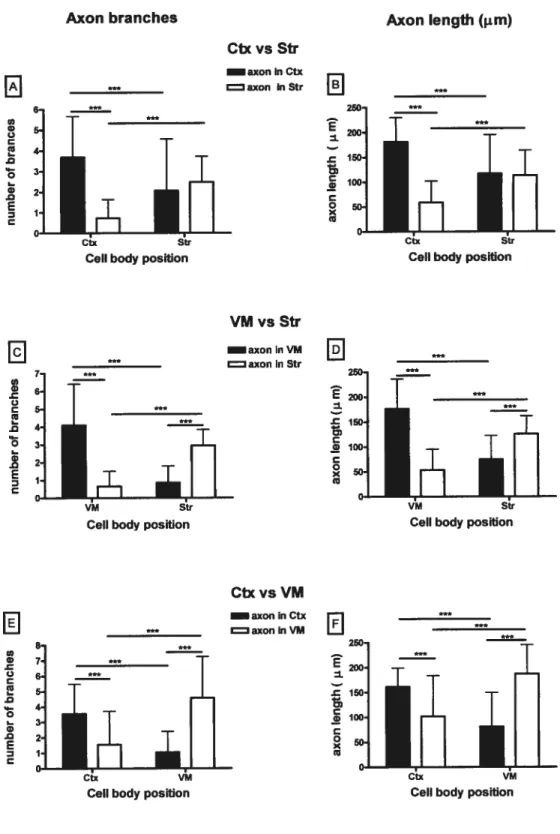
3.3. Axon growth on membranes from different brain regions
When challenged with altemating stripes of membranes from either Ctx, $tr or VM, 5-HT axons branched and grew preferentially in other stripes made with the same type of
30
membranes as those where their parent celi body was located (fig. 2B,B’). Non 5-HT neurons stiil made fewbranches and had short axons.
In ahemating membrane stripes of Ctx and Str. when the celi body was located in Ctx stripes (n=36), the number and total Ïength of 5-HT axon branches were, respectively, 5
and 3 times higher in Ctx than Str stripes (fig. 5AB, lefi side). However. 5-HT axons
showed no stripe preference when their celi bodies were located in Str membrane stripes (n = 29) (fig. 5A,B, right side). There is a significant interaction between the location of the perikarya in Ctx or $tr membrane stripes, and 5-HT axon branching in specific stripes (p<O.00l).
With dorsal raphe neurons cultured on alternating VM and Str stripes (fig. 5C,D), there was also a significant interaction between the location ofthe perikarya and the number and length of axon branches in any given type of stripes (p<O.00I). When the 5-HT ceil
bodies were located in VM stripes (n = 41). the number and total length of axon branches
were 6 and 3 times larger. respectively in VM than Str stripes (fig. 5C.D, lefi side). When
the perikarya were in $tr stripes (n = 22), the number and length of 5-HT axonal branches
were 3 and 1.7 time larger in $tr than VM stripes (fig.5C,D, right side).
With altemating stripes of VM and Ctx membrane, there was, again a significant interaction between ccli body location and number/length of 5-HT axonal branches in each type of stripes (Fig. 5 E,F). With 5-HT perikarya in Ctx stripes (n = 25), axons had 2.3 times more branches that were 1.6 times longer in Ctx than VM stripes; whereas those in VM stripes (n= 33) had 4 times more branches, 2.3 time longer in VM than Ctx stripes.
Thus, except for 5-HT neurons in Str stripes altemafing with Ctx stripes, 5-HT axons always branched preferentially on the saine type of membranes as those bearing their parent
perikarya. It is noteworthy that the large majority of these axons branched in the same type of membranes, but in different stripes than their celi body. They therefore had the choice to branch in similar or different types of membrane stripes.
3.4. Membranes treated with PI-PLC no longer induce 5-HT axon branching
The molecules involved in the guidance or branching of5-HT axons are presumed to
be proteins (Petit et al., 2005). Three major modes of protein association exist with celi membranes: transmembrane, GPI-anchored and peripheral proteins. The enzyme PI-PLC is known to remove the GPI-anchored proteins (Walter et al., 1987). To gain some insight into the molecules involved in the above phenomena, we first examined the effects of membranes treated with P1-PLC on axon branching.
for the present thesis, we could only test this treatment with VM membranes. Our resuits show that, with altemating stripes of VM membranes that were either treated or untreated with PI-PLC, 5-HT axons branched almost exclusively onto the untreated membrane stripes, whatever the location of their parent perikarya on treated or unfreated
membrane stripes (fig. 6A,B, n 17 and 14 5-HT neurons in VM and VMp.pLc stripes,
respectively.). Similar resuits were obtained with stripes of membranes from $tr altemating with VM membranes treated with PI-PLC, where branching occurred almost exclusively in untreated Str stripes (figs. 3A,A’, 6C,D, n 4 in Str and 6 inVMpp stripes).
The effect of PI-PLC was probably stronger than it appears here, since it also affected branching in untreated VM stipes. Indeed, there was a significant difference in the number
and length of branches in untreated VM stripes in these experiments compared to ail other
experiments with VM stripes (2.59 axon branches and a length of 142 .tm in VM altemating with VMPIpLC compared to 4.01-4.58 and 177-192 im in VM stripes altemating with Ctx,
32
Str or PLL). Perhaps some of the enzyme remained in the treated membranes when the stripes were prepared, and diffused to “untreated” stripes. A more likely explanation might 5e that axons growing on treated stripes were affected in some way by PI-PLC-treated membranes.
In the experiments where VMpI.pLc altered with Str stripes, branching in the latter was flot affected: the number of branches in $tr stripes, in other experiments, was 2.48-2.95
and their length varied between 96 and 139 11m, with flO significant difference between
experiments.
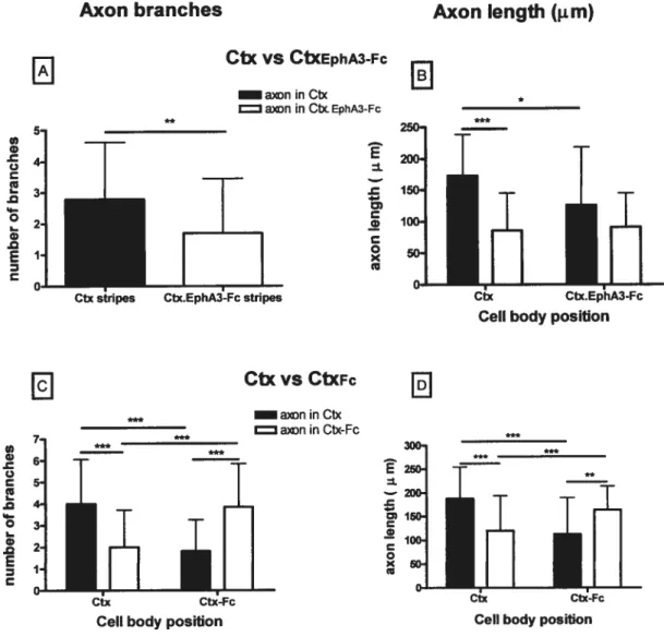
3.5. Treatment with EphA3-Fc affenuates 5-HT axon branchmg
Since epbrin-As are GPI-anchored proteins involved in axon branching (Gao et al.,
1999; Mann et aL, 2002), we next tested membranes treated with EphA3-Fc, a blocker of
ephrin-As. Up to now, we have only tested this treatment on membranes extracted from Ctx
(Fig. 3B.B’). With EphA3-fc-treated Ctx membranes altemating with stripes of untreated Ctx membranes 5-HT axons made 1.8 times more branches in untreated than EphA3-Fc-treated membrane stripes, whatever the location of their parent perikarya in unEphA3-Fc-treated (n = 34) or treated (n= 26) stripes (Fig. 7A). However, the number of branches made in untreated membrane stripes tended to be lower than in untreated Ctx membrane siripes in ail other experiments (2.79 vs 3.56-4.0; pO.O78); winch might indicate that the fusion protein, EphA3-fe diffused to “untreated” stripes, or that 5-HT axons were affected by their contact wiffi EphA3-Fc-treated membranes, even when they finally branched in untreated stripes.
On the other hand, the total length of 5-HT branches per axon, in treated versus
untreated membrane stripes, showed an interaction with ceil body location (p = 0.02 1).
stripes was twice that in EphA3-Fc-treated Ctx stripes (Fig. 73). The length of ffiese axon branches was also 1.4 times that of those in Ctx stripes, when the perilcarya were in CtxE
Fc stripes. The length of branches in EphA3-fc-treated stripes was comparable, whatever the
location of the perikarya. When the perikarya were in CtxE1F stripes, the length of their
axon branches showed no significant difference in Ctx vs CtxEt1F stnpes.
In suminary, the reduction in 5-HT axon branching in membranes treated wiffi EphA3-Fc was flot as strong as wiffi the PI-PLC treatment, but it was significant.
Control treatment of Ctx membranes with Fc alone had no effect on the number or length of 5-HT axon branches. Indeed, the number of branches and their total length was comparable (3.85 in Ctxf vs 4.00 in Ctx stripes) to that found in Ctx stripes in ail other experiments (Figs. 3C,C’ and 7C,D). However, there was an interaction between the number and length of branches and the stripe location of the parent perikarya (p<O.001). Indeed, axons from perikarya located in Ctx stripes branched in Ctx stripes, whule those from perilcarya located in CtxF stripes branched in this type of stripes. However, this interaction is due to a phenomenon different from experiments with untreated membranes stripes from different brain regions: the large majority of 5-HT axons branched in the same stripe as that oftheir parent ceil body, when stripes were treated with Fc. Thus, the growth ofthe axon out of both fc-treated and untreated was apparently hindered, but the axon branching was not affected, even in Fc-treated stripes, at variance wiffi EphA3-Fc-treated membranes. Future experiments with membranes from other brain regions treated with EphA3-Fc (or other EphA-Fc fusion proteins) or wiffi Fc alone will teil whether this effect of Fc will 5e con±ïrmed.
34
Nevertheless, we conclude that 5-HT axons branching is at least partly regulated by ephrin-As, in membranes extracted from the cerebral cortex.
FIGuRE2.DoRsAL RAPHE NEURONS CULTURED ON STRIPES 0FPLLOR CELL MEMBRANES
PREPARED FROM DIFFERENT BRAIN REGIONS
A,A’) Altemating stripes ofPLL and membranes prepared from the neonatal cerebral cortex. The membranes were labelled with fluorescent microbeads, which can be seen in A, together with 3j11-tubulin immunostaining. In A’, only 5-HT neurons are visualized. B,B’) Altemating stripes of membrane preparations from the striatum (Str) and ventral midbrain (VM), with microbeads and 111-tubu1in immunostaining being visible in B and 5-HT immunostaining in B’. Note that the 5-HT neuron, in B’ branches in a different Str stripe than the parent ce!! body, after crossing a VM stripe without branching.
36 VMç-pic VMic Str Ctx/[pIA3-Fc CtX/[pI,A3Ic Ctx Ctx Ctx/EphAi-Fc Ctx/EphAJ-Fc
fiiuRE3. DoRsALRAPHE NELTRONSONTOMEMBRANES TREATEDWITHPI-PLCOREPHA3-fc
A,A’) Altemating stripes of VM membranes treated with PI-PLC, and untreated Str membranes (labelled with fluorescent beads (A, beads and Fliti-tubulin immunostaining are shown; A’, 5-HI neurons). B,B’) Stripes of cerebral cortex (Ctx) membranes treated or not with EphA3-Fc. Note that a 5-HT neuron located in a treated Ctx membrane stripe branches
in an untreated Ctx membrane stripe. C,C’) $liipes of Ctx membranes treated or not with Fc, as control for experiment illustrated in B,B’. Treatment with Fc alone had no effect on the branching choices of 5-HT axons.
3$
Axon branches
Axon Iength (jm)
CtxvsPLL
9 350 ‘ni — a)n in Clx w 6-4 aoninPLL E 300 . I u 7j 250 c 61__
200 .0 5-1ri
• 4-I
_____
if’1
j’1
5
150 .- I 3-I 100 .0 I E 2j 0 50 z i] I________
Ceil body position Celi body position
VMvsPLL
8 300 ‘n ‘ 250]____
- 7__
____
. I 6__
200-) 150.1IrIi
lE,
w anin PLL 12f ____________ • 100-l o I sol z 1 o’ VM PLL VM P[LCeli body position Celi body position
6
StrvsPLL
6
_
5 *** 200, ‘n I axoninStr.5
C41
I c I L — I .0 I___
Iru,ÏH
.- I ‘ 1001 axoninPLL2
2-’ .l w I C I .0 O 50-I E 1-i z I__________
xl
C___________
___________ Str PLL Str P.LCelibody position Celi body position
FIGuRE4. SER0T0NERGIc AXON BRANCHING (NUMBER 0f “AXON BRANCHES” AND “AXON
LENGTH”), IN STRIPES 0f NEONATAL BRAIN CELL MEMBRANE, ALTERNATING WITH STRIPES 0F
POLY-L-LYS[NE(PLL),ACCORDING TO PARENT CELL BODY LOCATION IN MEMBRANE ORPLL
Stripes of membranes from the frontal cerebral cortex (Ctx, A.B), from the ventral midbrain (VM, CD), or from the striatum (Str, E,F), altemating with stripes of PLL, according ceil body location. Filled bars indicate branching in respective membrane stripes
and white bars indicate branching in PLE stripes. ANOVA with repeated measures; *


![FIGuRE 6. SER0T0NERGIc AXON BRANCHING (NUMBER 0f “AXON BRANCHES” AN]) “AXON](https://thumb-eu.123doks.com/thumbv2/123doknet/2051001.5384/56.918.168.790.152.609/figure-nergic-axon-branching-number-axon-branches-axon.webp)