Research Report
Pelvic-Floor Rehabilitation, Part 1: Comparison of
Two Surface Electrode Placements During
Stimulation of the Pelvic-Floor Musculature in
Women Who Are Continent Using Bipolar
Interferential Currents
Background and Purpose. Electrical stimulation of the pelvic jloor is used as an adjzmct in the conserva! ive treatment of urinal)' incontinence. No com·en sus exists, however, regarding electrode placements for optimal stimulation of the pelvic floor musculature. The purpose of this study was to compare two dijjèrent hipolar electrode placements, one suggested by Laycock and Green (L2) the other by Dumoulin (D2), during electrical stimulation with inter:feren tial currents
Q/
the pell•icfloor musculature in continent women, using a Iwo group crossover design. Subjects. Ten continent female volunteers, ranging in age from 20 to 39 years (X=27.3, 5D= 5.6), were randomly assigned ta one of two study groups. Methods. Bach s/u{{Y group received neuromuscular e!ectri cal stimulation (NMES) of the pelvic oor musculature using bath electrode placements, the arder of application heing reversed for each group. Force of contraction was measured as pressure (in centime/ers of water [cm H20}) ex erted on a vaginal pressure probe attached to a manometer. Data were ana!yzed using a twoway, mixedmodel analysis of variance. Results. No differ ence in pressure was observed between the two electrode placements. D[fferences in current amplitude were observed, with the D2 electrode placement requiring Jess current amplitude to produce a maximum recorded pressure on the ma nome/er. Subjective assessment by the subjects revealed a preference for the D2 electrode placement (7 of 10 subject:..). Conclusion and Discussion. Tbe lower current amplitudes required with the D2 placement ta obtain recordings com parable to those ohtained with the 12 technique suggest a more comjortab!e stimulation of the pelvic oor muscles. Tbe lower current amplitudes required also suggest that greater increases in pressure might he ohtained with the D2 placement by increasing the current amplitude white remaining within the comfort threshold. Tbese results will help to defi ne treatment guide/ines for a planned clinicat study investigating the ejfècts of NMES and exercise in the treatment of urinary stress incontinence in women postpartum. [Dumoulin C, Seahorne DE, QuirionDeGirardi C, Sullivan SJ. PelvicJloor rehabilitation, part
1: compan·son of two swface electrode placements dun'ng stimulation Q/ the peluic oor musculature in women who are continent using bipolar inter:feren
tial currents. Phys Tber. 1995; 75:106710 74.}
Key Words: Bipolar technique, Electrode position, Inteiferential currents, Pelvic oor electrostimulation, vaginal pressure probe.
Physical Therapy 1 Volume 75, Number 12/ December 1995
Chantale Dumoulin Derek E Seabome Cécile Quirion- DeGirardi S John Sullivan 1067/35
Urinary continence is the capacity to retain urine in the bladder between two voluntary micturitions. 1 Inconti nence is the involuntary Joss of urine, which can be demonstrated and which presents a social and hygienic problem. 2
Genuine urinary stress incontinence (GSI) results from urethral sphincter incompetence and is defined by the International Continence Society as "the involuntary Joss of urine occur ring when, in the absence of a detm sor contraction, intravesical pressure exceeds the maximal urethral pres sure."2 Genuine stress incontinence is the most common form of urinary incontinence, with an estimated 78% of cases of GSI being related to preg nancy and the birth process. 2 Persans with this condition experience inconti nence of urine when the intra abdominal pressure is raised, for ex ample, during coughing, sneezing, or any form of physical activity that in creases intraabdominal pressure ..l Neuromuscular electrical stimulation (NMES) has been shown to be effec tive in the treatment of GSI. Stimula tion via the pudendal nerve, at fre quencies of 20 to 50 Hz, improves urethral closure by activating the pelvicfloor musculature. 4 In addition, NMES can increase conscious aware ness of the action of these muscles, thus facilitating the ability to perform a voluntary muscle contraction." Severa} methods of stimulating the pelvicfloor
muscles have been described, includ ing the use of bath lowfrequency faradic currents6·7 and medium
frequency interferential currents.Kw The use of mediumfrequency interfer ential currents has been suggested as a means of overcoming the problem of stimulating deepseated stmctures more effectively, without using inva sive methods. The capacitive compo nent (reactance) of tissue resistance has been hypothesized to decrease inversely with the currcnt frequency. 11 By decreasing the reactance, the over all tissue resistance will diminish, thereby facilitating the stimulation of cleep stmctures.12
Regardless of the method used, the localization of the stimulating elec trodes is of critical importance in ob taining a maximal contraction. The intensity of an electrically induced muscle contraction is directly relatee! to the number of motor units activat ed.13 The number of motor units acti vated, in tum, is influencee! by the current amplitude and frequency and the placement of the stimulating electrodes. 13
In 1988, Laycock and Green 14 com pared different electrode placements during stimulation with interferential currents of the pelvicfloor muscles of female subjects. Using vaginally lo cated sensors, they measured peak currents and peak pressures evoked in the perivaginal tissues, as weil as tis sue resistance, for each of three elec
trode placements during stimulation of the pelvic floor. They concluded that a bipolar electrode placement, with one electrode placed between the ischial tuberosities (over the anus) and the other electrode placee! over the ante nor perineum, inferior to the pubic symphysis, produced an equally effec tive stimulation of the pelvic floor, as comparee! with a quadripolar elec trode placement. They recommended the bipolar placement, basee! on its ease of application. 14
Electrical stimulation at current intensi ties necessary to produce adequate muscle contractions can result in un pleasant or painful sensations. Be cause patient discomfort is otl:en the limiting factor during NMES,l'' this discomfort can reduce the effective ness of the treatments. In our clinical practice, we have utilized the bipolar technique suggested by Laycock and Green 14 for treating women with post
partum GSI. During stimulation, sorne of our patients have complained of intense discomfort due to high current concentration under the anterior electrode (in the region of the cli toris). We have therefore suggestecl an alternative electrode placement for the anterior electrode, to a position imme diately superior to the pubic symphy sis. We postulate thar this modifiee! electrode placement will decrease the discomfort and increase the efficacy of NMES of the pelvic floor.
The suggested alternative position produces a current spread estimated from anatomical measures to be 2 C Dumoulin, MSc, PT, b Physical Therapist, Hôpital SteJustine de Montrée1l, 3175 Côte Ste slightly greater than the 140 cm (ap Catherine, Montréal. Québec, Canada H3T !CS, and Teaching Assistant and Lecturer, L'Ecole de
Réadaptation, Faculté de Médecine, Université de Montréal, Montréal, Québec, Ce1nada H3C 3]7. Address al! correspondence to Ms Dumoulin at the second address.
DE Seaborne. MSc, PT, is Professor, Depe1rtment of Physiotherapy, L'Ecole de Réadaptation, Fac ulté de Médecine, Université de Montréal.
C QuirionDeGirardi, MA, PT. is Associate Professor (ret), L'Ecole de Réadaptation, Faculté de Médecine. Université de Montréal.
SJ Sullivan. PhD, is Associate Professor and Chair, Department of Exercise Science, Concordia University, Montréal, Québec, Canada H4B 1R6, and is affiliated with the Centre de Recherche, Institut de Réadaptation de Montréal, 6300 Darlington Ave, Montréal. Québec, Canada H3S 2]4, and L'Ecole de Réadaptation, Faculté de Médecine, Université de Montréal.
This study was approved by the Ethics Committee of Faculté de Médecine, Université de Montréal.
This article zms submitted Octoher 5. 1994, and was accepted Allf!.USI 15, 1995
proximate) reported by Laycock and Green. 14 The direction of current flow
follows closely that of the bipolar electrode placement suggested by Laycock and Green.14 By displacing
the anterior electrode to a point supe rior to the pubic symphysis, however, the current will theoretically penetrate deeper within the pelvis. 11 To avoid confusion between these two tech niques, for the purpose of this study only, we have reclassified the bipolar female electrode placement suggested
Subject No. Age {y) Height (m) Weight (kg) BMia (kg/m2 )
influenced our findings. Ail our sub jects had a BMI below 27. Persons
24 1.70 65.8 23.0 having a BMI greater than 27 are con
2 39 1.62 60.0 23.0
3 25 1.68 61.2 21.5 Instrumentation
4 25 1.57 52.1 21.0
5 24 1.68 53.5 19.0 The electrical stimulator used during
6 26 1.68 56.7 20.0 this study was an Endomed 433
7 20 1.60 51.3 20.5 mediumfrequency interferential cur
8 37 1.63 56.7 21.5 rent stimulator* with a medium
9 26 1.52 49.9 21.5 frequency output of either 2 or 4 kHz.
10 27 1.70 72.6 25.0 According to the manufacturer's speci
x
27.3 1.64 58.0 21.6 fications, this stimulator has an ampliSD 5.6 0.06 7.1 1.7 tude modulation frequency spectrum
1. Su!Jject Characteristics
and is derived from the formula: wt (kg)/ht (m2
). I3ecause fat hasan elec trical impedance of hetween 1,000 and 3,000 O!cm2 , 1 obesity could have sideree! dinically obese. 1il
"BMI=body mass index.
hy Laycock and Green 14 as L2 and the
alternative electrode placement as 02. The purpose of our stuùy was to com pare the two different electrode place ments (L2 and 02) in the stimulation of the pelvicfloor musculature, using hipolar interferential currents, to deter mine which of the two methods pro duœd a stronger contraction with the Iowest current amplitude. The force of contraction of the pelvicfloor muscu lature was measured indirectly as pressure (in centimeters of water [cm H2 0]) registered on a manometer attached to a vaginal pressure probe. We expected that 02 would be the more effective of the two electrode placements. The results ohtained from this study helped to determine treal ment guidelines for a clinical study of the effects of noninvasive electrical stimulation of the pelvicfloor muscu lature in women with postpartum urinary stress incontinence (see our companion article in this issue).
Method Subjects
Ten continent women aged between 20 and 39 years (X=27.3, 50=5.6), who were recruited from a population of clinicians and graduate and under graduate university students, volun teered as subjects for this study. Ail subjects demonstrated the ability to perform a voluntary pelvicfloor con traction. None of the subjects had any previous history of urinary inconti nence or any neuromuscular injury likely to influence our results. Ail subjects were nulliparous, and during the period of data acquisition, none were menstruating or had an intrauter ine deviee implanted. Descriptive statistics are shawn in Table 1. Before participating in the study, ali volun teers signed an approved informed consent form.
Age, weight (wt), and height (ht) were recorded and body mass index (BMI) was computed for each subject. Body mass index 16 is a measure of obesity
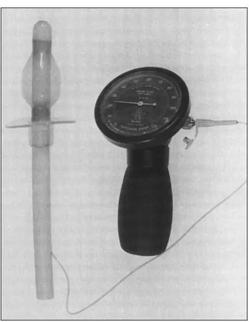
(interference frequency) continuously adjustable between 0 and 100Hz. A bipolar application implies thar the two medium frequencies are super posed within the stimulator and ap plied directly as an interferential cm rent at the preselected frequency. The force of contraction of the pelvicfloor musculature elicited by the stimulation was measured indirectly as pressure On centimeters of water [cm H20]) registerecl on a manornetert attachee! to a vaginal pressure probe.* The pressuresensitive manometer used in this study was capable of detecting and measuring changes in perivaginal pressure resulting from contractions of the pelvicfloor muscles. Prior to the experiment, the manometer was ex aminee! and tested by the hioengineer ing department of a major Montréal teaching hospital (Hôpital SteJustine de Montréal). which reported a high leve! of reliahility for this instrument. Both the manometer and vaginal probe are illustrated in Figure 1. Experimental Design
Our experimental design was a two group crossover design, with ali sub jects receiving stimulation with the two different electrode placements. To reduce any experimental effect result
ing from the orcier of stimulation, the
•EnrafNonius Delft, f:quipement de Physiothérapie P Gélinas Ltée CP68, Suce ''D," Montréal Qué
bec, Canada H3K 3R9. '
tMed0Gen !ne, Hll MetropolitainE, Montréal, Québec, Canada HlR 1Z7. 1Ponex Ltd, Hythe, Kent, England CT21 6jL
Physical Therapy 1Volume 75, Number 121 Oecember 1995
10 subjects were randomly assignee! to one of two groups (n=S per group) prior to the experiment. Each subject selected 1 of a series of 10 sealed envelopes containing an equal num
Figure 1. Pressuresensitive manometer attached to the uaginal pressure probe.
treatment table, with the knees and hips supported in flexion at approxi mately 70 degrees and bath hips in lateral (external) rotation. 14 This posi
tion facilitated the positioning of the electrodes, encouraged relaxation, and
reducecl intraabdominal pressure, Hl
which coule! register on the manome ter and thereby influence the results of the study.
Pelvic-floor assessment. The pelvic floor assessment was performecl by a physical therapist (CD), using a vagi nal examination technique clescribed
by Chiarelli and O'Keefe. 18 The thera
pist wore disposable, sterile surgical latex gloves. After palpating the me dial fibers of the subject's pubococcy geus muscle with the index finger, she instructed the subject to contract her pelvicfloor musculature to more accu rately identify its precise location.
Accorcling to Bo et al,19 the center of
pelvicfloor activity is located in an area approximately 3.5 cm from the introitus.
The disposable vaginal pressure probe was acljusted corresponding to the depth of each subject's musculature, as determinee! by vaginal palpation. Following instructions given by the therapist, the subject then inserted the probe herself, using a sterile water soluble jelly as a lubricating medium.§ The probe was then attachee! to a manometer. The subject was required to squeeze on the probe by contract ing her pelvicfloor muscles. At the same time, the therapist completed the ber of odd and even numbers. The 5
subjects who selected an envelope containing an even number began the experiment with the L2 electrode placement, followed by the 02 elec trode placement. The 5 subjects select ing an envelope with an odd number were treated in the reverse arder. Procedure
Detailed explanations were given to each subject regarding the aims of the study, the equipment to be used, and the techniques. The subject was al
lowed to test the effects of the stimu lating current on an exposed portion of her forearm, using a remote current amplitude control. It has been our experience thar patients will tolerate higher current amplitudes if the cur rent is selfregulated. Following this initial briefing, the subject was re quired to perform her perineal toiler using soap and water, in an adjoining private washroom. She was then in structed to disrobe the lower part of her body and assume a semisupine position (tnmk inclined at 50° from the horizontal) on a padded wooden
adjustment of the probe position to a site where maximum pressure was obtained, as indicated by the manometer.
Electrode placement and stimula- tion sequence. The electrodes were placed in position in the predeter minee! arder. The posterior (6X8cm) carbonsilicone electrode,* enclosed in a cellulose sponge pad* moistened with warm tap water, was placee! directly over the subject's anal region. The anterior electrode (4X6 cm), similarly enclosecl, was placee! in the median plane, immediately inferior to
the pubic symphysis for the L2 tech
!MUKO Luhricating ]elly, Ingram & Bell Medical, Don Mills, Ontario, Canada M3B 1L9. nique, or immediately superior to the
2. Maximum Prl!ssure Ohtained With Neummuscu!ar Electrica! Stimulation"
Order
Maximum Pressure (cm H20)
Subject No. L2 02
maximum intensity 20 Wedensky inhi bition is the state of incomplete repo larization of a nerve fiher when the nerve is stimulatec.l with a high intcnsity, mcdiumfrequcncy ( 2,000 Hz) current. Complete repolarization
can only occur if the current intensity
is reduccd periodically. 21 To limit L2/D2 D2/L2 2 2.5 10.0 4 2.0 3.0 6 2.0 3.0 8 3.0 5.0 10 40 4.0
x
2.7 5.0 SD 0.8 2.9 1 4.5 5.0 3 20 3.0 5 16.0 12.0 7 4.5 4.0 9 5.0 6.0x
6.4 6.0 SD 5.5 3.5 Grand X 4.6 5.5 Grand SD 4.2 3.1muscle fatigue, a 2minute rest period separated stimulatecl contractions and a 1)minute rest pl:'riod separated the two dectrode placement techniques. During this 15minute period, the electrodes werc removed and the pacls were moistened prior to being secured in the alternative position. At the same time, the vaginal probe was checked to ensure that its position remained unchanged. Of the three contractions elicited in each electrode placement, the strongest contraction was retained for use in the statistical analysis. All readings were verified hy two re scarchers, neither of whom was masked.
"Suh1ects with even numhers were treated with electrode placement suggcsted by Laycock ancl Green (L2) followecl hy electrode placement suggested by Dumoulin (])2); subjects with odd numbers weœ tested in the reverse order.
Data Analysis
Descriptive statistics were calculated for maximum pressure and current puhic symphysis for the D2 technique.
The electrodes were secured in posi tion by rne ms of a perforated ruhber band! passing between the legs of the subject < nd attachee! anteriorly and posteriorly to insulated metal rings on a lumbar traction belt* securecl around the suhject's waist. To avoid any possi bility of cross infection between sub jects, we used only new cellulose electrode envelopes. which were changed for each subject. The carbon silicone electrodes and leads were cleaned with alcohol and the pedo rated mhber band was disinfected in Cidexll solution following each application.
An amplitudemodulated medium frequem:y (AMF) cu1rent of 10Hz and a base frequcncy (carrier frequency) of 2 kHz were used throughout the stucly as the stimulating current. To achieve this AMF cutTent, two separate
mediumfrequency cwrents, one of 2,000 Hz and the other of 2,010 Hz, are superposee! within the stimulator. The result is an AMF current rising and falling in amplitude 10 times per second. The effect on the tissues is that of a lowfrequency stimulating current of 10 Hz. When muscle tissue is stimulated via the nerve at this fre quency, the result is a subtetanic mus cle contraction.
Three muscle contractions were elic ited wîth each electrode placement. U.;ing the remote control, the suhject, under the supervision of the therapist, increased the current amplitude gradu ally to a leve! of maximum tolerance, remaining at this leve! for 3 seconds. Maximum tolerance was defined as a point just below the pain threshold. To reduce the possibility of any \XTe densky inhibition, 30 seconds only was allowed for each subject to reach
amplitude. In addition. hoth maximum pressure and current amplitude were analyzed using a twoway, mixed model (one hetweengroup factor and one withinsubject factor) analysis of variance (ANOVA), 22 the betwcen
group factor heing the orùer of stimu lation (L2 folluwed by D2 or D2 fol lowed by L2) and the withinsubject factor being the electrode placement
(L2 and 02). The ANOVA procedures
were perfonned using the SYSTAT statistical package:" A probability leve! of :S:.05 was adoptee! for ali statistical tests.
Results
Descriptive statistics for maximum pressure and current amplitude are presented in Tables 2 and 3. No differ ence was ohserved betwcen the maxi mum pressures ohtained for both the L2 and D2 electrode placements
(P= .34), nor was there any effect due
to either the orcier of application or the interaction bctwecn the order and IIJohmon & Johnson Medical Products, Peterborough, Ontario, Can<tda K9J 7B9.
"SYSTAT !ne, !HOO Sherman Ave, Evanston, IL 60201.
Physical Therapy 1 Volume 75, Number 12/ December 1995
the electrode placement (Tah. 4). 1071 /39
Order 1 344.45 344.45 0.32 .59 electrodes and the motor nerve of a muscle will affect this impedance, thereby influencing the density of
Error 8 8686.60 1085.83
Placement 2668.05 2668.05 17.81 .003
Orderx placement 378.45 378.45 2.53 .151 current at the target site and thus the
Error 8 1199.00 149.88 quality of the muscle contraction. The
Current Amplitude Paired With Jvfaximum Pressure Generated hr Neuro
muscular Electrical Stimulation"
Current Amplitude (mA)
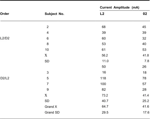
In contrast, the mean CUITent ampli tude required to produce the maxi mum recorded pressure was found to be less (.F=17.81, P<.003) with the D2 placement (41.6 mA) th an that re quired with the L2 placement (64.7
Order Subject No. L2 02 rnA). No elfects duc to the order of
presentation or the interaction be
tween order and placement were L2/D2 D2/L2 2 68 45 4 39 39 6 60 32 8 53 40 10 61 53
x
56.2 41.8 SD 11.0 7.8 50 26 3 16 18 5 118 78 7 100 57 9 82 28x
73.2 41.4 SD 40.7 25.2 Grand X 64.7 41.6 Grand SD 29.5 17.6observed (Tab. 5). These results sug gest that the D2 placement was capa ble of producing a contraction of comparable magnitude at a reduced CUITent amplitude. At the end of the session, when asked which technique they prefcrred, 7 of the 10 women indicated a preference for the D2 electrode placement.
Discussion
The objective of this study was to determine the most effective of two electrode placements in stimulating the pelvicfloor musculature in conti nent, nulliparous womcn. The results showed that bath electrode place "Suhject with en n numbers werc treJtecJ with electrode placement suggeslccl by Laycock and
Green (L2l followed lw eleurode placement suggestcd hy Dumoulin (D2); subjects with oclcJ ments achieved contractions of com parable force, as measured by the
were treatcd rn the reverse orcler. manometer. The current amplitude
Table 4. Ana(rsisC?fVariance Summary for Maximum Prt>ssun! OIJtai11ed WitiJ Neuromuscular Hlectrical Stimulation
Source df
ss
MS F prequired to achieve the contractions, however, was lower with the D2 elec trode placement than with the L2 electrode placement. Based on these results, we concluded that D2 was the more effective electrode placement for these subjects. The subjects' prefer ence for this electrode placement, Order Error 8 Placement Order x placement 1 Error 8 27.61 27.61 1.22 .30 180.75 22.60 4.51 4.51 1.35 .28 9.11 9.11 2.73 .14 26.75 3.34
which they expressed verhally, might also indicate that the D2 electrode placement is also the more acceptable of the two electrode placements, an important consideration in the treat 5. AnalysisofVariance Summaryfor Current Amplitude Paired With Maxi mcnt of female urinary incontinence.
mum Preswre Ohtained With ,\'euromuscular Electrical Stimulation
Source df
ss
MS F pA major problem encountered in at tempting to stimulate deepseated structures, using noninvasive tech niques, is the elecrrical impedance olfered by the intervening tissues, which resist the flow of the stimulating
current. 14 The depth and consistency
of the tissues between the stimulating
motor nerve of the pubococcygeus
the D2 electrode placement , if the
current amplitude had been increased
to the same levels thar were achieved with the L2 electrode placement, a more forceful contraction would have resulted .
02
L2
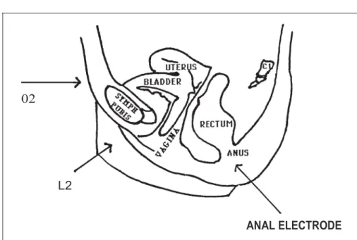
ANAL ELECTRODE
Figure 2. Diagrammatic representation qf'a median sagittal section through the
}i'male pelvis. Arrows indicate the anterior (D2, L2) and p osterior (anal) electrode placements. fAdapted.ficmzjohnston TB, Whillisj, eds. Gray's Anatomy: Descriptive
and Applied. 31st ed. New York, NY · Longmans, Green & Co; 1956.)
An alternative, or additional, explana tion for the submaximal responses is that, when the subjects sensed the
contraction , nervousness or the un usual sensation in a very sensitive and
intimate region of the body could
have caused them to stop increasing
the current amplitude , while still weil
helow the actual pain threshold, in both electrode placements . Delitto et al'" have shown thar the physical sensation of a muscle contracting as a
result of electrical stimulation, com
hined with the effect of stimulation of
local nociceptors , can lead to appre hension and fear. therehy reducing the
effectiveness of NMES as a means of eliciting a maximum or nearmaximum contracti on. Emotional factors, either
muscle (pudendal) is deeply situated
ar a depth of between 7.5 and 10 cm
in the pelvic cavity.6 Lowfrequenc..y
srimulating currents (050 pulses per second), using a noninvasive electrode
placement technique, are incapable of adequately stimulating the pelvicfloor muscles, unless current amplitudes are
increased to levels rhat can be ex tremely painful and porentially harm ful to the intervening tissues f>
The problem of tissue resistance has, seemingly, been overcome with the use of mediumfrequency interferential
currents. 12 The aim of obtaining a
maximal contraction of the pelvicfloor musculature with minimal current amplitudes is, however, still desirable,
and electrode placement can be of critical importance in achieving this
aim.
By displacing the anterior electrode
from the anterior perineum, interior to
the pubic symphysis (L2), to the re
gion immediately superior to the pu hic symphysis (D2), the depth of the
current field could , theorctically , he
increased. 1 1·12 Figure 2 illustrates this point diagrammatically . Assuming a gretter depth of penetration, a more
effective stimulation of the motor
ne1ve to the pubococcygeus muscle might be achieved. A!though no ex
perimental evidence exists to support
this hypothesis, a change in direction of the electrical field, produced by
displacing the anterior electr<XIe , could explain our results.
Our expectation thar the D2 electrode
placement would elicit a stronger
muscle contraction than the L2 elec
trode placement was not realized
during this study. No differences were observed between the force of con
trdction (measured as pressure in centimeters of water) provoked by the
tv..'O electr<XIe placements , nor was
rhere any carryover effect from D2 to L2. or vice versa (Tab. 2).
Two possible explanations might
account for the lack of any difference in the force of contraction. The in
stmctions given ro the subjects regard ing the effects of the stimulation could
have been misundersrood. Followup calls to ali subject<> , made in an at tempt to clarify this point, revealed
thar ali subjects ceased increasing the
current amplitude upon perception of an appreciable muscle contraction and
not necessarily for a maximum con
traction. Thus, it is possible thar for
consciously or subconsciously, may have influenced our subjects, affecting
their comprehension of the instruc
tions or causing them to overreact to the stimulus. In a normal treatment situation, this apprehension and mis
conception regarding the perceived
effects could gradually be overcome with repeated sessions. In such a
situation, and with encouragement and guidance, we fee! thar the D2
electrode placement would have re
sulted in a stronger muscle contraction at current amplitudes still below those obtained with the L2 electrode placement.
The choice of a nontetanizing fre
quency (10 Hz) for this study was
based on our concem with regard to
fatiguing the muscle. Stimulation at
higher frequencies (3050 Hz) may have resulted in a tetanie muscle con traction , further enhancing the efficacy of the stimulation. However, as any frequenc..y changes would have ap plied equally to both the L2 and D2 electrode placements, our results would have remained the same.
Dwyer and coworkers2 have demon strated thar there is a strong correla tion between a high BMI (ohesity) :md Physical Therapy
1Volume 75
, Number 12 / December 1995 1073 !41urinal)' stress incontinence in women. Adipose tissue offers a high resistance . to current flow,!'' and adipose tissue
tends to accumulate in the lower ab dominal and suprapubic regions in women. 23 None of our subjects were classified as obese, but this is a factor that could have influenced our results. Further research should take this as pect into consideration. Another limi tation of this study was the restricted number of subjects. Recruiting subjccts for this type of rcscarch is difficult, particularly whcn time constraints are imposed. Our study would have been strengthened had our groupings been larger. In spite of these drawbacks, however, we fee! that the results are encouraging and justify continued evaluation of the D2 technique in clinical trials of interferential currents for the treatment of female urinal)' stress incontinence.
Conclusion
Two electrode placements for NMES of pelvicfloor muscles have been described and compared, using conti nent female volunteers as subjects. Equivalent maximum pressures were observed with both electrode place ments. Current amplitudes required to obtain maximum pressure readings were Jess using the D2 electrode placement. Our interpretation of these findings is that the D2 electrode place ment produces a deeper, and there fore a more precise and effective, stimulation of the pelvicfloor muscu lature. This interpretation suggesto.; that a stronger muscle contraction might be obtained with the D2 electrode placement in subjects who become progressively more familiar with the
stimulation process while undergoing a treatment program. This hypothesis is examined in our companion article in this issue.
Acknowledgments
We express our appreciation to Dr Robert Gauthier, Department of Ob stetrics, and Dr Yves Homsy, Director, Department of Urology, Hôpital Ste Justine de Montréal, for their hclp in the selection and urologie evaluation of the patients participating in this research. Wc also thank Dr Jo Lay cock, Bradford Royal Infinnal)', Brad ford, England, for her helpful com ments and support in the preparation of this article.
References
1 Hilton P. Unstable urethral p ressure: to wards a more relevant definition. ln: Proceed ing s o( the international Continence Societ y , Boston Mass. 19H9:37 39.
2 Ahrams P, Blaivas J, Stanton S, Andersen J. The standardisation of the terminology of lower urinary tract function. Scand] Urol Nephrol. 19H8;114:17.
3 Beek RP, Hsu N. Pregnancy, childbirth, and menopause related to the development of stress incontinence. Am] Ohstet Gynecol. 1965;91:820 823.
4 Fall M, Lindstrom S. Electrical stimulation. Urol Clin N011h Am. 1991;18:393 407. 5 Leriche A, Leriche B. Place à la rééducation dans l'incontinence urinaire de la femme. ] Uro/ (Paris) 1988;94:285 288.
6 Scott B, Green V, Couldrey B. Pelvic faradism: investigation of methods. Physiother apy. 1969;55:302 305.
7 Plevnik S, Janez J, Vrtacnik P. Sh011term electrical stimulation: home treatment for uri nary incontinence. Worldj Urol. 1986;4:24 26. 8 McQuire W. Electrothera py and exercises for stress incontinence. Ph siothera py . 1975: 61:305 307.
9 Laycock J. Assessment and Treatment of Pelvic Floor Dy;function. Bradford, England:
Postgraduate School of Biomedical Sciences, Bradford University; 1992. Doctoral thesis. 10 Olah K, Bridges N, Denning J, et al. The conservative management of patients with symptoms of suess incontinence: a random ized, prospective study comparing weighted vaginal cones and interferenti<II therapy. Arnj Ohstet Gynecol. 1990;162:87 92.
11 MeyerWaarden K, Hansjurgens A, Fried mann B. Representation of electric fields in inhomogenous biological media. Biomed Tech (Berlin). 1980;25:295 297.
12 Goats GC. lnterferential current therapy. lJr] Sports Med. 1990;2:87 92.
13 Barnetr S, Cooney K, Johnston R. Elecrri cally elicited quadriceps femoris muscle torque as a function of various electrode placements . .f Clin Hlectrophysiol. 1991;3:5 8. 14 Laycock J, Green R. lnterferential rherapy in the treatment of incontinence. Phvsiother apy. 1988;74:161 168. · 15 Delitto A, Strube M, Shulman A, Minor S.
A study of discomfort with electrical stimula tion. Phys 1ber. 1992;72:410 424.
16 Health and Welfare Canada. Weight Levels Associated With Health: Canadian Guide/ines
1988. Ottawa, Ontario, Canada: Supply and Services Canada; 19il9:26. Catalogue no. H3'J 134.
17 Baker LL. Neuromuscular electrical stimu lation in the restoration of purposeful limb movement. ln: Wolf SL, ed. Hlectrotherapy. New York, NY: Churchill Livingstone !ne; 1981:2548.
18 Chiarelli P, O'Keefe D. Physiotherapy for the pelvic floor. Australian Journal of Physio therapy. 1981;27:103 108.
19 Bo K, Hagen R, Jirgensen .J, et al. The ef fects of two different pelvic floor muscle exer cise programs in the treatment of urinary stress incontinence in women. Neurology and Urodynamics. 1989;H:355 5S6.
20 Kloth L. Interference current. ln: Nelson
RM, Currier DP, eds. Clinicat Hlectrotberapv. East Norwalk, Conn: Appleton & Lange; 1987: 183 207.
21 Hansjurgens A, May liU. 'li·aditional and Modem Aspects oj'Electrotherapy 2nd ed. Temecula, Calif: Nemectron Medical !ne; 1984: 37
22 Myers J. Fundamentals oj'fuperimental Design. 2nd ccl. Boston, Mass: Allyn and Bacon !ne; 1973:1'!1 196.
23 Dwyer P, Lee E, Hay D. Ohesity and uri nary incontinence in women. Hrj Ohstet Gynaecol. 1988;95:91 96.